HOW I DO IT
Clipping of MCA aneurysms: how I do it
Philippe Bijlenga&Vitor Mendes Pereira&Karl Schaller
Received: 3 May 2011 / Accepted: 24 May 2011 / Published online: 4 June 2011 # Springer-Verlag 2011
Abstract
Introduction Aneurysms located at the middle cerebral artery bifurcation remain a clear neurosurgical indication. We detail here the steps necessary to enable safe surgery for Sylvian fissure aneurysms.
Methods An angiogram with 3D reconstruction is obtained and reviewed intraoperatively, just prior to the skin incision. During the exposure, the cistern is kept open by small cottonoids, thereby avoiding brain retraction. Continuous monitoring of MEPs along with ICG microscopic angio-fluorescence allows for detection of vascular compromise. Intraoperative angiography with 3D reconstruction allows for immediate correction of less than satisfactory surgical outcome.
Conclusions Careful planning of surgical strategy fol-lowed by a minimally invasive technique (with continuous neuro-monitoring) ensures safe surgery. The availability of intra-operative radiological guidance allows for optimal management.
Keywords Middle cerebral artery. Intracranial aneurysm . Clipping . Hybrid intervention . Neuro-monitoring . Indocyanine green angio-fluorescence . Per-operative angiography
Abbreviations
CSF Cerebrospinal fluid MCA Middle cerebral artery ICG Indocyanine green MEP Motor evoked potential
DRA Digital subtraction rotational angiography 3D-DRA 3D surface rendering reconstruction from DRA
Introduction
The field of vascular neurosurgery has undergone revolu-tionary developments that seek to improve patient care and outcomes. New treatment modalities and new devices continuously challenge the standard of care. The ISAT study showed that coiling of ruptured intracranial aneur-ysms is at least as effective and safe as microneurosurgical clipping performed within the first 72 h after ictus. As a result, most basilar tip and vertebral aneurysms are now coiled. On the other end of the spectrum, most middle cerebral artery (MCA) bifurcation aneurysms are still clipped and now secured within the first 24 h of ictus to reduce rebleeds [1, 4, 5]. As a result of increased opportunities and better quality cerebral imaging, more patients are being diagnosed with asymptomatic and unruptured intracranial aneurysms. The ISUIA study showed that some patients benefited more from conserva-tive management, others from open surgery and finally, there were those who benefited from endovascular
inter-Electronic supplementary material The online version of this article (doi:10.1007/s00701-011-1063-9) contains supplementary material, which is available to authorized users.
P. Bijlenga (*)
:
K. SchallerService de Neurochirurgie, Département de Neurosciences Cliniques, Hôpitaux Universitaire de Genève,
Faculté de médecine, Université de Genève, Rue Gabrielle-Perret-Gentil 4,
1211 Geneva 14, Switzerland e-mail: [email protected] V. Mendes Pereira
Service de Neuroradiologie Diagnostique et Interventionelle, Hôpitaux universitaire de Genève, Faculté de médecine, Université de Genève,
vention. The authors concluded that for aneurysms less than 7 mm in size located in the anterior circulation, there was a role for patient observation alone. However, no conclusion was drawn on a favored treatment modality should treatment be sought [8]. Recently, the advent of flow diverter stents has produced impressive results in the management of fusiform or side-wall saccular aneurysms, as well as ophthalmic aneurysms [3, 6]. Criteria to determine which patients and lesions are more appropri-ately treated by one method over the other remain ill-defined. Intuitively, patients with coagulopathy would clearly be better candidates for endovascular treatments while patients suffering vessel atherosclerosis with pla-ques, sub-occlusion or occlusion, are better candidates for surgery. Similarly, proximal lesions lying close to the circle of Willis are considered more accessible via the endolu-minal route while lesions more superficial and distal in the vascular tree tend to be more amenable to a surgical approach. Aneurysm morphology is also important. A saccular shape with a small neck allows for high coil compaction with little risk of parent vessel occlusion. Aneurysms with large necks or those involving both parent arteries and branches are reconstructed using stents or clips [2]. Current efforts are devoted to better identifying such factors, establishing their relative importance and quanti-fying their impact on decision-making, with the end goal of improving safety and long-term efficacy of treatments (Table1).
MCA aneurysms represent 25% of all intracranial aneurysms and more than 60% are incidental findings. Due to their position in the vascular tree and frequent incidence of large neck morphology, MCA aneurysms are mostly treated by microsurgical clipping [7]. We report advances introduced in our daily clinical practice to reduce peri-operative risks and offer an anatomical cure for such lesions affecting otherwise healthy patients with a median age between 55 and 60 years [8,9].
Operative technique (Table2)
Preoperative imaging and angiography (Fig.1)
Angiographic pre-operative images are studied by the treating team (comprising the senior and junior surgeons as well as the interventional neuroradiologist). The extent of sphenoid ridge to be removed is considered in relation to the superior orbital fissure. The anatomy of the circle of Willis is characterized with regards to the patency of left-to-right connections. This is to determine the optimal proximal clipping location in the event of precocious aneurysm rupture. Vascular and aneurismal anatomy to be
anticipated during surgery is studied using 3D recon-structions. The segmented volumes are rotated to visualize projections as encountered during surgery. The aneurysm neck and orientation are measured and the clipping strategy is defined accordingly (Fig. 1). In case of a very wide or atherosclerotic aneurysm neck, bifurca-tion reconstrucbifurca-tion may result in the occlusion of one or all branches. For those cases, a preventive bypass is discussed. Planning and operative technique of complex MCA aneurysms is out of the scope here. In order to prepare the pterional approach (see below), special attention is paid to the identification of pneumatized structures to be avoided during surgery. This extends from frontal sinuses to the anterior clinoid processes. The extent of Sylvian fissure opening is discussed and anatomical landmarks on MCA branches identified.
Table 1 Information for patient and family for non-complex MCA bifurcation aneurysm clipping
1. The operation is proposed if the risk of surgery is considered smaller than observation over a period of 5 years
2. Less than 5% risk of stroke
3. 2% risk of infection that may require antibiotic treatment for weeks and sometimes removal of the bone flap that will be replaced by prosthesis
4. 1% risk of a hematoma that would require reoperation for evacuation
5. Less than 1% epilepsy
6. Overall: 5% risk of significant handicap for patients less than 50 years of age, 10% for patients over 50 years 7. Overall: Less than 2% risk of death
8. Minor chewing discomfort for 2 months 9. Slight temporal muscle atrophy may be observed
10. Pins from head clamp may induce some discomfort for 1 or 2 days 11. Hematoma may be observed in the groin
12. Possible black eye for 1 week
Table 2 Key points
1. Examine and memorize the angio-architecture prior to skin incision 2. Identify key reliable landmarks, such as bifurcations and curves
of vessels
3. Optimize dural opening in relation to the size of the bony exposure 4. Assure venous epidural hemostasis by oxycellulose and
temporary cottonoid application
5. Avoid brain retraction, use small cottonoids to keep the Sylvian fissure open
6. Prepare a clipping site prior to dissection of the aneurysm dome 7. Puncture the aneurysm dome to assess complete exclusion 8. Perform high-resolution angiographic imaging as soon as
Positioning of the patient and general settings (Fig.2)
The patient lies supine in a^Le Corbusier^ lounge chair position. The shoulder on the operating side is elevated and the head is rotated 90° toward the opposite side. The head is fixed in a Mayfield head clamp using 3-pin fixation. Care is taken to keep the head above the level of the heart and to ensure both jugular veins remain patent. The groin is prepared for endovascular access. The anesthetist accesses the patient from the legs and left side of the body. The endovascular interventionist is positioned close to the right groin. The surgeon sits at the vertex.
We never use a lumbar drain. Patients receive antibiotics in the operating room approximately 20 min prior to the skin incision. Scalp electrodes are placed. Neurophysiological monitoring includes monitoring of motor evoked potential of the contra-lateral limbs.
Before draping, a^time-out^ (as per WHO guidelines) is performed in the presence of the anesthesiologist, interven-tionist, neurophysiologist, scrub nurse, and surgeons. The main stages of the procedure are reviewed and the instruments required for each stage are checked (emergency clips are chosen and prepared). Communication with all members of the team remains vital throughout the procedure.
Approach
A curved skin incision is carried-out beginning 1 cm anterior to the tragus at the level of the zygomatic arch and running behind the hairline, up to the midline. The pericranial flap is fashioned according to Yasargil’s descrip-tion of interfascial dissecdescrip-tion and reflected antero-inferiorly. The temporal muscle is detached from the bone sutures (starting from the orbito-zygomatic suture) and pterion, using a periosteal elevator. The upper insertion is sectioned using cutting monopolar. This technique allows preserva-tion of the periosteum, thereby allowing for a better cosmetic outcome due to reduced risk of temporal muscle atrophy. The muscle is reflected posteroinferiorly [9].
A burr hole is performed over the thin temporal bone and dura is detached antero-superiorly. An oval-shaped craniotomy is carried out, with the longer diameter being aligned with the Sylvian fissure. An average-sized craniot-omy is approximately 5 cm×4 cm wide. The anterior aspect of the craniotomy is drilled flush with the lateral wall of the orbit. Following elevation of the bone flap, the sphenoid ridge is removed using a Luer rongeur and drilled to achieve continuity with the lateral wall of the orbit. The meningo-orbital band is identified as the limit of bone removal. The bone flap is stored aside in normal saline for
Fig. 1 Pre-operative 3D-DRA should be used to prepare inter-vention (a). The 3D volume is oriented to match with the sur-gical field (b). Landmarks are identified on vessels to aid sur-gical exploration. Examples in-clude identifying the fissure opening point (B1) in order to establish the direction to the inferior trunk of M2 (B2). The bifurcation of the superior trunk is used as a proximity marker for a distal aneurysm (B3). The proximal control site is identi-fied (B4) relative to the proxi-mal aneurysm (B5). Clip shape and size is chosen for the prox-imal aneurysm that will be clipped first to avoid subsequent conflict when clipping the more superficial aneurysm (c). Clip-ping strategy is modeled for the second aneurysm (d)
later reinsertion. Perfect hemostasis is now achieved, prior to moving on to the intradural stage.
The dura is opened in a^U^-shape against the orbital wall and reflected anteriorly. In young patients with no cerebral atrophy or in cases with a ruptured aneurysm and a swollen brain, we follow the orbital roof down to the olfactory nerve and then laterally to the optic nerve. CSF is drained from the pre-optic cistern. This maneuver allows an early exposure of the internal carotid artery for proximal control if needed. A small hole is created in the arachnoid of the Sylvian fissure approximately 4 cm posterior to the drilled sphenoid ridge. Normal saline is gently injected under pressure until distal reflux is observed. The hole is then enlarged and the Sylvian fissure is opened by sharp dissection (e.g., using a diamond knife) in the direction of the limen insula. If the patient suffers an intraparenchymal hematoma most but not all of it
is removed to reduce mass effect but avoid rebleeding. The inferior trunk of M2 is identified and followed anteriorly towards the bifurcation. Very frequently, for proximal control, the M1 artery distal to the striate perforators, can be identified turning rostro-medially to the limen insula. Care is taken to gradually enlarge the arachnoids’ opening on the surface of the Sylvian fissure when progressing deeper. It is easier to open the Sylvian fissure from the depth aiming superficially. Veins are preserved as much as possible by cutting arachnoids’ adhesions both sides of the fissure allowing mobilization. If necessary, we favor sacrificing veins on the frontal side. The veins bridging to the sphenoparietal sinus are sometime wrapped with oxidized cellulose gaze. Once the distal M1 has been identified and the fissure opened sufficiently to avoid tension from retraction, cottonoids are used to keep the field open (thus
Fig. 2 Hybrid room configuration. The operating theatre is equipped with an Allura Xper FD20 (Philips Healthcare, Best, Netherlands). The head of the patient is fixed in a radiolucent Mayfield Infinity XR2 Scull Clamp (Integra Neuroscience Limited, Andover Hampshire, UK). During the procedure, MEPs are monitored by transcranial electric stimulation (Axon Eclipse, Axon Systems Inc, Hauppauge, NY) (X). Microsurgery is performed under a microscope using a OPMI® Pentero™ equipped with the INFRARED 800 option (M). The space is virtually divided into three regions. On the lower left of
the patient is the anesthetic (A) corner with equipment (E). On the lower right is the interventional radiologist (Int) corner with instrument table (T). The surgical team (S) and scrub nurse (N) are located at the head of the patient. The operative field is protected by horizontal laminar flow (L). Control of equipment, image processing, and analysis is performed in the “control zone” (C). Images can be projected on a screen on the left side of the room or viewed on a Digital Lightbox© (BrainLab, Feldkirchen, Germany) on the right side
negating the need for self-retaining retractors). The aneurysm is usually partially exposed at this stage.
Aneurysm neck dissection and clipping
The next step is to identify the superior trunk of M2 and expose the neck of the aneurysm. Care is taken to ensure that the instruments used are wet. Arachnoid is cleared from the vessel and aneurysm wall using microsurgical dissectors and micro-scissors. Movements are tangential to vascular structures. Following exposure of all the M2 branches, M1 trunk, and the aneurysm neck, the clip strategy is assessed and an appropriate clip is chosen. When high-resolution angiographic intra-operative imaging is not available, flow or blood velocity measurements of the M1 trunk (proximal to the aneurysm) and all M2 branches (immediately distal to the aneurysm) are performed in order to assess for potential vascular compromise post-clipping. Depending on the size of the aneurysm or per-operative rupture, a temporary clip may be applied proximally to deflate the dome or reduce the bleeding. When proximal clipping is intended, the anesthesiologist is informed, who then
increases the mean arterial blood pressure by 20%. MEPs are monitored.
Intra-operative quality control (Fig.3)
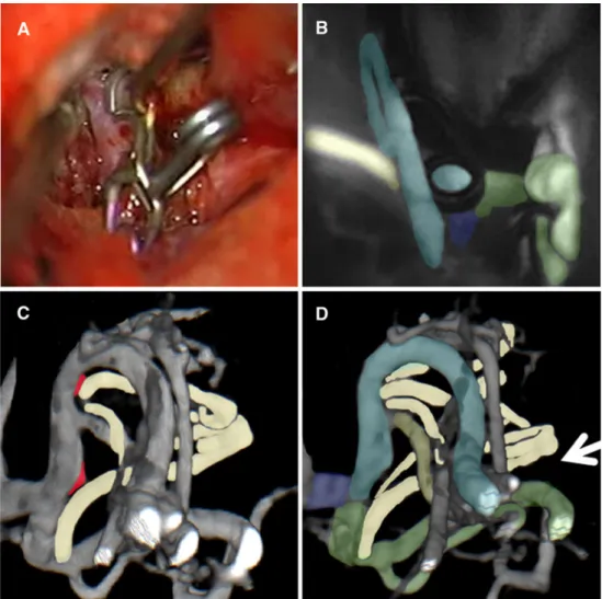
When the aneurysm clip is placed on the neck, the proximal clip is removed and the aneurysm dome is completely exposed. The clipping is checked by puncturing the aneurysm dome with a 25-G needle. The aneurysm should deflate and the neck is then explored on both sides to exclude remnants or occlusion of neighboring small perforators. ICG angio-fluorescence imaging is performed to assess the patency of all vessels. If ICG is not available, patency is confirmed using micro Doppler. Papaverine 40 mg diluted in 10 ml of normal saline is applied to the Sylvian fissure. The operating field is protected by a wet compress and the head of the patient is positioned within a transparent draped. The interventional neuroradiologist performs a conventional angiogram as well as a 3D rotational angiogram. Images are compared to preoperative data to verify that all branches and perforators are patent.
Fig. 3 The aneurysm clipping is checked visually (a). The aneurysm dome is punctured and flow in the different branches is assessed using indocyanine green angiofluores-cence (b). The inferior M2 trunk (green), the anterior branch of the superior M2 artery (light blue), the posterior branch of the superior M2 artery (yellow) and the M1 trunk (dark blue) are recognized and the rate of fluo-rescence increase is checked for all branches. A rotational angi-ography is carried out and reconstructed to allow 3D surface rendering. Clipping is carefully inspected (c). In the example small remnants (red) are corrected with two mini clips before final control is achieved (d)
3D vessel morphology is reconstructed from the acquired DRA and checked from all angles to exclude micro residuals, dog ears, or stenosis. Clips are repositioned if considered necessary and reassessed by 3D-DRA as required.
Closure
At the end of surgery, watertight closure of the dura is undertaken. A Gelfoam® sterile compressed sponge is applied between the dura and the bone flap in the event of CSF leak. The bone flap is replaced and maintained with titanium plates and low profile screws. Replacement and suture of the temporal muscle is done using absorbable sutures. Subgaleal drainage is usually avoided. Skin closure is carried out in two layers.
Postoperative course and instructions from the surgeon
The patient is observed overnight in an intensive care unit. Low-molecular-weight heparin is started 6 h post-craniotomy to prevent thromboembolic complications. The patient is usually discharged after 5 days (at which stage sutures are removed). A post-operative check is performed after 6 weeks. Patients are followed-up with CT angiography at 1 year, 5 years, and every 5 years thereafter.
Potential future evolutions
Integration of virtual images updated by ultrasound and endoscopic tools may provide a visual environment that would allow performing the operation through a few burr holes reducing tissue retraction damage even further.
Conclusions
The morbidity of MCA aneurysm clipping can be reduced by careful planning, adoption of minimally invasive microsurgi-cal techniques, and systematic continuous monitoring of MEPs. Intra-operative angiographic imaging allows timely correction of imperfections and allows for an optimal surgical result. Nevertheless, we still advocate long-term follow-up of
these patients with CT angiography to reduce the risk of hemorrhage from newly formed lesions.
Conflicts of interest None.
References
1. Bakker NA, Metzemaekers JD, Groen RJ, Mooij JJ, Van Dijk JM (2010) International Subarachnoid Aneurysm Trial 2009: endovascular coiling of ruptured intracranial aneurysms has no significant advantage over neurosurgical clipping. Neurosurgery 66(5):961–962
2. Bendszus M, Chapot R (2007) Balloon-assisted coil embolization. Surgical clip application should be considered as a first treatment option in large and wide-necked aneurysms. J Neurosurg 106 (4):734–735
3. Lylyk P, Miranda C, Ceratto R, Ferrario A, Scrivano E, Luna HR, Berez AL, Tran Q, Nelson PK, Fiorella D (2009) Curative endovascular reconstruction of cerebral aneurysms with the pipeline embolization device: the Buenos Aires Experience. Neurosurgery 64(4):632–642
4. Molyneux AJ, Kerr RS, Birks J, Ramzi N, Yarnold J, Sneade M, Rischmiller J, ISAT Collaborators (2009) Risk of recurrent subarachnoid haemorrhage, death, or dependence and standardised mortality ratios after clipping or coiling of an intracranial aneurysm in the International Subarachnoid Aneurysm Trial (ISAT): long-term follow-up. Lancet Neurol 8(5):427–433
5. Molyneux AJ, Kerr RS, Yu LM, Clarke M, Sneade M, Yarnold JA, Sandercock P, International Subarachnoid Aneurysm Trial (ISAT) Collaborative Group (2005) International subarachnoid aneurysm trial (ISAT) of neurosurgical clipping versus endovascular coiling in 2143 patients with ruptured intracranial aneurysms: a randomised comparison of effects on survival, dependency, seizures, rebleeding, subgroups, and aneurysm occlusion. Lancet 366(9488):809–817 6. Nelson PK, Lylyk P, Szikora I, Wetzel SG, Wanke I, Fiorella D
(2011) The pipeline embolization device for the intracranial treatment of aneurysms trial. AJNR Am J Neuroradiol 32(1):34–40 7. Regli L, Dehdashti AR, Uske A, de Tribolet N (2002) Endovas-cular coiling compared with surgical clipping for the treatment of unruptured middle cerebral artery aneurysms: an update. Acta Neurochir Suppl 82:41–46
8. Wiebers DO, Whisnant JP, Huston J 3rd, Meissner I, Brown RD Jr, Piepgras DG, Forbes GS, Thielen K, Nichols D, O_Fallon WM, Peacock J, Jaeger L, Kassell NF, Kongable-Beckman GL, Torner JC, International Study of Unruptured Intracranial Aneurysms Investigators (2003) Unruptured intracranial aneurysms: natural history, clinical outcome, and risks of surgical and endovascular treatment. Lancet 362(9378):103–110
9. Yasargil MG, Smith RD, Young PH, Teddy PJ, Roth P (1984) Microneursurgery, vol. 2. Georg Thieme Verlag, Stuttgart, p 385