Versatile Reactivity and Theoretical Evaluation of Mono- and
Dinuclear Oxidovanadium(V) Compounds of Aroylazines:
Electrogeneration of Mixed-Valence Divanadium(IV,V) Complexes
Subhashree P. Dash,
†,‡Satabdi Roy,
†Monalisa Mohanty,
†M. Fernanda N. N. Carvalho,
§Maxim L. Kuznetsov,
§Joa
̃o Costa Pessoa,
*
,§Amit Kumar,
§,∥Yogesh P. Patil,
⊥Aurélien Crochet,
¶and Rupam Dinda
*
,††Department of Chemistry, National Institute of Technology, Rourkela 769008, Odisha, India
‡Department of Chemistry, Indira Gandhi Institute of Technology, Sarang, Parjang, Dhenkanal 759146, India
§Centro de Química Estrutural, Instituto Superior Técnico, Universidade de Lisboa, Avenida Rovisco Pais, 1049-001 Lisboa, Portugal ∥Centre for Nano and Material Sciences, Jain University, Jain Global Campus, Jakkasandra Post, Bangalore 562112, India
⊥Department of Inorganic and Physical Chemistry, Indian Institute of Science, Bangalore 560012, India
¶Department of Chemistry, Fribourg Center for Nanomaterials, University of Fribourg, CH-1700 Fribourg, Switzerland
ABSTRACT: The substituted hydrazones H2L1−4(L1−4= dibasic
tridentate ONO2−donor ligands) obtained by the condensation of 2-hydroxy-1-naphthaldehyde and 2-aminobenzoylhydrazine (H2hnal-abhz) (H2L1) , hydroxy-1-naphthaldehyde and 2-hydroxybenzoylhydrazine (H2hnal-hbhz) (H2L2), 2-hydroxy-1-acetonaphthone and benzoylhydrazine (H2han-bhz) (H2L3), or 2-hydroxy-1-acetonaphthone and 2-aminobenzoylhydrazine
(H2han-abhz) (H2L4) are prepared and characterized. Reaction of
ammonium vanadate with the appropriate H2L1−4 results in the formation of oxidoethoxidovanadium(V) [VVO(OEt)(L1−4)] (1− 4) complexes. All compounds are characterized in the solid state and in solution by spectroscopic techniques (IR, UV−vis,1H,13C, and 51V NMR, and electrospray ionization mass spectrometry).
Single-crystal X-ray diffraction analysis of 1, 3, and 4 confirms the coordination of the corresponding ligands in the dianionic (ONO2−) enolate tautomeric form. In solution, the structurally characterized [VVO(OEt)(L)] compounds transform into the monooxido-bridged divanadium(V,V) [(VVOL)
2-μ-O] complexes, with the processes being studied by IR and1H,13C, and51V NMR. The density functional theory (DFT) calculated Gibbs free energy of reaction 2[VVO(OEt)(L4)] + H2O⇆ [(VVOL4)2- μ-O] + 2EtOH is only 2−3 kcal mol−1, indicating that the dinuclear complexes may form in a significant amount. The electrochemical behavior of the complexes is investigated by cyclic voltammetry, with the VV−VIVE1/2redvalues being in the range 0.27−0.44 V (vs SCE). Upon controlled potential electrolysis, the corresponding (L)(O)VIV−O−VV(O)(L) mixed-valence species are obtained upon partial reduction of the [(VVOL)2-μ-O] complexes formed in solution, and some spectroscopic characteristics of these dinuclear mixed-valence complexes are investigated using DFT calculations and by electron paramagnetic resonance (EPR), with the formation of VIV−O−VVspecies being confirmed by the observation of a 15-line pattern in the EPR spectra at room temperature.
■
INTRODUCTIONInterest in the coordination chemistry of vanadium, with particular emphasis on its biological,1 structural,1a,b,d,2 and catalytic properties,3−6has increased over the past 2 decades. Prospective therapeutic applications of vanadium compounds, namely, for the treatment of Diabetes mellitus and cancer and more recently promising antiamoebic activity against Entamoe-ba histolytica as well as antitrypanosomal activity against Trypanosoma cruzi,1c,estimulated research to better understand the behavior of vanadium complexes in solution.7−10
Vanadium complexes possess versatile oxidation states (from −III to +V). Among these, +IV and +V are the most common and can be accessed easily under aerobic conditions. A good number of structural, spectroscopic, and solution studies of such vanadium compounds, in which the element is present in these high oxidation states under assorted N/O-donor environments, have been reported.8a−c,g,11
http://doc.rero.ch
Published in "Inorganic Chemistry 55(17): 8407–8421,
2016"
which should be cited to refer to this work.
While the solid-state structural aspects and reactivity of the well-known vanadium(V) complexes containing VVO2+ units have been satisfactorily exposed, not so much is known of their solution behavior.1b,12Subjects of interest include the stability of mononuclear species in solution, possibility of formation of coordination isomers,12a,eformation of di-10,12b,cor polynuclear complexes,12d and electron-transfer capabilities of mono- and dinuclear species.10,12b
Mixed-valence metal complexes have received much attention,13 and one of the relevant aspects in this area includes an understanding of the role of the ligand environment in the extent of electron localization/delocalization among the metal centers and the potential use of mixed-valence systems in
molecular electronics and molecular computing.14 The
chemistry of mixed-valence vanadium incorporating O/N-coordinating ligands is of enduring interest.15In contrast to the well-known oxidovanadium(IV or V) complexes containing VIVO2+ or VVO3+ units,15b,16
relatively few mixed-oxidation (IV,V) complexes have been isolated and structurally characterized. In the context of the present study, it is relevant to mention that, although the chemistry of mixed-valence divanadium(IV,V) complexes generated through chemical synthesis is reasonably developed,15,17 there are only a few reported examples of compounds isolated through electro-synthesis.12b,15d,18
We describe herein the synthesis and characterization of mononuclear monoalkoxido-bound monooxidovanadium(V) complexes (1−4) of the ligands derived from the substituted hydrazones H2L1−4(H2L1−4= aroylhydrazones, where L1−4act as dibasic tridentate ONO-donor ligands; Scheme 1); these
ligands may be considered as azine analogues. We also show that the transformation of the structurally characterized
monooxidoalkoxidovanadium(V) compounds [VVO(OEt)(L)]
in solution lead to the corresponding monooxido-bridged divanadium(V,V) compounds [(VVOL)2-μ-O].12b The mono-oxido-bridged divanadium(IV,V) mixed-valence species are also obtained by controlled potential electrolysis of the correspond-ing divanadium(V,V) precursors in solution. These mixed-valence species are characterized by IR, electron paramagnetic resonance (EPR), and 51V NMR, and their formulation was corroborated by density functional theory (DFT) calculations. Theoretical calculations were also carried out for most complexes reported to support and substantiate the measured structural and spectroscopic properties. We should highlight that, in agreement with Young,15awe are designating here, as mixed-valence V2O33+compounds, both those that correspond to complexes with localized VIV and VV centers, exhibiting 8-line EPR spectra, and those where the unpaired electron is delocalized throughout the two V nuclei, exhibiting a 15-line EPR spectra due to the effective hyperfine coupling of the two 51V centers. It is demonstrated that spin localization/ delocalization takes place between two V atoms, and, hence, a different pattern of EPR spectra for various compounds (15-line vs 8-(15-line) is determined by the geometry of the V−O−V fragment (linear vs bent).
■
EXPERIMENTAL SECTIONMaterials and Methods. Elemental analyses were carried out on a PerkinElmer C, H, and N analyzer. IR spectra of solid compounds [VVO(OEt)(L)] (1−4) as KBr pellets and in a CHCl
3solution in a
NaCl cell were recorded on a PerkinElmer 783 spectrometer. 1H Scheme 1. Schematic Diagram of the Synthesis of Monooxidoalkoxidovanadium(V) [VVO(OEt)(L1−4)] (1−4) and the Various Pathways through Which the Compounds May Be Transformed in Solution
NMR spectra were recorded with a Bruker ADVANCE DPX 300 MHz spectrometer using SiMe4as an internal standard.51V NMR spectra
were obtained on a Bruker Avance III 400 MHz spectrometer in deuterated acetonitrile:dimethylsulfoxide (MeCN-d1:DMSO-d6), and
chemical shift51V values (δV) were referenced relative to neat VVOCl 3
as the external standard. Mass spectrometry (MS) spectra were obtained on a SQ-300 MS instrument operating in the electrospray ionization (ESI) negative mode. Electronic spectra were recorded on a Shimadzu UV−vis recording spectrophotometer. Electrochemical studies were made by cyclic voltammetry (CV) and controlled potential electrolysis (CPE) at room temperature under a nitrogen atmosphere using a PC-controlled VoltaLab PST 050 potentiostat. Measurements were made in a three-compartment cell provided with platinum wire (working and auxiliary) and silver wire (reference) electrodes using tetrabutylammonium tetrafluoroborate (0.10 M)/ CH3CN as the supporting electrolyte. Potentials were referred to a
saturated calomel electrode (SCE) using the ferrocene/ferrocenium couple ([Fe(η5-C
5H5)2]0/+; E1/2= 0.382 V) as the internal standard.
Electron paramagnetic resonance (EPR) spectra were recorded with a Bruker ESP 300E X-band spectrometer either at 77 K or at room temperature, and the spin Hamiltonian parameters were obtained by simulation of the spectra with the computer program of Rockenbauer et al.19The calibrant was 2,2-diphenyl-1-picrylhydrazyl (g: 2.0037).
Crystal Structure Determinations. Single crystals of compounds were mounted on a Bruker Smart Apex CCD diffractometer (1) equipped with a graphite monochromator and a Stoe IPDS 2 diffractometer (3 and 4) equipped with an Oxford Cryosystem open-flow cryostat and a Mo Kα radiation (λ = 0.71073 Å). The crystallographic data and details of refinement are given in Table 1. The unit cell dimensions and intensity data were measured at 293(2) K (1) and 200 (2) K (3 and 4). Absorption correction was partially integrated in the data reduction procedure for crystals of 3 and 4.20 The intensity data were corrected for Lorentz, polarization, and absorption effects. Absorption corrections were applied using SADABS, and the structures were solved by direct methods using the program SHELXS-9721 and refined using least squares with the SHELXL-9721software program. H atoms were either found or placed in calculated positions and isotropically refined using a riding model. The non-H atoms were refined anisotropically.
CV and CPE. The redox properties of complexes 1−4 were studied by CV, and the potentials were measured versus SCE using ferrocene as the internal standard. CPE was carried out at the potential of the reduction process (selected according to CV data) using a typical procedure: the addition of a weighed amount of complex [1 (5.0 mg), 2(5.0 mg), 3 (7.0 mg), and 4 (8.0 mg)] to the electrolyte solution (10 mL; NBu4BF4/CH3CN, 0.10 M, under a nitrogen atmosphere),
waiting ca. 1 h, and then proceeding with the chronocoulometric process until it reached the number of coulombs (Q) corresponding to transfer of half an electron per mole of complex. Then, a cyclic voltammogram was obtained, a sample of the electrolyzed solution was collected and immediately transferred to an EPR tube (previously purged with nitrogen gas), and the solution was frozen by emersion of the tube in liquid nitrogen.
Computational Details. Full geometry optimization of the molecular structures was carried out at the DFT level of theory using the B3P86 functional22awith the help of the Gaussian09 program package.23 This functional was found to be sufficiently accurate for theoretical studies of structural parameters and 51V NMR chemical
shifts of various vanadium complexes.24 For some calculations, B3LYP,22a,c M06,22d and TPSSH22e functionals were also used. Optimization was carried out in both gas and solution phases. In the latter case, the IEFPCM solvation model25 with the UAKS molecular cavity and dispersion, repulsion, and cavitation contribu-tions were applied, and DMSO, ethanol (EtOH), or MeCN were used as the solvent. No symmetry operations were applied for any of the structures calculated. Geometry optimization was carried out using a relativistic Stuttgart pseudopotential, which describes 10 core electrons, and the appropriate contracted basis set (8s7p6d1f)/ [6s5p3d1f] for the V atom26 and the 6-31G(d) basis set for other atoms. The Hessian matrix was calculated analytically for all optimized
structures to prove the location of correct minima (no imaginary frequencies) and to estimate the thermodynamic parameters, with the latter being calculated at 25°C.
Magnetic shielding was calculated for the equilibrium geometries using the GIAO method27at the IEFPCM-B3P86/6-311+G(2d,p)//6-31G(d) level. The 51V chemical shifts were estimated relative to
VVOCl
3calculated at the same level of theory.
The51V hyperfine coupling constants were estimated at the
single-point calculations using the BHandHLYP functional and 6-311G* basis set for all atoms on the basis of the equilibrium geometry obtained at the B3P86/6-31G*(V-ECP) level of theory. The anisotropic 51V hyperfine coupling constants A
x, Ay, and Az were
estimated as the sum of the isotropic Fermi contact term and corresponding dipolar hyperfine interaction term.28
It must be emphasized that for a vanadium(IV) species the hyperfine coupling constant (A) values may be negative, but in the literature, normally it is their absolute value that is reported. This simplification was also adopted in this work. Previously, it was found that half-and-half functionals such as BHandHLYP demonstrate better performance in calculations of the EPR parameters than many other hybrid functionals because of more accurate estimates of the Fermi contact term.28a−c
For the dinuclear vanadium(V,V) compounds [(VVOL)
2-μ-O], two
isomers were calculated, i.e., the trans isomer with the opposite positions of the VO bonds relative to the V−O−V bridge and the gauche isomer with the OV···VO torsion angle of 78−103°. The latter isomer is thermodynamically more stable, and mainly structures of this type are discussed below.
Table 1. Crystal Data and Refinement Details for 1, 3, and 4
1 3 4
empirical formula C20H18N3O4V C21H19N2O4V C21H20N3O4V
Mw 415.31 414.32 429.34
temperature (K) 293(2) 200(2) 200(2) wavelength (Å) 0.71073 0.71073 0.71073 cryst syst monoclinic monoclinic monoclinic space group P21/n P21/n P21/n unit cell dimens
a (Å) 7.5992(7) 10.0073(19) 10.305(2) b (Å) 12.7639(11) 15.0465(18) 10.502(2) c (Å) 19.5684(17) 13.023(2) 18.603(3) α (deg) 90 90 90 β (deg) 90.373(2) 91.581(15) 92.716(16) γ (deg) 90 90 90 volume (Å3) 1898.0(3) 1960.2(6) 2011.0(6) Z 4 4 4 density (calcd) (Mg m−3) 1.453 1.404 1.418 abs coeff (mm−1) 0.554 0.535 0.526 F(000) 856 856 888
θ range for data
collection (deg) 1.90−26.00 2.07−25.00 2.192−24.995 reflns collected 19292 24699 25250 indep reflns 3732 [R(int) = 0.0788] 3452 [R(int) = 0.0843] 3549 [R(int) = 0.1342] completeness toθ = 25.00° (%) 100.0 100.0 100.0 refinement method full-matrix least
squares on F2 full-matrix leastsquares on F2 full-matrix leastsquares on F2 data/restraints/ param 3732/1/262 3452/48/255 3549/0/265 GOF on F2 1.045 1.043 0.920 final R indices [I > 2σ(I)] R1 = 0.0939, wR2 = 0.2142 R1 = 0.0382, wR2 = 0.1034 R1 = 0.0497, wR2 = 0.1164 R indices (all data) R1 = 0.1668,
wR2 = 0.2584 R1 = 0.0512, wR2 = 0.1091 R1 = 0.1224, wR2 = 0.1521 largest diff peak and hole (e Å−3) 0.589 and −0.319 0.230 and−0.287 0.257 and−0.365
http://doc.rero.ch
Preparation of Compounds H2L1−4. The substituted hydrazones
H2hnal-abhz (H2L1), H2hnal-hbhz (H2L2), and H2han-bhz (H2L3),
and H2han-abhz (H2L4) were prepared by similar procedures; we
describe here the preparation of H2L1: a solution of
2-amino-benzoylhydrazine (1.51 g, 10.0 mmol) in EtOH (20 mL) was added to afiltered solution of 2-hydroxy-1-naphthaldehyde (1.72 g, 10.0 mmol) in EtOH (15 mL) with stirring. The obtained mixture was refluxed and stirred for 2 h. The resulting pale-yellow compounds were filtered, washed with EtOH, and dried over fused CaCl2. Elemental analysis
results and NMR (1H and 13C) and IR data for all compounds
corroborate with their formulation.
H2hnal-abhz (H2L1). Yield: 65%. Anal. Calcd for C18H15N3O2: C,
70.81; H, 4.95; N, 13.76. Found: C, 70.77; H, 4.98; N, 13.75. Selected IR peaks with proposed assignments (KBr pellet, νmax/cm−1): 3472
ν(O−H); 3367 ν(NH2)as; 3201ν(NH2)s; 3037ν(N−H); 1637 ν(C O); 1555ν(CN).1H NMR (400 MHz, DMSO-d 6):δ 12.97 (s, 1H, naphthyl−OH), 11.95 (s, 1H, NH), 9.47 (s, 1H, HCN), 8.18−6.63 (m, 10H, aromatic), 6.61 (s, 2H, NH2).13C NMR (100 MHz, DMSO-d6):δ 165.12, 158.33, 150.93, 146.33, 133.18, 132.91, 132.07, 129.45, 128.57, 128.25, 128.16, 123.96, 120.88, 119.42, 117.12, 115.14, 112.62, 109.06.
H2hnal-hbhz (H2L2). Yield: 75%. Anal. Calcd for C18H14N2O3: C,
70.58; H, 4.61; N, 9.15. Found: C, 70.59; H, 4.58; N, 9.12. Selected IR peaks with proposed assignments (KBr pellet,νmax/cm−1): 3466ν(O−
H); 3022ν(N−H); 1640 ν(CO); 1578 ν(CN).1H NMR (400 MHz, DMSO-d6):δ 12.74 (s, 1H, naphthyl−OH), 12.07 (s, 1H, NH), 11.87 (s, 1H, aryl−OH), 9.54 (s, 1H, HCN), 8.33−6.62 (m, 10H, aromatic).13C NMR (100 MHz, DMSO-d 6):δ 163.72, 158.51, 157.89, 147.44, 133.78, 132.68, 131.44, 128.69, 128.55, 127.57, 127.50, 123.33, 120.73, 118.92, 118.65, 117.05, 115.48, 108.36.
H2han-bhz (H2L3). Yield: 58%. Anal. Calcd for C19H16N2O2: C,
74.98; H, 5.30; N, 9.20. Found: C, 74.96; H, 5.32; N, 9.21. Selected IR peaks with proposed assignments (KBr pellet,νmax/cm−1): 3337ν(O−
H); 3071ν(N−H); 1659 ν(CO); 1573 ν(CN).1H NMR (400 MHz, DMSO-d6):δ 10.31 (s, 1H, naphthyl−OH), 9.55 (s, 1H, NH), 7.91−7.27 (m, 11H, aromatic), 2.34 (s, 3H, CH3). 13C NMR (100 MHz, DMSO-d6):δ 153.71, 152.63, 134.24, 131.82, 131.51, 130.21, 129.06, 128.76, 128.36, 128.23, 127.86, 127.43, 126.99, 123.67, 123.19, 118.99, 118.71, 113.74, 24.30.
H2han-abhz (H2L4). Yield: 64%. Anal. Calcd for C19H17N3O2: C,
71.46; H, 5.37; N, 13.16. Found: C, 71.48; H, 5.37; N, 13.14. Selected IR peaks with proposed assignments (KBr pellet, νmax/cm−1): 3473
ν(O−H); 3410 ν(NH2)as; 3329ν(NH2)s; 3221ν(N−H); 1663 ν(C
O); 1553ν(CN).1H NMR (400 MHz, DMSO-d
6):δ 10.35 (s, 1H,
naphthyl−OH), 9.17 (s, 1H, NH), 7.91−6.23 (m, 10H, aromatic), 6.05 (s, 2H, NH2), 2.35 (s, 3H, CH3).13C NMR (100 MHz,
DMSO-d6):δ 153.98, 152.47, 152.36, 149.84, 132.47, 131.56, 130.22, 129.09,
128.34, 127.98, 127.67, 123.78, 123.14, 118.94, 117.04, 115.46, 114.69, 113.59, 24.22.
Preparation of [VVO(OEt)(L)] (1−4). We describe here a general
synthetic procedure taking NH4VVO3 as the metal precursor.
NH4VVO3(1.0 mmol) was added to a hot solution of the appropriate
H2L1−4(1.0 mmol) in EtOH (20 mL); the color changed instantly to
brown, and after 3 h of refluxing, the reaction mixture was filtered off and kept for precipitation of the compounds. After 3−4 days, crystals of diffraction quality were obtained and used for X-ray diffraction (XRD) structure determination.
[VVO(OEt)(hnal-abhz)] (1). Yield: 61%. Anal. Calcd for
C20H18N3O4V: C, 57.84; H, 4.37; N, 10.12. Found: C, 57.83; H,
4.39; N, 10.08. Selected IR peaks with proposed assignments (KBr pellet, νmax/cm−1): 3461ν(NH2)as; 3322 ν(NH2)s; 1595ν(CN);
1255ν(C−O)enolic; 991ν(VO). IR (CHCl3, cm−1): 1002, 977 (V
O); 821 (V−O−V). UV−vis [CHCl3;λmax, nm (ε, M−1cm−1)]: 434
(15257); 336 (19651); 286 (26852); 248 (30614). ESI-MS (DMSO): m/z 386.16 (100%, [VVO 2(L1)]−, MW = 386.26), 756.13 (14%, [VVVIVO 3(L1)2]−, MW = 756.51).1H NMR (400 MHz, DMSO-d6):δ 9.99 (s, 1H, HCN), 9.94 (s, 1H, HCN), 8.59−6.43 (m, 20H, aromatic), 5.76−5.54 (m, 2H, CH2(OEt)), 4.32 (s, 2H, NH2), 3.48− 3.43 (q, 4H, CH2(EtOH)), 1.60−1.57 (t, 3H, CH3(OEt)), 1.08−1.05 (t, 6H, CH3(EtOH)). 13C NMR (100 MHz, DMSO-d6):δ 171.22, 170.93, 164.64, 164.46, 164.18, 149.78, 149.65, 148.26, 148.11, 135.71, 135.52, 135.15, 132.91, 132.78, 132.65, 132.41, 130.97, 130.57, 129.44, 129.34, 128.82, 128.54, 128.48, 124.71, 124.35, 122.45, 122.11, 119.90, 119.03, 116.39, 115.17, 111.82, 111.67, 111.05, 110.36, 81.80, 56.52, 19.03, 18.84.1H NMR (400 MHz, DMSO-d 6+ EtOH):δ 9.80 (s, 1H, HCN), 8.49−6.57 (m, 10H, aromatic), 5.73−5.54 (m, 2H, CH2(OEt)), 4.33 (s, 20H, NH2), 3.49−3.44 (q, 46H, CH2(EtOH)), 1.60−1.57 (t, 3H, CH3(OEt)), 1.09−1.05 (t, 67H, CH3(EtOH)).13C NMR (100 MHz, DMSO-d6 + EtOH): δ 171.06, 164.22, 149.65, 148.15, 135.04, 132.88, 132.27, 130.54, 129.23, 128.54, 128.39, 124.24, 121.94, 119.79, 116.31, 115.08, 111.65, 111.07, 81.70, 56.54, 18.75, 18.64. 51V NMR (DMSO-d 6/MeCN-d1 (1:10)): δ −548 (85.0%), −576 (15.0%).
[VVO(OEt)(hnal-hbhz)] (2). Yield: 65%. Anal. Calcd for C20H17N2O5V: C, 57.70; H, 4.12; N, 6.73. Found: C, 57.72; H,
4.08; N, 6.77. Selected IR peaks with proposed assignments (KBr pellet, νmax/cm−1): 3442 ν(O−H); 1597 ν(CN); 1254 ν(C−
O)enolic; 996ν(VO). IR (CHCl3, cm−1): 1006, 980 (VO); 823
(V−O−V). UV−vis [CHCl3;λmax, nm (ε, M−1cm−1)]: 425 (12649);
339 (20637); 273 (27392); 245 (27822). ESI-MS (DMSO): m/z 387.08 (100%, [VVO 2(L2)]−, MW = 387.02), 758.05 (30%, [VVVIVO 3(L2)2]−, MW = 758.48).1H NMR (400 MHz, DMSO-d6): δ 11.60 (s, 1H, OH), 11.42 (s, 1H, OH), 10.09 (s, 1H, HCN), 9.95 (s, 1H, HCN), 8.65−6.83 (m, 20H, aromatic), 5.86−5.66 (m, 2H, CH2(OEt)), 3.47−3.41 (q, 4H, CH2(EtOH)), 1.63−1.59 (t, 3H, CH3(OEt)), 1.07−1.04 (t, 6H, CH3(EtOH)).13C NMR (100 MHz, DMSO-d6):δ 169.95, 164.66, 164.29, 158.96, 158.90, 149.52, 136.06, 135.73, 133.98, 133.80, 133.01, 129.72, 129.51, 129.45, 129.34, 128.90, 128.62, 124.85, 124.53, 122.64, 122.29, 119.76, 119.57, 118.90, 117.31, 114.69, 114.30, 111.62, 111.46, 83.32, 56.50, 19.02, 18.81.1H NMR (400 MHz, DMSO-d6+ EtOH):δ 11.61 (s, 1H, OH), 9.94 (s, 1H,
HCN), 8.56−6.97 (m, 10H, aromatic), 5.86−5.66 (m, 2H, CH2(OEt)), 3.47−3.41 (q, 46H, CH2(EtOH)), 1.63−1.59 (t, 3H, CH3(OEt)), 1.07−1.04 (t, 68H, CH3(EtOH)).13C NMR (100 MHz, DMSO-d6+ EtOH):δ 170.25, 164.36, 159.04, 149.04, 135.35, 133.31, 132.92, 129.35, 129.05, 128.61, 128.34, 124.18, 121.80, 119.49, 119.05, 116.95, 114.67, 111.41, 83.14, 56.57, 18.25. 51V NMR (DMSO-d 6/ MeCN-d1(1:10)):δ −554 (80.0%), −594 (20.0%).
[VVO(OEt)(han-bhz)] (3). Yield: 66%. Anal. Calcd for
C21H19N2O4V: C, 60.88; H, 4.62; N, 6.76. Found: C, 60.86; H,
4.58; N, 6.77. Selected IR peaks with proposed assignments (KBr pellet,νmax/cm−1): 1595ν(CN); 1239 ν(C−O)enolic; 999ν(VO).
IR (CHCl3, cm−1): 1004, 966 (VO); 820 (V−O−V). UV−vis
[CHCl3; λmax, nm (ε, M−1 cm−1)]: 430 (7003); 343 (14511); 286 (21713); 234 (25207). ESI-MS (DMSO): m/z 385.10 (64%, [VVO 2(L3)]−, MW = 385.27), 754.19 (100%, [VVVIVO3(L3)2]−, MW = 754.53).1H NMR (400 MHz, DMSO-d 6):δ 7.98−7.22 (m, 22H, aromatic), 5.54−5.41 (m, 2H, CH2(OEt)), 3.47−3.42 (q, 4H, CH2(EtOH)), 2.92 (s, 3H, CH3), 2.84 (s, 3H, CH3), 1.52−1.48 (t, 3H, CH3(OEt)), 1.08−1.04 (t, 6H, CH3(EtOH)). 13C NMR (100 MHz, DMSO-d6):δ 170.91, 170.36, 166.64, 166.47, 166.16, 160.17, 134.85, 134.75, 133.89, 132.13, 131.82, 131.77, 130.94, 130.66, 130.01, 129.95, 129.56, 129.35, 128.97, 128.70, 128.61, 128.53, 128.00, 127.58, 126.54, 126.17, 124.99, 124.88, 124.34, 119.50, 118.75, 118.17, 118.03, 117.86, 80.23, 56.51, 24.09, 23.84, 19.01, 18.67.1H NMR (400 MHz,
DMSO-d6+ EtOH):δ 8.09−7.20 (m, 11H, aromatic), 5.54−5.41 (m,
2H, CH2(OEt)), 3.48−3.43 (q, 56H, CH2(EtOH), 2.92 (s, 3H, CH3), 1.53−1.49 (t, 3H, CH3(OEt)), 1.08−1.04 (t, 83H, CH3(EtOH)).13C NMR (100 MHz, DMSO-d6 + EtOH): δ 170.87, 166.13, 159.73, 133.80, 131.99, 131.75, 131.69, 129.58, 129.26, 128.86, 128.49, 127.47, 126.10, 124.25, 119.41, 118.16, 80.20, 56.53, 23.68, 18.77, 18.50.51V NMR (DMSO-d6/MeCN-d1(1:10)):δ −500 (100.0%).
[VVO(OEt)(han-abhz)] (4). Yield: 64%. Anal. Calcd for
C21H20N3O4V: C, 58.75; H, 4.70; N, 9.79. Found: C, 58.74; H,
4.73; N, 9.78. Selected IR peaks with proposed assignments (KBr pellet,νmax/cm−1): 1567ν(CN); 1238 ν(C−O)enolic; 996ν(VO).
IR (CHCl3, cm−1): 1004, 969 (VO); 821 (V−O−V). UV−vis
[CHCl3;λmax, nm (ε, M−1cm−1)]: 419 (14445); 307 (18634); 277
(24006); 253 (30231). ESI-MS (DMSO): m/z 400.02 (100%, [VVO
2(L4)]−, MW = 400.28), 784.30 (50%, [VVVIVO3(L4)2]−, MW
= 784.56).1H NMR (400 MHz, DMSO-d 6):δ 8.21−6.31 (m, 20H, aromatic), 5.56−5.42 (m, 2H, CH2(OEt)), 4.52 (s, 2H, NH2), 3.50− 3.45 (q, 4H, CH2(EtOH)), 2.95 (s, 3H, CH3), 2.87 (s, 3H, CH3), 1.54−1.51 (t, 3H, CH3(OEt)), 1.10−1.07 (t, 6H, CH3(EtOH)).13C NMR (100 MHz, DMSO-d6): δ 171.89, 171.39, 166.55, 166.22, 166.03, 159.23, 158.05, 150.01 149.87, 134.68, 133.78, 132.85, 132.59, 131.98, 131.72, 130.73, 130.61, 129.93, 129.56, 129.33, 127.99, 127.56, 126.52, 126.16, 124.95, 124.33, 119.51, 118.45, 118.16, 117.99, 117.89, 116.40, 115.38, 115.13, 111.26, 110.04, 80.15, 56.57, 24.19, 23.96, 19.03, 18.74.1H NMR (DMSO-d 6+ EtOH, 400 MHz):δ 8.09−6.60 (m, 10H, aromatic), 5.56−5.41 (m, 2H, CH2(OEt)), 4.51 (s, 2H, NH2), 3.52−3.47 (q, 48H, CH2(EtOH)), 2.86 (s, 3H, CH3), 1.55− 1.52 (t, 3H, CH3(OEt)), 1.11−1.07 (t, 72H, CH3(EtOH)).13C NMR (100 MHz, DMSO-d6 + EtOH): δ 171.97, 165.94, 158.21, 149.79, 133.63, 132.36, 131.91, 130.51, 129.14, 127.34, 126.02, 124.18, 119.28, 118.09, 116.28, 115.27, 111.23, 80.00, 56.61, 23.64, 18.50, 18.35.51V NMR (DMSO-d6/MeCN-d1(1:10)):δ −511 (100%).
■
RESULTS AND DISCUSSIONSynthesis. The reaction of NH4VV
O3 with an equimolar
amount of H2hnal-abhz (L1), H2hnal-hbhz (L2), H2han-bhz (L3), and H2han-abhz (L4) (cf.Scheme 1 andeq 1) in EtOH yielded the oxidovanadium(V) complexes [VV O(OEt)(hnal-abhz)] (1), [VVO(OEt)(hnal-hbhz)] (2), [VV O(OEt)(han-bhz)] (3), and [VVO(OEt)(han-abhz)] (4), respectively, in good yield. These compounds are soluble in CH2Cl2, N,N-dimethylformamide, and DMSO, sparingly soluble in methanol, EtOH, and CH3CN, and diamagnetic and nonconducting in solution. + + → + + NH VO H L EtOH [V O(OEt)(L)] NH OH H O 4 3 2 V 4 2 (1)
Transformation of the structurally characterized [VV O(OEt)-(L)] complexes in solution, leading to the formation of the corresponding monooxido-bridged divanadium(V,V)
com-pounds [{VVO(L)}
2-μ-O] (eq 2), is demonstrated. The mixed-valence vanadium(IV,V) species V2O33+ were generated in solution by constant potential electroreduction, at−0.10 V, of the corresponding [{VVO(L)}2-μ-O] species (eq 3).
+ ⇆ ‐μ‐ +
2[V O(OEt)(L)]V H O [{V O(L)} O] 2EtOH
2 V 2
(2)
‐μ‐ + −→ ‐μ‐ −
[{V O(L)}V 2 O] e [{V O(L)}{V O(L)} O]V IV
(3)
Scheme 1provides an overview of the compounds described in this work and of how they are obtained. The structural formulas of complexes are based on elemental analyses, spectroscopic (IR, UV−vis, EPR, 1H, 13C, and51V NMR, and ESI-MS) data, and single-crystal XRD analyses of compounds 1, 3, and 4.
Structure Description. The IR spectra of complexes 1−4 showed the characteristic VO stretching band at ca. 991−999 cm−1, and the observed elemental (C, H, and N) analytical data of all of them are consistent with their formulations as [VVO(OEt)(L)] compounds. The preliminary characterization data (microanalysis and IR) indicated the presence of the ligand, oxido, and ethoxido moieties in the complexes. To confirm the binding mode of the azine-type ligands in these compounds, the molecular structure of some of the monomeric complexes was determined by a single-crystal XRD technique. The atom numbering for complexes 1, 3, and 4 is given in Figure 1with the relevant bond distances and angles collected inTable 2. The ORTEP image of complex 4 identifies the C19
Figure 1.ORTEP (30%) diagrams of (a) 1, (b) 3, and (c) 4.
atom as slightly disordered, which is reflected as B-level CheckCIF alerts. In all compounds, the VVcenter is surrounded by a O4N binding set, constituting a distorted square pyramidal structure, as indicated by the geometric parameterτ: 0.25 (1), 0.33 (3), and 0.19 (4).29 The basal plane is made up of the phenolato O, enolato O, and imine N atoms from the ligand and the O atom from deprotonated alkoxide. The Schiff base ligands form six- and five-membered chelate rings, with the
O1−V1−N1 and O2−V1−N1 angles in the range of
82.50(2)−81.91(7)° and 75.10(1)−74.36(7)°, respectively. The apical positions of the square pyramids are occupied by the terminal oxido O3 atoms. The short V1−O3 distance, in the range of 1.606(5)−1.573(4) Å, indicates the presence of a V−O(oxido) bond, which is commonly found in five- and six-coordinated octahedral complexes of vanadium(IV) and vanadium(V).15b,16Considering compounds 1, 3, and 4, overall the V−O bond lengths follow the order V−O(oxido) < V− O(alkoxido) < V−O(naphthalato) < V−O(enolato). These data indicate stronger binding of the alkoxido moiety compared to those of naphthalato and enolato O atoms.
IR Spectra in Solution. The IR spectra of complexes 1−4
in chloroform/DMSO depict two VO stretching bands at
∼1004 and ∼970 cm−1, which may be indicative of the
presence of two distinct VO moieties in solution.12b A representative spectrum is shown in Figure S1. Another new
band observed in solution at around 820−823 cm−1 was
tentatively assigned to the ν(V−O−V)12b,17f,18b,24c,30 mode, which was not observed in the spectra of 1−4 in the solid state. These observations indicate that at least two distinct species may exist in solution. The IR spectra in solution depict many features already observed in the solid state, corresponding to the mononuclear complexes, as well as features that probably correspond to the simultaneous formation of the corresponding dinuclear complexes as second species.
Electronic Spectroscopy. The electronic spectral data of
1−4 are summarized in the Experimental Section, and a
representative spectrum is shown in Figure S2. Each displays four strong absorption bands in CHCl3in the wavelength range 435−235 nm. The lower-energy absorptions in the range 435− 420 nm are ascribed to the ligand-to-metal charge-transfer transitions, while the higher-energy absorptions in the range
340−235 nm are likely to be due to ligand-centered
transitions.12b,30 The electronic spectra depict similar features
but do not allow clear conclusions regarding the possibility of the existence of mononuclear− dinuclear equilibria.
NMR Spectral Studies. The 1
H NMR spectra of 1−4 in
DMSO-d6(which contained trace water) yielded two close but separate sets of bands in an approximate 1:1 ratio (part a, Figure 2A,B;1H NMR spectra of 1 are taken as representative). The set of sharp1H NMR signals marked with pink rectangles correspond to the monomeric form MVof species 1, which was isolated in the solid state and structurally characterized. The relatively less sharp signals marked with blue rectangles were assigned to a distinct species designated by MV-MV (the corresponding second species, probably a dinuclear μ-oxido complex) generated in situ in solution. So, as is also discussed below, the1H NMR results for 1−4 indicate that, in each case, at least two species exist in DMSO solution.
The spectra of 1 and 2 exhibit two sets of−CH singlets in the ranges of 9.99−9.94 and 10.09−9.95 ppm, respectively. The spectrum of 2 exhibits two sets of−OH (phenolic) resonances at 11.60 and 11.42 ppm. The spectra of 3 and 4 exhibit two sets of−CH3(methyl) resonances in the ranges of 2.92−2.84 and 2.95−2.87 ppm, respectively. The spectra of 1 and 4 exhibit a broad signal for two−NH2protons in the range of 4.52−4.32 ppm. Finally, the aromatic protons of 1, 2, and 4 (a total of 20) and of 3 (a total of 22) appear in the range of 8.65−6.31 ppm. In addition, all of the complexes exhibit separate bands corresponding to bound ethoxido in the ranges of 5.86−5.41 ppm (−CH2) and 1.63−1.48 ppm (−CH3),12b,d,17q and the sharp peaks observed in the ranges of 3.50−3.41 ppm (−CH2) and 1.10−1.04 ppm (−CH3) were assigned to free, nonligated EtOH that is most likely to be formed in situ in solution. Although the chemical shifts of the methylene and methyl protons of EtOH are very close to those of free EtOH, the alcoholic proton is not detected, probably because of the low concentration or overlap of signals.
Upon the addition of a few drops of EtOH to the DMSO solution of 1−4, the second set of bands (blue) disappeared and cleaner spectra were obtained (part b,Figure 2A,B); these exhibit only one−OH (phenolic) resonance at 11.61 ppm for 2, one−CH proton signal in the range of 9.94−9.80 ppm for 1 and 2, and signals for a total of 10 aromatic protons for 1, 2, and 4 and 11 aromatic protons for 3 in the range of 8.56−6.57 ppm. In addition, the coordinated ethoxido bands in the ranges
of 5.86−5.41 ppm (−CH2) and 1.63−1.49 ppm
(−CH3)12b,d,17q broadened, and the intensity of the free
EtOH bands in the ranges of 3.52−3.41 ppm (−CH2) and
1.11−1.04 ppm (−CH3) increased.
The13C NMR spectra of 1−4 in DMSO also showed two
sets of bands with an approximate ratio of 1:1; the second set of bands (blue) disappeared upon the addition of a small amount
of EtOH, as in the case of the 1H NMR spectrum. The
representative13C NMR spectra of 1 are included inFigure S3. Further characterization of the vanadium(V) complexes was obtained by measuring their 51V NMR spectra in solution; namely, the 51V NMR chemical shift values δ(51V) of the
compounds recorded in MeCN−DMSO (10:1) were obtained
to access their behavior/speciation upon modification of the composition of the solutions (Scheme 1 and Table 3). The assignment of the resonances was done by considering both the experiments described below and the results of the DFT calculations carried out; the latter also demonstrate a weak dependence of the 51V chemical shift on the solvent nature. The 51V NMR spectral data of these complexes show strong resonances atδ −548 (1), δ −554 (2), δ −500 (3), and δ −511 Table 2. Selected Bond Distances (Å) and Angles (deg) for
1, 3, and 4 parameter 1 3 4 V1−O1 1.834(4) 1.835(2) 1.810(3) V1−O2 1.914(4) 1.908(2) 1.897(3) V1−O3 1.606(5) 1.587(2) 1.573(4) V1−O4 1.730(7) 1.753(2) 1.712(5) V1−N1 2.075(5) 2.093(2) 2.088(3) N1−N2 1.384(6) 1.395(3) 1.396(5) O1−V1−O3 106.70(2) 108.11(8) 107.40(2) O1−V1−O4 99.40(3) 99.03(8) 96.90(2) O1−V1−N1 82.50(2) 81.91(7) 82.10(1) O2−V1−O3 107.10(2) 110.68(9) 106.90(2) O2−V1−O4 90.20(3) 89.39(8) 89.20(2) O2−V1−N1 74.40(2) 74.36(7) 75.10(1) O3−V1−O4 105.10(3) 105.20(9) 109.50(2) O3−V1−N1 97.00(2) 97.15(8) 97.40(2)
http://doc.rero.ch
ppm (4) in DMSO-d6:MeCN-d1 (1:10), values within those normally found for vanadium(V) complexes containing O/N-donor sets.1,8,31,32
Table 3summarizes the data of experimental and calculated 51V NMR chemical shifts obtained. Namely, it depicts data concerning the solvent-dependent behavior of the 51V NMR spectra. If coordination of a solvent molecule (DMSO, MeCN,
or EtOH) to vanadium in the monomeric complexes 1−4 is
considered, theoretical DFT calculations indicate that the
formation of 1·DMSO (δVcalc=−570 ppm), 1·MeCN (δVcalc= −579 ppm), and 1·EtOH (δVcalc
=−587 ppm) adducts from 1 and the corresponding solvent is significantly endergonic (by 13.1, 16.0, and 13.9 kcal/mol, respectively, in terms of the Gibbs free energy in solution). Thus, we do not expect the formation of these species to be favorable. Cartesian atomic coordinates (in Å) of the calculated equilibrium structures, for DMSO taken as the solvent, for the calculations of the bulky solvent effect are summarized inTable S1.
Figure 2.1H NMR spectra of complex 1 in DMSO-d
6: (A) full range; (B) aromatic region; (a) before the addition of EtOH; (b) after the addition of
EtOH.
The 51V NMR spectrum of 1 (ca. 4.0 mM) dissolved in
MeCN−DMSO has a major resonance at −548 ppm, a minor
one at−576 ppm, and one/two other very minor peaks [Figure 3A(a)]. Complex 1 (Scheme 1) probably corresponds to the
resonance at −548 ppm; this agrees reasonably with a δVcalc value of−566 ppm (Table 3). The minor species observed in Figure 3A(a) at−576 ppm may tentatively be assigned to 1V -1V·DMSO and 1V-1V·MeCN; in fact, the dinuclear complex 1V -1V(Scheme 1) corresponds to calculatedδVcalcvalues of−587
and−573 ppm.
Upon the addition of a few drops of EtOH, all minor peaks inFigure 3A(a) disappear and only the one atδV=−548 ppm
is observed [Figure 3A(b)]. After ca. 6 h, a clear major species
shows up at −536 ppm, and the resonance at −576 ppm is
again detected. Probably most VV-hnal-abhz complexes
decomposed with time upon the addition of EtOH, with the resonance at−536 ppm being compatible with those reported for vanadate ethanol esters (ROVO32−) or vanadates,31whose δVmay vary with the pH33and probably also slightly with the nature of the solvent.
Complex 2 in MeCN−DMSO depicts a resonance at −554
ppm [Figure 3B(a)]. The corresponding δVcalc = −553 ppm supports its assignment to 2. The resonance measured atδV= −594 ppm is tentatively assigned to the corresponding species 2V-2V (Scheme 1). The signal at δV = −594 ppm almost disappears upon the addition of EtOH [Figure 3B(b)], and after 6 h, only a resonance at−533 ppm is detected. Again we tentatively assign this resonance to vanadates or vanadate ethanol esters (ROVO32−).31,33The 51V NMR spectrum of 3 dissolved in MeCN−DMSO depicts a single resonance at −500 ppm (Figure 4A), which we assign to 3 (Scheme 1); for this
species,δVcalc=−529 ppm. After ca. 6 h, this peak decreases in intensity and a major resonance appears at −522 ppm, which we tentatively assign to the corresponding [{VVO(L3)}2-μ-O] (species 3V-3V; Scheme 1). After 12 h, several other minor peaks are detected; these may correspond to vanadate ethanol esters (ROVO32−and ROVO3H−) and/or to vanadates (V1, V2, or V4).31,33
In the case of 4 dissolved in MeCN−DMSO, it depicts a
resonance at δV = −511 ppm (Figure 4B), which may be
assigned to 4 (Scheme 1); for this species in DMSO, δVcalc = −539 ppm. After ca. 6 h, this peak disappears, and two other resonances are detected: a major one at−526 ppm and a minor one at−549 ppm. We tentatively assign the resonance at −526 ppm to the corresponding [{VVO(L4)}
2-μ-O] (species 4V-4V; Scheme 1) and the one at−549 ppm to vanadates or vanadate ethanol esters (ROVO32−).
31,33
After 12 h, the latter resonance disappears.
In agreement with the IR in solution, the NMR (1H,13C, and 51V) data also indicate that at least two species exist in a
Table 3. Summary of the Experimental51V NMR Data (δV,
ppm) and DFT Calculations (δVcalc, ppm, in parentheses, for DMSO solutions if not Stated Otherwise): See the Text and Scheme 1 compound MV M V-MV(dinuclear species) 1 −548 (−566), (−568),a(−568),b (−566)c −576 (−587, −573) 1·DMSO (−570) 1·EtOH (−587) 1·MeCN (−579) 2 −554 (−553) −594 3 −500 (−529) −522 4 −511 (−539), (−539)c −549 (−566) 4·DMSO (−543) 4·EtOH (−561)
aGeometry optimization with DMSO as the solvent, and single-point NMR calculations with MeCN as the solvent.bGeometry optimization with DMSO as the solvent, and single-point NMR calculations with EtOH as the solvent. cBoth geometry optimization and NMR calculations with EtOH as the solvent.
Figure 3.51V NMR spectra of 4.0 mM solutions of (A) 1 and (B) 2: (a) in MeCN:DMSO (10:1); (b) solutions of part a after the addition of a few drops of EtOH; (c) solutions of part b after 6 h.
Figure 4.51V NMR spectra of a 4.0 mM solutions of (A) 3 and (B) 4:
(a) in MeCN:DMSO (10:1); (b) solutions of part a after 6 h; (c) solutions of part b after 12 h.
DMSO/MeCN−DMSO solution (either immediately or after a few hours), and whatever the nature of the second species (here identified as MV-MV), it converts back to its monomeric form MVin the presence of a small amount of EtOH.
Alkoxides often form dinuclear complexes, so one of the possible structures for the dinuclear species is the formation of an alkoxido-bridged dimer.34However, we could exclude the possibility of an ethoxido-bridged dinuclear complex as the second species because there is only one set of bands for a coordinated ethoxido moiety. If the second species is an ethoxido-bridged dimer, there should be another set of bands for two bridging ethoxido groups, and as a six-coordinated complex, the VO stretching should have been found at lower frequency.12c Additionally, theoretical DFT calculations in-dicate that the Gibbs free energy of formation of such dinuclear
species 4−4a from two monomers of 4 (taken as
representative, Scheme 2) is highly endergonic in both
DMSO and EtOH solutions (ΔGs values are +35.0 and +32.4 kcal/mol, respectively). So, we may exclude the possibility of such alkoxido-bridged dimers as the second species.
Other possible structures for second species are a chain structure in which a O phenolate atom of the neighboring
monomeric species would link V centers of the monomeric complexes. We also exclude the possibility of such a phenolate-linked chain structure; if phenolate was coordinated to the V center in the minor species, a phenolate ligand to vanadium metal charge-transfer band would have disappeared when a small amount of EtOH was added to the chloroform solution of complexes 1−4. However, there were no such recognizable
changes in the UV−vis spectra of complexes 1−4 in the
chloroform solution after the addition of a small amount of EtOH. Additionally, theoretical DFT calculations demonstrate that such a dinuclear species 4−4b probably does not exist in solution because all attempts to locate the corresponding minimum on the potential energy surface resulted in the formation of two separate monomers of 4.
Finally, another of the plausible structures for the second species formed in solution is a dinuclear μ-oxido complex. From the IR results in solution, there was a significant change
in the VO stretches of complexes 1−4 in a chloroform/
DMSO solution compared to that of the monomeric species in the solid state, and another new band that may correspond to the ν(V−O−V)12b,30 mode is observed. This indicates that, along with monomeric species, the complexes may exist as an isolated dinuclear μ-oxido (second species) in solution, as reported in other recent publications.12f,30
On the basis of the above results, the mentioned solution behavior of 1−4 should be ascribed to the equilibria inScheme
3existing in chloroform/DMSO/MeCN−DMSO containing a
small amount of water, which also explains the simultaneous presence of the two species MV(monomer, isolated in the solid state and characterized by single-crystal XRD) and MV-MV (second species, proposed as a dinuclear μ-oxido complex) in solution.
Even though no water molecules were observed in the crystal structures of 1−4, possible proton sources may come from solvents used in the IR and NMR experiments or from water molecules in air that were probably adsorbed on the surface of the particles of the complexes. The role of water as well as of the free EtOH molecules for the existence of the above equilibria was inferred from the presence of the band at∼3.73 (br) ppm for water and the sharp peaks at∼3.47 and ∼1.07 ppm from the protons of free, nonligated EtOH in DMSO-d6. In some cases (2 and 3), the water peak is assigned, and in other cases (1 and 4), it is not clearly detected. However, these equilibria could be shifted more to the left by the addition of a few drops of EtOH, which was reflected in the change in the
Scheme 2. Formation of Dimeric Complex 4-4aa
aThe calculated Gibbs free energy of the reaction in DMSO solution is indicated. The molecular formula of 4-4b, for which no minimum was found in the calculation, is also shown (see the text).
Scheme 3. Schematic Diagram for the Interconversion of Monomeric and Dinuclear Species in Solution, Taking Complex 4 as Representativea
aTheΔG
svalues calculated using different functionals are indicated in kilocalories per mole.
NMR spectrum of [VVO(OEt)(L1−4)] upon the addition of EtOH.
These conclusions are supported by DFT calculations, which indicate that the Gibbs free energy of formation of a dinuclear complex of 4 shown in Scheme 3is only +2−3 kcal/mol in a DMSO solution. Such low positive values suggest that (i) the dinuclear complex may be easily formed in a DMSO solution in a noticeable amount, (ii) the monomer is still the predominant form in solution, in agreement with the higher intensity of its
signal in the 51V NMR spectra compared to that of the
dinuclear complex, and (iii) the addition of even a small amount of EtOH should efficiently shift this equilibrium toward monomeric structures, again in agreement with the experiment.
Therefore, we can conclude that, in a DMSO/MeCN
−DM-SO solution in the presence of EtOH, compounds 1−4 partly retain the structure in which they are present in the solid state, and the other second species, MV
-MV, which were also present in solution, may be reconverted into the mononuclear one, MV. Complexes MV-MV exhibit the characteristic spectroscopic properties of the dinuclearμ-oxido species, similar to those of the dinuclear μ-oxido complex reported in two of our recent publications.12f,30
ESI-MS. Complexes 1−4 in a DMSO solution gave two
peaks at m/z ∼400−385 and ∼784−754 in the ESI-MS
(negative mode) spectrum. The lower mass peak corresponds to the mononuclear complex anion, [VVO
2L]−, isolated in the solid state, and for the other peak, we propose it corresponds to the molecular anion [VVVIVO
3(L)2]−of their dinuclearμ-oxido species formed in solution. The representative ESI-MS spectrum for 2 is depicted in Figure 5. For complexes 1, 3,
and 4, rather similar MS results were obtained (Figures S4− S6). For 1, 2, and 4, the peak corresponding to the mass of the mononuclear ion is predominant (100%), whereas for 3, it is the opposite: the peak corresponding to the mass of the dinuclearμ-oxido ion is the predominant one. This difference in the behavior of 3 may be attributed to the absence of any H-bond-acceptor atom as the second substituent on the phenyl ring of the coordinated ligand, which diminishes the possibility of any intramolecular hydrogen bonding. The results of mass spectrometry give strong evidence in favor of the formation of μ-oxidodioxidodivanadium complexes as the second species in all systems.
Redox Properties. The redox properties of compounds
[VVO(OEt)(L)] (1−4) were studied by CV in MeCN using
[NBu4][BF4] as the electrolyte. The cyclic voltammograms of the complexes display quasi-reversible cathodic waves in the range of 0.27−0.44 V (vs SCE), assigned to the VV → VIV reduction processes (Figure S7).
When compound 2 is left in the electrolyte solution for about 1 h, a shift in the potential to lower values (by ca. 0.11 V) of the cathodic wave is observed (Figure 6). The potential of the new
wave is well in the range of that of the dinuclear
μ-oxidovanadium(V) compound [L5(O)VVOVV(O)L5] previ-ously reported for a complex with a similar ligand (L5, where H2L5 = 2-furoylazine of 2-hydroxy-1-acetonaphthone).30 In Figure 6, for comparative purposes, the cyclic voltammogram of [(VVOL5)2O] is also included; the similarity of the cyclic voltammograms of [L5(O)VVOVV(O)L5] and 2 (after standing ca. 1 h in the electrolyte solution) further confirms the feasibility of the formation of 2V-2Vin solutions of 2.
Chronocoulometry of 1−4 and Subsequent EPR
Studies. In MeCN, compounds 1−4 partly convert into the
monooxido-bridged divanadium(V,V) complexes MV-MV,
which most probably correspond to the precursors for monooxido-bridged divanadium(IV,V) compounds.
With the purpose of elucidating whether the mixed-valence species [{VVVIVO2(L)2}-μ-O] (designated as MV-MIV, where M = 1, 2, 3, or 4) could be obtained from complexes 1−4, CPE was undertaken at potentials selected according to the values measured by CV for the reduction of each complex (Table 4).
Figure 5. ESI-MS spectrum of complex 2 in DMSO: m/z 387.08 (100%, [VVO
2(L2)]−, MW = 387.02) and 758.05 (30%,
[VVVIVO
3(L2)2]−, MW = 758.48).
Figure 6. Cyclic voltammograms of 2 (---) immediately after dissolution and () after 1 h in the electrolyte solution and (•••) of [(VVOL5)
2O] (H2L5 = 2-furoylazine of
2-hydroxy-1-acetonaph-thone)30in [NBu4][BF4]/MeCN (0.10 M) solutions.
The chronocoulometric processes were undertaken until the charge supplied reached the value calculated for transfer of half an electron per mole of original complex (Q = 1/2nFc); then it was interrupted, and a sample of the electrolyzed solution was collected and frozen for EPR studies.
In accordance with DFT calculations, the lowest unoccupied molecular orbital (LUMO) of the dinuclear [{VVO(L)}
2-μ-O] compounds [{VVO(hnal-abhz)}2-μ-O] (1V-1V) and [{VV O-(han-abhz)}2-μ-O] (4V-4V) is centered at the V atoms, oxido ligands, and bridging O atom (Figure 7). In the singly reduced complex [VVO(hnal-abhz)−O−VIVO(hnal-abhz)]− (1V-1IV),
with both unrelaxed and optimized geometries, the spin density is mostly localized at one of the V centers (0.95−1.10 e;Figure 8). The negative spin density values at the bridging O atom and
oxido ligands (−0.01 to −0.11 e) indicate some spin
polarization and delocalization between the V and O atoms. All of these data suggest that both vertical and adiabatic
reductions affect only one metal center and lead to the
formation of a mixed-valence complex.
In contrast, in the monoreduced compound [{VO(han-abhz)}2-μ-O]− (4V-4IV) with unrelaxed geometry, the spin density is localized at both metal centers (ca. 0.60 e for each of the V atoms; Figure 8), indicating that the vertical reduction (i.e., that occurring at the initial moment before the geometry relaxation) equally affects both V atoms. In the partially optimized structure of 4V-4IVwith the frozen V−Obridgebonds and the V−O−V angle, the spin density is also delocalized between two V atoms (ca. 0.67 e for each of the V atoms). However, the fully optimized complex 4V-4IV corresponds to the mixed-valence species with the spin electron density concentrated at one V center (1.08 e). Therefore, the adiabatic reduction (i.e., that allowing the geometry relaxation) affects only one V center and leads to the formation of a mixed-valence complex.
These results also indicate that the spin delocalization between two V atoms is determined by the geometry of the V− O−V fragment. Taking into account that the energy difference between the partially and fully optimized structures of 4V-4IVis only 2.2 kcal/mol, structural changes leading to delocalization of the spin density between two V atoms can occur easily in a solution of 4V-4IVat room temperature due to thermal motion. All of these results correlate with the EPR data, indicating 8-line patterns at both 77 K and room temperature for 1V-1IV, while for 4V-4IV, an 8-line pattern at 77 K and a 15-line pattern at room temperature were obtained.
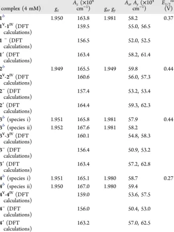
The EPR spectra of the solutions containing the electro-reduced species [{VVVIVO2(L)2}-μ-O]− were measured in Table 4. Spin Hamiltonian Parameters Obtained from 4.0
mM Solutions of the Reduced Forms of Compounds 1−4 in
MeCN and Those Calculated by Simulation of the
Experimental First-Derivative EPR Spectra Recorded at 77 K and DFT-Calculated Values for the Dinuclear Species [{VO(L1−4)}2-μ-O]−(1V-1IV, 2V-2IV, 3V-3IV, and 4V-4IV), Monomeric [VIVO(OEt)(L1−4)]−(Designated by 1−, 2−, 3−, and 4−), and [VIVO(L1−4)(EtOH)] (Designated by 1′, 2′, 3′, and 4′) Compoundsa complex (4 mM) gz Az(×104 cm−1) gx, gy Ax, Ay(×104 cm−1) E1/2red (V) 1b 1.950 163.8 1.981 58.2 0.37 1V-1IV(DFT calculations) 159.5 55.0, 56.5 1−(DFT calculations) 156.5 52.0, 52.5 1′ (DFT calculations) 163.4 58.2, 61.4 2b 1.949 165.5 1.949 59.8 0.44 2V-2IV(DFT calculations) 160.6 56.0, 57.3 2−(DFT calculations) 157.4 53.2, 53.4 2′ (DFT calculations) 164.4 59.3, 62.3 3b(species i) 1.951 165.8 1.981 57.9 0.44 3b(species ii) 1.952 167.6 1.981 58.2 3V-3IV(DFT calculations) 160.1 54.8, 58.3 3−(DFT calculations) 156.4 50.9, 53.2 3′ (DFT calculations) 163.4 57.2, 62.8 4b(species i) 1.951 165.1 1.980 58.7 0.27 4b(species ii) 1.950 167.0 1.980 59.4 4V-4IV(DFT calculations) 159.0 53.6, 57.5 4−(DFT calculations) 156.0 50.4, 53.0 4′ (DFT calculations) 163.2 57.0, 62.5
aThe column on the right displays the experimental E
1/2red(vs SCE)
obtained by CV studies. bFrom EPR spectra recorded after one-electron reduction for each of the two VVcenters present in solution.
Figure 7.Plots of the LUMO of the 1V-1Vand 4V-4Vcomplexes.
Figure 8.Spin-density distribution in 1V-1IVand 4V-4IVwith unrelaxed (A), partially optimized (B), and fully optimized (C) geometries.
liquid (at room temperature) as well as frozen (77 K) MeCN. The EPR spectra of frozen solutions of 3 and 4 after one-electron electroreduction for each of the two V centers indicate the presence of (at least) two species in solution. The frozen
solution EPR spectra were simulated,19 and the spin
Hamiltonian parameters thereby obtained are included in Table 4. The DFT calculated values of the 51V hyperfine coupling constants28 are in reasonable agreement with the experimental values (Table 4). Additionally, calculations demonstrate noticeable differences of the |Az| value for the
reduced mononuclear structures (i) [VIVO(OEt)(L)]−
[(156.0−157.4) × 10−4 cm−1] and (ii) [VIVO(L)(EtOH)] [(163.2−164.4) × 10−4 cm−1] and dinuclear structure [{VO-(L)}2-μ-O]−[(159.0−160.6) × 10−4cm−1].
The room temperature (300 K) X-band EPR spectra of complexes 1 and 2 (thus, 1V-1IV and 2V-2IV) after electro-reduction exhibited an 8-line pattern (Figures 9 and S8). In
these cases, the unpaired electron is localized on one of the V centers. In a frozen solution (77 K), the EPR spectra also exhibit patterns corresponding to a localized VIVO species. However, in the cases of 3 and 4, the EPR spectra at room temperature exhibit a 15-line pattern centered around g = 2.00, with Aisoof ca. 48 and 50 G (Figure 10). This corresponds to the g values and hyperfine pattern anticipated for delocalization
of a single, unpaired electron to two equivalent51V nuclei (2nI + 1 = 15 for two51V centers with I =7/2). These EPR spectra are indicative of delocalization of the unpaired electron over both metal centers; therefore, the generation of mixed-valence VIV−O−VV species is confirmed by EPR. Rather similar EPR spectra were reported by Dinda et al.18b and some other groups.17c,o,18a
Interestingly, in the ESI-MS (negative mode) obtained for 1−4, important peaks with m/z values closely corresponding to one-electron reduction of the dinuclear MV-MVcomplexes were detected. This confirms the plausibility and propensity of formation of both the MV-MV and MV-MIV species in these systems.
The formation of mixed-valence VV−O−VIVspecies is clearly demonstrated in these experiments for 3V-3IVand 4V-4IVfor the spectra at 77 K. It is noteworthy that the calculated Az values for the distinct reduced complexes MV-MIV, VIVO(OEt)(L)− (M−), and VIVO(L)(EtOH) (M′) differ significantly. The observed Az values obtained in the cases of 3 and 4 upon reduction mostly differ from these Azcalc values, with 3′ and 4′ being those that correspond to closer values, while the Azvalues obtained for 1 and 2 upon reduction are quite close to those of 1′ and 2′ (differences of less than 1%).
■
CONCLUSIONSFour substituted hydrazones H2L1−4 were prepared by the condensation of aroylhydrazines and naphthol derivatives and
characterized. When each of the H2L1−4 compounds was
reacted with the vanadium(V) precursor NH4VVO3, the
monomeric [VVO(OEt)(L)] complexes 1−4 were obtained
and characterized in the solid state and in solution by spectroscopic techniques (IR, UV−vis, 1H, 13C, and 51V NMR, and ESI-MS). Single-crystal XRD analysis of 1, 3, and 4confirmed coordination of each of the (L1−4)2−ligands with a O2N binding set, each corresponding to a distorted square pyramidal geometry around the VVcenter [τ values of 0.25 (1), 0.33 (3), and 0.19 (4)], where the O,N,O-donor atoms of L1−4 and the O ethoxido atoms constitute satisfactory O3N basal planes.
The structurally characterized [VVO(OEt)(L)] compounds transform in solution into the corresponding monooxido-bridged divanadium(V,V) compounds. Formation of the dinuclear μ-oxido species [{VVO(L1−4)}
2-μ-O] generated in
situ in solution was confirmed by several spectroscopic
techniques (IR and 1H, 13C, and 51V NMR) as well as by ESI-MS and DFT calculations. For example, in the case of
Figure 9.First-derivative EPR spectra of 1 in MeCN after CPE (upon passing the amount of charge corresponding to one-electron reduction for each of the two VV centers present): (a) spectrum at room temperature; (b) spectrum of the solution frozen at 77 K. An 8-line pattern is obtained at room temperature. The spin Hamiltonian parameters obtained for the spectrum at 77 K are inTable 4; the Aiso
value for the spectrum at room temperature is 93.2× 10⁻⁴cm−1.
Figure 10.First-derivative EPR spectra measured for solutions of 3 and 4 in MeCN after CPE (upon passing the amount of charge corresponding to one-electron reduction for each of the two VVcenters present): (a) spectra at room temperature; (b) spectra of the solutions frozen at 77 K. In both
cases, a 15-line pattern is obtained at room temperature. The spin Hamiltonian parameters obtained for the spectra at 77 K are inTable 4; the Aiso
values for the spectra at room temperature are 48× 10−4cm−1(for 3) and 50× 10−4cm−1(for 4).
compound 4, DFT allowed one to predict that its Gibbs free energy of formation of a dinuclear complex, 2[VVO(OEt)(L4)] + H2O⇆ [(VVOL4)2-μ-O] + 2EtOH, is only +2−3 kcal/mol in a DMSO solution, supporting the proposal that the dinuclear complex 4V-4Vmay be easily formed in a DMSO solution in a noticeable amount and that the addition of even a small amount of EtOH should be enough to shift this equilibrium towards its monomeric form, in agreement with what was observed in NMR (1H,13C, and51V) experiments.
The 8-line EPR pattern observed in the EPR spectra at 77 K
of the electrolyzed solutions of 1−4 showed that upon
reductive transfer of half an electron per mole to complexes, there was a reduction of VV to VIV. In the cases of the reductions with solutions of 3 and 4, a 15-line EPR pattern was recorded at room temperature, which indicates that the unpaired electron is extensively delocalized between the two V centers in the formed mixed-valence complex. In contrast, upon similar reductive transfer with solutions of 1 and 2, an 8-line EPR pattern was obtained at room temperature. It is plausible that mixed-valence compounds [{VO(L1−2)}2-μ-O]− formed but, in these two cases, with the unpaired electron localized on one of the V atoms. DFT calculations globally allowed rationalization of the data obtained for several systems. Moreover, the DFT-calculated Azvalues for the proposed VIV -containing species are in reasonable agreement with the experimental ones.
Further supporting the formation in solution of dinuclear
MV-MV species and their propensity to form MV-MIV
compounds is the detection of peaks in the ESI-MS (negative mode) of 1−4 with m/z values very closely matching those expected for oxido-bridged MV-MIVcomplexes.
The crystallographic data for the structural analysis of 1, 3, and 4 have been deposited with the Cambridge
Crystallo-graphic Data Centre (CCDC 1471661for 1,CCDC 1471662
for 3, and CCDC 1471663for 4). A copy of this information may be obtained free of charge from the CCDC, 12 Union Road, Cambridge CB2 1EZ, U.K. [Tel. +44 (0) 1223 762911; e-maildeposit@ccdc.cam.ac.uk].
■
ASSOCIATED CONTENT*
S Supporting InformationThe Supporting Information is available free of charge on the
ACS Publications website at DOI:
10.1021/acs.inorg-chem.6b01001.
IR spectra of 3 (Figure S1), UV−vis spectrum of 2
(Figure S2),13C NMR spectra of complex 1 (Figure S3), ESI-MS spectra of complexes 1, 3, and 4 (Figures S4−
S6), cyclic voltammograms of 1−4 (Figure S7), EPR
spectra of 2 (Figure S8), Cartesian atomic coordinates (Table S1) (PDF)
X-ray crystallographic data in CIF format for 1 (CIF) X-ray crystallographic data in CIF format for 3 (CIF) X-ray crystallographic data in CIF format for 4 (CIF)
■
AUTHOR INFORMATIONCorresponding Authors
*E-mail:joao.pessoa@ist.utl.pt(J.C.P.). *E-mail:rupamdinda@nitrkl.ac.in(R.D.). Author Contributions
The manuscript was written through contributions of all authors. All authors have given approval to thefinal version of the manuscript.
Notes
The authors declare no competingfinancial interest.
■
ACKNOWLEDGMENTSFunding for this research was provided by the Department of Science and Technology, India [Grants SR/WOS-A/CS-145/ 2011 (to S.P.D.), SR/FT/CS-016/2008 (to R.D.), and SB/FT/ CS-100/2013 (to A.K.)]. R.D. thanks Prof. H. Reuter, Prof. M. Nethaji, and Prof. S. Chattopadhyay for assistance with XRD analysis. J.C.P. thanks the Portuguese Foundation for Science and Technology [Grants UID/QUI/00100/2013, RECI/QEQ-QIN/0189/2012, RECI/QEQ-MED/0330/2012, and SFRH/ BPD/90976/2012 (to A.K.)].
■
REFERENCES(1) (a) Rehder, D. Bioinorganic Vanadium Chemistry; John Wiley & Sons, Ltd.: Chichester, U.K., 2008. (b) Crans, D. C.; Smee, J. J.; Gaidamauskas, E.; Yang, L. Chem. Rev. 2004, 104, 849−902. (c) Pessoa, J. C.; Etcheverry, S.; Gambino, D. Coord. Chem. Rev. 2015, 301−302, 24−48. (d) Costa Pessoa, J.; Garribba, E.; Santos, M. F. A.; Santos-Silva, T. Coord. Chem. Rev. 2015, 301−302, 49−86. (e) Kioseoglou, E.; Petanidis, S.; Gabriel, C.; Salifoglou, A. Coord. Chem. Rev. 2015, 301− 302, 87−105. (f) Rehder, D. Metallomics 2015, 7, 730−742.
(2) McLauchlan, C. C.; Peters, B. J.; Willsky, G. R.; Crans, D. C. Coord. Chem. Rev. 2015, 301−302, 163−199.
(3) Maurya, M. R.; Kumar, A.; Costa Pessoa, J. Coord. Chem. Rev. 2011, 255, 2315−2344.
(4) (a) da Silva, J. A. L.; da Silva, J. J. R. F.; Pombeiro, A. J. L. Coord. Chem. Rev. 2011, 255, 2232−2248. (b) Sutradhar, M.; Martins, L. M. D. R. S.; Guedes da Silva, M. F. C.; Pombeiro, A. J. L. Coord. Chem. Rev. 2015, 301−302, 200−239.
(5) Amadio, E.; Di Lorenzo, R.; Zonta, C.; Licini, G. Coord. Chem. Rev. 2015, 301−302, 147−162.
(6) Pellissier, H. Coord. Chem. Rev. 2015, 284, 93−110.
(7) (a) Noblía, P.; Baran, E. J.; Otero, L.; Draper, P.; Cerecetto, H.; González, M.; Piro, O. E.; Castellano, E. E.; Inohara, T.; Adachi, Y.; Sakurai, H.; Gambino, D. Eur. J. Inorg. Chem. 2004, 2004, 322−328. (b) Gambino, D. Coord. Chem. Rev. 2011, 255, 2193−2203.
(8) (a) Maurya, M. R.; Khan, A. A.; Azam, A.; Kumar, A.; Ranjan, S.; Mondal, N.; Pessoa, J. C. Eur. J. Inorg. Chem. 2009, 2009, 5377−5390. (b) Maurya, M. R.; Khan, A. A.; Azam, A.; Ranjan, S.; Mondal, N.; Kumar, A.; Avecilla, F.; Pessoa, J. C. Dalton Trans. 2010, 39, 1345− 1360. (c) Maurya, M. R.; Agarwal, S.; Abid, M.; Azam, A.; Bader, C.; Ebel, M.; Rehder, D. Dalton Trans. 2006, 937−947. (d) Maurya, M. R.; Kumar, A.; Bhat, A. R.; Azam, A.; Bader, C.; Rehder, D. Inorg. Chem. 2006, 45, 1260−1269. (e) Maurya, M. R.; Kumar, A.; Abid, M.; Azam, A. Inorg. Chim. Acta 2006, 359, 2439−2447. (f) Maurya, M. R.; Haldar, C.; Khan, A. A.; Azam, A.; Salahuddin, A.; Kumar, A.; Costa Pessoa, J. Eur. J. Inorg. Chem. 2012, 2012, 2560−2577. (g) Benítez, J.; Cavalcanti de Queiroz, A.; Correia, I.; Alves, M. A.; Alexandre-Moreira, M. S.; Barreiro, E. J.; Lima, L. M.; Varela, J.; González, M.; Cerecetto, H.; Moreno, V.; Pessoa, J. C.; Gambino, D. Eur. J. Med. Chem. 2013, 62, 20−27. (h) Fernández, M.; Varela, J.; Correia, I.; Birriel, E.; Castiglioni, J.; Moreno, V.; Costa Pessoa, J.; Cerecetto, H.; González, M.; Gambino, D. Dalton Trans. 2013, 42, 11900−11911.
(9) Rehder, D.; Costa Pessoa, J.; Geraldes, C.; Castro, M.; Kabanos, T.; Kiss, T.; Meier, B.; Micera, G.; Pettersson, L.; Rangel, M.; Salifoglou, A.; Turel, I.; Wang, D. JBIC, J. Biol. Inorg. Chem. 2002, 7, 384−396.
(10) Dash, S. P.; Pasayat, S.; Bhakat, S.; Roy, S.; Dinda, R.; Tiekink, E. R.; Mukhopadhyay, S.; Bhutia, S. K.; Hardikar, M. R.; Joshi, B. N.; Patil, Y. P.; Nethaji, M. Inorg. Chem. 2013, 52, 14096−14107.
(11) (a) Sutradhar, M.; Pombeiro, A. J. L. Coord. Chem. Rev. 2014, 265, 89−124. (b) Galloni, P.; Conte, V.; Floris, B. Coord. Chem. Rev. 2015, 301−302, 240−299. (c) Honzicek, J.; Vinklárek, J. Inorg. Chim. Acta 2015, 437, 87−94.


![Figure 5. ESI-MS spectrum of complex 2 in DMSO: m/z 387.08 (100%, [V V O 2 (L 2 )] − , MW = 387.02) and 758.05 (30%, [V V V IV O 3 (L 2 ) 2 ] − , MW = 758.48).](https://thumb-eu.123doks.com/thumbv2/123doknet/14950400.668056/10.892.467.831.461.727/figure-esi-ms-spectrum-complex-dmso-mw-iv.webp)