HAL Id: hal-03179496
https://hal.univ-angers.fr/hal-03179496
Submitted on 24 Mar 2021
HAL is a multi-disciplinary open access archive for the deposit and dissemination of sci-entific research documents, whether they are pub-lished or not. The documents may come from teaching and research institutions in France or abroad, or from public or private research centers.
L’archive ouverte pluridisciplinaire HAL, est destinée au dépôt et à la diffusion de documents scientifiques de niveau recherche, publiés ou non, émanant des établissements d’enseignement et de recherche français ou étrangers, des laboratoires publics ou privés.
Dodecyl creatine ester and lipid nanocapsule: a double
strategy for the treatment of creatine transporter
deficiency.
Alexandra Trotier-Faurion, Catherine Passirani-Malleret, Jérôme Bejaud,
Sophie Dézard, Vassili Valayannopoulos, Fréderic Taran, Pascale de Lonlay,
Jean-Pierre Benoit, Aloïse Mabondzo
To cite this version:
Alexandra Trotier-Faurion, Catherine Passirani-Malleret, Jérôme Bejaud, Sophie Dézard, Vassili Valayannopoulos, et al.. Dodecyl creatine ester and lipid nanocapsule: a double strategy for the treatment of creatine transporter deficiency.. Nanomedicine (London, England), 2014, 10, pp.185-91. �10.2217/nnm.13.205�. �hal-03179496�
part of
Dodecyl creatine ester and lipid
nanocapsule: a double strategy for
the treatment of creatine transporter
deficiency
Background: Creatine transporter (CT) deficiency is characterized by mutations in the gene encoding CT, leading to impaired transport of creatine at the cell membrane. Patients with this disease would thus benefit from replenishment of creatine inside the brain cells. Aim: We report a therapeutic strategy based on the use of dodecyl creatine ester incorporated into lipid nanocapsules (LNCs). Materials & methods: The dodecyl
creatine ester was incorporated in the shells of LNCs using Transcutol® (Gattefossé
SAS, Saint-Priest, France). The interactions of dodecyl creatine ester encapsulated in LNCs with an in vitro cell-based blood–brain barrier model was studied. The entry of the dodecyl creatine ester encapsulated in LNCs and the conversion of dodecyl creatine ester to creatine in the cells were also studied in the pathological context of CT deficiency. Results & discussion: We showed that these LNCs can cross the blood–brain barrier and enter brain endothelial cells. In human fibroblasts lacking functional CT, all or part of the dodecyl creatine ester was released from the LNCs and biotransformed to creatine, thus indicating the value of this strategy in this therapeutic context.
Original submitted 2 April 2013; Revised submitted 28 October 2013
Keywords: blood–brain barrier • creatine • creatine transporter deficiency • dodecyl creatine ester • lipid nanocapsule
Creatine metabolism deficiencies result from either an enzymatic defect of creatine bio synthesis (arginine:glycine amidinotrans ferase and guanidinoacetate methyltrans ferase deficiencies by recessive autosomal transmission) or a defect in the transport of creatine through cell membranes (Xlinked creatine deficiency caused by mutations in the SLC6A8 gene) [1]. In both cases, patients suffer from severe developmental disabil ities with language delay, extra pyramidal syndrome, behavioral disorders and some times epileptic seizures [2–4]. Therapies based on creatine supplementation or on creatine pre cursors larginine and lglycine significantly improve creatine bio synthesis deficiencies, in terms of both clinical and bio chemical findings. However, in a 4 to 6year follow up of patients suffering from
the creatine transporter deficiency, there was no clinical progress or increase in intra cerebral creatine levels [5]. Patients are thus in a clinical situation where the absence of a functional creatine transporter at the blood–brain barrier (BBB) and in the brain parenchyma cells, particularly in neurons and astrocytes, prevents the entry and dif fusion of creatine in the CNS [6–8]. There fore, an evaluation of new therapeutic strat egies for this cerebral metabolic disorder is now necessary. We previously reported that dodecyl creatine ester would be a good drug candidate to develop as a therapeutic option for patients suffering from creatine trans porter deficiency [9,101]. However, dodecyl creatine ester would be degraded by plasma esterases in all biological fluids. An efficient delivery system that targets dodecyl creatine
Alexandra Trotier-Faurion‡1,
Catherine Passirani2,3, Jérôme
Béjaud‡2,3, Sophie Dézard4,
Vassili Valayannopoulos5,
Fréderic Taran4, Pascale de
Lonlay5, Jean-Pierre Benoit2,3
& Aloïse Mabondzo*1
1CEA, Direction des Sciences du Vivant,
iBiTec-S, Service de Pharmacologie et d’Immuno Analyse, Equipe Pharmacologie Neurovasculaire, Gif-sur-Yvette, France
2L’Université Nantes Angers Le Mans–
Université d’Angers, F-49933, Angers, France
3INSERM U1066–Micro et
Nanomédecines Biomimétiques, Angers, France
4CEA, Direction des Sciences du Vivant,
iBiTec-S, Service de Chimie Bio-organique et de Marquage, Gif-sur-Yvette, France
5Centre de Référence des Maladies
Héréditaires du Métabolisme de l’Enfant et de l’Adulte, Hôpital Necker-Enfants Malades, Université Paris Descartes, Paris, France
*Author for correspondence: Tel.: +33 169 081 321 aloïse.mabondzo@cea.fr
10.2217/NNM.13.205 Nanomedicine (Epub ahead of print) future science group
Preliminary communication Trotier-Faurion, Passirani, Béjaud et al.
ester to the brain parenchyma has yet to be devel oped. By overcoming creatine transporter deficiency at the BBB, it would enable the delivery of dodecyl creatine ester inside brain cells thus restoring the cre atine pool and improving neuronal functions. Lipid nanocapsules (LNCs) have already been described as a promising approach to the specific delivery of lipophilic therapeutic agents [10–12], especially into the brain. Prepared according to a solventfree process, these spherical LNCs made of biocompatible materi als exhibit good stability in suspension. Their surface contains polyethylene glycol (PEG), which affects the vascular residence time of the nanocapsules, providing stealth properties. The PEG is contained in Solutol® HS15 (BASF, Levallois Perret, France),
used in the LNC preparation. The PEG is known to increase the vascular residence time of the LNCs and avoid the capture by the reticulo endothelial system. These two actions provide stealth properties to the LNC. This is required for an in vivo administration in order to get the best efficiency. Unpublished find ings from our laboratory demonstrate that such LNCs cross the BBB in an in vitro cellbased rat model [Trotier-Faurion A, Mabondzo A, Unpublished Data].
However, several investigations showed that the high susceptibility of dodecyl creatine ester to hydro lysis in aqueous media at high temperatures (above 37°C) rendered it unsuitable for the standard LNC preparation process. In this study, Transcutol®
(Gattefossé SAS, SaintPriest, France) was used to dissolve the dodecyl creatine ester at ambient temper ature and the mixture was added in the last heating step. Transcutol has interesting surfactant properties [13] and is thus incorporated into the shell of LNCs. The LNCs produced (dodecyl creatine ester encap sulated in LNCs [LNCC12]) have properties suit able for intravenous injection (size: 48.31 ± 1.77 nm; polydispersity index: 0.07 ± 0.01; zetapotential: 0.17 mV; LNCC12 theoretical concentration: 260 mg.ml1; and theoretical concentration of dode
cyl creatine ester: 300 µg.ml1). We then studied the
translocation of LNCC12 across an in vitro cell based rat BBB model [14] and the delivery of dodecyl creatine ester in endothelial and astroglial cells. We investigated whether, in the patho logical context of creatine transporter deficiency, entry of LNCC12 is possible and would result in an increase of the creatine pool in the cells.
Materials & methods
Dodecyl creatine ester synthesis
Dodecyl creatine ester was synthesized according to a process patented in July 2012 and experiments were conducted as described previously [9]. Briefly, the first
step consisted of an activation of the electrophilicity of the carbonyl moiety of creatinine by double protection of the cyclic guanidine (carbamate derivative). Then, in the presence of dodecanol, the creatinine ring opened spontaneously. A carbamate deprotection generated dodecyl creatine ester [9].
Preparation of LNCs
LNCs were prepared according to Heurtault et al. [15] with a minor modification derived from Roger et al. 2011 [13], consisting of the solubilization of 300 µg of dodecyl creatine ester in Transcutol added at the last heating step. Briefly, 0.3 g of Labrafac CC® (Gattefossé
SAS), 0.9 g of Labrafil M1944CS® (Gattefossé SAS),
1.0 g of Solutol HS15, 0.1 g of NaCl and 1.8 g of 4(2hydroxyethyl)1piperazineethanesulfonic acid buffer were heated at 90°C under magnetic stirring, then cooled to 60°C. Two cycles of progressive heat ing and cooling between 60 and 90°C were then per formed. At 90°C, just before the last cooling, 0.3 g of Transcutol containing the dodecyl creatine ester was added and at 75°C an irreversible shock was induced by dilution with cold 4(2hydroxyethyl)1pipera zineethanesulfonic acid buffer (5.6 ml at 2°C). The LNCs were analyzed for size distribution by photon correlation spectroscopy and zetapotential using the Malvern Zetasizer®, NanoSeries ZS (Malvern, Orsay,
France) after filtration through a 0.20µm filter from Sartorius (Les Ulis, France). The theoretical payload was 300 µg in 1 g of LNC suspension and the encap sulation efficiency was 90% corresponding to a final loading of 270 µg/g of the drug.
In vitro studies
BBB translocation of LNC-C12
The in vitro cellbased BBB model consisted of a coculture of primary rat endothelial and astroglial cells. Primary rat astroglial cells were seeded at a density of 2 × 10⁴ cells/well in 1500 µl on a 12well
plate. The astroglial culture medium was a mixture of minimum essential mediuma and Ham’s F12 nutrient mixture supplemented with 5% fetal bovine serum, 1% human serum, 1% penicillin/ streptomycin/neomycin and 0.4% FGF. A total of 24 h later, Costar® Transwell® (SigmaAldrich, Saint
Quentin Fallavier, France) inserts (pore size 0.4 µm; diameter 12 mm; surface area 1.12 cm²) were placed inside the wells and primary rat endothelial cells were plated on the upper layer at a density of 8 × 10⁴
cells/insert in 500 µl endothelial basal medium2 supplemented with the EGM®2MV kit (Lonza,
LevalloisPerret, France). The chambers containing endothelial cells and astroglial cells were considered as the apical and basolateral compartments, respectively.
The plates were incubated at 37°C in an atmosphere containing 5% CO2 and the BBB model formed confluent monolayers within 12 days [14,16]. After 12 days, the integrity of this BBB model was assessed. The apical and basolateral media were replaced by specific transport buffer (150 mM NaCl; 5.2 mM KCl; 2.2 mM CaCl2; 0.2 mM MgCl2; 6 mM NaHCO3; 2.8 mM glucose and 5 mM 4(2hydroxyethyl)1 piperazineethanesulfonic acid) without (negative control, vehicle) or with LNCC12 dissolved at 2, 5 and 10 mg.ml1. After 60 min of incubation,
cells’ incubation media were diluted fivefold in 95% acetonitrile/5% formic acid and cells were scraped in a mixture of 20% water/76% acetonitrile/4% formic acid. After centrifugation (13,000 × g, 10 min, 4°C), HPLC tandem mass spectrometry was used to detect the dodecyl creatine ester in each compartment and in endothelial and astroglial cell lysates. The integrity of the cellbased BBB models was demonstrated by measuring the flux of [14C]sucrose, [3H]vinblastine
and [3H]propranolol through the monolayer.
Transwells with rat endothelial cell monolayers were transferred to new 12well plates. A specific transport buffer was added: 500 µl to the apical compartment and 1500 µl to the basolateral compartment. After 60 min of incubation at 37°C of 0.1 µCi.ml1 [14C]labeled
sucrose, 1 µCi.ml1 [3H]propranolol in the apical
compartment and 0.1 µCi.ml1 [3H]vinblastine
in the apical and basolateral compartment, cells’ incubation media from both apical and basolateral compartments were collected. The amount of tracer that passed through the endothelial monolayer was determined by scintillation counting and the permeability of each compound X from the apical to the basolateral compartment (PappXA→B), and was
assessed using Equation 1:
[ ] [ ] P X T S X X V app A B basolateral B 0 # # # = " (Equation 1)
where X is the compound for which the permeabil ity is assessed, [Xbasolateral] is the concentration of the
compound X in the basolateral compartment at the end of the incubation, VB is the total volume of the
basolateral compartment (1.5 ml), T the time of the incubation, S is the transwell surface area and [X0]
is the concentration of compound X at initial time, T0. Validated BBB models have sucrose permeability below 8 × 106 cm.s1, propranolol permeability above
16 × 106 cm.s1 and a vinblastine permeability ratio
above 2.
The Lucifer yellow (LY) permeability test was used to study the effect of LNCC12 on BBB integrity. LY was diluted in transport buffer to a final concentration of 100 µM and added to the apical compartment
during LNCC12 incubation. Fluorescence leakage was determined for LY with 485 nm excitation and 530 nm emission using a fluorescence plate reader. The LY permeability was then calculated: a value below 5 × 106 cm.s1 indicated that the LNCC12
did not damage BBB integrity.
Uptake of LNC-C12 in fibroblasts
Human fibroblasts were obtained from skin biopsy specimens, a gift from the Centre de Référence des Maladies Héréditaires du Métabolisme at the Necker Hospital (Paris, France). Three patients with cerebral creatine deficiency caused by lack of creatine transporter and one control patient were studied. All of the mutations were previously described by Valayannopoulos et al. in 2013 [17]: p.Asn336del c.1006_1008delAAC (patient 1, DTp1) and p.(Gly499del) c.1497_1500delGAG (patient 2, VLp2) as P3 and P4, respectively, or by Valayannopoulos et al. in 2012 [18] as P2 for p.(G414del) c.1221_1223delTTC (patient 3, CTp3). The fibroblasts were plated out at 30,000 cells per well in sixwell plates in a DMEM supplemented with 10% fetal bovine serum, 1% penicillin/ streptomycin/neomycin, 1% sodium pyruvate and 1% lglutamine. They were cultured for 6 days by replacing the medium every 2–3 days. The incubation of LNCC12 consisted of replacing the medium by Hanks’ balanced salt solution in which the compound was diluted to 2, 5 and 10 mg.ml1.
After 1 h at 37°C, 5% CO2, the cells’ incubation media was removed and diluted fivefold in 95% acetonitrile/5% formic acid, and the cells were scraped in 20% water/76% acetonitrile/4% formic acid. After centrifugation (13,000 × g, 10 min, 4°C), HPLC tandem mass spectrometry was performed on supernatants to detect creatine and dodecyl creatine ester in the cell lysates.
HPLC tandem mass spectrometry identification of dodecyl creatine ester
Liquid chromatography (an HPLC system, LC20AD Shimadzu, MarnelaVallée, France) with a 2.0 × 150mm Uptisphere® Diol HPLC column
(UP6OH; Interchim, Montluçon, France) was used for elution of dodecyl creatine ester and creatine. The mobile phase was isocratic at 40/60 (detection of creatine) or 20/80 (detection of dodecyl creatine ester) solvent A/B (where solvent A was H2O containing 0.1% formic acid and solvent B was acetonitrile containing 0.1% formic acid); the flow rate was 0.4 ml/min. Analyte (10 µl) was injected onto the column placed in an oven at 40°C. The total run time was 6 min.
10.2217/NNM.13.205 Nanomedicine (Epub ahead of print) future science group
Preliminary communication Trotier-Faurion, Passirani, Béjaud et al.
Tandem mass spectrometry (Finnigan™ TSQ Quan tum Discovery with Xcalibur and LC Quan softwares; Thermo, Illkirch, France) in positive electrospray mode was used for detection. Spray voltage was 3.0 kV, and sheath and auxiliary gas pressures were 50 and 20 a.u., respectively. The insource collisioninduced dissocia tion energy was fixed at 12 V and the capillary tem perature was 350°C. The tube lens (creatine: 100 a.u.; dodecyl ester: 110 a.u.) and collision energy (creatine: 10 a.u.; dodecyl ester: 25 a.u.) values were optimized for each compound. Multiple reaction monitoring was used for the detection of the ion transitions: m/z 132.156 to 90.185 (creatine) or m/z 300.285 to 90.125 (dodecyl creatine ester). The standard curves showed linearity for creatine over a range of 0.05–10 and 0.01–05 µg.ml1
for dodecyl creatine ester. Creatine fatty esters and cre atine concentrations and amounts were determined in each compartment and in endo thelial cells, astroglial cells and fibroblast lysates. The amount of creatine fatty esters and the amount of creatine were standardized to the amount of protein in each lysate.
Results & discussion
Here, we show that the LNCC12 were incorporated into brain endothelial cells since we found 0.55 ± 0.14, 0.82 ± 0.43 and 0.87 ± 0.31 nmoles per mg of protein of dodecyl creatine ester in the endothelial cell lysates after a 60min incubation with 2, 5 and 10 mg.ml1 of LNCC12, respectively
(Table 1). No difference was detected between the three concentrations of LNCC12. Some LNCC12 were also able to diffuse through the BBB model and enter the glial cells (Table 1) since almost 2 nmoles per mg of protein of dodecyl creatine ester was found in the cell lysates for the three concentrations. This means that the dodecyl creatine ester had been protected from endothelial cell degradation by crossing the BBB. An interesting point is that even dodecyl creatine ester alone is also able to cross the BBB, but ester levels are almost twofold higher in glial cells than in endothelial cells when treated with LNCC12, whereas the reverse is true with the ester alone. This suggests that the LNCs would favor this
Table 1. Uptake of dodecyl creatine ester associated with lipid nanocapsules, in blood–brain barrier endothelial cells and blood–brain barrier glial cells, during translocation of dodecyl creatine ester encapsulated in lipid nanocapsules throughout the blood–brain barrier.
LNC-C12 concentration (mg.ml-1) Endothelial cells (nmoles/mg proteins) Astroglial cells (nmoles/mg proteins)
2 0.551 ± 0.14 2.550 ± 2.45
5 0.818 ± 0.43 2.262 ± 1.25
10 0.870 ± 0.31 1.394 ± 0.96
The amount of dodecyl creatine ester as nmoles per mg protein quantified in the blood–brain barrier endothelial cell lysate and blood–brain barrier glial cell lysate after an incubation with 2, 5 or 10 mg.ml-1 dodecyl creatine ester encapsulated in lipid nanocapsules.
LNC-C12: Dodecyl creatine ester encapsulated in lipid nanocapsules.
Endothelial cr eatine cont en t (nmoles/mg pr ot ein) 4 3 2 1 0 Vehicle 2 5 10 LNC-C12 concentration (mg.ml-1) Vehicle 2 5 10 * A str oglial cr eatine cont ent (nmoles/mg pr ot ein) 15 10 5 0 * A B LNC-C12 concentration (mg.ml-1)
Figure 1. Transport of dodecyl creatine ester encapsulated in lipid nanocapsules across an in vitro cell-based blood–brain barrier model. Creatine content in the blood–brain barrier (A) endothelial cell lysate and
(B) astroglial cell lysate. The amount of creatine is in nmoles per mg of protein quantified in blood–brain barrier
endothelial cell and astroglial lysates after a 60-min incubation with 2, 5 or 10 mg.ml-1 LNC-C12 compared with
the vehicle alone. The error bars represent the standard deviation. *p < 0.05 (analysis of variance plus Dunnett’s post-test).
passage to the brain and validates the applicability of LNCC12 to cross the BBB.
Not all of the tested conditions increased LY per meability, an internal standard of BBB integrity, as its values were below the limit range of 5 × 106 cm.s1[14]
(0.48 ± 0.10 × 10⁶, 0.41 ± 0.02 × 10⁶ and
0.82 ± 0.57 × 106 cm.s1 for 2, 5 and 10 mg.ml1
LNCC12, respectively, compared with the vehicle alone: 0.39 ± 0.17 × 106 cm.s1), suggesting that the
LNCC12 did not compromise the integrity of the
in vitro cellbased rat BBB model and that the cell
monolayer was intact.
When we quantified the creatine released from the dodecyl creatine ester, we showed that 2 mg.ml1
LNCC12 induced a slight increase of creatine content in the brain endothelial cell lysate (p < 0.05, Figure 1A). However, this was not the same for the higher concentrations and we could not find any dose–response correlation. No significant change was observed in the glial lysate creatine pool (Figure 1B). However, our experiments to analyze the esterase activity showed that they were functional so the glial cells were able to cleave the ester. Despite this lack of evidence of an increase in creatine in glial cells, we detected an increase of creatine in the apical and basolateral cells’ incubation media. Albeit detectable, this increase was not quantifiable because it was under the lowest limit of quantification of the bioanalytical method.
First, this indicates that as we tested the LNCC12 in a nonpathological BBB model, the creatine produced by the enzymatic conversion of dodecyl creatine ester could have been excreted in extracellular media through the functional SLC6A8, preventing us from determining an increase in creatine content in glial cells. Second, as the metabolic pathways in these cells were functional, the creatine released from the dodecyl creatine ester could have been involved in the metabolic pathway of the creatine/phosphocreatine shuttle. This is why we studied internalization of LNCC12 in a pathological human fibroblast model expressing a nonfunctional SLC6A8.
To confirm the relevance of our strategy, since our interest resided in developing a new pharmacological strategy to treat creatine transporter deficiency, we evaluated the release of dodecyl creatine ester and its conversion to creatine in human fibroblasts lacking functional creatine transporter. First, we demonstrated that LNCC12 was equivalently incorporated in fibroblasts from both control patients (28.9 ± 0.68 and 22.0 ± 1.27% of initial quantity) and patients with creatine transporter deficiency: DTp1 (27.7 ± 1.85 and 19.2 ± 0.86%), VLp2 (20.3 ± 8.12 and 19.3 ± 1.18%) and CTp3 (8.85 ± 0.34 and 18.2 ± 0.89%) at concentrations of 5 and 10 mg.ml1, respectively. This
indicates that the LNCC12 did not depend on creatine transporter to enter the cells. A significant increase in creatine content was detected in both nonpathological and pathological fibroblasts (Figures 2 & 3). This led
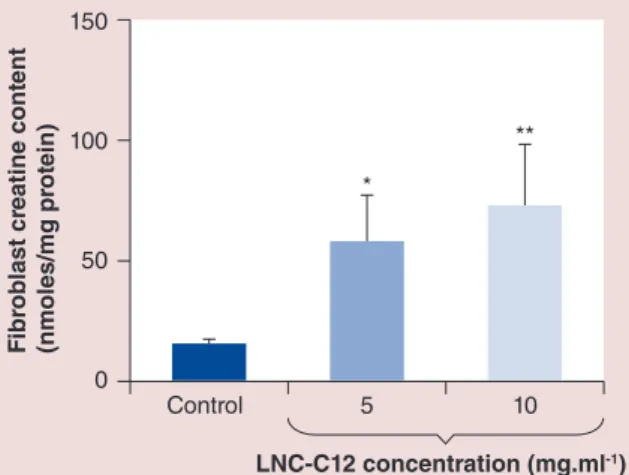
Fi br oblast cr eatine cont ent (nmoles/mg pr ot ein) 150 100 50 0 * ** Control 5 10 LNC-C12 concentration (mg.ml-1)
Figure 2. Creatine content in human fibroblasts of a control patient. A 60-min incubation of 5 or 10 mg.ml-1
LNC-C12 led to a significant increase compared with the vehicle alone in creatine content in fibroblasts of the control patient. The error bars represent standard deviation.
*p < 0.05, **p < 0.005 (analysis of variance plus Dunnett’s post-test).
LNC-C12: Dodecyl creatine ester encapsulated in lipid nanocapsules. Fi br oblast cr eatine cont ent (nmoles/mg pr ot ein) 15 10 5 0 C 5 10 C 5 10 C 5 10 LNC-C12 (mg.ml-1) LNC-C12 (mg.ml-1) LNC-C12 (mg.ml-1) ** ** ** *** *** *** DTp1 VLp2 CTp3
Figure 3. Significant increase of creatine content in human fibroblasts showing SLC6A8 deficiency.
A 60-min incubation of 5 or 10 mg.ml-1 LNC-C12
(concentration of LNC-C12 in mg.ml-1) led to a
significant increase compared with the control corresponding to untreated cells in creatine content in fibroblasts of patients with nonfunctional creatine transporter. The error bars represent standard deviation.
**p < 0.005, ***p < 0.001 (analysis of variance plus Dunnett’s post-test).
C: Control; LNC-C12: Dodecyl creatine ester encapsulated in lipid nanocapsules.
10.2217/NNM.13.205 Nanomedicine (Epub ahead of print) future science group
Preliminary communication Trotier-Faurion, Passirani, Béjaud et al.
to the assumption that, once the LNCC12 entered the fibroblasts, all or part of the dodecyl creatine ester released was biotransformed to creatine by esterases, which points to the great potential of this system in this particular therapeutic context. The same positive results were obtained with the dodecyl creatine ester alone, but it cannot be used in human therapy due to its degradation in the plasma.
These results suggest that our dodecyl creatine ester delivery system seems to be of particular interest in the pathological context of creatine transporter deficiency. Addition of Transcutol to the formulation is the only way to solubilize the dodecyl creatine ester, which has an amphiphilic chemical structure.
LNCC12 cross the BBB in vitro and deliver dodecyl creatine ester to the astrocytes in the brain parenchyma compartment. Although it did not increase the intra cellular creatine pool in nonpathological brain cells, LNCC12 limited the efflux of creatine outside of these cells compared with the dodecyl creatine ester alone. By contrast, in human fibroblasts from patients with creatine transporter deficiency, LNCC12 increased levels of creatine, the essential energy compound. We strongly believe that LNCC12 should be further investigated in in vivo models to assess the protection of dodecyl creatine ester from esterases. This formulation would be of particular interest in a twostep therapeutic strategy: first, the LNCC12 would cross the BBB and could be delivered itself or just release dodecyl creatine ester into the brain parenchyma; and second, in both cases, LNCC12 or dodecyl creatine ester
would penetrate neuronal cells and increase their creatine content, thus restoring neuronal functions in creatine transporter deficiency.
Future perspective
This study reports the first double strategy to overcome the BBB and increase the delivery of dodecyl creatine ester into the brain parenchyma. We describe here the in vitro proof of concept for the treatment of the creatine transporter deficiency by LNCC12. In the future, it will be of major interest to evaluate the therapeutic efficiency of this new device in an in vivo model of the pathology (SLC6A8/ knockout mouse
model [19]). The LNCC12 could be fluoro or radio labeled in order to be detected in vivo and follow the therapeutic efficiency of the dodecyl creatine ester.
Acknowledgements
The authors thank Mickaël Kempf for his liquid chromatography – tandem mass spectrometry support and the patients included in this study.
Financial & competing interests disclosure
This work was supported by the Fondation Jérôme Lejeune and by the CEA, Life Division of Sciences, Institut of Biology and Technology of Saclay (Gif-sur-Yvette, France). The authors have no other relevant affiliations or financial involvmement with any organization or entity with a financial interest in or financial conflict with the subject matter or materials discussed in the manuscript.
No writing assistance was utilized in the production of this manuscript.
References
Papers of special note have been highlighted as: l of interest
l l of considerable interest
1 Stockler S, Schutz PW, Salomons GS. Cerebral creatine deficiency syndromes. Clinical aspects, treatment and pathophysiology. Subcell. Biochem. 46, 149–166 (2007). l Overview of the clinical features of creatine deficiency
syndromes.
2 Schulze A. Creatine deficiency syndromes. Mol. Cell. Biochem. 244(1), 143–150 (2003).
l l First study to report a link between the defect in the
X-linked creatine transporter and developmental delay, mild epilepsy and several language impairments.
3 Degrauw TJ, Cecil KM, Byars AW, Salomons GS, Ball WS, Jakobs C. The clinical syndrome of creatine transporter deficiency. Mol. Cell. Biochem. 244(1–2), 45–48 (2003).
Executive summary
• The surfactant Transcutol® (Gattefossé SAS, Saint-Priest, France) dissolves the dodecyl creatine ester, an amphiphilic chemical structure, at ambient temperature and thus led to the incorporation of the creatine prodrug in the shell of lipid nanocapsules (LNCs) with suitable intravenous injection properties (size: 48.31 ± 1.77 nm; polydispersity index: 0.07 ± 0.01; and zeta-potential: -0.17 mV).
• This new device (dodecyl creatine ester encapsulated in LNCs) crosses the blood–brain barrier and delivers dodecyl creatine ester to the brain parenchyma, even in the context of a nonfunctional creatine transporter.
• In human fibroblasts from patients with creatine transporter deficiency, dodecyl creatine ester encapsulated in LNCs increased levels of creatine – the essential energy compound.
4 PooArguelles P, Arias A, Vilaseca Ma et al. XLinked creatine transporter deficiency in two patients with severe mental retardation and autism. J. Inherit. Metab. Dis. 29(1), 220–223 (2006).
5 Van De Kamp JM, Pouwels PJ, Aarsen FK et al. Longterm followup and treatment in nine boys with Xlinked creatine transporter defect. J. Inherit. Metab. Dis. 35(1), 141–149 (2012).
6 Ohtsuki S, Tachikawa M, Takanaga H et al. The blood– brain barrier creatine transporter is a major pathway for supplying creatine to the brain. J. Cereb. Blood Flow Metab. 22(11), 1327–1335 (2002).
l l First study to report the major contribution of creatine
transporter at the blood–brain barrier in supplying creatine to the brain.
7 Braissant O, Henry H, Beard E, Uldry J. Creatine deficiency syndromes and the importance of creatine synthesis in the brain. Amino Acids 40(5), 1315–1324 (2011).
8 Lunardi G, Parodi A, Perasso L et al. The creatine transporter mediates the uptake of creatine by brain tissue, but not the uptake of two creatinederived compounds. Neuroscience 142(4), 991–997 (2006).
9 TrotierFaurion A, Dezard S, Taran F, Valayannopoulos V, De Lonlay P, Mabondzo A. Synthesis and biological evaluation of new creatine Fatty esters revealed dodecyl creatine ester as a promising drug candidate for the treatment of the creatine transporter deficiency. J. Med. Chem. 56(12), 5173–5181 (2013).
l l Reports a new promising drug candidate for the treatment
of the creatine transporter deficiency.
10 Lamprecht A, Bouligand Y, Benoit JP. New lipid nanocapsules exhibit sustained release properties for amiodarone. J. Control. Release 84(1–2), 59–68 (2002).
11 Laine AL, Huynh NT, Clavreul A et al. Brain tumour targeting strategies via coated ferrociphenol lipid nanocapsules. Eur. J. Pharm. Biopharm. 81(3), 690–693 (2012).
12 Huynh NT, Passirani C, Saulnier P, Benoit JP. Lipid nanocapsules: a new platform for nanomedicine. Int. J. Pharm. 379(2), 201–209 (2009).
l Describes the promising nanovector for oral administration.
13 Roger E, Lagarce F, Benoit JP. Development and characterization of a novel lipid nanocapsule formulation of Sn38 for oral administration. Eur. J. Pharm. Biopharm. 79(1), 181–188 (2011).
14 Lacombe O, Videau O, Chevillon D et al. In vitro primary human and animal cellbased blood–brain barrier models as a screening tool in drug discovery. Mol Pharm 8(3), 651–663 (2011).
15 Heurtault B, Saulnier P, Pech B, Proust JE, Benoit JP. A novel phase inversionbased process for the preparation of lipid nanocarriers. Pharm. Res. 19(6), 875–880 (2002).
16 Brun E, Carriere M, Mabondzo A. In vitro evidence of dysregulation of blood–brain barrier function after acute and repeated/longterm exposure to TiO(2) nanoparticles. Biomaterials 33(3), 886–896 (2012).
17 Valayannopoulos V, Bakouh N, Mazzuca M et al. Functional and electrophysiological characterization of four non truncating mutations responsible for creatine transporter (SLC6A8) deficiency syndrome. J. Inherit. Metab. Dis. 36(1), 103–112 (2013).
18 Valayannopoulos V, Boddaert N, Chabli A et al. Treatment by oral creatine, larginine and lglycine in six severely affected patients with creatine transporter defect. J. Inherit. Metab. Dis. 35(1), 151–157 (2012).
19 Skelton Mr, Schaefer Tl, Graham Dl et al. Creatine
transporter (CrT; Slc6a8) knockout mice as a model of human CrT deficiency. PLoS ONE 6(1), e16187 (2011).
l l First study to report the in vivo model of the creatine
transporter deficiency.
Patent
101 Dezard D, Taran F, TrotierFaurion A, Mabondzo A: EP2692719A1 (2014).