HAL Id: inserm-01192850
https://www.hal.inserm.fr/inserm-01192850
Submitted on 3 Sep 2015
HAL is a multi-disciplinary open access
archive for the deposit and dissemination of
sci-entific research documents, whether they are
pub-lished or not. The documents may come from
teaching and research institutions in France or
abroad, or from public or private research centers.
L’archive ouverte pluridisciplinaire HAL, est
destinée au dépôt et à la diffusion de documents
scientifiques de niveau recherche, publiés ou non,
émanant des établissements d’enseignement et de
recherche français ou étrangers, des laboratoires
publics ou privés.
Madrid
Beatriz Lara, Maria Teresa Martínez, Ignacio Blanco, Cristina
Hernández-Moro, Eladio Velasco, Ilaria Ferrarotti, Francisco Rodriguez-Frias,
Laura Perez, Irene Vazquez, Javier Alonso, et al.
To cite this version:
Beatriz Lara, Maria Teresa Martínez, Ignacio Blanco, Cristina Hernández-Moro, Eladio Velasco, et al..
Severe alpha-1 antitrypsin deficiency in composite heterozygotes inheriting a new splicing mutation
QO Madrid. Respiratory Research, BioMed Central, 2014, 15, pp.125. �inserm-01192850�
R E S E A R C H
Open Access
Severe alpha-1 antitrypsin deficiency in
composite heterozygotes inheriting a new
splicing mutation QO
Madrid
Beatriz Lara
1, Maria Teresa Martínez
2, Ignacio Blanco
3, Cristina Hernández-Moro
4, Eladio A Velasco
4,
Ilaria Ferrarotti
5, Francisco Rodriguez-Frias
6, Laura Perez
7, Irene Vazquez
7, Javier Alonso
8, Manuel Posada
9and Beatriz Martínez-Delgado
7*Abstract
Background: Severe Alpha-1 Antitrypsin (AAT) deficiency is a hereditary condition caused by mutations in the SERPINA1 gene, which predisposes to lung emphysema and liver disease. It is usually related to PI*Z alleles, and less frequent to rare and null (QO) alleles. Null-AAT alleles represent the end of a continuum of variants associated with profound AAT deficiency and extremely increased risk of emphysema.
Methods: A family with severe AAT deficiency was analyzed to achieve genetic diagnosis. The complete exons and introns of the SERPINA1 gene were sequenced and transcriptional analysis by RT-PCR was performed to characterize the effect of splicing variants found in the patients. In addition, a minigene MGserpa1_ex1b-1c was cloned into the pSAD vector to in vitro investigate the independent impact of variants on splicing process.
Results: We report a new identified null allele (PI*QOMadrid) in two adult siblings with practically no detectable
serum AAT. The PI*QOMadridallele consist of a duplication of the thymine (T) in position +2 of the donor splice site
of exon 1C (+2dupT). In these two subjects, PI*QOMadridoccurred in compound heterozygote combination with the
previously described variant PI*QOPorto. Both QOMadridand QOPortovariants are located very close together in a
regulatory region of the SERPINA1 gene. Analysis of transcripts revealed that QOMadridvariant prevented the
expression of transcripts from exon 1C, and then normally spliced RNA products are not expected in the liver of these patients. In addition, aberrant splicing patterns of both variants were clearly distinguished and quantified by functional in vitro assays lending further support to their pathogenicity.
Conclusion: Finding pathogenic mutations in non-coding regions of the SERPINA1 highlight the importance that regulatory regions might have in the disease. Regulatory regions should be seriously considered in discordant cases with severe AAT deficiency where no coding mutations were found.
Keywords: Alpha-1 antitrypsin, Allelic variants, Null alleles, QO alleles, Splicing, Minigenes Background
Human alpha-1 antitrypsin (AAT), also named alpha-1 protease inhibitor (α1-PI) and SERPINA1 (Serine Protease Inhibitor, group A, member 1), is a circulating glycoprotein with a broad spectrum antiserine-protease activity, includ-ing the inhibition of free elastase from neutrophil. AAT
acts mainly as an acute phase reactant but also has anti-inflammatory, anti-infectious and immunomodulation effects [1,2]. Deficiency of AAT leads to lung tissue damage and emphysema due to uncontrolled elastase activity, or liver disease caused by accumulation within the hepatocytes of misfolded, aggregated AAT protein [3].
Severe AAT deficiency is an inherited condition char-acterized by AAT serum levels below 35% (or 50 mg/dL) the normal value. The protein is codified at the protease inhibitor locus (14q32.1), by theSERPINA1 gene, which is organized into three non-coding exons (1A, 1B and
* Correspondence:bmartinezd@isciii.es
7Molecular Genetics Unit, Instituto de Investigación en Enfermedades Raras
(IIER), Instituto de Salud Carlos III (ISCIII), Carretera Majadahonda-Pozuelo Km 2,200, Majadahonda, Madrid 28220, Spain
Full list of author information is available at the end of the article
© 2014 Lara et al.; licensee BioMed Central Ltd. This is an Open Access article distributed under the terms of the Creative Commons Attribution License (http://creativecommons.org/licenses/by/4.0), which permits unrestricted use, distribution, and reproduction in any medium, provided the original work is properly credited. The Creative Commons Public Domain Dedication waiver (http://creativecommons.org/publicdomain/zero/1.0/) applies to the data made available in this article, unless otherwise stated.
1C) and four coding exons (2–5) [4]. The SERPINA1 gene is inherited following an autosomal co-dominant pattern [5].
Over 80% of AAT is synthesized in the liver, although other cell types such as blood monocytes, neutrophils, macrophages, pancreas, endothelium, enterocytes, lung alveolar and some cancer cells are also capable of secret-ing additional quantities [6,7]. Transcriptional regulation occurs in at least two different sites within the gene: the hepatocyte promoter located upstream the transcription start site of exon 1C, and the monocyte promoter located upstream of exon 1A [8].
More than one hundred different genetic variants have been already described in detail and a wide knowledge about how these alterations affect the protein conform-ation and function has been learnt in the last decades [9]. Allelic variants of AAT are conventionally classified as normal and deficient [1,3,5,10]. The most common normal AAT alleles are PI*M1 (rs6647; NM_000295.4: c.710 T > C; p.Val213Ala, mature protein), PI*M2 (rs709932; c.374G > A; p.Arg101His) and PI*M3 (rs1303; c.1200A > C; p.Glu376Asp). The allelic frequency of these polymorphisms varies among populations being M1 (24%), M2 (13%), and M3 (23%) (minor allele frequency source:1000 Genomes). In contrast to normal AAT alleles, there are two categories of genetic variants that cause AAT deficiency: the deficient and the null alleles. The most common deficient alleles are PI*S (rs17580: c. 863A > T; p. Glu264Val) and PI*Z (rs28929474; c.1096G > A; p.Glu342Lys), with a PI*S prevalence in Caucasians of 5-10% [11] (3%, 1000 Genomes) and a PI*Z prevalence of 1-3% (0.7%, 1000 Genomes). Normal serum levels are associated with M alleles. In contrast reduced levels are associated with the PI*S and PI*Z alleles with AAT serum levels of 40% and 10-20%, respectively. Then, the PI*Z allele is related to severe deficiency and is the phenotype most often associated with the disease.
There are also other rare variants, with a lower fre-quency ranging from 1 × 10⁻⁴to 2.5 × 10⁻⁵, and around 15% serum AAT [10,12]. Both PI*S and PI*Z, and rare deficiency alleles MMalton, MDuarte, and SIiyama produce
misfolded proteins which are retained into hepatocytes forming polymers, which can cause cell stress and liver damage, and on the other hand, as a result of polymerization/retention into hepatocytes, reduced blood and tissues concentration of AAT, insufficient to protect tissues against proteases [9,13].
In the last two decades about 25 null variants, associated with trace amounts (<1%) of plasma AAT, have been discovered (Table 1) [4,10,12,14-26]. Although little information about their prevalence is available, it is thought that they are extremely infrequent, with an estimated combined frequency of 1.4 × 10⁻⁴among Caucasians. In
fact, the few published cases of carriers of null alleles have been found on European and European-American individ-uals, with only three carriers found in descendants of Egyptians, African-Americans and Chinese. Notably, des-pite their low prevalence, null variants have a strong effect on phenotype, conferring an extraordinary risk to develop severe pulmonary emphysema [19,27,28]. The majority of these variants cause premature stop codons in the SERPINA1 gene leading to either unstable mRNA or trun-cated, unstable proteins (i.e. QOGranite Falls, QOMattawa,
QOHong Kong, QOBellingham). Other mechanisms include
complete gene deletion (QOIsola di Procida), missense
mutations which probably destabilize the AAT protein (QOLudwigshafen and QONew Hope), or mutations affecting
the RNA splicing process (QOWest, QOBonny Blue, QOPorto)
[4,29]. Although infrequent, AAT null variants have allowed a better understanding of the molecular basis of the disease and revealed functionally critical regions of the gene sequence [27].
Although other previously described AAT null alleles have been reported in Spain [10], now we describe the first new AAT null allele in Spain, in a Caucasian fam-ily from Madrid, designated as QOMadrid, as this city
was the place of birth and residence of the index-case, as well as his three siblings and parents. These family cases showed a combination of two rare splicing vari-ants in the donor splice site of intron 1C, QOPortoand
the new allele QOMadrid. Molecular characterization of
these mutations located in regulatory region of the gene, allowed as to better understand the mechanisms of transcription and alternative splicing of SERPINA1 gene.
Methods
Patients
The family was composed by four siblings, three males and one female, two of them PI*QOPorto/QOMadrid
compound heterozygotes with severe AAT deficiency, and the remaining ones PI*M/QOPorto as well
hetero-zygotes but with moderate AAT deficiency (Figure 1). Their parents died years ago, and none of them had de-scendants or other blood relatives. Signed informed consent for the study was obtained from patients.
All these patients were previously characterized for AAT serum levels, AAT protein phenotype and genotyping of the common PI*S and PI*Z alleles. The determination of serum AAT was conducted by immunonephelometry with an autoanalyzer ArrayTM Protein System (Beckman-In-struments, Brea, California, USA).
Protease inhibitor (PI) typing was made by means of isoelectric focusing (IEF) technique as previously de-scribed [10,35]. All four siblings were studied for the existence of AAT protein variants by IEF but conclusive patterns were not found.
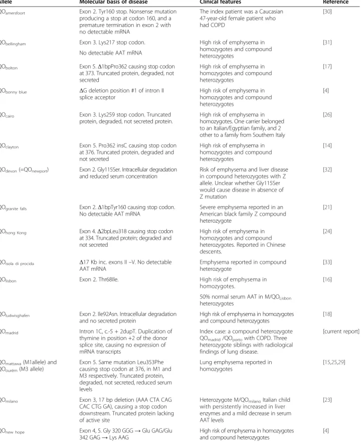
Table 1 Molecular and clinical features of known 21 PI*QO (Null) alleles
Allele Molecular basis of disease Clinical features Reference
QOamersfoort Exon 2. Tyr160 stop. Nonsense mutation
producing a stop at codon 160, and a premature termination in exon 2 with no detectable mRNA
The index patient was a Caucasian 47-year-old female patient who had COPD
[30]
QObellingham Exon 3. Lys217 stop codon. High risk of emphysema in
homozygotes and compound heterozygotes
[31] No detectable AAT mRNA
QObolton Exon 5.Δ1bpPro362 causing stop codon
at 373. Truncated protein, degraded, not secreted
High risk of emphysema in homozygotes and compound heterozygotes
[17] QObonny blue ΔG deletion position #1 of intron II
splice acceptor
High risk of emphysema in homozygotes and compound heterozygotes
[4] QOcairo Exon 3. Lys259 stop codon. Truncated
protein, degraded, not secreted protein.
High risk of emphysema in homozygotes. One carrier belonged to an Italian/Egyptian family, and 2 other to a family from Southern Italy
[26]
QOclayton Exon 5. Pro362 insC causing stop codon
at 376. Truncated protein, degraded and not secreted
High risk of emphysema in homozygotes and compound heterozygotes
[14] QOdevon(=QOnewport) Exon 2. Gly115Ser. Intracellular degradation
and reduced serum concentration
Risk of emphysema and liver disease in compound heterozygotes with Z allele. Unclear whether Gly115Ser would cause disease in absence of Z mutation
[32]
QOgranite falls Exon 2.Δ1bpTyr160 causing stop codon.
No detectable AAT mRNA
Severe emphysema reported in an American black family Z compound heterozygote
[21] QOhong Kong Exon 4.Δ2bpLeu318 causing stop codon
at 334. Truncated protein; degraded and not secreted
High risk of emphysema in homozygotes and compound heterozygotes. Reported in Chinese descents.
[24]
QOisola di procida Δ17 Kb inc. exons II –V. No detectable
AAT mRNA
Emphysema reported in compound heterozygote
[33] QOlisbon Exon 2. Thr68Ile. High risk of emphysema in
homozygotes.
[16] 50% normal serum AAT in M/QOLisbon
heterozygotes QOludwisghafen Exon 2. Ile92Asn. Intracellular degradation
and no secreted protein
High risk of emphysema in homozygotes and compound heterozygotes
[18] QOmadrid Intron 1C, c.-5 + 2dupT. Duplication of
thymine in position +2 of the donor splice site, causing no expression of mRNA transcripts
Index case: a compound heterozygote QOmadrid/QOportowith COPD. Three
heterozygote siblings with radiological findings of lung disease.
[current report]
QOmattawa(M1allele) and
QOourém(M3 allele)
Exon 5. Same mutation Leu353Phe causing stop codon at 376, in M1 and M3 respectively. Truncated protein, degraded, not secreted, reduced serum levels
Lung emphysema reported in homozygotes
[15,25,29]
QOmilano Exon 3, 17 bp deletion (AAA CTA CAG
CAC CTG GA), causing a stop codon downstream. Truncated protein lacking of active site
Heterozygote M/QOmilanoItalian child
with persistently increased in liver enzymes and a mild decrease in serum AAT levels
[23]
QOnew hope Exon 4, 5. Gly 320 GGG→ Glu GAG/Glu
342 GAG→ Lys AAG
High risk of emphysema in homozygotes and compound heterozygotes
Table 1 Molecular and clinical features of known 21 PI*QO (Null) alleles (Continued)
QOporto Intron 1C, c.-5 + 1G > A. Splicing site
variant, causing no expression of mRNA transcripts.
High risk of emphysema in homozygotes. [29] QOriedenburg Whole gene deletion. No AAT gene
expression
High risk of emphysema in homozygotes and compound heterozygotes
[22] QOsaarbueken Exon 5. 1158dupC causing stop codon
at 376. Truncated protein; not secreted
High risk of emphysema in homozygotes.
[16] 50% normal serum AAT in
M/QOSaarbuekenheterozygotes
QOsoest Exon 2. Thr102delA, which produces a
TGA stop signal at codon 112
Index case: a homozygote 46-year-old man with severe COPD
[30] QOtrastevere Exon 3. Try194 stop codon. Intracellular
degradation of truncated protein; not secreted
Emphysema reported in an Italian compound heterozygote
[20]
QOwest G→ T position +1 of intron 2 splice
donor substitution.ΔGly164- Lys191. Aberrant mRNA splicing, intracellular degradation and no detectable protein
Emphysema reported in a compound heterozygote
[34]
Moderate DAAT
Severe DAAT
II-1
II-2
II-3
II-4
I-1
I-2
67 mg/dL
72 mg/dL
8,7 mg/dL
9 mg/dL
[AAT]:
QOPorto M2(A) M3 (C) QOPorto M2 (A) M3 (C) QOMadrid QOPorto M2 (A) M3 (C) QOMadrid QOPorto M2(A) M3 (C)Figure 1 Pedigree of the family studied. Both parents (I-1 and I-2) were dead when the AAT study was performed. The four siblings studied correspond to 3 males (II-1, II-2 and II-4) and a female (II-3). The index case is II-4, indicated by the arrow. None of them had any children. Allele combination of the QOPortoand QOMadridmutations and the normal variants, M2 (G/A, Arg101His) and M3 (A/C, Glu376Asp) found in each
In addition, all subjects were initially genotyped for the commonest defective variants PI*Z and PI*S by a LightCycler PCR, using primers and hybridization probes as described before [36]. None of these com-mon deficient variants were found in our cases.
Sequence analysis of the entireSERPINA1 gene
Since the low serum AAT concentration of the subjects could not be attributed to PI*Z genotypes, the entire coding sequence of the SERPINA1 gene was analyzed (Reference sequences NG_008290.1, NM_000295.4). Exons 2 to 5 and exon-intron junctions were analyzed by means of a Sanger automated sequencing, using previously described primers [37]. After that, to exclude the exist-ence of other rare variants outside coding regions of the gene, primers for amplification of additional gene fragments to cover the whole gene were designed (Additional file 1: Table S1). The entire gene sequence were analyzed by Sanger automated sequencing (ABI PRISM 377 Applied BioSystems) in the four studied in-dividuals of the family.
Expression analysis by RT-PCR
RNA extraction from peripheral blood was performed using RNAeasy kit (Quiagen) following manufacturer’s recommendations. Then, cDNA synthesis was carried out by reverse transcription PCR (RT-PCR) using the Maxima First Strand cDNA Synthesis kit (Thermo Scientific). To analyze transcripts and splicing (from normal hepatocytes and peripheral blood samples) oc-curring in patients and controls, the regions between E1A/E2, E1B/E2 and E1C/E2 or E1C/E5 were amplified using several primers located in exon 1A, 1B, 1C, exon 2 and exon 5 as follows: E1A-F: 5′-TCCTGTGCCTGCC AGAAGAG-3′; E1B-F, 5′-ATCAGGCATTTTGGGGTG ACT-3′ [29]; E1C, 5′CTGTCTCCTCAGCTTCAGGC3′; E2-R, 5′-TTCTTGGCCTCTTCGGTGTC-3′ and E5-R, 5′-CCATGAAGAGGGGAGACTTGG-3′. In addition, a primer localized in the initial region of intron 1C INT1C, 5′-GGGGATGGAGAATGTGAGCC-3′ was also used to analyze the existence of possible transcripts using intron sequence in mutant cases. PCR amplification of the different products was performed under the following conditions: 35 cycles of 94°C for 45 s, 60°C for 45 s, and 72°C for 45 s. Amplified products were visualized in 1-2% agarose gels, purified by PCR purification Kit (Qiagen) and subsequently cloned into pGEM-T easy vector (Promega, Madison WI, USA) or directly ana-lyzed by sequencing with an ABI PRISM 377 sequen-cer. Ligation reactions were used to transform DH5α competent cells. Clones containing PCR products were selected by blue/white colony and standard ampicillin selection. Positive transformants were analyzed by PCR and sequenced using AAT primers.
Computational predictions
By using different prediction methods integrated in Alamut 1.3 software we evaluated splice signal detec-tion (SpliceSiteFinder-like, MaxEntScan, GeneSplicer, Human Splicing Finder or Known constitutive signals) and Exonic Splicing Enhancers ESE binding site detection (ESEFinder) comparing reference and mutated sequences.
Construction of the Minigene MGserp1a_ex1b-1c
In order to analyze the effect of the splicing variants, a minigene containing exons 1B and 1C with the flanking intronic sequences were constructed. Exons 1B and 1C and part of the flanking intronic sequences (1,252 bp; Additional file 2: Figure S2) were amplified with Phusion High Fidelity polymerase (Fisher Scientific, Madrid, Spain) and forward, 5′ GCTCTAGAACTAGTGGATCCCCCGGG GAGCAAAAACAGAAACAGG 3′ and reverse, 5′ ATAA GCTTGATATCGAATTCCTGCACTTTGTTGCTGTTG CTGTATC 3′ primers (cloning tails are in italics). This fragment was cloned into the pSAD® splicing vector (patent# P201231427, Consejo Superior de Investiga-ciones Científicas, Spain) by overlap extension PCR [38], transformed into the DH5α strain of Escherichia coli (Life Technologies, Carslbad, CA, USA), and plated on LB-agar with ampicillin (Fisher Scientific) at 100μg/μL, X-Gal (5-bromo-4-chloro-3-indolyl-beta-D-galactopyranoside, Fisher Scientific) at 40μg/μL and IPTG 0.1 mM (isopropyl beta-D-thiogalactopyranoside, Fisher Scientific), where re-combinant white colonies were selected. This construct constituted the minigene MGserp1a_ex1b-1c.
In vitro expression analysis of splicing variants QOPorto and QOMadridby site directed mutagenesis of the Minigene MGserp1a_ex1b-1c
To include in the minigene the studied genetic variants, mutagenesis was carried out according to PCR muta-genesis protocol over the wild type minigene by using Pfu UltraHF polymerase (Agilent, Santa Clara, CA) and primers for variant c.-5 + 1G > A (QOPorto) (F:5′ACTG
ACCTGGGACAGTGAATCATAAGTATGCCTTTCA CTGCGA3′, R: 5′TCGCAGTGAAAGGCATACTTA TGATTCACTGTCCCAGGTCAGT3′) and for variant QOMadrid, c.-5 + 2dupT (F:5′CTGACCTGGGACAGTG
AATCGTTAAGTATGCCTTTCACTGCGAGA3′, and R:5′TCTCGCAGTGAAAGGCATACTTAACGATTCA CTGTCCCAGGTCAG3′).
For transfection, approximately 105 HeLa (human cervical carcinoma) cells were grown to 90% confluency in 0.5 mL of medium (DMEM, 10% Fetal Bovine Serum, 1% glucose and 1% Penicillin/Streptomycin; Fisher Scientific) in 4-well plates (Nunc, Roskilde, Denmark). Cells were transfected with 1 μg of each minigene and 2 μL of Lipofectamine 2000 (Life Technologies). To inhibit non-sense mediated decay (NMD), cells were incubated with
cycloheximide (Sigma-Aldrich, St. Louis, MO) 300μg/mL (4 hours).
To analyze the expression products generated by the insertion of the variants, firstly
RNA from transfected cells was purified with the Gene-MATRIX Universal RNA purification kit (EURx, Gdansk, Poland) with DNAse I treatment. Then, retrotranscription was carried out with 400 ng of RNA and the Transcriptor first strand cDNA synthesis kit (Roche Applied Science, Penzberg, Germany) and sequence specific primer SA2-PSPL3_RTREV (5′TGAGGAGTGAATTGGTCGAA3′) that retrotranscribes only RNA produced by the mini-gene. Two to five μL of cDNA were amplified with Platinum Taq polymerase (Life Technologies) and spe-cific RT-PCR primers of the vector exons, SD6-PSPL3_RTFW (5′-TCACCTGGACAACCTCAAAG-3′) and SA2-PSPL3_RTREV. Samples were denatured at 94°C for 5 min, followed by 35 cycles consisting of 94°C for 30 sec, 59°C for 15 sec, and 72°C (1 min/kb), and a final extension step at 72°C for 5 min.
Semiquantitative fluorescent RT-PCRs were done with FAM-labelled SD6-PSPL3_RTFW primer as previously reported [39]. OneμL of a dilution 1/10-1/20 of the RT-PCR products was mixed with 18.5 μL of Hi-Di Form-amide (Life Technologies) and 0.5 μL of Genescan 500 Rox Size Standard (Life Technologies). To visualize the expression products, samples were run on an ABI3130 sequencer and analyzed with Peak Scanner (Life Technologies).
Results
Clinical and phenotypic characteristics of patients
The PI*QOPorto/QOMadrid index case (II-4) was a male
former smoker of 27 pack-years, diagnosed with severe COPD at age of 44, and currently receiving home oxy-gen therapy. He had dyspnea on moderate and small exertion, and radiological findings of diffuse panlobu-lar emphysema and isolated bronchiectasis. Liver function tests were normal. He presented no detectable IEF bands and his serum AAT levels ranged from 9 to 17 mg/dL (Figure 1).
The other PI*QOPorto/QOMadridsibling (case II-3) was
a never smoker asymptomatic female that maintained a nearly normal respiratory function, but early functional signs of lung damage ((Transfer coefficient (diffusion constant) of CO diffusion (KCO) slightly decreased), and mild signs of lung emphysema, detected by computed tomography (CT) scan at the age of 41. She also showed absence of IEF bands, and AAT serum levels between 8 and 15 mg/dL. Her liver function tests were within normal parameters.
The remaining two siblings (II-1 and II-2) aged 51 and 55, with PI*M/QOPorto genotypes had a moderate AAT
deficiency, with AAT serum values around 70 mg/dL, and IEF revealing a normal electrophoretic pattern, com-patible with an M phenotype. This partial reduction in the AAT levels but a normal protein phenotype corresponds to their heterozygous genotype, constituted by one normal (M) and one null allele (QOPorto). These subjects suffered
from a moderate congenital mental retardation of unknown etiology. None of them showed symptoms or signs of respiratory disease, and their lung function tests remain normal at present, despite both are former smokers (smoking exposure of 10 pack-years each one). The CT scan detected mild basal emphysema in II-1 and no patho-logical findings in II-2.
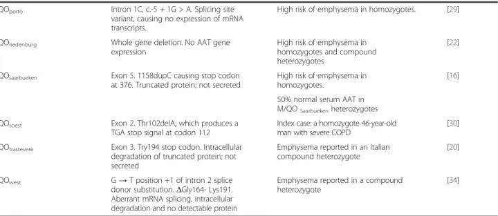
Identification of the QOMadridmutation
The entire sequence of the SERPINA1 gene, including coding and non-coding regions, was analyzed. DNA se-quencing of the exon 1C and intron 1C revealed in the four siblings a heterozygous base substitution G to A at position +1 of the splice donor site of intron 1C (NM_000295.4:c.-5 + 1G > A), previously described as QOPortosplicing mutation [29]. Analysis of the complete
sequence of theSERPINA1 gene showed that this was the only pathogenic mutation found in individuals II-1 and II-2 (Figure 2). The heterozygous status for QOPortoallele
in these patients was compatible with their moderate AAT deficiency.
Interestingly, the other two family members, II-3 and II-4, who showed a severe AAT deficiency, in addition to the QOPorto mutation were carriers of a duplication of
the thymine (T) in position +2 of the same splice donor site of intron 1C (NM_000295.4: c.-5 + 2dupT) (Figure 2). Since splicing sites are highly conserved sequences, this variation presumably disrupts the splicing donor site of this intron. This effect would be compatible with the strong AAT deficiency that both subjects presented. Since the whole sequence of the gene, coding and noncoding re-gions, was analyzed in these two patients, and there was no other pathogenic change, we assumed reasonably that this change is a deficient allele, not previously described, that was termed QOMadrid following the classical
no-menclature agreed by experts in the field, with QO representing a non-expressing gene at the protein level, and Madrid the name of the place of origin of the first carrier of the allele [40,41].
In addition, all four patients were heterozygous for the M2 (G/A, Arg101His, rs709932) and M3 (A/C, Glu376Asp, rs1303) common polymorphisms of the SERPINA1 gene. Sequencing of the expression products generated by using primers located in exon 1C and exon 5 demonstrated that patients II-1 and II-2, carriers of the QOPortoallele, only expressed transcripts with the M3 (C,
Asp376) variant. Since QOPortomutation affects the
patients occur in the other not expressed allele carrying the M2 (A, His101) but not the M3 (C, Asp376) variant (Figure 1). Consequently, the QOMadridmutation found in
patients II-3 and II-4 segregated with the M3 (C, Asp376) variant.
QOMadridimpairs normal splicing inin silico analysis
Thymine duplication at the site of splicing of intron 1C (i.e., the QOMadridmutation) is located in a highly
con-served splicing sequence of vertebrate splice donor sites and may lead to deregulation of gene expression. Com-putational predictions of splicing signals demonstrated disappearance of the constitutive donor splicing signals in the case of the QOMadrid mutation, similarly to what
happened for the QOPorto splicing mutation. In addition
several putative binding sites for Exonic Splicing En-hancers (ESE elements), such as SC35, SRp55 or SRp40 splicing factors, were predicted to be also affected by the duplication of T in position +2 of the QOMadrid allele.
Thus, SC35 site would be disrupted by QOMadrid
muta-tion and the predicted scores for SRp55 or SRp40 were reduced (Figure 3).
Expression analysis of severe AAT deficiency cases carrying QOPortoand QOMadrid
To further investigate the molecular effect on splicing and expression of theSERPINA1 gene with the QOMadrid
mutation which co-occurred with QOPorto mutation in
patients II-3 and II-4, expression products generated by different primer pairs in exons 1A, 1B, 1C and exons 2 and 5 were analyzed (Figure 4A). RT-PCR expression analysis of fragment E1C-E2 showed normally spliced products in control liver tissue and peripheral blood, as well as in the two moderate AAT deficiency patients II-1 and II-2 with PI*M/QOPorto genotype (Figure 4B). In
these patients sequence analysis demonstrated only one expressed allele based on the Arg101His polymorphism, corresponding to the normal M3 allele. However, no expression products were detected in patients carrying both splicing variants QOPorto and QOMadrid, clearly
indicating that QOMadrid mutation also impairs normal
splicing of intron 1C and represents a new null allele of theSERPINA1 gene.
We also checked using primers in the E1C-Int1C whether transcripts including intron 1C sequence were
Wildtype
QOPorto
QOPorto/QOMadrid
Exon 1C
Intron 1cE1A
E1B E1C
E2
E3
E4
E5
M2 (Arg101His) M1 (Val213Ala) M3 (Glu376Asp)
Figure 2 Results of direct sequencing of the exon 1C-inton 1C boundary region. Schematic representation of SERPINA1 gene is represented in the top showing position of the non-coding exons 1A, 1B and 1C and coding exons E2 to E5. Location of the common polymorphisms M1 (exon 3), M2 (exon 2) and M3 (exon 5) of this gene are also displayed. Position of the variants QOPortoand QOMadridis
marked with an arrow. Sequencing results of the patients are shown below. Comparison between a reference sequence from a normal individual (top sequence) and sequence from individuals II-1 and II-2 (middle panel) showing a heterozygous G to A change corresponding to the QOPorto. Bottom panel corresponds to direct sequencing of cases II-3 and II-4 that reveals heterozygosity for both QOPortoand the
expressed because of disruption of the splicing site due to the presence of both intron 1C splicing mutations, QOPortoand QOMadrid. Similar to what was described for
the QOPorto mutation [29], no expression products
retaining intron 1C sequence were detected indicating that if these transcripts are synthesized they might be probably rapidly degraded.
Transcripts that include E1A and E1B were also analyzed. Amplification of the E1A/E2 fragment in all patients, normal peripheral blood samples, and in a normal liver sample analyzed, produced one single band visible in agarose gel in all cases. Sequencing analysis revealed that the only mRNA product resulted from the direct splice of exon 1A to exon 2 (Figure 4C). Alternative spliced forms, previously described by others in mono-cytes/macrophages [8,42], were not detected in our sam-ples. Moreover, in all patients transcripts which include E1A showed expression of both alleles, since both variants Arg101 and His101 (G/A) were found. This indicates that QOPortoand QOMadridmutations do not impede
transcrip-tion not requiring the splice donor site of intron 1C. Regarding transcripts which included exon 1B, multiple expression products were detected after amplification with primer pair E1B-E2. After cloning, five different products were identified in patients II-1 and II-2 (Figure 4D). The most frequent species, as shown by the number of col-onies, were those including exons 1B joined to 1C and to exon 2 (named as product [1] in Figure 4D). Alternative
products ([2] and [3] in Figure 4D) generated using a cryptic splice donor site in exon 1B previously described, producing a 18 bp shorter fragment at the 3′ end of the exon 1B, were also found [43]. In addition, alternatively spliced products formed by direct splice of exon 1B to E2 ([4] in Figure 4D), as well as transcripts retaining the in-tron 1B ([5] in Figure 4D), were also found but in less amount.
In patients with QOPortoand QOMadridmutations (II-3
and II-4) only two different transcripts species were detected by using E1B and E2 primer pair. In these cases both fragments corresponded to RNA products contain-ing only E1B joined to E2 (Figure 4D), one of the products previously described using the cryptic splice donor site in exon 1B [44]. However, these patients do not include E1C in transcription products since splice donor site of intron 1C is completely disrupted by the presence of QOPortoand
QOMadridsplicing mutations.
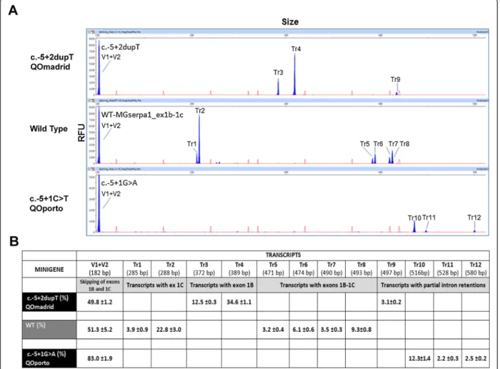
Functional assays by hybrid minigenes
The wild type (wt) and the mutant minigenes were func-tionally assayed in HeLa cells. The wt construct produced at least 7 different transcript as well as a wide range of rare splicing isoforms (Figure 5), where the skipping of both exons 1B and 1C (only with vector exons v1 + v2) was the most frequent event (51.3%). This isoform resem-bles the most abundant transcript of AAT in macrophages without exons 1B and 1C [43]. We also identified four
Figure 3 Schematic representation of the region E1C-Intron 1C containing the mutations QOPortoand QOMadrid. The consensus donor
splicing sequence is disrupted in both the QOPortomutation and the QOMadridmutation. Boxes represent the scores of splicing factors obtained
by bioinformatic tools. Several putative binding sites for splicing enhancer elements, SC35, SRp55 or SRp40, were predicted to be affected by these mutations. In the top panel, duplication of T of the QOMadridvariant cause that site for SC35 disappear, and in the bottom panel the
QOPortovariant cause a reduction of the score value for the SC35 from 3.48 in the reference sequence to 3.12 in the mutated sequence.
E1C_E5
E1C_E2
E1A_E2
1A 2E1B_E2
1C 2 1B II-3 II-4 II-1 II-2 2 1BA
B
C
D
1C 2 3 4 5 1C 2 [1] [2] [3] [4] [5]Figure 4 Expression analysis in the family patients carrying the QOPortomutation (II-1 and II-2) and the ones carrying both QOPortoand
QOMadridvariants (II-3 and II-4). A) Schematic representation of the SERPINA1 gene. To amplify different transcripts some forward primers in
exons 1A, 1B, and 1C and reverse primers in exons 2 and 5 were designed (arrows). B) RT-PCR amplification of mRNA using primers located in exon 1C and the reverse primers in exon 2 or exon 5, analyzed in normal hepatocytes (H), the AAT cases, and in a normal peripheral blood sample (PB). No expression products were found in cases II-3 and II-4 when using exon 1C primer. All the other cases showed a single band of 587 bp corresponding to expression products containing the exon 1C directly spliced to exon 2, or in the case of E1C-E5 the expression product corresponded to a fragment of 1318 bp with the exon 1C joined to all the coding exons (2 to 5). C) Fragments generated by amplification of expression products using primers in exon 1A and exon 2. All cases showed expression of a transcript including the exon 1A directly joined to exon 2. No other alternative splicing variants were found. D) Expression analysis using primers in exon 1B revealed multiple bands meaning that alternative splicing occurred between exons 1B and 1C. After cloning, we differentiated five different splicing forms, some showed the use of alternative splicing sites on exon 1B (3 and 4) previously described. One of the transcription species retained the intron 1B (5). Severe DAAT cases only express transcripts without exon 1C.
distinct isoforms with exons 1B and 1C that resulted from the combination of four splice sites: the canonical donor and acceptor sites, and one donor (18 nucleotides down-stream in intron 1B) and one acceptor (3 nucleotides downstream in exon 1C) cryptic sites (transcripts Tr5, Tr6, Tr7 and Tr8, together accounting for 22.1%; Figure 5). Finally, Tr1 and Tr2 would represent transcripts lacking exon 1B. These results are in accordance to the transcripts described by Rollini and Fournier (2000) [43]. Therefore, the splicing pattern of wild type MGserp1a_ex1b-1c mim-icked that of macrophages cell lines.
Interestingly, mutations c.-5 + 1G > A and c.-5 + 2dupT triggered the elimination of six transcripts of the wild type minigene (Tr1, 2, 5, 6, 7, 8) and generated six new ones, Tr10, Tr11 and Tr12 (c.-5 + 1G > A), and Tr3, Tr4 and
Tr9 (c.-5 + 2dupT). The V1 + V2 transcript was signifi-cantly enriched in c.-5 + 1G > A (83.0% vs. 51.3% in wt) although other three aberrant transcripts (Tr10-12) would correspond to partial retentions of intron 1C. The new mutation QOMadrid (c.-5 + 2dupT) induced isoforms Tr3
and 4 that were relatively abundant (12.5 and 34.6%, respectively) and corresponded to exon 1C skipping and alternative usage of the two donor sites of intron 1B. This result reproduced the splicing profile of patient RNA.
Discussion
We have characterized a new splicing variant in the SERPINA1 gene producing a null allele that we named PI*QOMadrid following the traditional terms used to
Figure 5 Transcripts detection in wt and splicing mutation minigenes. A) Fluorescent capillary electrophoresis of RT-PCR products generated by the wild type, c.-5 + 2dupT and c.-5 + 1G > A minigenes. Screenshots of Peak Scanner electropherograms are shown. Fluorescent RT-PCRs (blue peaks) of wt and mutant minigenes were run in an ABI3130 DNA sequencer with Genescan ROX 500 (red peaks) as size standard. RFU: Relative Fluorescence Units. B) Quantification of all detected of transcripts (Tr1 to 12) generated by the wild type, or mutants c.-5 + 1G > A (QOPorto)
and c.-5 + 2dupT (QOMadrid) minigenes of the SERPINA1 gene are represented with the mean proportion of each one. Sizes were calculated by the Peak
Scanner software. Depending the use of the alternative splicing sites described for exon 1B and 1C, the deduced transcript composition is: Tr1: V1-1Cs -V2; Tr2: V1-1Cl -V2; Tr3: V1-1Bs -V2; Tr4: V1-1Bl -V2; Tr5: V1-1Bs -1Cs-V2; Tr6: V1-1Bs -1Cl-V2; Tr7: V1-1Bl -1Cs-V2; Tr8: V1-1Bl -1Cl-V2; Tr9-12: partial intron retentions. (1Bs and 1Bl: exon1B short and long, respectively; 1Cs and 1Cl: exon 1C short and long, respectively).
designate the new genetic variants found in this condition. This new allele increases the short list of null alleles reported to date, which are characterized by the almost absence serum AAT [4].
As shown in Table 1, several molecular mechanisms have been described to be responsible for the lack of expression of AAT protein including deletion of AAT coding exons, nonsense mutations producing formation of premature stop codons or splicing mutations. The QOMadrid variant corresponded to the fourth splice site
alteration described so far in the SERPINA1 gene. The three other reported splicing variants were QOWest,
QOBonny blueand QOPorto. The QOWestand QOBonny blue
alleles are caused by alterations in the splice donor and acceptor sites of intron 2 [4,34], while QOPorto
corre-sponded to a G to A transition in the donor splice site of intron 1C [29]. Therefore, the QOMadridadds a new
splicing variant (+2dupT) to the same splicing donor site of intron 1C.
This new QOMadridmutation, which occurred in a M3
background, enlarge the number of pathogenic muta-tions outside the coding sequence of theSERPINA1 gene and highlight the importance that regulatory regions might have in the disease. Studies of these regions should be seriously considered in discordant cases with severe AAT deficiency where no coding alterations are found.
In the two severe AAT deficiency patients of this family in Madrid, the new splicing mutation occurred in het-erozygous state in combination with another splicing mutation previously described as QOPorto, curiously
both affecting the donor splice site of intron 1C. Both composite heterozygotes PI*QOPorto/QOMadrid
individ-uals showed extremely low levels of serum AAT and a practically total absence of detectable plasma protein by isoelectric focusing, indicating that these changes strongly affect the AAT synthesis by disrupting the normal splicing of the intron 1C. In this regard, we demonstrate that these patients lacked of transcripts with exon 1C, and therefore of the properly matured mRNA synthesized by the liver which is the major source of the AAT mRNA. Nevertheless, these two composite heterozygotes were able to produce AAT mRNA transcripts from exons 1A and 1B, which are probably originated from the macrophage-specific pro-moter localized in the gene upstream of the liver propro-moter [8]. Is it possible then, that the low level of serum AAT protein detected in severe patients was produced by mac-rophages or other cells than hepatocytes. However, these transcripts represent a small proportion of the total AAT transcription, and may be insufficient to compensate the negative effects of the splicing mutations in liver cells.
In the index case, the presence of both QOPorto and
QOMadridmutations likely caused the genetic
susceptibil-ity to early onset emphysema after smoking exposure,
similarly to which occur with other null alleles [27]. The siblings presenting a genotype PI*M/QOPorto showed
AAT serum levels concordant with the presence of a normal and a mutated allele. Heterozygous status for a null allele is generally considered to produce enough normal protein to keep enough antielastase activity in the lung, and hence the risk of lung disease might be mild. These patients also presented moderate degree of mental retardation. Dementia-like syndrome associated with serpinopaties has been reported [9] but there is no evidence suggesting that QOPorto could be related with
mental impairment detected in these individuals. Although splicing variants have been rarely described causing AAT deficiency, their existence show that correct splicing events are important for the accurate function of this gene. Splicing alterations have been described to originate genetic diseases such as spinal muscular atrophy, neuro-fibromatosis type I or myotonic dystrophy, among many others. Numerous disease causing genes undergo alter-native splicing to regulate expression, and it is estimated that overall 15% of mutations are located within splicing sites and more than 20% of missense mutations lie within predicted splicing elements [45].
Computational analysis of the effect of the new QOMadrid
revealed the disruption of the consensus donor splicing site sequence. In addition, using exonic splicing enhancers (ESE) motif predictions it was observed that the variant mainly eliminate a potential SC35 factor binding site. This and other ESE sequences have been described to play a key role in regulating splicing events [46].
Transcriptional analysis of the AAT deficiency family members allowed us to verify that transcription from exon 1C, still detected in siblings with PI*M/QOPorto,
was totally impaired in the compound heterozygotes PI*QOPorto/QOMadrid. The QOMadrid mutation highlights
the importance of exon 1C for the correct expression of AAT in the liver, in order to ensure the production of appropriate serum AAT concentrations. Even though transcripts from the exons 1A and 1B were detected in the compound heterozygotes, these RNA species might not be abundant enough to provide acceptable AAT serum levels.
Moreover, we have constructed and validated the mini-gene MGserpa1_ex1b-1c in the pSAD vector where the splicing patterns of mutant and wt minigenes were clearly distinguished. Consequently, it constitutes a very valuable tool for the functional and clinical classification of DNA variants from disease genes, facilitating the dis-crimination between neutral and deleterious changes. This approach also allows quantifying the impact on splicing of a single variant without the interference of another allele as it occurs in patient RNA. Moreover, at least 13 different transcripts from the AAT construct were detected supporting the high sensitivity and resolution of
fluorescent capillary electrophoresis of RT-PCR products as previously reported [39]. Furthermore, the wild type construct and several cell lines displayed similar splicing patterns [43] and variant c.-5 + 2dupT in minigene and patient RNA was associated with exon 1C skipping in both cases, lending further support to the reproducibility of minigene assays. Indeed, all the transcripts detected in macrophage cell lines were identified in our wild type minigene, including the alternative selection of cryptic splice sites in intron 1B and exon 1C. 3.
Conclusion
In summary, a new variant in the 5′UTR promoter region of theSERPINA1 gene causing a new null allele QOMadrid
has been described for a first time. This new variant is added to the previously described null variants that affect splicing causing the disease. The minigene system is a powerful approach to detect variants with an impact on splicing and have contributed to a better knowledge of this gene expression step by quantification of normal and ab-errant alternative transcripts of theSERPINA1 gene.
Similar to what occur with deficient variants located in exons, which allowed a better understanding of the AAT structure-function relationships, splicing variants provide us with more insight into the expression regulation of the SERPINA1 gene.
Additional files
Additional file 1: Table S1. List of primers used to amplify all fragments of the entire SERPINA1 gene (Ensembl: ENSG00000197249). Additional file 2: Figure S1. Scheme of minigene MGserpa1_ex1b-1c. Abbreviations
AAT:Alpha-1 antitrypsin; QO: Null allele; IEF: Isoelectric focusing; ESE: Exonic Splicing Enhancers; CT: Computed tomography.
Competing interests
The authors declare that they have no conflict of interests. Authors’ contributions
BL, participated in the design of the study, helped to collect clinical data and draft the manuscript. MTM, carried out clinical evaluation and interpretation of the results. IB, participated in interpretation of the results, involved in drafting the manuscript and revising it critically. CHM and EV, designed and performed the transcriptional in vitro study using minigenes. IF, carried out part of phenotypic and molecular genetic studies and critically review the results. FRF, carried out phenotypic and molecular genetic studies. LP, carried out sequence and transcriptional analysis. JA, participated in interpretation of results and helped to draft the manuscript. MP, helped in the design and critical revision of the manuscript. BMD participated in the design and coordination of the study and draft the manuscript. All authors read and approved the final manuscript. Acknowledgements
This work has been partially funded by the Instituto de Salud Carlos III grants TPY1250/12 (BMD) and PI13/1749 (EAV) (Spanish Ministry of Economy and Competitiveness) and BIO/VA08/13 (Consejería de Sanidad, Junta de Castilla y León). CHM was supported by a fellowship from Fundación Villalar (Cortes de Castilla y León, Spain). We thank collaborators from the REDAAT (Spanish Registry of Alpha-1 Antitrypsin deficiency patients) and all members of the Human Genetics Area of the ISCIII for their support.
Author details
1
Servicio de Neumología, Hospital Universitario Arnau de Vilanova, Lleida, Spain.
2Servicio de Neumología, Hospital Universitario Doce de Octubre, Madrid, Spain. 3
Board of Directors of the Alpha-1 Antitrypsin Deficiency Spanish Registry, Lung Foundation Breathe, Spanish Society of Pneumology (SEPAR), Barcelona, Spain.
4
Grupo de Splicing y Cáncer, Instituto de Biología y Genética Molecular (CSIC-UVa), Valladolid, Spain.5Center for Diagnosis of Inherited Alpha-1
Antitrypsin Deficiency, Department of Molecular Medicine, Section of Pneumology, IRCCS San Matteo Hospital Foundation, University of Pavia, Pavia, Italy.6Servicio de Bioquímica, Hospital Universitario Vall d’ Hebron, Barcelona, Spain.7Molecular Genetics Unit, Instituto de Investigación en
Enfermedades Raras (IIER), Instituto de Salud Carlos III (ISCIII), Carretera Majadahonda-Pozuelo Km 2,200, Majadahonda, Madrid 28220, Spain.
8
Human Genetics Area, Instituto de Investigación en Enfermedades Raras (IIER), Instituto de Salud Carlos III (ISCIII), Madrid, Spain.9Instituto de
Investigación en Enfermedades Raras (IIER), Instituto de Salud Carlos III (ISCIII), Spain RDR and CIBERER, Madrid, Spain.
Received: 27 June 2014 Accepted: 1 October 2014
References
1. Janciauskiene S, Ferrarotti I, Laenger F, Jonigk D, Luisetti M: Clinical utility gene card for: alpha-1-antitrypsin deficiency. Eur J Hum Genet 2011, 19. doi:10.1038/ejhg.2010.246.
2. Knappstein S, Ide T, Schmidt MA, Heusipp G: Alpha 1-antitrypsin binds to and interferes with functionality of EspB from atypical and typical enteropathogenic Escherichia coli strains. Infect Immun 2004, 72:4344–4350.
3. Janciauskiene SM, Bals R, Koczulla R, Vogelmeier C, Kohnlein T, Welte T: The discovery of alpha1-antitrypsin and its role in health and disease. Respir Med 2011, 105:1129–1139.
4. Lee JH, Brantly M: Molecular mechanisms of alpha1-antitrypsin null alleles. Respir Med 2000, 94(Suppl C):S7–S11.
5. American Thoracic Society and European Respiratory Society: American Thoracic Society/European Respiratory Society statement: standards for the diagnosis and management of individuals with alpha-1 antitrypsin deficiency. Am J Respir Crit Care Med 2003, 168:818–900.
6. du Bois RM, Bernaudin JF, Paakko P, Hubbard R, Takahashi H, Ferrans V, Crystal RG: Human neutrophils express the alpha 1-antitrypsin gene and produce alpha 1-antitrypsin. Blood 1991, 77:2724–2730.
7. Perlmutter DH, Cole FS, Kilbridge P, Rossing TH, Colten HR: Expression of the alpha 1-proteinase inhibitor gene in human monocytes and macrophages. Proc Natl Acad Sci U S A 1985, 82:795–799. 8. Hafeez W, Ciliberto G, Perlmutter DH: Constitutive and modulated
expression of the human alpha 1 antitrypsin gene. Different transcriptional initiation sites used in three different cell types. J Clin Invest 1992, 89:1214–1222.
9. Lomas DA, Mahadeva R: Alpha1-antitrypsin polymerization and the serpinopathies: pathobiology and prospects for therapy. J Clin Invest 2002, 110:1585–1590.
10. Rodriguez-Frias F, Miravitlles M, Vidal R, Camos S, Jardi R: Rare alpha-1-antitrypsin variants: are they really so rare? Ther Adv Respir Dis 2012, 6:79–85. 11. Blanco I, de Serres FJ, Fernandez-Bustillo E, Lara B, Miravitlles M: Estimated
numbers and prevalence of PI*S and PI*Z alleles of alpha1-antitrypsin deficiency in European countries. Eur Respir J 2006, 27:77–84.
12. Cox DW, Billingsley GD: Rare deficiency types of alpha 1-antitrypsin: electro-phoretic variation and DNA haplotypes. Am J Hum Genet 1989, 44:844–854. 13. Lomas DA, Evans DL, Finch JT, Carrell RW: The mechanism of Z alpha
1-antitrypsin accumulation in the liver. Nature 1992, 357:605–607. 14. Brantly M, Lee JH, Hildesheim J, Uhm CS, Prakash UB, Staats BA, Crystal RG,
Hildeshiem J: alpha1-antitrypsin gene mutation hot spot associated with the formation of a retained and degraded null variant [corrected; erratum to be published]. Am J Respir Cell Mol Biol 1997, 16:225–231. 15. Curiel D, Brantly M, Curiel E, Stier L, Crystal RG: Alpha 1-antitrypsin deficiency
caused by the alpha 1-antitrypsin Nullmattawa gene. An insertion mutation rendering the alpha 1-antitrypsin gene incapable of producing alpha 1-antitrypsin. J Clin Invest 1989, 83:1144–1152.
16. Faber JP, Poller W, Weidinger S, Kirchgesser M, Schwaab R, Bidlingmaier F, Olek K: Identification and DNA sequence analysis of 15 new alpha
1-antitrypsin variants, including two PI*Q0 alleles and one deficient PI*M allele. Am J Hum Genet 1994, 55:1113–1121.
17. Fraizer GC, Siewertsen M, Harrold TR, Cox DW: Deletion/frameshift mutation in the alpha 1-antitrypsin null allele, PI*QObolton. Hum Genet 1989, 83:377–382.
18. Frazier GC, Siewertsen MA, Hofker MH, Brubacher MG, Cox DW: A null deficiency allele of alpha 1-antitrypsin, QOludwigshafen, with altered tertiary structure. J Clin Invest 1990, 86:1878–1884.
19. Fregonese L, Stolk J, Frants RR, Veldhuisen B: Alpha-1 antitrypsin Null mutations and severity of emphysema. Respir Med 2008, 102:876–884. 20. Lee J, Novoradovskaya N, Rundquist B, Redwine J, Saltini C, Brantly M: Alpha
1-antitrypsin nonsense mutation associated with a retained truncated protein and reduced mRNA. Mol Genet Metab 1998, 63:270–280. 21. Nukiwa T, Takahashi H, Brantly M, Courtney M, Crystal RG: alpha 1-Antitrypsin
nullGranite Falls, a nonexpressing alpha 1-antitrypsin gene associated with a frameshift to stop mutation in a coding exon. J Biol Chem 1987, 262:11999–12004.
22. Poller W, Faber JP, Weidinger S, Olek K: DNA polymorphisms associated with a new alpha 1-antitrypsin PIQ0 variant (PIQ0riedenburg). Hum Genet 1991, 86:522–524.
23. Rametta R, Nebbia G, Dongiovanni P, Farallo M, Fargion S, Valenti L: A novel alpha1-antitrypsin null variant (PiQ0Milano ). World J Hepatol 2013, 5:458–461.
24. Sifers RN, Brashears-Macatee S, Kidd VJ, Muensch H, Woo SL: A frameshift mutation results in a truncated alpha 1-antitrypsin that is retained within the rough endoplasmic reticulum. J Biol Chem 1988, 263:7330–7335. 25. Vaz RL, Costa F, Marques P, Mendonca C, Rocha J, Seixas S: Severe alpha-1
antitrypsin deficiency caused by Q0(Ourem) allele: clinical features, haplotype characterization and history. Clin Genet 2012, 81:462–469. 26. Zorzetto M, Ferrarotti I, Campo I, Balestrino A, Nava S, Gorrini M, Scabini R,
Mazzola P, Luisetti M: Identification of a novel alpha1-antitrypsin null variant (Q0Cairo). Diagn Mol Pathol 2005, 14:121–124.
27. Salahuddin P: Genetic variants of alpha1-antitrypsin. Curr Protein Pept Sci 2010, 11:101–117.
28. Fregonese L, Stolk J: Hereditary alpha-1-antitrypsin deficiency and its clinical consequences. Orphanet J Rare Dis 2008, 3:16.
29. Seixas S, Mendonca C, Costa F, Rocha J: alpha1-Antitrypsin null alleles: evidence for the recurrence of the L353fsX376 mutation and a novel G->A transition in position +1 of intron IC affecting normal mRNA splicing. Clin Genet 2002, 62:175–180.
30. Prins J, van der Meijden BB, Kraaijenhagen RJ, Wielders JP: Inherited chronic obstructive pulmonary disease: new selective-sequencing workup for alpha1-antitrypsin deficiency identifies 2 previously unidentified null alleles. Clin Chem 2008, 54:101–107.
31. Satoh K, Nukiwa T, Brantly M, Garver RI Jr, Hofker M, Courtney M, Crystal RG: Emphysema associated with complete absence of alpha 1- antitrypsin in serum and the homozygous inheritance [corrected] of a stop codon in an alpha 1-antitrypsin-coding exon. Am J Hum Genet 1988, 42:77–83. 32. Graham A, Kalsheker NA, Bamforth FJ, Newton CR, Markham AF: Molecular
characterisation of two alpha-1-antitrypsin deficiency variants: proteinase inhibitor (Pi) Null(Newport) (Gly115––Ser) and (Pi) Z Wrexham (Ser-19––Leu). Hum Genet 1990, 85:537–540.
33. Takahashi H, Crystal RG: Alpha 1-antitrypsin Null(isola di procida): an alpha 1-antitrypsin deficiency allele caused by deletion of all alpha
1-antitrypsin coding exons. Am J Hum Genet 1990, 47:403–413. 34. Laubach VE, Ryan WJ, Brantly M: Characterization of a human alpha
1-antitrypsin null allele involving aberrant mRNA splicing. Hum Mol Genet 1993, 2:1001–1005.
35. Holmes MD, Brantly ML, Crystal RG: Molecular analysis of the
heterogeneity among the P-family of alpha-1-antitrypsin alleles. Am Rev Respir Dis 1990, 142:1185–1192.
36. Rodriguez F, Jardi R, Costa X, Cotrina M, Galimany R, Vidal R, Miravitlles M: Rapid screening for alpha1-antitrypsin deficiency in patients with chronic obstructive pulmonary disease using dried blood specimens. Am J Respir Crit Care Med 2002, 166:814–817.
37. Zorzetto M, Russi E, Senn O, Imboden M, Ferrarotti I, Tinelli C, Campo I, Ottaviani S, Scabini R: von, Eckardstein A., Berger, W., Brandli, O., Rochat, T., Luisetti, M., and Probst-Hensch, N. SERPINA1 gene variants in individuals from the general population with reduced alpha1-antitrypsin concentrations. Clin Chem 2008, 54:1331–1338.
38. Bryksin AV, Matsumura I: Overlap extension PCR cloning: a simple and reliable way to create recombinant plasmids. Biotechniques 2010, 48:463–465.
39. Acedo A, Sanz DJ, Duran M, Infante M, Perez-Cabornero L, Miner C, Velasco EA: Comprehensive splicing functional analysis of DNA variants of the BRCA2 gene by hybrid minigenes. Breast Cancer Res 2012, 14:R87. 40. Cox DW, Johnson AM, Fagerhol MK: Report of Nomenclature Meeting for
alpha 1-antitrypsin, INSERM, Rouen/Bois-Guillaume-1978. Hum Genet 1980, 53:429–433.
41. Shows TB, McAlpine PJ: The 1981 catalogue of assigned human genetic markers and report of the nomenclature committee. Oslo Conference (1981): Sixth International Workshop on Human Gene Mapping. Cytogenet Cell Genet 1982, 32:221–245.
42. Perlino E, Cortese R, Ciliberto G: The human alpha 1-antitrypsin gene is transcribed from two different promoters in macrophages and hepatocytes. EMBO J 1987, 6:2767–2771.
43. Rollini P, Fournier RE: Differential regulation of gene activity and chromatin structure within the human serpin gene cluster at 14q32.1 in macrophage microcell hybrids. Nucleic Acids Res 2000, 28:1767–1777. 44. Rollini P, Xu L, Fournier RE: Stable expression and cell-specific chromatin
structure of human alpha1-antitrypsin cosmid transgenes in rat hepatoma cells. Nucleic Acids Res 2000, 28:3605–3614.
45. Singh RK, Cooper TA: Pre-mRNA splicing in disease and therapeutics. Trends Mol Med 2012, 18:472–482.
46. Blencowe BJ: Exonic splicing enhancers: mechanism of action, diversity and role in human genetic diseases. Trends Biochem Sci 2000, 25:106–110.
doi:10.1186/s12931-014-0125-y
Cite this article as: Lara et al.: Severe alpha-1 antitrypsin deficiency in composite heterozygotes inheriting a new splicing mutation QOMadrid.
Respiratory Research 2014 15:125.
Submit your next manuscript to BioMed Central and take full advantage of:
• Convenient online submission
• Thorough peer review
• No space constraints or color figure charges
• Immediate publication on acceptance
• Inclusion in PubMed, CAS, Scopus and Google Scholar
• Research which is freely available for redistribution
Submit your manuscript at www.biomedcentral.com/submit