HAL Id: hal-02372075
https://hal-polytechnique.archives-ouvertes.fr/hal-02372075
Submitted on 17 Nov 2020
HAL is a multi-disciplinary open access
archive for the deposit and dissemination of sci-entific research documents, whether they are pub-lished or not. The documents may come from teaching and research institutions in France or abroad, or from public or private research centers.
L’archive ouverte pluridisciplinaire HAL, est destinée au dépôt et à la diffusion de documents scientifiques de niveau recherche, publiés ou non, émanant des établissements d’enseignement et de recherche français ou étrangers, des laboratoires publics ou privés.
An actin-based viscoplastic lock ensures progressive
body-axis elongation
Alicia Lardennois, Teresa Ferraro, Flora Llense, Michel Labouesse, Gabriella
Pásti, Julien Pontabry, David Rodriguez, Samantha Kim, Christelle Gally,
Pierre Mahou, et al.
To cite this version:
Alicia Lardennois, Teresa Ferraro, Flora Llense, Michel Labouesse, Gabriella Pásti, et al.. An actin-based viscoplastic lock ensures progressive body-axis elongation. Nature, Nature Publishing Group, 2019, 573 (7773), pp.266-270. �10.1038/s41586-019-1509-4�. �hal-02372075�
1
1
An actin-based viscoplastic lock ensures progressive body
2
axis elongation
3
4
5
6
7
8
9
10
11
12
13
14
15
16
17
18
19
# Corresponding author: [email protected]
20
Alicia Lardennois, Teresa Ferraro, Flora Llense & Michel
Labouesse
CNRS UMR7622, Institut de Biologie Paris–Seine (IBPS), Sorbonne Université, Paris, France
Gabriella Pásti, Julien Pontabry, David Rodriguez, Samantha
Kim, Christelle Gally & Michel Labouesse
IGBMC –CNRS UMR 7104, INSERM U964, Development and Stem Cells Department, Université de Strasbourg, Illkirch, France
Pierre Mahou & Emmanuel Beaurepaire
INSERM U1182 – CNRS/ UMR7645, Laboratoire d’Optique et Biosciences, Ecole Polytechnique, Paris, France
Julien Pontabry
RS2D, Mundolsheim, France
Shoichiro Ono
Departments of Pathology and Cell Biology, Winship Cancer Institute, Emory University School of Medicine, Atlanta, GA, USA
2
21
22
Abstract
23
24
25
A key step in animal development is the process of body axis elongation, laying out the
26
final form of the entire animal. This critically depends on polarized cell shape changes1,
27
which rely on the interplay between intrinsic forces generated by molecular motors2-5,
28
extrinsic forces due to adjacent cells pulling or pushing on the tissue6-9, and mechanical
29
resistance forces due to cell and tissue elasticity or friction10-12. Understanding how
30
mechanical forces influence morphogenesis at the cellular and molecular level remains
31
a critical challenge2. Recent work outlined that cell shape changes occur through small
32
incremental steps2,4,5,13, suggesting the existence of specific mechanisms to stabilize cell
33
shapes and counteract cell elasticity. Here, we identify a spectrin-kinase-formin network
34
required to stabilize embryo shape when repeated muscle contractions promote C.
35
elegans embryo axis elongation. Its absence induces complete axis retraction due to
36
damage of epidermal actin stress fibers. Modeling predicts that a mechanical
37
viscoplastic deformation process can account for embryo shape stabilization. Molecular
38
analysis suggests that the physical basis for viscoplasticity originates from the progressive
39
shortening of epidermal microfilaments induced by muscle contractions and FHOD-1
40
formin activity. Our work thus identifies an essential molecular lock acting in a
41
developmental ratchet-like process.
42
3
44
45
C. elegans provides a simple and integrated model to study the cellular impact of
46
mechanical forces. Its embryonic elongation only relies on cell shape changes and
47
includes two phases depending on tension and stiffness anisotropies in the epidermis12,
48
and beyond the 2-fold stage on muscle activity8 (Fig. 1a; Supplementary material).
49
Importantly, neither stage relies on pulsatile actomyosin flows12, as observed in the early
50
C. elegans zygote, or during Drosophila gastrulation and germband extension2,4,5,13.
51
Because muscles are tightly mechanically coupled to the epidermis through epidermal
52
hemidesmosomes14, their contractions also displace the epidermis. This can be
53
monitored by tracking the anterior-posterior displacement of muscle nuclei and the
54
circumferentially oriented epidermal actin filaments (Fig. 1b-b’’’). Importantly, all
55
muscles do not contract simultaneously (X. Yang and M. Labouesse, unpublished).
56
Hence, when some areas of the epidermis are longitudinally compressed (red line in Fig.
57
1c’), others are stretched (green line in Fig. 1c’) before eventually relaxing (Fig. 1c-c”;
58
Fig. S1). Importantly, we previously established that the tension generated by muscle
59
activity triggers a mechanotransduction pathway in the epidermis, which promotes axis
60
elongation8.
61
The relaxation observed after each muscle contraction raises a conundrum: how can
62
muscle activity power embryonic elongation from 100 µm to 200 µm within an hour if cell
63
elasticity brings cells back to their initial length after each contraction (Fig. 1d). One
64
simple hypothesis would be that some mechanism stabilizes the transient body
65
deformations induced by muscle activity (Fig. 1d’), as was proposed for Drosophila
66
gastrulation or germband extension in processes involving myosin II5,15,16. To uncover this
67
mechanism, we focused on the kinase PAK-1, which lies at the crossroads of
68
4
hemidesmosome remodelling9 and actomyosin force regulation17,18. We first performed
69
a feeding RNAi screen in a strong yet viable pak-1 mutant, looking for enhancers of
70
embryonic lethality and/or body morphology defects (Fig. 2a). This screen identified the
71
gene spc-1 encoding α-spectrin as a strong pak-1 genetic enhancer leading to short
72
hatchlings (58 µm on average), which were significantly shorter than pak-1(tm403) (178
73
µm) or spc-1(RNAi) (91 µm) hatchlings (Fig. 2b, Table S1). The study of this genetic
74
interaction was further motivated by the identification of the central Src Homology 3
75
domain (SH3) of SPC-1 as an interactor of the N-terminal domain of PAK-1 in a yeast
76
two-hybrid screen (Fig. 2c, Table S2). Both screens thus point to an interaction between
77
SPC-1 and PAK-1.
78
To understand why pak-1(tm403) spc-1(RNAi) are shorter than spc-1(RNAi) embryos, we
79
examined their elongation rate using DIC microscopy. Wild-type and pak-1(tm403)
80
embryos initially elongated at the same rate, whereas spc-1 defective embryos
81
elongated slower and stopped around the 2-fold stage (100 µm) as previously
82
described19 (Fig. 2d). By contrast, spc-1(ra409) 1(tm403) and spc-1(RNAi)
pak-83
1(tm403) embryos reached ≈65 µm at a slow rate, but then could not maintain their
84
shape, retracting back to ≈50 µm, which neither spc-1(ra409) nor pak-1(tm403) embryos
85
did (Fig. 2d, Movie 1). Two observations suggest that this phenotype is linked to muscle
86
activity. First, spc-1 knock-down in git-1 or pix-1 mutants, two other players involved in
87
the same mechanotransduction pathway as PAK-19, also induced retraction (Fig. S2).
88
Second, spc-1(RNAi) pak-1(tm403) embryos started to retract at the onset of active
89
muscle contraction in control embryos (pink box in Fig. 2d). To directly prove this
90
hypothesis, we abrogated muscle function in spc-1(ra409) pak-1(tm403) embryos by
91
knocking-down the kindlin homolog UNC-11220. Strikingly, spc-1(ra409) pak-1(tm403)
92
5
embryos defective for unc-112 no longer retracted (Fig. 2e; Movie 2). We conclude that
93
the mechanical input provided by muscles to the epidermis induces the retraction
94
phenotype observed in spc-1 pak-1 double mutants.
95
The simplest interpretation of the retraction phenotype described above is that a
96
cellular structure maintaining embryo shape fails to emerge or collapses in spc-1 pak-1
97
double mutants once muscles become active. Two arguments lead us to consider that
98
this structure corresponds to the actin cytoskeleton. First, SPC-1/α-spectrin and its
99
binding partner SMA-1/ß-spectrin form an actin-binding hetero-tetramer colocalizing
100
with actin21 and partially with PAK-1 in epidermal cells (Fig. S3). Second and foremost, it
101
has long been known that treating C. elegans embryos with the actin-depolymerizing
102
drug cytochalasin-D induces a retraction phenotype very similar to that presented
103
herein22. We thus characterized actin filaments by spinning-disk confocal imaging of a
104
LifeAct::GFP probe12. Segmentation analysis of the fluorescence signal associated with
105
actin filaments in the dorso-ventral epidermis (Fig. 3a-a’’’) revealed more discontinuity in
106
spc-1 pak-1 double deficient embryos (Fig. 3d-d’’’) compared to the control genotypes
107
(Fig. 3a-c’’’). Moreover, Fourier transform analysis indicated that their degree of
108
anisotropy relative to the circumferential axis was abnormal (Fig. 3d’’-d’’’). Note that
109
both phenotypes were visible mainly at mid elongation, i.e. after muscles become
110
active (Fig. S4), suggesting that a well-structured actin network emerges too slowly in
111
double mutants rather than it collapses.
112
Collectively, the results described so far, together with the retraction phenotype of
113
cytochalasin-D treated embryos22, suggest that the actin filament defects account for
114
spc-1 pak-1 embryo retraction, and further link muscle activity with these defects.
115
6
Significantly, as the wild-type embryo lengthens, its circumference decreases by roughly
116
20% due to embryo volume conservation (Fig. S5) and thus, the length of actin filaments
117
in dorso-ventral cells should decrease. Hence, we suggest that muscle activity normally
118
promotes actin filament shortening, probably through sliding or shortening of filaments
119
relative to each other after their bending (Fig. S1). We further suggest that SPC-1 and
120
PAK-1 stabilize cell shape by maintaining actin bundle integrity. We could not define the
121
shortening mechanism by spinning-disk microscopy, probably because each muscle
122
contraction results in changes beyond the time and space resolution of the microscope.
123
However, we suggest that it goes awry in the absence of SPC-1 and PAK-1, due to the
124
lack of a capping or bundling activity (Fig. 4b-b’).
125
To rationalize the role of muscles in the process of actin bundle shortening and
126
stabilization, we described the C. elegans embryo as a Kelvin-Voigt material (a spring in
127
parallel to a dashpot) submitted to forces acting in the epidermis and muscles (Fepid and
128
Fmuscles) (Fig. 4c equations-a and -b; Supplementary material). Note that Fepid is written as
129
the product of an active force, Fseam, and a passive component resulting from actin
130
bundle stiffness, 𝛼!" (Supplementary material and ref. 11). Since muscle-defective
131
mutants cannot elongate beyond the 2-fold stage, then Fepid can only extend embryos
132
until that stage (due to the spring restoring force; Fig. S6a-a’). Simply adding the force
133
Fmuscles should not trigger any further extension, because it oscillates between a positive
134
and negative input (Fig. 1bc, Fig. S6b-b’).
135
Recently, several studies have suggested that systems exposed to a stress can undergo
136
a permanent rearrangement, which can be described as a plastic deformation23 or as
137
a change in the spring resting length24,25. Accordingly, we incorporated an increase of
138
7
the spring resting length λ in the equations described above by writing that it changes
139
by a factor β (Fig. 4c equation-c; Fig. 4d). Thereby, we could accurately predict the
140
elongation pattern of wild-type embryos (Fig. 4f, Fig. S6d-d’; Supplementary material).
141
Conversely, in spc-1 pak-1 defective embryos, the continuing damage to actin
142
filaments should reduce their stiffness (component 𝛼!" in Fig. 4c equation-b), which we
143
expressed by writing that it depends on a tearing factor γ (Fig. 4c equation-d, Fig. 4e);
144
thereby, we could accurately predict their retraction pattern (Fig. 4f, Fig. S6e-e’). We
145
thus propose that SPC-1/α-spectrin and PAK-1 regulate a process of mechanical
146
plasticity in the physical sense. From a cellular standpoint, having a changing resting
147
length means that body elasticity does not bring the embryo back to its initial shape
148
upon muscle relaxation, enabling progressive lengthening.
149
To further define the molecular basis of viscoplasticity, we performed a small-scale RNAi
150
screen to search for gene knockdowns inducing retraction of spc-1(ra409) embryos (Fig.
151
4g; Table S3). This screen identified the atypical formin FHOD-1 (Fig. 4h-i; Fig. S7, movie
152
3), which has previously been linked to actin dynamics in the epidermis26. We confirmed
153
that fhod-1(tm2363); spc-1(RNAi) embryos also showed a penetrant retraction
154
phenotype (Fig. 4h; Fig. S7). The identification of this specific formin was intriguing
155
because vertebrate FHOD1 promotes actin capping and bundling rather than
156
nucleation and elongation27. It thus raised the tantalizing possibility that FHOD-1 activity
157
stabilizes the actin cytoskeleton while it gets remodeled under the influence of muscle
158
activity during embryo circumference reduction. Furthermore, the genetic interaction
159
suggests that FHOD-1 acts with SPC-1 and PAK-1. To examine this possibility, we tested
160
whether FHOD-1 derivatives removing at least the C-terminal DAD domain, predicted to
161
auto-inhibit formins28, can rescue the retraction phenotype of spc-1 pak-1 deficient
162
8
embryos. Strikingly, after epidermis-specific expression of a form lacking the FH2 and
163
DAD domains, transgenic spc-1(RNAi) pak-1(tm403) embryos did not retract and were
164
significantly longer than non-transgenic siblings; rescue was better than with the
full-165
length protein. By contrast, the DAD deleted form did not rescue and deletion of the
166
FH1-FH2-DAD domains marginally rescued retraction, arguing that the FH2 F-actin
167
nucleation domain is not necessary for rescue but that the FH1 is. Truncation of the
C-168
terminal DAD domain or of the FH1-FH2-DAD domains marginally rescued retraction (Fig.
169
4j). Importantly, an FH2-DAD truncation in the mammalian FHOD1 still enables it to
170
bundle actin27, further strengthening the notion that FHOD-1 bundling activity is indeed
171
required and providing a potential molecular basis for viscoplasticity. It also indicates
172
that muscle-induced actin remodeling would primarily result from sliding and
re-173
bundling (see Fig. 4a-a’). Furthermore, we conclude that the retraction of spc-1 pak-1
174
deficient embryos mainly results from a lack of FHOD-1 activation.
175
Several factors could contribute to improperly regulate the activity of PAK-1 and
FHOD-176
1. First, SPC-1 could help recruit FHOD-1 and PAK-1, since FHOD-1::GFP and PAK-1::GFP
177
made small aggregates in SPC-1 defective embryos (Fig. S8). Second, we found that
178
the cycles of actin filament displacement induced by muscle contractions were almost
179
twice as short in spc-1(RNAi) pak-1(tm403) embryos compared to pak-1(tm403) and
180
wild-type controls (3 sec against 5.7 sec; Fig. S9, Movie 4). These shorter muscle
181
contractions might still induce actin filament shortening but not give enough time for
182
their stabilization. Third, PAK-1 might directly activate FHOD-1 downstream of the
183
mechanotransduction pathway induced by muscles, since git-1 and pix-1 mutations
184
combined with spc-1 RNAi-knockdown also induced a retraction (see Fig. S2a-g).
185
9
Altogether, our data identify three proteins involved in stabilizing cell shapes in a system
186
involving two mechanically interacting tissues submitted to repeated stress. We propose
187
that the progressive shortening of actin filaments under the control of these factors
188
mediates a cellular viscoplastic process promoting axis elongation. A similar
189
spectrin/p21-activated kinase/FHOD1 system might operate in vertebrate tissues
190
comprising an epithelial layer surrounded by a contractile layer, such as our internal
191
organs. Interestingly, high FHOD1 expression correlates with poor prognosis of breast
192
cancer patients29. Thus, a similar viscoplastic process might also influence the metastatic
193
properties of tumor cells positioned next to contractile cells.
194
195
ACKNOWLEDGEMENTS
196
The authors thank Anne Spang, Stephan Grill, Yohanns Bellaïche for critical comments
197
on the manuscript, and Melanie Gettings for improving the English. We also thank the
198
Caenorhabditis Genetics Center (funded by the NIH Office of Research Infrastructure
199
Programs P40 OD010440) for strains, and the IBPS Imaging Facility for advice. This work
200
was supported by an Agence Nationale pour la Recherche, European Research
201
Council (grant #294744), Israël-France Maïmonide exchange program grants, and
202
installation funds from the Centre National de la Recherche Scientifique (CNRS) and
203
University Pierre et Marie Curie (UPMC) to ML. This work was also made possible by
204
institutional funds from the CNRS, University of Strasbourg and UPMC, the grant
ANR-10-205
LABX-0030-INRT which is a French State fund managed by the Agence Nationale de la
206
Recherche under the framework programme Investissements d’Avenir labelled
ANR-10-207
IDEX-0002-02 to the IGBMC.
208
10
209
AUTHORS
’ CONTRIBUTIONS
210
ML conceived the project, GP initiated many aspects of the study while AL performed
211
most experiments, with contributions from TF for modelling, TF and JP for image analysis,
212
FL for the generation of FHOD-1 variants, CG shared data from a related screen, SK for
213
the spc-1(ra409) mini-RNAi screen, DR for technical assistance. ML wrote the manuscript
214
and all authors commented and proofread it (except SK, who was an intern), AL
215
assembled figures, TF wrote the Supplementary mathematical modelling material, and
216
FL the Materials section.
217
218
219
220
FIGURE LEGENDS221
Figure 1: Muscle contractions deform the epidermis to their mechanical coupling
222
(a) C. elegans embryonic elongation from comma to 2-fold stages depends on a
223
ROCK-promoted actomyosin force in seam cells (cyan) and actin-promoted stiffness in
224
dorso-ventral cells (orange); elongation beyond the 2-fold stage requires repeated
225
muscle contractions (red flash), which induce a PAK-1-dependent
mechano-226
transduction pathway. Open cross-sections (bottom) show muscle positions. (b-b’’’)
227
Epidermis actin filament (green) and muscle nucleus (red) tracking in a wild-type 2-fold
228
embryo. (b’) Kymographs from the yellow rectangle area (b) showing the concurrent
229
11
displacement of epidermal actin and muscle nuclei. (b’’) Resulting displacement
230
curves; (b’’’) quantification of the area between them; its low value underlines the tight
231
mechanical coupling between both tissues. Scale bar,
10 µm.
(c-c'') A muscle232
contraction/relaxation cycle illustrating its local impact on epidermal actin filaments in a
233
wild-type 2-fold embryo (timing in left corner). Yellow (relaxation), red (compression)
234
green (stretching) distances between landmarks denoted 1-4: (c) [1-2], 7.8 μm; [2-3],
235
19.8 μm; [3-4], 24.6 μm. (c') [1-2], 9.4 μm; [2-3], 13.6 μm; [3-4], 26.2 μm. (c'') [1-2], 8.0 μm;
236
[2-3], 19.2 μm; [3-4], 25.0 μm. In (b-c) the Pepidermis promoter is Pdpy-7. (d) Hysteresis
237
graph of an idealized elastic material returning to its initial shape after deformation
238
(top), or showing permanent deformation30 (bottom).
239
240
Figure 2: Loss of PAK-1 and SPC-1 triggers a muscle-dependent retraction of embryos
241
(a) RNAi screen in a pak-1 mutant identified spc-1 as an enhancer (Table S1). (b) DIC
242
micrographs of newly hatched wild-type, pak-1(tm403) (scale bar: 10 µm), spc-1(RNAi)
243
and spc-1(RNAi) pak-1(tm403) (scale bar: 25 µm). Quantification of L1 hatchling body
244
length: wild-type (n=38); 1(tm403) (n=32); spc-1(RNAi)(n=26); spc-1(RNAi)
pak-245
1(tm40) (n=36). (c) A yeast two-hybrid screen using the PAK-1 N-term domain as a bait
246
identified the SPC-1 SH3 domain as a prey (orange background) (Table S2). (d)
247
Elongation profiles and corresponding terminal phenotypes of wild-type (n=5),
pak-248
1(tm403) (n= 5), spc-1(RNAi) (n=8), spc(RNAi) pak-1(tm403) (n=8). (e) Elongation profiles
249
in a muscle defective background. 112(RNAi) (n=5); spc-1(RNAi) (n=8);
unc-250
112(RNAi); pak-1(tm403) spc-1(ra409) (n=5); spc-1(RNAi) pak-1(tm403) (n=8). Right
251
bracket (d, e), extent of retraction for spc-1(RNAi) pak-1(tm403) embryos. Scale bars, 10
252
µm. Error bars, SEM.
253
12
254
Figure 3: Actin filament defects in SPC-1 and PAK-1 defective embryos
255
(a-d) Epidermal actin filaments visualized with the Pdpy-7::LifeAct::GFP reporter
256
construct in wild-type (a-a’’’), pak-1(tm403) (b-b’’’), spc-1(RNAi) (c-c’’’) and spc-1(RNAi)
257
pak-1(tm403) (d-d’’’) embryos at mid-elongation (2-fold equivalent) stage. Yellow
258
rectangle, region of interest (ROI). Scale bar, 10 µm. (a’-d’) ROI after binarisation (green)
259
and major axis detection (red), based on (a’’’) three steps of image treatment for
260
continuity and orientation analysis. (a’’-d’’) Actin continuity: distribution of actin
261
segments based on their length. Wild-type (n=16); pak-1(tm403) (n=21);
spc-262
1(RNAi) (n=21) ; spc-1(RNAi) pak-1(tm403) (n=17) (b’’’-d’’’) Actin filament orientation:
263
the curves represent the number of actin filaments oriented perpendicular to the
264
elongation axis (90° angle in wild-type) based on the Fast Fourier Transformation (FFT in
265
a’’’). Wild type (n=18) ; 1(tm403) (n=20) ; spc-1(RNAi) (n=18) ; spc-1(RNAi)
pak-266
1(tm403) (n=18). Scale bars, 10 µm (c, d, e, f), or 1 µm (c’, d’, e’, f’). P values, *<0,05;
267
**<0,001; ***<0,0001; ns not significant.
268
269
Figure 4: An actin-remodeling network providing mechanical plasticity ensures embryo
270
elongation
271
(a-b’) Cellular model of embryo elongation. (a-a’) In control embryos, muscle
272
contractions (red arrows) provoke actin filament shortening in the dorso-ventral
273
epidermis, probably through sliding or shortening, followed by SPC-1/PAK-1-dependent
274
actin stabilization. Whether spectrin is found along (scenario 1) or between (scenario 2)
275
actin filaments is unknown (a). (b-b’) In spc-1 pak-1 deficient embryos, actin remodeling
276
goes uncontrolled. (c-f) Viscoplastic mechanical model of embryo elongation. The
277
embryo is represented as a Kelvin-Voigt solid (spring stiffness k, resting length λ, viscosity
278
13
η) submitted to the forces Fepid and Fmuscle. System equations for the model. (d) Wild-type
279
case: an increasing resting length during stretching phases imparts mechanical
280
plasticity. (e) spc-1 pak-1 mutants: Fepid progressively decreases. (f) Comparison of
281
experimental and predicted elongation curves taking the constitutive equations shown
282
in (c). (g) A retraction screen in a spc-1 mutant identifies fhod-1. (h) Snapshot at three
283
time-points of spc-1 deficient embryos in control, pak-1 or fhod-1 backgrounds; (i)
284
terminal body length at hatching: spc-1(ra409) after feeding on L4440 control (n=21), or
285
fhod-1(RNAi) (n=25) bacteria. (j) Pdpy-7 driven epidermis expression of truncated
FHOD-286
1 variants and terminal body length at hatching: spc-1(RNAi)(n=26);
spc-1(RNAi)pak-287
1(tm403) no transgene (n=36), 1(full length) (n=16), 1(∆DAD) (n=17),
FHOD-288
1(∆FH2-DAD) (n=38) and non-transgenic siblings (n=78), FHOD-1(∆FH1-FH2-DAD) (n=18).
289
Scale bar, 15 µm. Error bars, SEM. P values: *<0,05; **<0,001; ***<0,0001; ns, not significant.
290
291
292
293
14
294
295
References
296
1
Tada, M. & Heisenberg, C. P. Convergent extension: using collective cell
297
migration and cell intercalation to shape embryos. Development 139,
298
3897-3904, doi:10.1242/dev.073007 (2012).
299
2
Gilmour, D., Rembold, M. & Leptin, M. From morphogen to morphogenesis
300
and back. Nature 541, 311-320, doi:10.1038/nature21348 (2017).
301
3
Keller, R. Developmental biology. Physical biology returns to
302
morphogenesis. Science 338, 201-203, doi:10.1126/science.1230718 (2012).
303
4
Martin, A. C., Kaschube, M. & Wieschaus, E. F. Pulsed contractions of an
304
actin-myosin network drive apical constriction. Nature 457, 495-499,
305
doi:nature07522 [pii]
306
10.1038/nature07522 (2009).
307
5
Rauzi, M., Lenne, P. F. & Lecuit, T. Planar polarized actomyosin contractile
308
flows control epithelial junction remodelling. Nature 468, 1110-1114,
309
doi:nature09566 [pii]
310
10.1038/nature09566 (2010).
311
6
Collinet, C., Rauzi, M., Lenne, P. F. & Lecuit, T. Local and tissue-scale forces
312
drive oriented junction growth during tissue extension. Nat Cell Biol 17,
313
1247-1258, doi:10.1038/ncb3226 (2015).
314
7
Desprat, N., Supatto, W., Pouille, P. A., Beaurepaire, E. & Farge, E. Tissue
315
deformation modulates twist expression to determine anterior midgut
316
differentiation in Drosophila embryos. Dev Cell 15, 470-477,
317
doi:10.1016/j.devcel.2008.07.009 (2008).
318
8
Lye, C. M. et al. Mechanical Coupling between Endoderm Invagination
319
and Axis Extension in Drosophila. PLoS Biol 13, e1002292,
320
doi:10.1371/journal.pbio.1002292 (2015).
321
9
Zhang, H. et al. A tension-induced mechanotransduction pathway
322
promotes
epithelial
morphogenesis.
Nature
471,
99–103,
323
doi:10.1038/nature09765 (2011).
324
10
Behrndt, M. et al. Forces driving epithelial spreading in zebrafish
325
gastrulation. Science 338, 257-260, doi:10.1126/science.1224143 (2012).
326
11
Dierkes, K., Sumi, A., Solon, J. & Salbreux, G. Spontaneous oscillations of
327
elastic contractile materials with turnover. Phys Rev Lett 113, 148102,
328
doi:10.1103/PhysRevLett.113.148102 (2014).
329
12
Vuong-Brender, T. T., Ben Amar, M., Pontabry, J. & Labouesse, M. The
330
interplay of stiffness and force anisotropies drive embryo elongation. Elife
331
6, doi:10.7554/eLife.23866 (2017).
332
13
Munro, E., Nance, J. & Priess, J. R. Cortical flows powered by asymmetrical
333
contraction transport PAR proteins to establish and maintain
anterior-334
15
posterior polarity in the early C. elegans embryo. Developmental cell 7,
335
413-424, doi:10.1016/j.devcel.2004.08.001 (2004).
336
14
Vuong-Brender, T. T., Yang, X. & Labouesse, M. C. elegans Embryonic
337
Morphogenesis.
Curr
Top
Dev
Biol
116,
597-616,
338
doi:10.1016/bs.ctdb.2015.11.012 (2016).
339
15
Simoes Sde, M., Mainieri, A. & Zallen, J. A. Rho GTPase and Shroom direct
340
planar polarized actomyosin contractility during convergent extension.
341
The Journal of cell biology 204, 575-589, doi:10.1083/jcb.201307070 (2014).
342
16
Vasquez, C. G., Tworoger, M. & Martin, A. C. Dynamic myosin
343
phosphorylation regulates contractile pulses and tissue integrity during
344
epithelial morphogenesis. The Journal of cell biology 206, 435-450,
345
doi:10.1083/jcb.201402004 (2014).
346
17
Gally, C. et al. Myosin II regulation during C. elegans embryonic
347
elongation: LET-502/ROCK, MRCK-1 and PAK-1, three kinases with different
348
roles. Development 136, 3109-3119, doi:10.1242/dev.039412 (2009).
349
18
Vuong-Brender, T. T. K., Suman, S. K. & Labouesse, M. The apical ECM
350
preserves embryonic integrity and distributes mechanical stress during
351
morphogenesis. Development, doi:10.1242/dev.150383 (2017).
352
19
Norman, K. R. & Moerman, D. G. Alpha spectrin is essential for
353
morphogenesis and body wall muscle formation in Caenorhabditis
354
elegans.
The
Journal
of
cell
biology
157,
665-677,
355
doi:10.1083/jcb.200111051 (2002).
356
20
Rogalski, T. M., Mullen, G. P., Gilbert, M. M., Williams, B. D. & Moerman, D.
357
G. The UNC-112 gene in Caenorhabditis elegans encodes a novel
358
component of cell-matrix adhesion structures required for integrin
359
localization in the muscle cell membrane. The Journal of cell biology 150,
360
253-264 (2000).
361
21
Praitis, V., Ciccone, E. & Austin, J. SMA-1 spectrin has essential roles in
362
epithelial cell sheet morphogenesis in C. elegans. Dev Biol 283, 157-170
363
(2005).
364
22
Priess, J. R. & Hirsh, D. I. Caenorhabditis elegans morphogenesis: the role of
365
the cytoskeleton in elongation of the embryo. Dev Biol 117, 156-173 (1986).
366
23
Bonakdar, N. et al. Mechanical plasticity of cells. Nat Mater 15, 1090-1094,
367
doi:10.1038/nmat4689 (2016).
368
24
Doubrovinski, K., Swan, M., Polyakov, O. & Wieschaus, E. F. Measurement of
369
cortical elasticity in Drosophila melanogaster embryos using ferrofluids.
370
Proc Natl Acad Sci U S A 114, 1051-1056, doi:10.1073/pnas.1616659114
371
(2017).
372
25
Munoz, J. J. & Albo, S. Physiology-based model of cell viscoelasticity. Phys
373
Rev
E
Stat
Nonlin
Soft
Matter
Phys
88,
012708,
374
doi:10.1103/PhysRevE.88.012708 (2013).
375
26
Vanneste, C. A., Pruyne, D. & Mains, P. E. The role of the formin gene
fhod-376
1 in C. elegans embryonic morphogenesis. Worm 2, e25040,
377
doi:10.4161/worm.25040 (2013).
378
16
27
Schonichen, A. et al. FHOD1 is a combined actin filament capping and
379
bundling factor that selectively associates with actin arcs and stress fibers.
380
J Cell Sci 126, 1891-1901, doi:10.1242/jcs.126706 (2013).
381
28
Kuhn, S. & Geyer, M. Formins as effector proteins of Rho GTPases. Small
382
GTPases 5, e29513, doi:10.4161/sgtp.29513 (2014).
383
29
Jurmeister, S. et al. MicroRNA-200c represses migration and invasion of
384
breast cancer cells by targeting actin-regulatory proteins FHOD1 and
385
PPM1F. Mol Cell Biol 32, 633-651, doi:10.1128/MCB.06212-11 (2012).
386
30
Vincent, J. in Structural Biomaterials: Third Edition 1-28 (Princeton
387
University Press, 2012).
388
389
390
391
392
d c
a b
Area between curves (a.u)
Pepidermis::LifeAct::GFP Pmuscle::HIS-24::mCherry b’ b’’ LifeAct HIS-24 Time 0 0.1 1 0 0.01 0.02 0.03 0.04 0.05 0.06 0.07 0.08 0.09 0.1 b’’’ Contraction ratio
Full contraction cycle Area between the curves Merge Actin contraction Muscle contraction Time (sec.)
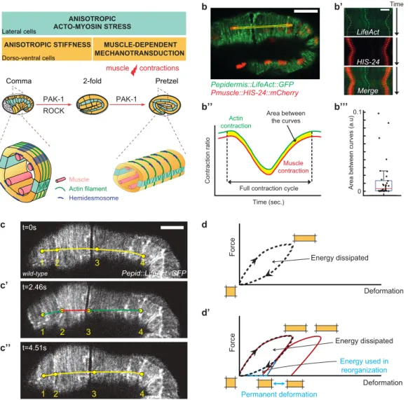
Figure 1: Muscle contractions deform the epidermis to their mechanical coupling
(a) C. elegans embryonic elongation from comma to 2-fold stages depends on a ROCK-promoted actomyosin force in seam cells (cyan) and actin-promoted stiffness in dorso-ventral cells (orange); elongation beyond the 2-fold stage requires repeated muscle contractions (red flash), which induce a PAK-1-dependent mechano-transduction pathway. Open cross-sections (bottom) show muscle positions. (b-b’’’) Epidermis actin filament (green) and muscle nucleus (red) tracking in a wild-type 2-fold embryo.
(b’) Kymographs from the yellow rectangle area (b) showing the concurrent displacement of epidermal actin and muscle nuclei. (b’’) Resulting displacement curves;
(b’’’) quantification of the area between them; its low value underlines the tight mechanical coupling between both tissues. Scale bar, 10 µm. (c-c'') A muscle contraction/relaxation cycle illustrating its local impact on epidermal actin filaments in a wild-type 2-fold embryo
(timing in left corner). Yellow (relaxation), red (compression) green (stretching) distances between landmarks denoted 1-4: (c) [1-2], 7.8 μm; [2-3], 19.8 μm; [3-4], 24.6 μm.
(c') [1-2], 9.4 μm; [2-3], 13.6 μm; [3-4], 26.2 μm. (c'') [1-2], 8.0 μm; [2-3], 19.2 μm; [3-4], 25.0 μm. In (b-c) the Pepidermis promoter is Pdpy-7.
(d) Hysteresis graph of an idealized elastic material returning to its initial shape after deformation (top), or showing permanent deformation30 (bottom).
PAK-1 MUSCLE-DEPENDENT MECHANOTRANSDUCTION ANISOTROPIC STIFFNESS PAK-1 2-fold ROCK Actin filament Hemidesmosome Muscle ANISOTROPIC ACTO-MYOSIN STRESS Lateral cells Dorso-ventral cells Comma Pretzel muscle contractions Force Force Deformation Deformation Energy dissipated Energy dissipated Permanent deformation Energy used in reorganization c’ c’’ d’ 1 2 3 4 Pepid::LifeAct::GFP t=0s t=2.46s t=4.51s wild-type 1 2 3 4 1 2 3 4
45 65 85 105 125 145 165 185 205 225 0 120 150 180 210 240 Time (minutes) Length (µm) 4-fold 2-fold d 100 80 60 30 0 60 90 120 150 180 210 240 unc-112(RNAi)
spc-1(ra409) spc-1(ra409) pak-1(tm403)unc-112(RNAi); spc-1(ra409) pak-1(tm403)
105 85 125 65 45 165 185 205 225 145 wild-type pak-1(tm403) spc-1(RNAi) spc-1(RNAi) pak-1(tm403) Length (µm) Time (minutes) unc-112(RNAi) unc-112(RNAi); spc-1(ra409) pak-1(tm403) e 30 0 60 90 120 150 180 210 240 pak-1(tm403) wild-type spc-1(RNAi) pak-1(tm403) spc-1(RNAi) 2-fold 1.5-fold 1.4-fold muscle contractions c
a YEAST TWO HYBRID SCREEN
prey bait SR9 KINASE domain CRIB domain 100AA SR10 SR8 SH3 Length (µm) *** *** *** *** *** *** 300 wild-type pak-1(tm403) spc-1(RNAi) spc-1(RNAi) pak-1(tm403) PAK-1 SPC-1 200 100 0 Primary readout : enhanced lethality
Secondary readout : elongation defect Liquid assay
dsRNA +
b
RNAi ENHANCER SCREEN in pak-1mutant
45
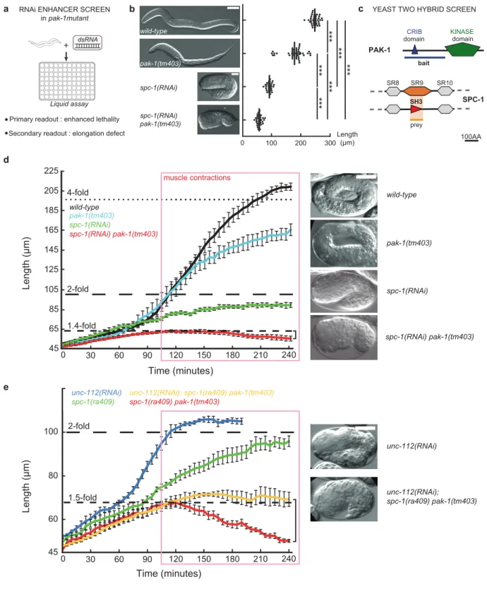
Figure 2: Loss of PAK-1 and SPC-1 triggers a muscle-dependent retraction of embryos
(a) RNAi screen in a pak-1 mutant identified spc-1 as an enhancer (Table S1).
(b) DIC micrographs of newly hatched wild-type, pak-1(tm403) (scale bar: 10 µm), spc-1(RNAi) and spc-1(RNAi) pak-1(tm403) (scale bar: 25 µm). Quantification of L1 hatchling body length: wild-type (n=38); pak-1(tm403) (n=32); spc-1(RNAi)(n=26); spc-1(RNAi) pak-1(tm40) (n=36).
(c) A yeast two-hybrid screen using the PAK-1 N-term domain as a bait identified the SPC-1 SH3 domain as a prey (orange background) (Table S2). (d) Elongation profiles and corresponding terminal phenotypes of wild-type (n=5), pak-1(tm403) (n= 5),
spc-1(RNAi) (n=8), spc(RNAi) pak-1(tm403) (n=8). (e) Elongation profiles in a muscle defective background.
unc-112(RNAi) (n=5); spc-1(RNAi) (n=8); unc-112(RNAi); pak-1(tm403) spc-1(ra409) (n=5); spc-1(RNAi) pak-1(tm403) (n=8). Right bracket (d, e), extent of retraction for spc-1(RNAi) pak-1(tm403) embryos. Scale bars, 10 µm. Error bars, SEM.
Angle (degrees) b’’ c’’ d’’ c’’’ d’’’ wild-type Pepid::LifeAct::GFP a’’ spc-1(RNAi) pak-1(tm403) pak-1(tm403) a b d a’ b’ d’ spc-1(RNAi) c c’ a’’’ 0 50 100 150 Angle (degrees) b’’’ pak-1(tm403) wild-type 0 50 100 150 Angle (degrees) wild-type 50 100 150 6 5 4 3 2 0 1 0 spc-1(RNAi) pak-1(tm403) wild-type spc-1(RNAi) x10-3 x10⁻³ peak p-value wt vs pak-1(-) 0.33 peak p-value spc-1(-) vs spc-1(-)pak-1(-) peak p-value wt vs spc-1(-) 6 5 4 3 2 0 1 6 5 4 3 2 0 1 x10⁻³ Angular coef ficient Angular coef ficient Angular coef ficient ROI Binary Ellipse fit FFT + high-pass filter Integration along each direction Angular coefficient spc-1(RNAi) peak p-value wt vs spc-1(-)pak-1(-) θ = 0 θ = 75 -2 1.5x10 -6 2.1x10 -3 2x10 # of segments 10 20 30 40 0 Length (pixels) 6 4 2 0 10 20 30 40 0 6 4 2 0 # of segments 10 20 30 40 0 6 4 2 0 # of segments 10 20 30 40 0 6 4 2 0 # of segments or Length (pixels) Length (pixels) Length (pixels) mean : 21,21 error of the mean : 0,87
mean : 16,56 error of the mean : 0,84
mean : 16,61 error of the mean : 0,64
mean : 12,89 error of the mean : 0,48
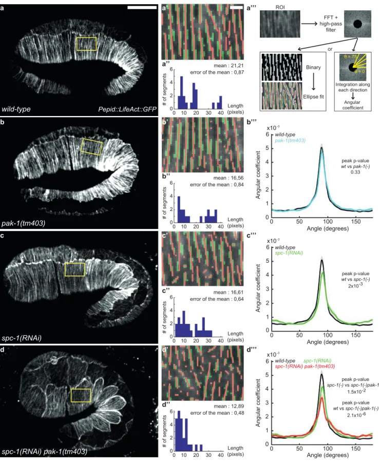
Figure 3: Actin filament defects in SPC-1 and PAK-1 defective embryos
(a-d) Epidermal actin filaments visualized with the Pdpy-7::LifeAct::GFP reporter construct in wild-type (a-a’’’), pak-1(tm403) (b-b’’’), spc-1(RNAi) (c-c’’’) and spc-1(RNAi) pak-1(tm403) (d-d’’’) embryos at mid-elongation (2-fold equivalent) stage.
Yellow rectangle, region of interest (ROI). Scale bar, 10 µm. (a’-d’) ROI after binarisation (green) and major axis detection (red),
based on (a’’’) three steps of image treatment for continuity and orientation analysis. (a’’-d’’) Actin continuity: distribution of actin
segments based on their length. Wild-type (n=16); pak-1(tm403) (n=21); spc-1(RNAi) (n=21) ; spc-1(RNAi) pak-1(tm403) (n=17) (b’’’-d’’’) Actin filament orientation: the curves represent the number of actin filaments oriented perpendicular to the elongation axis (90° angle in wild-type) based on the Fast Fourier Transformation (FFT in a’’’).
Wild type (n=18) ; pak-1(tm403) (n=20) ; spc-1(RNAi) (n=18) ; spc-1(RNAi) pak-1(tm403) (n=18).
h g
Readout : 1-fold embryo
in liquid assay
dsRNA +
RNAi ENHANCER SCREEN in spc-1(ra409)
fhod-1(RNAi); spc-1(ra409) spc-1(RNAi) pak-1(tm403) spc-1(RNAi)
lima bean lima bean + 130 min lima bean + 220 min
fhod-1(RNAi); spc-1(ra409) spc-1(ra409) + L4440 i j fhod-1(tm2363); spc-1(RNAi) *** 60 80 100 120Length(µm) spc-1(RNAi) pak-1(tm403) spc-1(RNAi) pak-1(tm403) ; FHOD-1(Full Length) spc-1(RNAi) pak-1(tm403) ; FHOD-1(ΔDAD) spc-1(RNAi) pak-1(tm403) ; FHOD-1(ΔFH2/DAD)
spc-1(RNAi) pak-1(tm403) ; FHOD-1(ΔFH1-DAD)
Length (µm) 60 100 *** n.s c dλ dl dt dt = βdldt if > 0 l - λ > Fepid = FseamαDV dαDV dl dt dt = γdldt if < 0 and αDV > 0 dl - k (l - λ) Fepid Fmuscles dt = + + a) b) c) d) Fold change 2 1
mech. model spc-1(-)pak-1(-)spc-1(RNAi) pak-1(tm403) mech. model wild-typewild-type
Cyclic stress (muscles) 3 4 Time (minutes) 0 50 100 150 200 η f d e Plastic spring k, λ, β,Fc η Fepid Fmuscles k, λ, β 0 Fepid Fmuscles wild-type PROPER REMODELING Contraction Relaxation d d - δd L + δl L spc-1 (-) pak-1 (-) short contraction cycles REMODELING DEFECTS L - δl L d + δd d normal contraction cycles a b PAK-1 SPC-1
actin binding protein
Legend: GBD FH3(DID) FH1 FH2 DAD
η Fc k a’ b’ 140 spc-1(RNAi)
spc-1(RNAi) pak-1(tm403) ; FHOD-1(ΔFH2/DAD) (siblings not carrying the construction)
* H(αDV) Hill(λ) *** 1 2 0<β<1
{
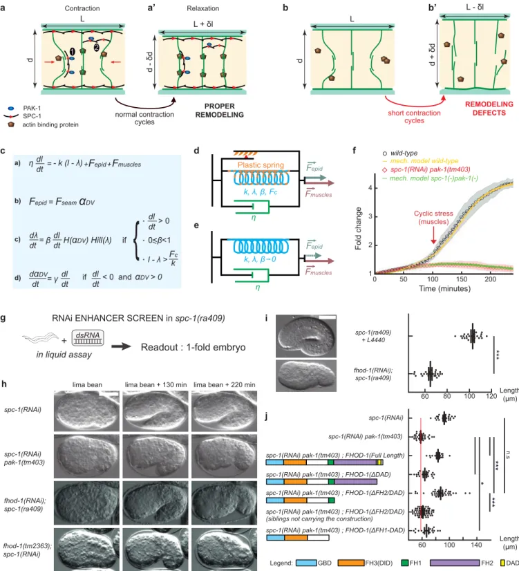
Figure 4: An actin-remodeling network providing mechanical plasticity ensures embryo elongation
(a-b’) Cellular model of embryo elongation.
(a-a’) In control embryos, muscle contractions (red arrows) provoke actin filament shortening in the dorso-ventral epidermis,
probably through sliding or shortening, followed by SPC-1/PAK-1-dependent actin stabilization. Whether spectrin is found along (scenario 1) or between (scenario 2) actin filaments is unknown (a). (b-b’) In spc-1 pak-1 deficient embryos, actin remodeling goes uncontrolled.
(c-f) Viscoplastic mechanical model of embryo elongation. The embryo is represented as a Kelvin-Voigt solid (spring stiffness k, resting length λ, viscosity η) submitted to the forces Fepid and Fmuscle. System equations for the model.
(d) Wild-type case: an increasing resting length during stretching phases imparts mechanical plasticity. (e) spc-1 pak-1 mutants: Fepid progressively decreases.
(f) Comparison of experimental and predicted elongation curves taking the constitutive equations shown in (c). (g) A retraction screen in a spc-1 mutant identifies fhod-1.
(h) Snapshot at three time-points of spc-1 deficient embryos in control, pak-1 or fhod-1 backgrounds;
(i) terminal body length at hatching: spc-1(ra409) after feeding on L4440 control (n=21), or fhod-1(RNAi) (n=25) bacteria. (j) Pdpy-7 driven epidermis expression of truncated FHOD-1 variants and terminal body length at hatching: spc-1(RNAi)(n=26); spc-1(RNAi)pak-1(tm403) no transgene (n=36), FHOD-1(full length) (n=16), FHOD-1(∆DAD) (n=17), FHOD-1(∆FH2-DAD) (n=38) and non-transgenic siblings (n=78), FHOD-1(∆FH1-FH2-DAD) (n=18). Scale bar, 15 µm. Error bars, SEM. P values: *<0,05; **<0,001; ***<0,0001; ns, not significant.
1
SUPPLEMENTARY MATERIAL
1
An actin-based viscoplastic lock ensures progressive body axis elongation
2
Alicia Lardennois1*, Gabriella Pásti2*, Teresa Ferraro1, Julien Pontabry2,3, David
3
Rodriguez2, Samantha Kim2, Flora Llense1, Christelle Gally2, Michel Labouesse1,2 #
4
5
Content
6
1- Supplementary Mechanical Modeling
7
2- Methods
8
3- Supplementary Figures S1-S8 with legends
9
4- Captions for movies S1-S8
10
5- Supplementary References
11
12
13
1-Supplementary Mechanical Modeling
14
1.1 Background information. During embryogenesis the C. elegans embryo
15
undergoes a process of elongation whereby it becomes four times as long as the
16
long axis of the eggshell (50 µm). Cell proliferation and cell intercalation are absent,
17
therefore the process of axis elongation relies only on the ability of the embryo to
18
extend in the anterior-posterior direction. The outer epithelium (epidermis) plays an
19
essential role in this process.
20
Changing the status of any physical entity requires the involvement of a force
21
(mechanical or chemical), and the C. elegans embryo is no exception to this rule of
22
physics. During the first phase of elongation and until muscles become active, the
23
2
machinery driving elongation involves an active force in the lateral epidermis (also
24
called seam cells), and a passive force exerted by the dorsal and ventral epidermal
25
cells (called DV cells) adjacent to the seam cells (Fig. 1a). Seam cells have a high
26
concentration of non-muscle myosin II, which has a non-polarized distribution and
27
does not display pulsatile flows1,2, as observed for instance during Drosophila
28
germband elongation3. Nevertheless, the stress generated by the seam cells is
29
anisotropic and globally oriented along the DV axis (see cyan box in Fig. 1a)2. The
30
stress anisotropy results mainly from the presence of circumferential F-actin filament
31
bundles in DV epidermal cells, which create a global stiffness anisotropy (see yellow
32
box in Fig. 1a). The DV epidermal cells do not contribute to generate active stress
33
(Fig. 1a), as their myosin II is kept mostly silent through the activity of the RhoGAP
34
RGA-21,2,4. The interplay between stress anisotropy in seam cells, stiffness anisotropy
35
from the DV epidermis, and hydrostatic pressure resulting from the reduction of
36
embryo diameter, induces a force oriented along the AP direction that is sufficient to
37
extend the embryo until it reaches the 2-fold stage2.
38
Note that here as well as in the main text we refer to each elongation phase based
39
on the ratio between the actual embryo length and that of the eggshell long axis,
40
i.e. 1.7-fold or 2-fold means that the embryo has reached roughly 85 µm or 100 µm,
41
respectively. Importantly, in mutant embryos which extend slower, we refer to
42
embryo stages based on the length that a wild-type embryo would reach after the
43
same time duration with t0 corresponding to the beginning of elongation (see
44
extension curves in Fig. 2d and Movie 1).
45
For simplicity, let us call the net force in the AP direction produced by the epidermis
46
the epidermal cell force (Fepid). This force is not enough to explain the elongation up
47
to the 4-fold stage, since genetic analysis has established that embryos with
non-48
3
functional muscles do not elongate beyond the 2-fold stage (Fig. 2b)5. Therefore,
49
muscles provide a second active force driving elongation beyond the 2-fold stage,
50
which we will call Fmuscles.
51
During embryogenesis, muscles organize and assemble in four rows located under
52
the epidermis (Fig. 1a). Muscles are attached to the extracellular matrix that
53
separates them from the epidermis, and that in turn serves to anchor the epidermis
54
through hemidesmosome-like junctions1. Muscle organization and maturation is a
55
progressive process, such that muscle activity starts with small contractions at the
1.7-56
fold, which progressively become more robust. The mechanical activity of muscles
57
can be summarized as an alternation of contractions followed by relaxation. Since
58
muscles are tightly connected to the epidermis, their contractions locally and
59
repeatedly induce an anterior-posterior compression and extension of the epidermis,
60
which can be visualized through the displacement of the actin cables (Fig. 1b-c, Fig.
61
S7). The stress exerted by muscle contractions on the epidermis induces a
62
mechanotransduction pathway (2nd yellow box in Fig. 1a), which is essential to
63
promote hemidesmosome maturation and embryo elongation8.
64
65
1.2 Viscoplastic model. The C. elegans epidermis can be modelled as a visco-elastic
66
body, more specifically as a Kelvin-Voigt system with a spring and dashpot in
67
parallel, subject to two main active forces: the epidermal force Fepid, which is a
68
continous positive force, and the muscle force Fmuscles, which is a pulsatile force since
69
muscles alternatively contract and relax. The first force is present since the beginning
70
of elongation, whereas the second force starts only after the 1.7-fold stage. The
71
elastic reaction of the epidermis to active forces can be captured by Hooke’s law;
72
the damped nature of the reaction can be expressed by a viscous term. Overall the
73
4
length of the embryo over time l(t) can be captured by the equation:
74
𝜂 !"!"= −𝑘 𝑙 − 𝜆 + 𝐹!"#$+ 𝐹!"#$%&# (1)
75
where k is the body stiffness, λis the worm resting length and η is the coefficient of
76
viscosity. Inertia has been neglected given the low Reynolds number of the system.
77
Eq. (1) corresponds to the so-called Kelvin-Voigt viscoelastic model6,7 that captures
78
the behavior of viscoelastic solids under stress or deformation. For constant forces,
79
the solution of Eq. (1) is given by:
80
𝑙 𝑡 =!!"#$! !!"#$%&#
! (1 − 𝑒!! !) + 𝜆 (2)
81
meaning that the length of the system relaxes to the plateau value !!"#$! !!"#$%&#
! in a
82
relaxation time of 𝜏 = 𝜂/𝑘.
83
Fepid can promote elongation until the 2-fold stage (Fig. S5a’). Beyond, the pulsatile
84
force originating from muscles, Fmuscles, alternates periods of positive, negative or null
85
contribution, so that its temporal average < 𝐹!"#$%&# >! is null:
86
< 𝐹!"#$%&# >!= ! ! 𝐹!"#$%&#(𝑡) ! ! 𝑑𝑡 = 0, for 𝜃 = 𝑛𝑇 (3)87
where θ is the integration period, T is the period and n is a positive integer. As a
88
consequence, on average, Fmuscles will not contribute tothe steady state length (𝑙!!)
89
of the embryo (see Fig. S5a-c) that is set by 𝑙!!=!!"#$! + 𝜆.
90
A way to introduce a positive contribution to embryo lengthening is to allow some
91
plasticity, in the physical sense, or ability of the system to get reorganized. For
92
example, let consider a stretching pulse due to muscle activity during which the
93
embryo increases its length l(t) with an increment dl, such that the embryo will be
94
5
temporarily l(t)+dl long at the end of the pulse. During the subsequent relaxation
95
phase, due to elasticity in the system, the embryo should return to the initial length l(t)
96
it had before muscles had locally extended it. However, if it undergoes a permanent
97
plastic deformation, then the body will permanently keep a portion of the stretched
98
length. This situation corresponds to a permanent rearrangement, and it has been
99
observed and modeled in biological systems undergoing stresses8,9. Similarly to9, a
100
simple mathematical solution to introduce plasticity consists in having an adjustable
101
resting length λ that increases linearly with the length l(t) according to:
102
!" !" = 𝛽 !" !"H(𝛼!") if !" !"> 0 and 𝑙 − 𝜆 > 𝐹! 𝑘 (4)103
where 0≤β<1 is a proportionality factor called ‘plasticity factor’; the case of β=0
104
corresponds to an absence of plasticity. The condition !"
!"> 0 ensures that there is
105
rearrangement only during the extension phases and the condition; 𝑙 − 𝜆 > 𝐹! 𝑘
106
means that the rearrangement takes place only if the applied force exceeds a
107
critical force 𝐹!. The term H(𝛼!") is the Haeviside step function, which expresses that
108
dorso-ventral rearrangement is possible only in presence of resistance. For the
109
description of 𝛼!"term, see next paragraph. Note that Eq. (4) is equivalent to
110
𝜆 = 𝜆 0 1 − 𝛽 + 𝛽 𝑙 with 𝜆 0 being both the length and the resting length at time
111
zero and 𝐹! = 0. With these choices and with a constant positive force F Eq. (1) has
112
the following solution:
113
𝑙(𝑡) =(!!!)! !!(1 − 𝑒!!(!!!)!) + 𝜆(0) (5)
114
Hence, the plasticity condition effectively reduces the body stiffness 𝑘 to 𝑘(1 − 𝛽)
115
enabling the system to reach a longer final size compared to the one allowed by the
116
Kelvin-Voigt system alone, and increasing the relaxation time. By introducing
117
6
plasticity like in Eq. (4), the body progressively gains length at each stretching phase
118
(Fig S5d-d’).
119
120
1.3 The consequences of actin stability defects. As reminded above, the intensity of
121
Fepid relies on two components: the constant contractility of the seam cell actomyosin
122
network, and the stiffness of the actin cables in DV cells. We can then represent it
123
like:
124
𝐹!"#$= 𝐹!"#$ 𝛼!" (6)
125
where the force 𝐹!"#$ represents the active force generated by myosin II in the
126
lateral cells and 𝛼!" is the passive component given by the presence of actin
127
filament bundles in the DV cells. The biomechanical significance of equation (6) is
128
that both 𝐹!"#$ and 𝛼!" positively contribute to Fepid , and that if one is absent Fepid=0.
129
This captures the fact that in the absence of myosin II there will be no pulling force
130
because the active component is absent, and that if actin cables are lost myosin II is
131
missing the resistance structure onto which it can pull, resulting in a null epidermis AP
132
force.
133
As shown in the main text (Fig. 3), the absence of SPC-1 and PAK-1, combined with
134
the mechanical input from muscles, induces actin integrity defects in DV cells. To
135
translate this situation in mathematical terms, we chose to write the passive
136
component 𝛼!" as follows:137
!!!" !" = 𝛾 !" !" 𝑖𝑓 !" !"< 0 and 𝛼!" ≥ 0 (7)138
where γ≥0 is a proportionality factor defined as a ‘tearing factor’. The condition
139
7 !"(!)
!" < 0 means that 𝛼!"decreases stepwise with time. In addition, we are imposing
140
that γ=0 corresponds to an absence of tearing, like in wild-type embryos. The
141
biological significance of this choice is the following: in phases of length decrease
142
(net negative force), the overall circumference of the embryo should increase due
143
to volume conservation (Fig. S4). In a background of unstable actin filaments (like in
144
spc-1 pak-1 double mutants) their resistance to stress originating from muscle activity
145
would not be maintained. For this reason, 𝛼!" should progressively decrease at each
146
cycle. The condition 𝛼!" ≥ 0 prevents 𝛼!" from assuming negative values. Thus with
147
𝛼!" decreasing, Fepid will progressively decrease, and as consequence the system
148
length will shorten (see Fig. S5e and Fig. 4e).
149
150
1.4 Equations summary. In summary, we describe the embryo body as a plastic
151
Kelvin-Voigt solid according to the following system of equations:
152
𝜂 !"
!"= −𝑘 𝑙 − 𝜆 + 𝐹!"#$+ 𝐹!"#$%&# (1) (Kelvin-Voigt with adjusting resting length
153
subject to Fepid+Fmuscles)
154
!" !" = 𝛽 !" !" H(𝛼!") if !"!"> 0 , 0 ≤ β < 1 and 𝑙 − 𝜆 > 𝐹! 𝑘 (5) (plasticity condition)
155
!!!" !" = 𝛾 !" !" 𝑖𝑓 !"!"< 0 and 𝛼!" ≥ 0 (7) (tearing condition)
156
With 𝐹!"#$= 𝐹!"#$ 𝛼!" as described in Eq.(4).
157
We assume that γ=0 for wild-type and unc-112, meaning that the resistance of
dorso-158
ventral actin filament bundles remains unaffected by the body length changes
159
caused by muscle activity.
160
8
The equations (1), (4), (5) and (7) have seven parameters: η, k, Fseam, 𝛼!"(0), Fmuscles, β
161
and γ. In order to reduce the parameter space we fixed some of them:
162
- for simplicity we set k=1;
163
- from the laser ablation experiments performed in reference2, the relaxation
164
time of epidermal actin filaments following the laser cut is in the order of a few
165
seconds. Being the relaxation time in a Kelvin-Voigt system given by 𝜏 = 𝜂/𝑘,
166
we set η=3 so that the relaxation time is 3 seconds.
167
- Fepid is the multiplication of two parameters, and thus from the parameter
168
point of view can be considered as a single parameter that we formally set to
169
Fseam=1 letting 𝛼!"(0) as a free parameter.
170
- The size of the critical force 𝐹! has been chosen to be half Fseam (𝑭𝒄 = 𝟎. 𝟓).
171
Thereby, we consider that low intensity forces cannot trigger a plastic
172
response.
173
- Regarding Fmuscles, we specified its details on the basis of the measured
174
contraction durations for embryos between 1.7 and 2-fold stages (Fig. S7). For
175
wild-type embryos, the duration of positive and negative periods has been set
176
to 6 seconds and the period of null contribution has been set to 15 seconds,
177
whereas for spc-1 mutants and spc-1 pak-1 double mutants the duration of
178
non-null activity has been set to 3 seconds and the duration of null
179
contribution is set to 15 seconds. In wild-type embryos, the intensity of Fmuscles
180
has been left as a fit parameter together with 𝛼!", β and γ in order to be
181
determined by comparing with the data. The muscle force amplitude for
spc-182
1 and spc-1 pak-1 mutants has been set to 50% of the wt intensity according
183
to our experimental observations.
184
To better adapt to the experimental observations, both Fseam and Fmuscles have been
185
modified by introducing an initial transient that sets their behavior from zero to the