https://doi.org/10.1002/hbm.23613
Neural correlates of experimental trauma memory retrieval
Geraldine Gvozdanovic,1,2 Philipp Stämpfli1, Erich Seifritz1,4 & Björn Rasch3,4*
1 Department of Psychiatry, Psychotherapy and Psychosomatics, Psychiatric Hospital, University of Zürich, Zürich Switzerland
2 Institute of Psychology, University of Zürich, Zürich, Switzerland
3 Department of Psychology, University of Fribourg, Fribourg, Switzerland 4 Zurich Center for Interdisciplinary Sleep Research (ZiS), University of Zürich,
Switzerland
*Corresponding author:
Björn Rasch, Department of Psychology, Division of Cognitive Biopsychology and Methods, University of Fribourg, Rue P.-A.-Faucigny 2, CH-1701 Fribourg, Switzerland;
Email: [email protected] Phone: +41 26 300 7637
Words abstract: 188 Words article body: 3741 Tables: 2
Figures: 3
Keywords: trauma, retrieval, analogue study, intrusive memories, fMRI Abstract
Objectives
Traumatic memories such as intrusions and flashbacks play a major role in the development and maintenance of Posttraumatic Stress Disorder (PTSD). A thorough understanding of the neural mechanisms underlying traumatic memories is indispensable for precise diagnosis, for personalized treatment and prevention. In particular, the identification of early neural
predictor variables for intrusion development shortly after trauma exposure requires detailed investigation.
Experimental design
Here we examined the neural correlates of early experimental trauma memory retrieval in a traumatic film paradigm in 42 young healthy females, using both implicit and explicit retrieval tasks.
Principal observations
We show that implicit experimental trauma retrieval specifically involved the retrosplenial cortex and the anterior cingulate cortex (ACC), while both retrieval tasks resulted in trauma-related activity in the posterior cingulate cortex (PCC) and the precuneus. Importantly, neural activity early after experimental-t auma exposure predicted later intrusion development, with independent contributions from activity in the retrosplenial cortex (implicit retrieval) and the PCC (explicit retrieval). Additional PPI analyses revealed stronger connectivity between the bilateral amygdala and the supplementary motor area, pre- and paracentral lobule for the control group compared to the experimental trauma group.
Our study gives new insights in the neural correlates of experimental trauma memory retrieval and their predictive value for subsequent symptom development. Our results could provide the basis for personalized early treatment and prevention of PTSD.
Introduction
Traumatic memories are a crucial symptom of Posttraumatic Stress Disorder (PTSD) and play a major role in the development and maintenance of PTSD (APA, 2013; Elbert and Schauer, 2002; Michael, et al., 2005). Traumatic memories represent a major distress to patients as they are reactivated involuntarily after exposure to internal or external cues. They are rendered highly intrusive and disturbing. Several theoretical approaches describe PTSD as a memory disorder. For example, . Brewin and colleagues (Brewin, et al., 1996) propose in their dual representation model that traumatic memories are mainly associated with a situationally accessible memory system (SAM). The SAM system contains lower level perceptual
processing and associated emotions, which are easily reactivated by memory cues and lead to intrusions, flashbacks and nightmares. In contrast, the verbally accessible memory (VAM) is a more complex system which enables voluntary verbal recall and interacts with
autobiographical memories to achieve an integration of the trauma into personal context (Brewin, 2014; Brewin, et al., 1996). Along similar lines, Ehlers and Clark (Ehlers and Clark, 2000) assume that trauma memories are inadequately integrated into context and other
autobiographical memories. The authors propose that a traumatic memory is processed as if an individual would still be under current threat (Ehlers and Clark, 2000).This leads to a strong implicit memory bias (perceptual priming) for stimuli associated with the trauma and a reduced threshold for trauma reactivation by traumatic memory cues. Also Elbert and Schauer (Elbert and Schauer, 2002) hypothesize that the isolation and lack of integration of traumatic memories might be a major cause for the intrusive and involuntary nature of traumatic memories. They assume that autobiographical memories can be divided into cold (fact knowledge of life events) and hot (emotional and sensory) memories, which are typically
interconnected for “healthy”, non-traumatic memories (Elbert and Schauer, 2002). However, exposure to a traumatic event can induce unusually high arousal responses associated with strong physiological and neurophysiological responses such as stress hormone releases and activation of brain structures involved in emotional processing (Ozer, et al., 2003). For example, exaggerated amygdala response during encoding of negative stimuli has been associated with increased hippocampal activity and PTSD symptom severity (Brohawn, et al., 2010). This “hyperactivity” during encoding of a traumatic event might lead to an
“overconsolidation” of traumatic memories (Pitman, 1989). During this process, memory-related brain areas such as the amygdala and hippocampus can lose their interconnectivity (Elbert and Schauer, 2002), creating an isolated fear network of strong, integrated hot memories.
In sum, several theoretical accounts describe PTSD as a memory disorder (Brewin, 2014), mainly associated with a reduced integration of traumatic memories in the autobiographical memory network. PTSD patients typically exhibit an enhanced recall of trauma-related stimuli, an altered retrieval of autobiographical memories in response to cue words and also show an enhanced implicit memory in complex tasks. (McNally, 1997). Despite their intense vividness, traumatic memories have the capacity of changing their structure (McNally, 2005) and can even be strongly suppressed by patients. As memory processes are assumed to play a major role for PTSD, a thorough understanding of the neural mechanisms underlying the development of traumatic memories is indispensable and not yet achieved.
Previous research focusing on traumatic memory retrieval was mainly conducted in patients with full psychiatric manifestations of PTSD. It revealed altered neural correlates during the presentation of trauma-related stimuli and trauma memory in a variety of brain regions. Most prominent are brain activations in the amygdala, ventromedial prefrontal cortex, anterior cingulate cortex (ACC) and insular cortex during trauma related stimuli (Pitman, et al., 2012). A recent meta-analysis of trauma memory in PTSD revealed an increased activation in the
retrosplenial cortex, precuneus, ACC and amygdala (Sartory, et al., 2013). Moreover, patients who implicitly processed trauma stimuli exhibited an increased activation in the amygdala, ventrolateral prefrontal cortex and fusiform gyrus (Morey, et al., 2009) in a working memory task with trauma related distractors. In contrast, patients who explicitly focused on a trauma during a script-driven imagery task exhibited increased brain activation in thalamus, ACC and medial frontal gyrus and enhanced connectivity in the posterior cingulate cortex, parietal lobe and occipital lobe, specifically on the right hemisphere (Lanius, et al., 2001; Lanius, et al., 2004). A recent study that directly compared conscious and subconscious processing of trauma reported that, while subconscious trauma processing resulted in an increased activity in the posterior cingulum, conscious trauma processing was correlated with an increased activity in the basal forebrain (Rabellino, et al., 2016).
Results indicate the necessity of differentiating between different memory types in PTSD. Moreover, as the distress of traumatic memories is also due to involuntary intrusiveness, differences in neural correlates of implicit vs. explicit retrieval attempts might also be functionally relevant for development and maintenance of the disorder.
The theories on trauma memory in PTSD mentioned before (Brewin, et al., 1996; Ehlers and Clark, 2000; Elbert and Schauer, 2002; McNally, 1997) all assume that there are dysfunctions involved in different types of memories in PTSD. Presumably, these different memory
systems also underlie diverse neural correlates in the brain (Brewin, 2001). Implicit memory is induced by trauma cues and typical for the induction of intrusions. Explicit memory on the other hand involves a direct trauma retrieval and is used in trauma therapy to improve trauma memory integration. A recent study (Rabellino, et al., 2016) directly compared responses to conscious and unconscious (subliminal) processing of trauma-related cues and reported fast defensive reactions of an inate alarm system that are not processed in a conscious way (Rabellino, et al., 2016). In the current study, we will directly compare neural responses to incidentally processes trauma cues (implicit memory) and intentional processing and
imagination of trauma scripts (explicit memory) using established paradigms on implicit (Morey, et al., 2009; Oei, et al., 2012) and explicit (Lanius, et al., 2001; Lanius, et al., 2004) memories to shed new light on memory processing in PTSD.
In addition, our study in healthy participants evaluates experimentally-induced trauma
memory retrieval at a very early stage. Studies on patients with full psychiatric manifestations usually focused on traumatic memory retrieval in later stages of the disorder. However, neural correlates of trauma memory in early stages might have a crucial predictive value for later symptom development. Thus, in order to understand the neural mechanisms of the
development of traumatic memories, an examination of the early period after trauma exposure is required, which can be achieved by an analogue study in healthy participants using the trauma-film paradigm. The trauma-film paradigm was first developed over 50 years ago (Horowitz, 1969; Lazarus, 1964) and has been validated in over 70 studies only in the past years (James, et al., 2016). It is an experimental medicine model that evaluates deviant behavior in non-patient samples in order to analyze processes involved in the development of PTSD (James, et al., 2016). Responses comparable to real-life trauma have been proven to be observed in healthy subjects after an experimental trauma (i.e. trauma film) (James, et al., 2016). Responses include hallmark symptoms of PTSD (James, et al., 2016). Specifically, physiological arousal, intrusive memories, negative cognitions and mood have been reported (APA, 2013; Butler, 1995; Holmes, et al., 2004; James, et al., 2016; Weidmann, et al., 2009). However, in contrast to PTSD, symptoms after an experimental trauma only last for hours or days and finally decrease within one week (Holmes, et al., 2004; James, et al., 2016). In our study, healthy participants watched a traumatic film consisting of a rape scene, which resembles witnessing a traumatic event. According to the diagnosis criteria for PTSD, both direct exposure to a traumatic event and witnessing trauma can result in PTSD (APA, 2013), and particularly witnessing sexual assaults have been associated with PTSD symptoms (Witte, et al., 2016). Thus, the trauma film paradigm used in the current study provides an optimal
setting to bridge clinical and non-clinical samples in a highly controlled environment in order to examine PTSD symptoms (Clark and Mackay, 2015).
Concerning the neural correlates in analogue studies, there has only been one functional magnetic resonance imaging (fMRI) study focusing on the encoding of an experimental traumatic event itself (Bourne, et al., 2013). Neural correlates during encoding involved brain regions of the amygdala, striatum, thalamus and anterior cingulate cortex. These brain regions significantly predicted development of experimental trauma memory intrusions (Bourne, et al., 2013).
In contrast, brain areas and processes involved in early retrieval attempts of traumatic memories and their relation to traumatic intrusion development as a predictor variable have not yet been identified. The present study aims at filling this gap by identifying neural correlates of experimental trauma memory retrieval shortly after exposure in an analogue paradigm with healthy participants. In particular, we directly compare neural correlates of implicit and explicit experimental rauma memory retrieval and their predictive values for the development of subsequent intrusive memories.
We hypothesize that different neural correlates are involved in implicit and explicit trauma film memory retrieval, both predicting later intrusive memories in a distinct way. We predict implicit memories to be associated with brain regions characteristic of emotional processing and emotional memory such as the ACC, amygdala and posterior cingulate cortex. During the explicit memory task, we predict both emotional-memory associated brain regions and
episodic/autobiographical memory regions such as the retrosplenial cortex, the PCC and the precuneus as there is a deliberate focus on the trauma film memory itself.
Methods and Material
Forty-five healthy, non-smoking, right-handed female participants underwent a general screening to exclude present and past medical and psychiatric disorders and for standard MRI exclusion criteria. Due to technical problems, three participants had to be discarded from the experiment (one participant fell asleep in the scanner and data of two participants could not be analyzed due to technical difficulties), leaving forty-two healthy participants in the sample (mean age 23.3, range 19 – 33 years). Subjects provided their written informed consent after the study protocol had been fully explained. Additionally, subjects were reimbursed for their participation (25 CHF/h). The study protocol was in line with the Declaration of Helsinki and was approved by a local ethical review board.
Experimental procedure and design
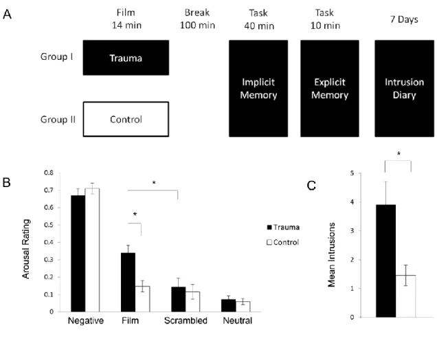
Participants arrived at the imaging center and the procedure was explained. First, they were trained for the experimental paradigms (implicit and explicit memory task, see below). Afterwards one half of the subjects (n = 21) watched a trauma movie (experimental-rauma group) and the other half (n = 21) watched a neutral movie (neutral group), according to a between subject design. 100 min after the movie, participants performed first an implicit and then an explicit memory task. Meanwhile fMRI was performed. After the measurements, participants received an intrusion diary with a detailed explanation for the following seven days and were sent home (see Figure 1A, for a summary of the procedure).
--Insert Figure 1 about here--
Film paradigm:
According to the trauma film paradigm of (Holmes and Bourne, 2008), participants either watched a trauma film or a control film (duration of both film clips: 14 min). In the trauma
film, rape and further physical assault was depicted (movie called “irreversible”, Gaspar Noe, 2002). In the neutral movie, a collection of neutral scenes with the same characters was displayed. Both movies had the same duration. Participants had to rate feelings on an emotionality scale before and after the movie. Moreover, saliva samples were taken before and after the movie.
Implicit memory recall: Sternberg task with emotional distractors
For the implicit paradigm, we used a Sternberg working memory task with emotional distractors (Oei, et al., 2012) (see Supplementary Material for details). The emotional
distractors were presented for 4 s and consisted of four different picture categories. Categories included 20 film pictures, 20 negative and 20 neutral pictures taken from the international affective pictures system (Lang, et al., 2008) and 20 scrambled pictures. Both neutral and negative IAPS pictures were matched in brightness to the film pictures. Scrambled pictures consisted of a collage of all pictures and were blurred with a Gaussian filter. Notably, our film pictures consisted of emotionally neutral pictures taken from both movies equally to prevent familiarity effects (trauma and neutral films). They included no action but rather served as a (initially neutral) reminder cue (e.g. tunnel where the protagonist was raped). Throughout all trials, participants were explicitly asked to focus on the Sternberg task and not on the
distractors, so that the emotional distractors solely trigger implicit memories of the film watched before. After completion of the Sternberg task, all pictures were presented again and rated according to vividness and arousal on an 11-point Likert scale.
In the explicit memory tasks, participants listened to an audiotape retelling the traumatic and neutral movie clip, according to a classical script-driven imagery design (Pitman, et al., 1987) (see supplementary information for details).
The intrusion diary consisted of a seven-day diary which had to be filled in at home.
Participants were educated about intrusive memories and asked to write down every intrusive memory that came to their mind. This included content, vividness, distress, and intrusive characteristics for every memory that was experienced daily during this time. Intrusion diaries were analyzed and the sum of reported intrusions per week was used for further analyses. Scoring criteria for intrusions included ratings on distress, vividness and the sudden occurrence of the reported memories.
Functional MRI data acquisition and analysis.
Measurements were performed on a Philips Achieva 3.0 Tesla TX whole-body magnetic resonance scanner (Philips Healthcare, Best, The Netherlands) (see supplementary
information for details). All tasks and films were presented on MR compatible video goggles (Resonance Technology, Northridge, USA). Furthermore, for good sound quality, headphones especially designed for MR measurements (MR confon GmbH, Magdeburg, Germany) were used. Standard image data preparation, preprocessing, and statistical analysis were performed with the software SPM8 (www.fil.ion.ucl.ac.uk/spm). Functional data preprocessing included a correction for slice-scan timing acquisition, realignment to the first volume using rigid body transformation and normalization to the EPI template in Montreal Neurological Institute (MNI) space. Images were subsequently resliced to a voxel resolution of 2 × 2 × 2 mm3 and smoothed using a 6 mm full width at half maximum Gaussian kernel. A general linear model was performed. In the first level analysis of the implicit memory task (Sternberg-task), stimulus-onsets and duration of each emotional distractor category (neutral film pictures, negative and neutral IAPS pictures and scrambled pictures) were included in the model. The main contrast of interest on the first level was the difference in activity for film pictures > scrambled pictures. In the explicit memory task (script driven imagery), first level fMRI-models included stimulus-onsets and duration of each audiotape (both trauma audiotape and
control film audiotape). The main contrast of interest was the difference between the trauma audiotape and the neutral audiotape. In addition, six affine motion correction regressors were included as regressors of no interest in both analyses. On the second level, random effects analyses were performed for statistical inference on the group level. Statistical significance was set at pFWE.cluster < 0.05, with a cluster defining threshold (CDT) > of 0.001.
Psychophysiological interaction effects and brain behavioral correlates
For psychophysiological interaction (PPI) analyses and the brain-behavior correlates, we extracted seeds from second level group results for the implicit (ACC, PCC and retrosplenial cortex) and explicit memory task (precuneus, PCC) using a 6 mm sphere around the center coordinates of the second level cluster peaks (see supplementary information).
Additionally for the PPI, we used the hippocampus and amygdala as predefined regions of interest (ROIs) from the Wake Forest University (WFU) Pickatlas. Statistical significance was set at pFWE.cluster < 0.05, with a cluster defining threshold (CDT) > of 0.001 (Eklund, et al., 2016).
For the brain behavior correlates a stepwise linear regression was performed with the sum of intrusions as an independent variable and peak levels (6 mm sphere) of brain regions found in the second level analysis of both implicit and explicit tasks as dependent variables (please see below). Stepwise selection criteria for inclusion: probability of F < 0.05 an exclusion:
probability of F < 0.10.
Results
As expected, participants who had seen the traumatic movie (trauma group) rated the film pictures as more arousing and negatively valenced as the neutral film group (t (40) = 3.5 p = 0.001, see Figure 1B, for ratings see Supplementary Table I. Note that all film pictures showed neutral scenes from both the trauma and the neutral movies, so no explicit traumatic action was shown. Thus, participants from the experimental trauma group rated the same (initially neutral) film pictures as more negative and more arousing than the neutral group due to watching the traumatic movie before. In addition, participants from the experimental trauma group rated the film pictures as more negative and more arousing than neutral IAPS pictures (t20 = 6.6, p < 0.001), while participants from the control group did not do that. Subjective valence and arousal ratings did not differ significantly between the experimental- trauma and the control group for negative, neutral and scrambled pictures (all p > 0.10). The pattern was substantiated by a significant interaction effect in the between-subject factor of film type (experimental trauma vs. neutral) and picture type (negative, neutral, film and scrambled pictures; valence: F3,120 = 7.80, p = 0.003 and arousal F3,120 = 5.44, p < 0.001). Performances in the Sternberg-Task did not differ significantly, neither between the two experimental groups nor between pictures types (all p > 0.10).
Main effects of brain activity underlying experimental trauma memory retrieval
In accordance with previous studies in PTSD patients, implicit retrieval of experimental trauma memories was associated with robustly increased brain activity in the retrosplenial cortex/precuneus, posterior cingulate cortex (PCC) and anterior cingulate cortex (ACC, all p < 0.05, family-wise error corrected for multiple comparisons for the whole brain on cluster level; see Figure 2 A and B and Table I).
When looking at experimental trauma > control and specifically at the film pictures > negative IAPS pictures contrast, the retrosplenial cortex and the precuenus were still
significant. However, the PCC and ACC were not found in this contrast (see Supplementary Figure 3).
Interestingly, also in the explicit memory retrieval task (script-driven imagery), brain activation was increased in the experimental trauma group compared to the neutral group. Importantly, brain regions were partly similar to the implicit retrieval task, including the left and right precuneus and the posterior cingulate cortex (p < 0.05, family-wise error corrected for multiple comparisons on the cluster level, see Figure 2 C and D and Table II). No
increases in brain activity were observed in the control group compared to the experimental trauma group, neither in the implicit nor the explicit retrieval task. Note that between groups (experimental trauma and control) exactly the same stimuli were presented in both retrieval tasks (i.e. implicit: film pictures; explicit: trauma scripts). Thus, any difference in brain activity between the experimental trauma and the control group can only be explained by the fact that the experimental trauma group previously saw the trauma film and retrieved
memories of this film during the implicit and explicit retrieval tasks.
--Insert Table I and II about here--
--Insert Figure 2 about here--
Psychophysiological interaction analyses of experimental trauma memory retrieval
In the implicit retrieval task, PPI analyses with a bilateral amygdala seed revealed a higher connectivity with the supplementary motor area, pre- and paracentral lobule for film pictures > scrambled pictures (pFWE.cluster < 0.05, see Supplementary Figure 1), which was significantly stronger in the control group compared to the experimental trauma group. Interestingly, the
higher connectivity with the supplementary motor area and pre- and paracentral lobule was found also with a bilateral hippocampus seed, an ACC and a PCC seed. There were no significant connectivities with any of the reported seeds that were stronger for the experimental trauma compared to the control group.
In the explicit retrieval task, the PPIs with a bilateral amygdala and hippocampus seed
revealed no significant differences in connectivity. However, a PCC seed revealed significant higher connectivity in the left and right superior-, middle- and inferior temporal lobe, and supramarginal gyrus (pFWE.cluster < 0.05, see Supplementary Material, Figure 1). This result was found in the trauma audio file > control audio file for the control group compared to the experimental trauma group. There were no significant PPI results in any of these seeds in the experimental trauma group > control group contrast.
Brain activity during experimental trauma memory retrieval predicts later intrusions
Finally, we examined the important question whether brain activity identified during implicit and explicit retrieval early after experimental trauma exposure has any predictive value for later development of intrusive memories. First, eigenvariates were extracted from the three activity clusters identified in implicit memory retrieval task (ACC, retrosplenial cortex and PCC). In addition, data was also extracted from the two activity clusters identified in the explicit memory retrieval task (PCC, precuneus). All five variables were entered as predictors in a step-wise linear regression analysis. This analysis revealed a significant prediction of the number of intrusive memories by activity in the retrosplenial cortex in the implicit retrieval task (R2 = .263) (p< 0.05) and by activity in the posterior cingulate cortex in the explicit memory task (R2 = .152) (p< 0.05). The combined prediction was significantly more robust compared to the predictive value of each of the two predictors alone (R2 = .452). Subsequent
partial correlation analyses confirmed that both predictors independently contributed to the prediction of the number of intrusions when controlling for the other predictor, respectively (retrosplenial cortex (implicit retrieval task) and number of intrusions r = -.467 (controlled for PCC); PCC (explicit retrieval task) and number of intrusions: r = -.570, p < 0.05, controlled for retrosplenial cortex, both p < 0.05), see Figure 4). Thus, both brain activities during implicit and explicit experimental trauma memory retrieval significantly predict later intrusion development, but they explain distinct aspects of the variance. Further variables such as anxiety ratings, and arousal - and valence ratings did not contribute to the model. As the film pictures > negative IAPS pictures revealed a trauma related significant activation only in the precuneus and retrosplenial cortex, we have also controlled whether these
activations predicted intrusions development. Stepwise linear regression analyses revealed that also the slightly altered peak of the retrosplenial cortex ([X Y Z] = -6 -56 4) predicted later intrusion development (R2 = .207) (p< 0.05).
--Insert Figure 3 about here--
Intrusions taken as a covariate during the explicit memory task revealed additional
correlations with neural activity in the precentral lobe, cerebellum, vermis (pFWE.cluster < 0.05) and orbitofrontal cortex (pFWE.cluster < 0.10), see Supplementary Figure 2. No additional brain-behavior correlations were found when intrusions were taken as a covariance in the implicit memory task.
Discussion
Our findings reveal a highly specific network of increased activation in the cingulate cortex, retrosplenial cortex and precuneus during retrieval of memories associated with a traumatic film. This network was specific for retrieval of experimental trauma memories, as the
activation in this network critically depended on the existence of memory representation of the traumatic movie: The activation only occurred in participants that saw and witnessed traumatic scenes compared to a control group that was presented with neutral scenes from the same movie and thus did not witness a traumatic event. In addition, this experimental trauma memory retrieval network also depended on the type of retrieval access: While implicit and automatic retrieval activated the retrosplenial cortex and the anterior as well as posterior cingulate cortex, explicit and intentional retrieval activated the posterior cingulate cortex and the precuneus. Interestingly, the retrosplenial cortex and precuneus seem to be specific for the experimental trauma material, whereas the ACC and PCC were more associated with overall heightened responses to negative stimuli. The strongest overlap of both types of memory retrieval access was observed in the posterior cingulate cortex. Particularly the retrosplenial cortex and the posterior cingulate cortex have been strongly implicated in self-referential processing as well as in episodic memory encoding and retrieval, particularly with respect to autobiographical memories (Foster, et al., 2013; Maddock, et al., 2001). The PCC is
hypothesized to be of high importance in successful memory retrieval. Furthermore, the posterior cingulate cortex has been reported to be implicated in the processing of emotional salience (Maddock, et al., 2003). Both the PCC and the retrosplenial cortex have been proposed as mediators for the interaction between emotions and memory (Maddock, 1999; Maddock, et al., 2003).
Interestingly, the ACC has been associated with detection of unfavorable outcomes, response errors, response conflict, and decision uncertainty (Ridderinkhof, et al., 2004), which might explain the additional activation in this region during implicit and involuntary retrieval cues by the distractor pictures presented during the Sternberg task. Importantly, all of the identified brain regions have been previously implicated also in retrieval of trauma memories in patients suffering from PTSD: A recent meta-analysis of trauma memory in PTSD patients with full psychiatric manifestation reported also activation in the retrosplenial cortex, precuneus, ACC
and PCC (Rabellino, et al., 2016; Sartory, et al., 2013). Thus neural correlates in healthy subjects at an early stage after a trauma film resemble neural correlates found in PTSD
patients with full psychiatric manifestation. In contrast, here we did not observe any increased amygdala activation during retrieval of experimental trauma memories. This accounts for both implicit and explicit retrieval, which might point to an even stronger emotional reactivation during exposure to traumatic cues in patients compared to healthy females. However, we observed a significant decrease in connectivity between the amygdala and the supplementary motor area during implicit retrieval of experimental trauma memories. The supplementary motor area has been associated with readiness for action in previous literature (Cunnington, et al., 2005). In case of an inability to either fight or flee, less activity in the supplementary motor area can be seen as a consequence of this inability. The displayed phenomenon can be observed in PTSD patients, where freezing or fainting occurs during trauma exposure and reliving, as suggested by dissociations theories in PTSD (Schauer and Elbert, 2010). However, this interpretation cannot be proven as we have not measured symptoms on
dissociation scales to support this claim. Moreover, the decrease in connectivity has only been observed in the control > experimental trauma group and therefore might not be specific for experimental trauma.”. The, decreased connectivity between the amygdala and the
supplementary motor area in our data again points to a similar neural mechanism as in PTSD patients already early after experimental trauma exposure. This might be particularly
important during implicit and involuntary retrieval conditions.
Finally, we show that activation in the experimental trauma retrieval network predicted later intrusion development in a highly specific manner: While activation in the retrosplenial cortex predicted intrusions when experimental trauma memory retrieval was implicit, activation in the posterior cingulate cortex independently predicted intrusive memories under explicit retrieval conditions. Our results confirm that there is a high predictive potential of neural correlates for the development of experimental trauma memories, together explaining almost
40% of the variance of the number of experimental trauma memory intrusion during seven days after experimental trauma exposure. As intrusive memories are a critical risk factor for the development and maintenance of PTSD (Michael, et al., 2005), a possible control and change in activity in these brain regions might be highly relevant for early intervention strategies. Interestingly, the predictions were negative in both the implicit and explicit retrieval condition: Higher activation in the regions predicted a smaller number of intrusions during the subsequent 7 days. Thus, activation in these regions might be indicative for successful memory integration within autobiographical networks (including the appropriate contextual information) rather than “overconsolidation” of traumatic memories. Our findings suggest that facilitating proper memory integration in regions of the retrosplenial cortex and posterior cingulate cortex early after experimental trauma exposure might support the formation of a controllable memory representation of the traumatic event in appropriate autobiographical contexts. Increased participation of the brain regions involved in long-term memory storage might thereby avoid or at least reduce the chance of
distinct“overconsolidation” and “decontextualisation”, which might underlie the intrusive and uncontrollable character of traumatic memories.
A clear strength of the presented study is that we gained new insights into neural processing early after experimental trauma exposure due to our elaborated study design. Longitudinal studies in patients are hard to conduct but necessary for identifying predictor variables. Additional high heterogeneity in the type of trauma makes it difficult to find key predictor variables in PTSD patient studies. In our study with healthy participants and an equal “trauma exposure”, implicit and explicit memory recall was distinguished and intrusions could be predicted in a unique way. Moreover, we directly used stimuli of the “trauma” itself, which served as cues for memory recall. So far there has been no study directly using the exact same visual stimuli in a retrieval task as during the experimental trauma itself. Results of our
experimental trauma-specific neural correlates and their prediction value therefore give new relevant perspective for the field of neuroscience and PTSD.
A major limitation of this study is that we only evaluated healthy participants. Moreover, the experimental trauma rather includes features of witnessing a trauma. Patient samples with PTSD will reveal additional and stronger neural correlates during “real” trauma memory exposure. Therefore, a replication of our findings has to be made in a longitudinal perspective after trauma exposure until the development of full PTSD psychiatric manifestation.
Moreover, both the functional as well as structural neural correlates of PTSD patients might differ already before traumatization and this may lead to altered processing of a traumatic event. As we were not able to address this topic in our study, this further limits the relevance of our findings. Future studies should focus more on predefined vulnerability factors such as hippocampal atrophy (Gilbertson, et al., 2002) before traumatization in order to further specify predictive factors and potential preventive treatments in PTSD.
Furthermore, also differences in encoding during experimental trauma exposure might alter traumatic memory processing and subsequently PTSD symptoms. Therefore, our findings are also limited in respect to predefined differences in encoding. Future studies should also focus on pre-traumatic differences in encoding and their prediction towards altered memory
processing and PTSD symptoms.
Our study gives new insights in the neural correlates of experimental trauma memory and their predictive value for symptom development. With the identification of the relevance of PCC and retrosplenial cortex, preventive and targeted treatment can be developed. Future research should focus on interventions that might support memory integration to diminish these trauma-specific neural correlates and therefore reduce the occurrence of later symptom development. A possible intervention that targets memory integration can be potentially found by enhancing memory consolidation and integration during sleep ((Stickgold, 2002),(Kleim, et al., 2016)).
Acknowledgements
This work was supported by the Clinical Research Priority Programs ‘Sleep and Health’ and 'Molecular Imaging' of the University of Zurich and a grant from the Swiss National Science Foundation (SNF 100014_162388). We thank Sarah Schoch, Julia Frey, Anja Betschart, and Marco Bigica for assistance in data collection.
Financial Disclosures
References
Al-Nuaimi, M.A., Hamad, R.A., Lafta, R.K. (2015) Effects of witnessing or exposure to community violence on mental health of Iraqi men. Qatar Med J, 2015:10. APA, A.P.A. (2013) Diagnostic and statistical manual of mental disorders (5th ed.).
Washington, DC.
Atwoli, L., Platt, J., Williams, D.R., Stein, D.J., Koenen, K.C. (2015) Association between witnessing traumatic events and psychopathology in the South African Stress and Health Study. Soc Psychiatry Psychiatr Epidemiol, 50:1235-42.
Bourne, C., Mackay, C.E., Holmes, E.A. (2013) The neural basis of flashback formation: the impact of viewing trauma. Psychol Med, 43:1521-32.
Brewin, C.R. (2001) A cognitive neuroscience account of posttraumatic stress disorder and its treatment. Behaviour research and therapy, 39:373-93.
Brewin, C.R. (2014) Episodic memory, perceptual memory, and their interaction: foundations for a theory of posttraumatic stress disorder. Psychological bulletin, 140:69-97.
Brewin, C.R., Dalgleish, T., Joseph, S. (1996) A dual representation theory of posttraumatic stress disorder. Psychol Rev, 103:670-86.
Brohawn, K.H., Offringa, R., Pfaff, D.L., Hughes, K.C., Shin, L.M. (2010) The neural correlates of emotional memory in posttraumatic stress disorder. Biological psychiatry, 68:1023-30.
Butler, G., Wells, A., & Dewick, H. . (1995) Differential Effects of Worry and Imagery After Exposure to a Stressful Stimulus: A Pilot Study. Behavioural and Cognitive
Psychotherapy, 23:45–56.
Clark, I.A., Mackay, C.E. (2015) Mental Imagery and Post-Traumatic Stress Disorder: A Neuroimaging and Experimental Psychopathology Approach to Intrusive Memories of Trauma. Front Psychiatry, 6:104.
Cunnington, R., Windischberger, C., Moser, E. (2005) Premovement activity of the pre-supplementary motor area and the readiness for action: studies of time-resolved event-related functional MRI. Human movement science, 24:644-56.
Ehlers, A., Clark, D.M. (2000) A cognitive model of posttraumatic stress disorder. Behaviour research and therapy, 38:319-45.
Eklund, A., Nichols, T.E., Knutsson, H. (2016) Cluster failure: Why fMRI inferences for spatial extent have inflated false-positive rates. Proc Natl Acad Sci U S A, 113:7900-5.
Elbert, T., Schauer, M. (2002) Burnt into memory. Nature, 419:883.
Foster, B.L., Kaveh, A., Dastjerdi, M., Miller, K.J., Parvizi, J. (2013) Human retrosplenial cortex displays transient theta phase locking with medial temporal cortex prior to activation during autobiographical memory retrieval. The Journal of neuroscience : the official journal of the Society for Neuroscience, 33:10439-46.
Gilbertson, M.W., Shenton, M.E., Ciszewski, A., Kasai, K., Lasko, N.B., Orr, S.P., Pitman, R.K. (2002) Smaller hippocampal volume predicts pathologic vulnerability to psychological trauma. Nat Neurosci, 5:1242-7.
Holmes, E.A., Bourne, C. (2008) Inducing and modulating intrusive emotional memories: a review of the trauma film paradigm. Acta psychologica, 127:553-66.
Holmes, E.A., Brewin, C.R., Hennessy, R.G. (2004) Trauma films, information processing, and intrusive memory development. J Exp Psychol Gen, 133:3-22.
Horowitz, M.J. (1969) Psychic trauma. Return of images after a stress film. Arch Gen Psychiatry, 20:552-9.
James, E.L., Lau-Zhu, A., Clark, I.A., Visser, R.M., Hagenaars, M.A., Holmes, E.A. (2016) The trauma film paradigm as an experimental psychopathology model of
Kleim, B., Wywowsky, J., Schmid, J., Seifritz, E., Rasch, B. (2016) Effects of sleep after experimental trauma on intrusive emotional memories. Sleep, in press.
Lang, P.J., Bradley, M.M., Cuthbert, B.N. (2008) International affective picture system (IAPS): Affective ratings of pictures and instruction manual. Technical report A-8. Lanius, R.A., Williamson, P.C., Densmore, M., Boksman, K., Gupta, M.A., Neufeld, R.W.,
Gati, J.S., Menon, R.S. (2001) Neural correlates of traumatic memories in
posttraumatic stress disorder: a functional MRI investigation. The American journal of psychiatry, 158:1920-2.
Lanius, R.A., Williamson, P.C., Densmore, M., Boksman, K., Neufeld, R.W., Gati, J.S., Menon, R.S. (2004) The nature of traumatic memories: a 4-T FMRI functional connectivity analysis. The American journal of psychiatry, 161:36-44.
Lazarus, R.S. (1964) A laboratory approach to the dynamics of psychological stress. American Psychologist, , 19:400–411.
Maddock, R.J. (1999) The retrosplenial cortex and emotion: new insights from functional neuroimaging of the human brain. Trends Neurosci, 22:310-6.
Maddock, R.J., Garrett, A.S., Buonocore, M.H. (2001) Remembering familiar people: the posterior cingulate cortex and autobiographical memory retrieval. Neuroscience, 104:667-76.
Maddock, R.J., Garrett, A.S., Buonocore, M.H. (2003) Posterior cingulate cortex activation by emotional words: fMRI evidence from a valence decision task. Human brain mapping, 18:30-41.
McNally, R.J. (1997) Implicit and explicit memory for trauma-related information in PTSD. Ann N Y Acad Sci, 821:219-24.
McNally, R.J. (2005) Debunking myths about trauma and memory. Can J Psychiatry, 50:817-22.
Michael, T., Ehlers, A., Halligan, S.L., Clark, D.M. (2005) Unwanted memories of assault: what intrusion characteristics are associated with PTSD? Behaviour research and therapy, 43:613-28.
Morey, R.A., Dolcos, F., Petty, C.M., Cooper, D.A., Hayes, J.P., LaBar, K.S., McCarthy, G. (2009) The role of trauma-related distractors on neural systems for working memory and emotion processing in posttraumatic stress disorder. Journal of psychiatric research, 43:809-17.
Oei, N.Y., Veer, I.M., Wolf, O.T., Spinhoven, P., Rombouts, S.A., Elzinga, B.M. (2012) Stress shifts brain activation towards ventral 'affective' areas during emotional distraction. Soc Cogn Affect Neurosci, 7:403-12.
Ozer, E.J., Best, S.R., Lipsey, T.L., Weiss, D.S. (2003) Predictors of posttraumatic stress disorder and symptoms in adults: a meta-analysis. Psychological bulletin, 129:52-73. Patki, G., Solanki, N., Salim, S. (2014) Witnessing traumatic events causes severe behavioral
impairments in rats. Int J Neuropsychopharmacol, 17:2017-29.
Perkonigg, A., Kessler, R.C., Storz, S., Wittchen, H.-U. (2000) Traumatic events and post-traumatic stress disorder in the community: prevalence, risk factors and comorbidity. Acta Psychiatr Scand, 101:46±59.
Pitman, R.K. (1989) Post-traumatic stress disorder, hormones, and memory. Biological psychiatry, 26:221-3.
Pitman, R.K., Orr, S.P., Forgue, D.F., de Jong, J.B., Claiborn, J.M. (1987) Psychophysiologic assessment of posttraumatic stress disorder imagery in Vietnam combat veterans. Arch Gen Psychiatry, 44:970-5.
Pitman, R.K., Rasmusson, A.M., Koenen, K.C., Shin, L.M., Orr, S.P., Gilbertson, M.W., Milad, M.R., Liberzon, I. (2012) Biological studies of post-traumatic stress disorder. Nat Rev Neurosci, 13:769-87.
Rabellino, D., Densmore, M., Frewen, P.A., Theberge, J., Lanius, R.A. (2016) The innate alarm circuit in post-traumatic stress disorder: Conscious and subconscious processing of fear- and trauma-related cues. Psychiatry research, 248:142-50.
Ridderinkhof, K.R., Ullsperger, M., Crone, E.A., Nieuwenhuis, S. (2004) The role of the medial frontal cortex in cognitive control. Science, 306:443-7.
Sartory, G., Cwik, J., Knuppertz, H., Schurholt, B., Lebens, M., Seitz, R.J., Schulze, R. (2013) In search of the trauma memory: a meta-analysis of functional neuroimaging studies of symptom provocation in posttraumatic stress disorder (PTSD). PLoS One, 8:e58150.
Schauer, M., Elbert, T. (2010) Dissociation Following Traumatic Stress. Journal of Psychology, Vol. 218:109–127.
Stickgold, R. (2002) EMDR: a putative neurobiological mechanism of action. J Clin Psychol, 58:61-75.
Weidmann, A., Conradi, A., Groger, K., Fehm, L., Fydrich, T. (2009) Using stressful films to analyze risk factors for PTSD in analogue experimental studies--which film works best? Anxiety Stress Coping, 22:549-69.
Witte, T.H., Casper, D.M., Hackman, C.L., Mulla, M.M. (2016) Bystander interventions for sexual assault and dating violence on college campuses: Are we putting bystanders in harm's way? J Am Coll Health:1-9.
Figure 1. Study design and behavioral results: (A) Participants were randomly allocated to
either a trauma film group or a control film group which was presented inside the MR scanner. After a 100 min break, experimental trauma memory retrieval was accessed by both an implicit and an explicit memory task. The implicit retrieval task consisted of a Sternberg task with film, negative, neutral and scrambled pictures as distractors. The explicit retrieval task was a script-driven imagery task using an auditory representation of the traumatic movie. During the following seven days participants filled in an intrusion diary. (B). Subjective ratings of the pictures used in the implicit memory tasks revealed a significantly higher arousal rating for participants in the trauma film group compared to the control group. Note that pictures used in the implicit memory task were identical for both groups. Thus, difference in arousal ratings can only be attributed to memories of the movie. Arousal ratings of other picture categories did not differ significantly between the groups. (C) During the 7-day period after the movie presentation, participants in the trauma film group reported significantly more experimental trauma memory intrusions compared to the control group. *: p < 0.05; mean ± standard error are indicated.
Figure 2. Implicit memory task (2A and 2B) and explicit memory task (2C and 2D). Neural
correlates during the implicit memory task revealed activation the ACC (1), PCC (2) and retrosplenial cortex (3). During the explicit memory task activations were found in the PCC (4) and precuneus (5). Coronal and sagittal slices are shown with a threshold of pFWE.cluster =
0.05, superimposed on a canonical normalized image of SPM8 (mean t1 image, avg152T1.nii).
Figure 3. (A) Conjunction analyses of both the implicit (red) and the explicit memory task
(blue) revealed an overlap in the posterior cingulate cortex (violet) associated with memory retrieval of the traumatic film. Coronal (left) and sagittal (right) slices are shown thresholded at pFWE.cluster = 0.05, superimposed on a canonical normalized image of SPM8 (mean t1
image, avg152T1.nii). (B) Data extraction from the three clusters of activity observed in the implicit task (PCC, ACC and retrosplenial cortex) revealed a significant prediction of the number of intrusive memories only for the retrosplecial cortex. (C) For the explicit retrieval task, only activity in PCC predicted later intrusion development. These predictions were independent of each other as confirmed by partial correlation analyses (retrosplenial cortex (implicit retrieval task) and number of intrusions r = -.467 r2 (controlled for PCC); PCC (explicit retrieval task) and number of intrusions: r = -.570, p < 0.05, controlled for retrosplenial cortex, both p < 0.05).