HAL Id: tel-01763165
https://tel.archives-ouvertes.fr/tel-01763165
Submitted on 10 Apr 2018
HAL is a multi-disciplinary open access
archive for the deposit and dissemination of sci-entific research documents, whether they are pub-lished or not. The documents may come from teaching and research institutions in France or abroad, or from public or private research centers.
L’archive ouverte pluridisciplinaire HAL, est destinée au dépôt et à la diffusion de documents scientifiques de niveau recherche, publiés ou non, émanant des établissements d’enseignement et de recherche français ou étrangers, des laboratoires publics ou privés.
Epidemiology and dynamic of dengue and chikungunya
in several provinces in Vietnam
Kim Lien Pham Thi
To cite this version:
Kim Lien Pham Thi. Epidemiology and dynamic of dengue and chikungunya in several provinces in Vietnam. Microbiology and Parasitology. Université Montpellier; National Institute of Hygiene and Epidemiology (Hanoi, Viet Nam), 2015. English. �NNT : 2015MONTS095�. �tel-01763165�
Délivré par Université de Montpellier
Préparée au sein de l’école doctorale GAIA –ED 584
Et de l’unité de recherche Intertryp
Spécialité : Biologie des Interactions
Présentée par Pham Thi Kim Lien
Soutenue le 15 Décembre 2015 devant le jury composé de
Mme Sofia MUBARIKA, Prof, Universitas Gadjah Mada Rapporteur
M Toan NGO VAN, Prof, Hanoi Medical University Rapporteur
M Christian DEVAUX, DR, CNRS Rapporteur
Mme Laurence BRIANT, CNRS Examinateur
Mme Nga PHAN THI, Prof, NIHE Examinateur
M Duong TRAN NHU, Prof, NIHE Directeur
M Roger FRUTOS, DR, CIRAD Directeur
Epidemiology and dynamic of dengue and
chikungunya in several provinces in Vietnam
TABLE OF CONTENTS
ACKNOWLEDGEMENTS
LIST OF ABBREVIATIONS
LIST OF FIGURES AND TABLES
GENERAL INTRODUCTION ... 1
PART I. LITERATURE REVIEW ... 5
1. Arboviruses ... 5
2. Different of Arbovirus families ... 6
2.1. Family Flaviviridae ... 6
2.1.1. Classification ... 6
2.1.2 Genome structure ... 9
2.1.3 Vector ... 11
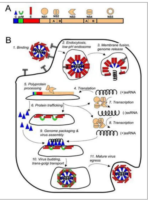
2.1.4 Flavivirus replication cycle ... 16
2.1.5 Main disease ... 17
2.2. Family Togaviridae (genus Alphavirus) ... 20
2.2.1. Classification ... 20
2.2.2. Genome structure ... 21
2.2.3. Vector ... 22
2.2.4. Togaviridae replication cycle ... 22
2.2.5. Main diseases ... 24
2.3. Family Bunyaviridae ... 27
2.3.1. Classification ... 27
2.3.2. Genome structure ... 27
2.3.3. Bunyaviridae replication cycle ... 28
2.3.4. Vector ... 29
2.3.5. Main disease ... 30
3. Flavivirus and Dengue ... 32
3.2. Genetic evolution ... 33
3.3. Vectors ... 34
3.1.1 Aedes aegypti and Aedes albopictus ... 34
3.1.2 Transmission cycle ... 35
3.4. Dengue epidemiology ... 37
3.4.1. History and origin of Dengue ... 37
3.4.2. Global burden ... 38
3.5. Diagnosis ... 52
3.5.1. Clasification in clinical ... 52
3.5.2. Disease course ... 54
3.5.3. Laboratory diagnosis... 56
3.5.4. Treatment and prevention ... 60
3.5.5. DHF immunopathogenesis ... 63
4. Alphaviruses and Chikungunya virus ... 70
4.1. Classification ... 70
4.2. Genetic evolution ... 71
4.3. Replication cycle ... 73
4.4. Vector ... 73
4.5. Chikungunya Epidemiology ... 75
4.5.1. History and origin of chikungunya ... 75
4.5.2. Global burden ... 76
4.5.3. Epidemiology trend in several regions of the world ... 78
4.5.4. Diagnosis... 83
4.5.5. Treatment and prevention ... 86
4.5.6. Chikungunya immunopathogenesis ... 88
5. Combinated arboviral infections ... 89
5.1. Combinated Flavivirus infections ... 89
PART II. DENGUE AND CHIKUNGUNYA IN VIETNAM ... 93
CHAPTER 1. ROLE OF AEDES SPECIES IN THE TRANSLISSION
OF DENGUE IN URBAN AREAS IN NORTH VIETNAM ... 94
1.1. Context of study ... 94
1.2. Objective ... 95
1.3. Discussion and conclusions ... 96
CHAPTER 2. SURVEILLANCE OF DENGUE AND CHIKUNGUNYA
VIRUS INFECTION IN DONG THAP ... 98
2.1. Context of the study ... 98
2.2. Objective ... 99
2.3. Discussion and conclusions ... 99
CHAPTER 3. DENGUE VIRUS, CHIKUNGUNYA VIRUS AND RISK
FACTORS IN SEVERALS PROVINCES OF VIETNAM. ... 101
3.1. Context of study ... 101
3.2. Objective ... 103
3.3. Discussion and conclusions ... 103
GENERATION CONCLUSSION AND PERSPECTIVES ... 107
ACKNOWLEDGEMENTS
To write this thesis, I am greatly indebted to program, institutes and many people for their support and encouragements.
First of all, I would like to my thanks to the Eramus Mundus program, granted me the scholarship to pursue this doctoral program; To Ecole doctoralle Sibaghe, Montpellier 2 University, French. To Centre d’études d’agents Pathogènts et Biotechnologies pour la Santé, center National de la recherche scientique (CPBS, CNRS), Montpellier, France, and National Institute of Hygiene and Epidemiology, Ha Noi, Vietnam for giving me an opportunity to follow this academic program and for equipping me with such an updated knowledge to make this thesis possible.
I want to express my deep gratitude to Dr Christian Devaux (IRD), Professor Sofia Mubarika (U. Gadjah Mada) and Professor Ngo Van Toan (Hanoi Medical University) for having accepting and making me the honor to review this PhD work.
My deep gratitude goes to Professor Roger Frutos, from French Agricultural Research center, and Centre d’études d’agents Pathogènts et Biotechnologies pour la Santé, center National de la recherche scientique (CPBS, CNRS), Montpellier, France for accepting to be my supervisor, for his great support, effort and help in articles and thesis writing, for making several round trips between France and Vietnam.
My special thanks goes to my cosupervisor, Associate Professor Tran Nhu Duong, deputy of National Institute of Hygiene and Epidemiology, Ha noi Viet Nam for firstly allowing me to work as part of his project, for his great support, encouragement to complete the PhD program.
My huge thanks and appreciation to my cosupervisor, Dr. Laurence Briant, Head of Virus host cell interactions Laboratory, Centre d’études d’agents Pathogènts et Biotechnologies pour la Santé, center National de la recherche scientique (CPBS, CNRS), Montpellier, France accepting me to her laboratory, for transferring her expertise in biomolecular necessary for the accomplishment of the work in this thesis.
My special thanks to my cosupervisor, Prof. Phan Thi Nga , former Head of Training and Science Management Department, National Institute of Hygiene and Epidemiology, Ha Noi Viet Nam for her support and allowing me to work as part of her team and help me in her laboratory.
I would like to thanks to Dr Laurent Gavotte, teacher of Montpellier 2 University (UM 2) for allowed me to his lab in Parasitology, Evolutionary Biology, Ecology Department, UM 2, French for the accomplishment of the work, for his help and support in an article.
My thanks to Dr Pierrick Labbé, teacher of Evolution, Vector, Adaptation in Symbioses Department, Montpellier 2 University, French for generously accepting me to his laboratory at Montpellier 2 University for sharing his precious expertise in DNA mosquitoes extraction, for his kindness.
I would like to express my sincere gratitude and appreciation to Dr Babatunde Olowokure, Director of Surveillance, Disease Prevention and Control, Caribbean Public Health Agency for his patient and encouragement to improve myself, valuable comments enabling me to attend and complete the PhD program.
I would like to thanks the medical doctor and nursing staff of the General Hospital in Dong Thap for their care of patients, supported, and their cooperation for this research.
My grateful to the Preventive Medicine Centers in three regions in the North, Center, South were Ha Noi, Ha Tinh, Quang Tri, Thua Thien Hue, Dak nong and Long An, Vietnam for their cooperation during conducted of this study.
My thanks to Arbovirus laboratory, Virologcal Department National Institute of Hygiene and Epidemiology, Hanoi, Vietnam for allowed me to work as part of their team, and for their help.
I would like to thank the following Professors, friends and colleagues:
To my colleagues at Entomology Department, National Institute of Hygiene and Epidemiology, Hanoi, Viet Nam thanks to Le Thi Yen, Tran Chi Cuong, Dang Duc Dong, Nguyen Van Soai for their help collected vector and serum samples, shared to me the hard trips and very interested in provinces of three regions in Vietnam, for their generosity and for being so helpful in all the works in order to generate data used in this thesis.
My thanks to Eric Bernard, Patrict Eldin, Nathalie Chazal, Eymeric Neyret, Simon Fontanel, Roy Matkovic in the Virus host cell interactions Laboratory, Centre d’études d’agents Pathogènts et Biotechnologies pour la Santé, center National de la recherche scientique, Montpellier, France allowed me to work as part of their team, and for their help in Montpellier, France.
Special many thanks mention to Marco Perriat Sanguinet, Evolutionary Biology, Ecology Department, Montpellier 2 University, France for his help during the time in France.
To Dr. Emmanuel Cornillot, CPBS, CNRS for talking interest in this work.
To Dr.Vu Trong Duoc, Vice head of Entomology department, National Institute of Hygiene and Epidemiology, Hanoi, Vietnam for his support and help on data analysis.
To Dr. Truong Ba Tang, General Dong Thap hospital for his cooperation to collected sample in General Dong Thap hospital.
All teachers, friends and colleagues for their help during the course.
Last but not a least, my heartfelt thanks and love to my parents, my daughter, my young brother, my niece, and sister who have helped me in every possible way to my completion of this Doctor of physolophy study.
LIST OF ABBREVIATIONS
AIDSt Acquired immune deficiency syndome
Ae Aedes
APCs Antigen precenting cells
BALB/c An Albino, laboratory-bred strain of the House Mouse
CCD Charge couple device
CCHFV Crimean- Congo Heamorrhagic fever virus
cDNA Complementary Deoxyribonucleic acid
CDC Centers for Disease Control and Prevention
CHIKV Chikungunya virus
CPBS Center Agents Pathogens and Biotechnology for Health study CNRS The French National Centre for Scientific Research
CNS Central nervous system
CTF Colorado tick fever
CTL Cytotoxic T-Lymphocyte
CTLA 4 Cytotoxic T-Lymphocyte associated protein 4
DC-SIGN Dendritic Cell-Specific Intercellular adhesion molecule-3-Grabbing Non-integrin
DENV Dengue virus
DF Dengue fever
DHF Dengue hemorrhage fever
DSS Dengue shock syndrome
E Envelope protein
EEE Eastern Equine Encephalitis
ER Endoplasmic reticulum
GAC IgG antibody capture
G6PD Gluco 6-phosphate dehydrogenase
HI Hemagglutination Inhibiton assay
HLA Human leucocyte antigen
ICTV International Committee on Taxonomy of Virus
IgG Immunoglobulin G
IgM Immunoglobulin M
IHC Immunohistochemical
IVM Intergrate vetor management
JE Japanise Encephalitis
KUN Kunjin virus
LACV5 La crosse virus
MAC-ELISA M antibody- capture Enzyme Link Immuosorbent Assay
MBL2 Mannose- binding lectin 2
MRC Medical Research Council
MVE Murray Valley Encephalitis virus
NHEK Normal Human Epidermal Keratinocytes
NS Nontructural protein
NSS Non-structural protein NS-S
NIHE National Institute of Hygien and Epidemiology
OD Optical Density
ORF Openreading frame
RNA Ribonucleic Acid
RT- PCR Reverse Transcriptase Polymerase Chain Reaction
RVFV Rift Valley fever virus
SDS-PAGE Sodium dodecyl sulfate polyacrylamide gel electrophoresis
SEA Southeast Asia
SLE St. Louis Encephalitis
SNV Sin Nombre virus
SSRNA Single Stranded ribnucleic Acid
TBE Tick born encephalitis
TDR Tropical disease research
TNF Tumor necrosis factor
TGF Transforming growth factor
TV Tetravalent vaccine
US United state
VCAM-1 Vascular cell adhesion molercular -1
WEE Western Enquin Encephalitis
WHO World Health Organizaion
WNV West Nile Virus
LIST OF FIGURES AND TABLES
Figue 1: Three genera of the Flaviviridae family in the Phylogenetic tree.
... 7
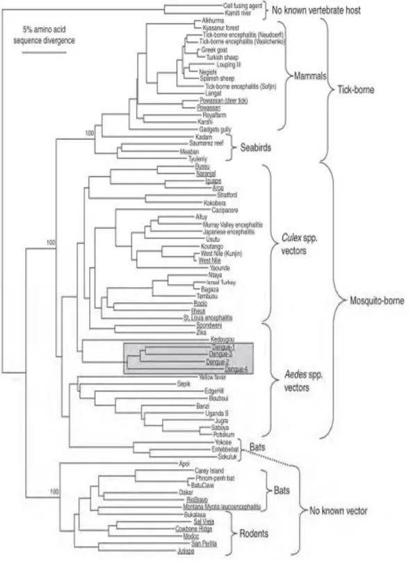
Figure 2: Devided from the GenBank library, phylogenetic tree of the flaviviruses derived from partial Non-structural protein NS-S sequences.
... 8
Figure 3: Schematic structure of flavivirus virion
... 9
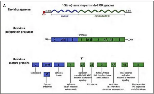
Figue 4: The single open reading frame is depicted with the structural and non structural protein coding region (colored is blue and green repectively).
... 10
Figure 5: The detail of cleavage sites for cellular proteases, NS2B/ NS3 and unknown protease are indicated .
... 10
Figure 6: Transmission cycle for tick-born encephalistic virus.
... 11
Figure 7: Life cycle of Culex mosquito
... 13
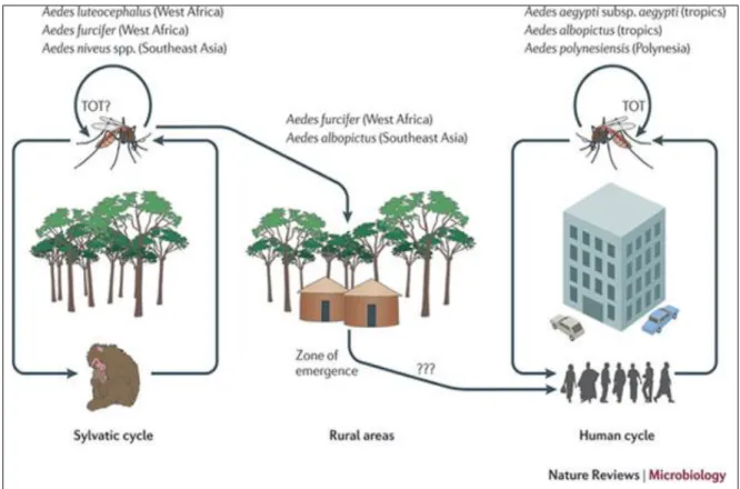
Figure 8: Depicting the hypothetical evolutionary history of endemic or epidemic DENV emergence from zoonotic transmission cycles .
... 16
Figure 10: Genome structure of Togaviruse
... 21
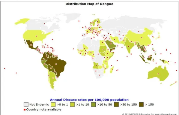
Figure 14: Endemic or potentially endemic to 144 countries of Dengue transmission, 2015
.. 38
Figure 15: Worldwide. Dengue, cases.
... 39
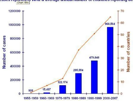
Figure 16: Average annual number of DF/DHF case reported to WHO, and average of countries reporting dengue, 1955-2007 .
... 40
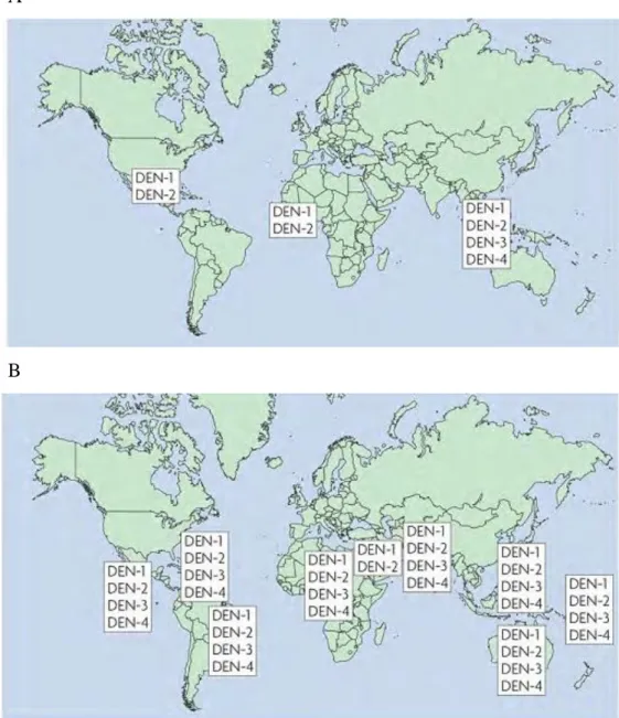
Figure 17: The change in distribution of dengue serotype. The figure shows the distribution in 1970 (A) and 2004(B)
... 41
Figure 18: Western Pacific region. Dengue cases (A), deaths (B).
... 43
Figure 19: Southeast Asian region. Dengue, cases (A) deaths (B).
... 45
Figure 20: Vietnam map.
... 46
Figure 21: Distribution of DF/DHF in Vietnam in 2009.
... 47
Figure 22: Vietnam. Dengue / DHF, cases (A) and death (B)
... 48
Despite the existence of a National dengue control Program since 1998, dengue remains a major health problem in Vietnam. Over the past 15 years, the number of DENV cases has been increasing.
... 48
Figure 23: European Union, Dengue cases
... 49
Figure 24: Americas region. DHF, cases (A) and deaths (B).
... 51
Figure 25: Dengue case classification and levels of severity
... 53
Figure 26: The Course of Dengue disease
... 54
Figure 27: Comparison of diagnostic tests according to their accessibility and confidence
57
Figure 28: Dengue research and training supported and encouraged by the UNICEF-UNDP-World Bank-WHO.... 61
Figure 29: Model of antibodies- dependent enhancement
... 68
Figure 30: Chikungunya virus Genetic and Physical Structure
... 71
Figure 31: Chikungunya virus and dispersal and evolution.
... 72
Figure 32: Origin, spread, and distribution of chikungunya virus and its vector.
... 74
Figure 33: Predicted dispersal pattern of Chikungunya virus from Africa to the Indian Ocean and Europe during the past 20 to 50 years
... 76
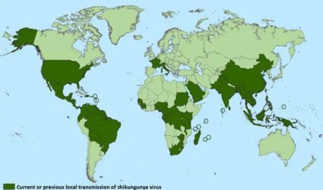
Figure 34: Chikungunya active transmission regions established from published data illustrate geographic and temporal knowledge of previous outbreaks.
... 77
Figure 35: Endemic or potentially endemic to 96 countries.
... 78
Figure 36: Map showing the distribution of chikungunya virus enzootic strains in Africa and the emergence and spread of the Asian lineage (red arrows and dots) and the Indian Ocean lineage (yellow arrows and dots) from Africa
... 79
Figure 37: Outbreak of Chikungunia, 2004-2007
... 81
Figure 38: European Union, chikungunya, case.
... 82
Figure 39: Introduction of chikungunya to the Caribbean by epidemiological week first reported in December 2013 to August 2014.
... 83
Figure 40: Timeline of Infection, Symptom, and Biomarkers
... 84
Figure 41: Distribution of Dengue, Yellow Fever, and their principal vector, the Aedes aegypti mosquito.
... 90
Figure 43: Location of the eight selected study districts in Hanoi, Vietnam
... 94
Figure 42: Location of the five selected study provinces in the north, center, south of Vietnam.
... 101
Table 1: The geographic distribution of spatially unique occurrence for the Americas, Europe/ Africa, and Asia/ Oceania
... 14
Table 2: Summarize of current dengue vaccines .... 63
TÓM TẮT
Sốt xuất huyết Dengue (SXHD) là bệnh truyền nhiễm cấp tính, gây dịch do vi rút Dengue gây nên. Dịch bệnh Sốt xuất huyết đã và đang là một đại dịch nguy hiểm, đe dọa đến sức khỏe và tính mạng của hàng triệu người dân trên toàn Thế giới. Bệnh sốt xuất huyết lưu hành chủ yếu ở vùng nhiệt đới và cận nhiệt đới. Hơn 50 năm qua, sốt xuất huyết dengue lan truyền với tốc độ rất nhanh, từ 9 nước báo cáo dịch bệnh vào năm 1970, cho đến nay vi rút dengue lan rộng tới 144 nước. Theo ước tính của WHO, hàng năm có khoảng 50 đến 100 triệu người nhiễm virus Dengue, trong đó có hơn 500.000 người phải nhập viện.
Chikungunya bệnh lần đầu tiên được phát hiện vào năm 1952 ở châu Phi tại cao nguyên Makonde. Đây là một khu vực biên giới giữa Tanzania và Mozambique. Bệnh cảnh lâm sàng của bệnh sốt Chikungunya có dấu hiệu gần giống với bệnh sốt xuất huyết, vì vậy nó có thể khó chẩn đoán ở những nơi có bệnh sốt xuất huyết lưu hành phổ biến. Cũng như bệnh sốt xuất huyết, bệnh sốt do vỉ rút Chikungunya chưa có vaccine phòng bệnh và thuốc điều trị đặc hiệu. Khi bị mắc bệnh, biện pháp điều trị chỉ tập trung vào việc giải quyết triệu chứng và biến chứng. Ở những nơi điều kiện thuận lợi, muỗi truyền bệnh Aedes aegypti và
Aedes albopictus và có tập tính sống gần gũi với người là yếu tố nguy cơ quan trọng cho
bệnh sốt Chikungunya và dengue lưu hành, phát triển và gây bệnh. Bệnh xảy ra ở châu Phi, châu Á và tiểu lục địa Ấn Độ. Trong nhiều thập kỷ gần đây, muỗi truyền bệnh sốt chikungunya đã có cơ hội lây lan sang cả châu Âu và châu Mỹ. Trong năm 2007, sự lây truyền bệnh này đã được thông báo phát hiện lần đầu tiên tại châu Âu trong một ổ dịch nội địa ở phía đông bắc nước Italia. Năm 2013, 2014 những vụ dịch lớn xảy ra của vi rút chikungynya tại Ametican và Caribean.
Mặc dù chương trình phòng chống sốt xuất huyết quốc gia ở Vietnam đã được thực hiện từ năm 1999, tuy nhiên sốt xuất huyết ở Việt Nam được coi là vùng dịch lưu hành địa phương, chủ yếu ở các tỉnh miền Nam và Nam Trung Bộ. Thống kê của Bộ Y tế Việt Nam cho thấy, hiện nay sốt xuất huyết Dengue đứng thứ 7 trong số 10 nguyên nhân nhập viện hàng đầu. Còn chikungunya vi rút được phát hiện ở Việt nam từ 10 người lính Mỹ vào năm 1967, nhưng cho tới nay, bệnh sốt xuất huyết do vi rút chikungunya tại Việt nam vẫn còn là một vấn đề cần được quan tâm. Ở Việt nam, tập quán trữ nước trong các dụng cụ chứa nước sinh hoạt để đối phó với thời tiết khô của người dân cũng là yếu tố làm gia tăng quần thể muỗi truyền bệnh và gia tăng nguy cơ xảy ra dịch lớn, đặc biệt là tại Miền Nam. Việc
sử dụng các hoá chất diệt côn trùng không được kiểm soát làm ĵtăng tính kháng của vector truyền bệnh. Sự gia tăng các hoạt động giao lưu, buôn bán và du lịch giữa các vùng miền trong nước, ngoài nước như Cambodia và Lào góp phần làm tăng nguy cơ lan truyền SXH trong cộng đồng.
Mục tiêu nghiên cứu của luận văn này nhằm hiểu rõ hơn về bệnh sốt xuất huyết và tình hình vi rút chikungunya ở một số tỉnh tại Việt nam, đặc biệt ở một số tỉnh có chung đường biên giới với Lào và Campuchia, nghiên cứu chỉ số Stegomya của hai véc tơ truyền bệnh sốt xuất huyết Ae. aegypti và Ae. Albopictus, với những tác động của véc tơ sốt xuất huyết dengue và chikungunya khác nhau tại năm huyện tại miền Bắc, miền Trung và miền Nam, Việt Nam. Ngoài ra nghiên cứu cũng mô tả vai trò của bệnh sốt xuất huyết ở những bệnh nhân thu thập được ở bệnh viện Đồng tháp, Việt Nam. Mặt khác, để hiểu rõ hơn về sự tương quan giữa mật độ của Ae. aegypti và Ae. Albopictus gây sốt xuất huyết Dengue bùng phát tại thủ đô Hà nội, Việt Nam. Bên cạnh đó, phân tích kiểu gen của quần thể muỗi được thực hiện để cung cấp thông tin rõ ràng về khả năng lan truyền của muỗi với xuất huyết dengue và chikungunya ở Việt Nam. Kết quả cho thấy rằng sự phát hiện vi rút chikungunya ở muỗi Ae. aegypti nên được coi là cảnh báo vi rút chikungunya xuất hiện tại Việt Nam và có thể lưu hành cùng với bốn týp huyết thanh khác nhau là một vấn đề của sức khỏe cộng đồng và xã hội.
Résumé étendu en français
Depuis la seconde moitié du 20ème Siècle, avec les progrès de la médecine, en particulier dans le domaine de la vaccination, des antibiotiques, et avec l’amélioration des conditions de vie, la disparition des maladies infectieuses était espérée. Il n’en est toutefois rien. Dans les pays développés les efforts se sont concentrés sur les maladies non infectieuses, délaissant les maladies infectieuses. Ces maladies infectieuses, paludisme, SIDA, tuberculose, dengue, chikungunya, ont continué à s’étendre dans les pays en développement mais émergent aussi en zone tempérée, et en particulier en Europe, à la faveur des changements environnementaux et d’une société globalisée.
Parmi ces maladies, la dengue est l’une des plus dynamiques en termes de propagation. Elle est particulièrement présente en Asie du Sud Est qui est sa zone d’origine. Elle est transmise par des moustiques du genre Aedes particulièrement adaptés à un environnement anthropisé. La dengue demeure un problème majeur de santé publique malgré des efforts de lutte antivectorielle et cela pour plusieurs raisons : 1) Il n’y a actuellement aucun traitement ni aucun vaccin connu, 2) Son incidence annuelle globale est estimée à 50 millions de cas entraînant plusieurs dizaines de milliers de morts, 3) Plus de 2.5 milliards de personnes sont à risque, 4) La maladie existe également sous deux formes sévères, la dengue hémorragique (DHF) qui peut évoluer en syndrome de choc (DSS) dont l’issue peut être mortelle, 5) Depuis les années 1950, l’incidence des formes graves (DHF/DSS) a été multipliée par 30, 6) L’infection est souvent asymptomatique, ce qui conduit à de mauvais diagnostics et une sous-estimation du nombre de cas. La dengue est causée par 4 virus ou sérotypes distincts, DENV1, DENV2, DENV3 etDENV4. Un cinquième serotype, DENV5, a récemment été isolé. Un patient infecté par un sérotype devient résistant à ce sérotype (immunité homologue) mais redevient sensible aux autres sérotypes après un délai d’environ12 semaines et présente alors un risque accru de développer une forme sévère suite à une seconde infection.
Le chikungunya est une autre maladie arbovirale transmise par les mêmes vecteurs que la dengue. Le chikungunya et la dengue sont également très similaires du point de vue
des symptômes (en particulier les symptômes précoces), de la période d’incubation et de la zone géographique. Le terme « chikungunya », utilisé à la fois pour le virus et la maladie, signifie « marcher avec le dos courbé » en Swahili et Makonde, et fait référence à l’effet incapacitant des arthralgies. Ce virus a été isolé pour la première fois en 1952 en Tanzanie. Des épidémies de chikungunya ont été décrites en Birmanie, Thaïlande, Cambodge, Vietnam, Inde, Sri Lanka, et Philippines. Les épidémies récentes causes par un virus variant ont causé plusieurs millions de cas dans l’Océan Indien et en Inde. La forme reémergente s’est étendue en Thaïlande et au Cambodge et plus récemment dans les Caraïbes, le Pacifique et l’Europe où plusieurs cas autochtones ont été observés à Montpellier en 2014.
La dengue et le chikungunya sont parmi les maladies les plus difficiles à distinguer, en particulier en phase précoce, du fait de symptômes similaires, de co-infections et de la transmission par les mêmes moustiques péridomestiques du genre Aedes. Aedes aegypti est considéré comme le vecteur principal en zone urbaine avec Aedes albopictus et d’autres moustiques anthropophiles du genre Aedes comme vecteur secondaires.
Le Vietnam est en Asie du Sud Est situé entre le Golfe du Tonkin et le Golfe de Thaïlande et est limitrophe de la Chine, de Laos et du Cambodge. Le climat est tropical au sud et sous régime de mousson au nord avec quatre saisons marquées. Cette distribution climatique affecte l’apparition des épidémies de dengue. La dengue a pour la première fois été décrite au Vietnam en 1958 dans le nord puis à partir de 1960. En raison de la différence climatique, la dengue est présente toute l’année dans le sud alors qu’elle est liée à la saison des pluies dans les autres régions. Le chikungunya a lui été détecté pour la première fois en 1967 chez des soldats américains durant la guerre du Vietnam. Le chikungunya a cependant été détecté dans certaines études chez des patients présentant des symptômes de dengue.
La dynamique de ces deux maladies n’est toutefois pas claire et de nombreuses informations manquent encore sur les facteurs d’influence et les mécanismes. Ces connaissances sont essentielles pour comprendre les dynamiques en jeu et améliorer les capacités de gestion et maîtrise de ces maladies. Si la dengue est depuis longtemps
hyperendémique au Vietnam, ce n’est pas encore le cas du chikungunya qui est très présent au Cambodge et au Laos et présente donc un risque d’émergence. Les échanges très importants entre le Vietnam, le Cambodge et le Laos, tant au niveau des biens que des personnes, constituent donc une voie d’entrée très plausible.
L’objectif de ce travail de thèse était donc de conduire un travail de détection et de surveillance de la dengue et du chikungunya à la fois au niveau clinique chez des patients hospitalisés avec des symptômes fébriles aigus. Ces études se sont concentrées à la frontière avec le Cambodge et le Laos. Ce travail a été réalisé en partie au Vietnam dans le cadre du NIHE et en partie en France au CPBS (UMR 5236 UM-CNRS).
Les objectifs étaient les suivants :
1. Etablir une surveillance et une étude épidémiologique et entomologiques dans des zones frontalières avec le Laos et le Cambodge.
2. Mieux comprendre la dynamique de circulation de la dengue au Vietnam et identifier les serotypes en cause.
3. Evaluer la présence et la dynamique de circulation du chikungunya au Vietnam
4. Comparer les incidences et dynamiques entre les diverses régions du Vietnam.
5. Etudier le rôle des diverses populations de moustiques dans la transmission de la dengue et du chikungunya en période d’épidémie et hors période d’épidémie.
6. Etudier la dynamique de transmission lors de l’épidémie de Hanoi en 2011.
7. Evaluer le système hospitalier de surveillance
8. Analyser la structure génétique de populations de moustiques potentiellement vectrices.
Cette thèse est donc présentée sous forme d’articles organisés en chapitres. Ces chapitres et articles sont les suivants :
Partie 1: Etude bibliographique
Pham Thi Kim Lien, Roger Frutos. Joint arboviral infections: risk and prospective. (Article en préparation)
Partie 2 : Etudes de terrain et de laboratoire
Chapitre 1 - Role of Aedes species in the transmission of dengue in urban areas in North Vietnam
Pham Thi Kim Lien, Vu Trong Duoc, Laurent Gavotte, Emmanuel Cornillot, Phan Thi Nga, Laurence Briant, Roger Frutos, Tran Nhu Duong. Role of Aedes aegypti and Aedes
albopictus during the 2011 dengue fever outbreaks in Hanoi, Vietnam. Asian Pacific
Journal of Tropical Medicine, 2015; 8 (7); 543-548.
L’objectif de ce travail était d’évaluer les correlations entre les cas de dengue et les densités de population de moustiques du genre Aedes durant l’épidémie de dengue de Hanoi en 2011. 24 foyers épidémiques ont été analysés sur 8 districts entre Août et Décembre 2011. 140 patients ont été hospitalisés suite à un diagnostic de dengue avec une prédominance masculine (59.3%) et une classe d’âge comprise entre 15 et 34 ans. Seuls DENV1 (11.27%) et DENV2 (88.79%) ont été détectés à partir des échantillons cliniques. L’échantillonnage des moustiques conduit dans et autour des habitations des patients a montré la prédominance d’A. aegypti (95.15%) par rapport à A. albopictus (4.85%). En
conclusion, il y a une correlation positive entre les densités de population d’A. aegypti, le nombre de cas humains et la durée des épidémies. Ceci n’a pas été observé avec A.
albopictus. Trois lots d’A. aegypti ont été trouvé positifs à la dengue. Deux avec DENV1 et
un avec DENV2.
Chapitre 2 – Surveillance of dengue and chikungunya virus infection in Dong Thap
Pham Thi Kim Lien, Laurence Briant, Truong Ba Tang, Bui Minh Trang, Laurent Gavotte, Emmanuel Cornillot, Tran Nhu Duong, Roger Frutos, Phan Thi Nga. Virologic and serologic surveillance of arbovirus infection in Dong Thap, Vietnam with special reference to dengue and chikungunya viruses: a 13 months study. Asian Pacific Journal of Tropical Medicine, In Press.
Cette étude visait à établir une surveillance dans la province de Dong Thap, qui est
frontière avec le Cambodge, afin d’évaluer la présence de virus de la dengue et du chikungunya parmi les patients hospitalisés à l’hôpital général de Dong Thap. Une analyse
descriptive a été conduite sur 131 patients hospitalisés avec une fièvre aiguë et des symptômes compatibles avec la dengue et le chikungunya. L’étude a été conduite de
Janvier 2012 à Février 2013. Le tableau clinique complet a été établi, ainsi que la détection sérologique et moléculaire. L’analyse sérologique a été conduite de façon séquentielle sur
des échantillons de sang collectés à l’admission et sept jours après admission. Les virus de
la dengue et du chikungunya ont été recherché par ELISA et PCR. 101 patients sur
131(77%) ont été confirmés positifs pour la dengue. Les quatre sérotypes ont été détectés avec une prédominance de DENV2 et DENV4. Aucun virus du chikungunya n’a été
détecté. Un différentiel d’efficacité a été observé. L’efficacité de détection sérologique sur
les mêmes patients était de 29% à l’admission et de 53% après 7 jours. 30 patients sur 131
risque de sous-estimation et le chikungunya n’est pas systématiquement détecté. Des
changements devraient être opérés dans les procédures de surveillance afin de permettre un
meilleur suivi de ces maladies.
Chapitre 3 - Dengue virus, chikungunya virus and risk factors in several provinces of Vietnam
Pham Thi Kim Lien, Laurence Briant, Laurent Gavotte, Pierrick Labbe, Marco Perriat-Sanguinet, Laurent Gavotte, Emmanuel Cornillot, Vu Trong Duoc, Nguyen Thi Yen, Tran Vu Phong, Nguyen Van Soai, Tran Duc Dong, Tran Chi Cuong, Phan Thi Nga, Tran Nhu Duong, Roger Frutos. Aedes mosquitoes mobility, diversity and risk factors for the diffusion of dengue and chikungunya in Vietnam. In preparation for submission to Emerging Infectious Diseases
Une surveillance active a été conduite au Vietnam pour évaluer la présence de la dengue et du chikungunya chez des patients hospitalisés avec une fièvre aiguë dans cinq provinces frontières avec le Laos et el Cambodge. Ce travail, conduit de 2012 à 2014, a été complété par une évaluation des populations de moustiques collectées dans la même zone. Un total de 558 sérums humains a été prélevé ainsi que 1104 moustiques adultes (991 A.
aegypti et 113 A. albopictus) et 10995 larves (8542 A. aegypti et 2453 A. albopictus)
collectés à partir de 2250 foyers. Le virus de la dengue a été trouvé sur 17 (3%) serums humains. Le virus du chikungunya n’a pas été détecté dans les échantillons humains. Des densités de population variables ont été trouvées, la plus élevée étant dans la province sud de Long An frontière avec le Cambodge. Le virus de la dengue a été détecté essentiellement chez A. albopictus. Le virus du chikungunya a été détecté chez A. aegypti. L’analyse phylogénétique des moustiques adultes collectés a montré une large diversité de génotypes, tous ayant été décrits dans d’autres parties du globe. De nouveaux haplotypes ont été détectés. Un moustique de l’espèce Culex vishnui a été trouvé positif pour la dengue, faisant de cette espèce un vecteur potentiel.
Ce travail sur la surveillance et la dynamique de la dengue et du chikungunya au Vietnam apporte un regard nouveau sur la situation actuelle et sur un certain nombre d’idées préconçues. En premier lieu cette étude est à notre connaissance la première analyse comparative à être conduite à une telle échelle sur le pays. Toutes les études précédentes concernaient des événements et épidémies à l’échelle locale et bien que correctes les conclusions ne reflètent qu’une vision locale et partielle de la dynamique. Une vision plus large est nécessaire pour mieux comprendre la dynamique de ces maladies.
La première conclusion de ce travail est qu’il est très difficile de tirer des conclusions sur le principal vecteur de la dengue. Dans ce travail, deux conclusions opposées ont été exprimées en fonction du lieu et peut être de la période. Durant l’épidémie de dengue de Hanoi en 2011, A. aegypti a été clairement identifié comme responsable de la propagation de la maladie. Une corrélation a été établie entre les densités de population locales d’A. aegypti et le nombre de cas cliniques. A. albopictus ne jouait aucun rôle. Quand une analyse globale a été réalisée dans plusieurs provinces du Nord, Centre et Sud Vietnam, le vecteur principal est apparu être A. albopictus. Encore plus important, les haplotypes impliqués ont été décrits dans d’autres parties du monde et un haplotype en particulier a été décrit récemment comme une population invasive en Europe. Plusieurs hypothèses peuvent émerger de cette apparente contradiction. La différence pourrait s’expliquer par la situation spécifique de Hanoi qui est une métropole urbaine à forte densité de population, un environnement favorable à A. aegypti par rapport à A.
albopictus. A. aegypti est en effet plus anthropophile qu’A. albopictus et il est logiquement
plus attendu dans une zone urbaine intense comme la capitale. L’étude conduite au sein de plusieurs provinces a montré à l’inverse qu’A. albopictus était le vecteur principal mais cette étude a été conduite en zone rurale où ce dernier est plus compétitif. Une autre explication pourrait être liée à la date des études. L’étude sur l’épidémie d’Hanoi a été conduite en 2011 alors que celle sur les diverses provinces a été réalisée entre 2012et 2014. Il n’est donc pas possible d’exclure un remplacement d’espèce dominante. Toutefois, cette hypothèse n’apparait pas très solide étant donné que dans l’étude sur plusieurs province, A.
aegypti était très largement l’espèce dominante. Une dernière explication pourrait être
trouvée dans la manière dont ces analyses ont été construites, ce qui représente aussi une forme d’autocritique. La conclusion issue de l’étude sur Hanoi était basée essentiellement
sur une corrélation entre densité et période de pic des populations de moustiques et le nombre de cas cliniques. Cette étude était basée sur un nombre d’échantillon faible. Dans l’étude sur plusieurs provinces, un plus grand nombre d’échantillons a été analysé et A.
albopictus a été identifié comme le vecteur principal alors que sa densité de population
était nettement plus faible que celle d’A. aegypti. Ceci est particulièrement vrai dans la province de Long An où A. albopictus représentait 0.9 % des captures et 50% des moustiques infectés. Avec un plus faible échantillon, cette étude aurait pu conduire à la conclusion d’un rôle prédominant d’A. aegypti.
Ce travail de thèse montre le besoin d’une approche globale et coordonnée lors de l’analyse de la dynamique de la dengue et du chikungunya. De telles études ne peuvent pas être conduites à partir d’événements ponctuels et localisés du fait que la somme d’études ponctuelles ne conduit pas nécessairement une vision globale exacte. Les études de surveillance devraient être conçues au niveau national et basée sur un plan maître incluant à la fois des études cliniques et entomologiques avec détection de virus dans tous les échantillons. Le génotypage des populations de moustiques devrait être conduit de façon obligatoire ainsi que le génotypage des virus isolés d’échantillons humains et entomologiques afin de conduire les analyses de dynamique au niveau des lignées virales. La lignée est le véritable niveau de granulométrie à considérer. L’espèce et le sérotype ne sont pas suffisamment précis pour permettre une analyse de la circulation et une cartographie d’association. Une première recommandation issue de ce travail serait donc de développer une telle approche intégrée et de la proposer au NIHE ainsi qu’au niveau national. Ceci doit inclure une base de données et un système de gestion des bases de données homogènes et intégrés permettant une analyse spatio-temporelle.
Ceci est particulièrement pertinent si l’on considère une autre conclusion de ce travail de thèse, à savoir que les moustiques impliqués dans la vection ne sont pas des populations locales mais des populations circulant à l’échelon du globe. Une analyse de la dynamique ne doit donc pas se concentrer sur les populations locales de moustiques, bien qu’elles puissent néanmoins jouer un rôle, mais plutôt sur le monitoring régulier des populations de moustiques et en particulier aux points d’entrée comme les ports. De telles procédures devraient aussi être développées et cela pourrait être une autre perspective de
ce travail. Génotypage, bases de données adaptées et analyses comparative des séquences de moustiques et de virus devront être développées ainsi que des analyses géographiques et des mouvements commerciaux internationaux.
Un dernier aspect mis en évidence par ce travail est l’adaptation limitée des procédures actuelles de surveillance et de dépistage mises en œuvre à l’hôpital et dans les services de santé publique. Comme cela a été montré dans le cas de l’hôpital de Dong Thap, les tests sérologiques réalisés à l’admission sous-estiment le nombre de patients positifs pour la dengue. De plus, ils ne permettent pas l’identification des lignées virales. Le chikungunya n’est en outre pas dépisté du tout. Il serait trop onéreux et difficilement réalisable de mettre en place un test sérologique multi-étapes avec au final des résultats peu satisfaisants. La dernière recommandation et perspective issues de ce travail seraient donc la mise en place d’un test PCR multiplex pouvant détecter les quatre sérotypes de dengue ainsi que les deux variants, ancien et reémergent du chikungunya. Ce test devrait être associé au séquençage des échantillons positifs. Le cout de la PCR et du séquençage est désormais très compétitif et le bénéfice attendu pour la santé publique compense très largement l’investissement en surveillance. Ceci devrait être lié au même système intégré de bases de données dynamiques que pour la surveillance des moustiques. Un projet devra être soumis en ce sens au NIHE et aux instances nationales.
En conclusion, ce travail de thèse a mis en évidence des points important à aborder de façon coordonnée afin de développer un système de surveillance efficace pour la dengue et le chikungunya au Vietnam. Développer ces systèmes et outils représentera un défi stimulant à relever et une valorisation intéressante de ce travail.
SUMMARY
Arthropod-borne viral infections (or arboviral infections) are common causes of fever syndromes worldwide, more than 130 arboviruses are known to cause disease in humans. Dengue fever (DF) caused by Flavivirus belong to the family Flaviviridae, and chikungunya is caused by an Alphavirus in the family Togaviridae. Both dengue and chikungunya diseases are transmitted by mosquitoes mainly Aedes aegypti (Ae. aegypti) and Aedes albopictus (Ae. albopictus) and can cause potentially severe and or debilitating chronic disease. Over the past 50 years, dengue has spread inexorably, with nine countries reporting dengue transmission prior to 1970 compared to over 144 now, and incidence having increased 30 fold. While dengue is the commonest and the most rapidly spreading mosquito - borne viral disease in the world, chikungunya (CHIK) has recently re - emerged after an interval of several decades to affect and place at risk millions of people in the Indian Ocean areas, Africa, Southeast Asia and more recently has spread to the Caribbean region. There are evidence countries in Asia, Africa and the Pacific of patients exhibiting co - infection with dengue and chikungunya simultaneously. Without a licensed vaccine, therapeutic druges and effective of vector transmission, the increasing number of case dengue virus (DENV), chikungunya virus (CHIKV) is associated with expanding geographyphic rage and increasing intensity of transmission in affected areas.
Vietnam is one of five countries in the Southeast Asia with the higtest dengue burden. Cambodia and Laos countries share a long and extensive border with Vietnam that have endemic for dengue, reported increasing chikungunya activity in Laos and Cambodian provinces bordering Vietnam. Dengue is highly endemics in tropical southern Vietnam, while increasing larger seasonal epidemics have occured in northern Vietnam over the last decade. Biomolecular level used to describes DENV serotypes. We have revealed that DENV serotypes circulating in Vietnam are similar to those observed in neighboring countries - in particular Cambodia and China. All four dengue virus serotypes founded circulating in Vietnam with dominant one varying through in southern and northern.
Although CHIKV affected areas often overlap with DENV - endemic areas, however, stimultaneous outbreaks are rare. So far, no case of chikungunya in human reported in Vietnam, excepted 10 cases in 1967 of American soldiers in Vietnam. This
study is the first to prospective investigate the circulation of the CHIKV and detected two
Ae. Aegypti mosquitoes infected with CHIKV in the south of Vietnam.
This thesis manuscript reports data obtained by multidisciplinary and integrative aproaches aiming to better understand dengue and chikungunya virus situation in several provinces shares the border with Laos and Cambodia, and found that the proportion of
Stegomya index of two vectors Ae. aegypti and Ae. albopictus, with the potential effects of
DENV and CHIKV vector differed at the communes belong five districts in the north, center and south of Vietnam. Additionally, we described that a significant the role of DENV as cause of febrile illness in human, and also identified high proportion of dengue infection among patients in Dong Thap hospital, in south of Vietnam. On the other hand, to better understand on the positive correlation between the population density of Ae. aegypti and the number of DENV human cases of outbreaks in Hanoi capital, north of Vietnam. Beside, an integrative analysis encompassing the genetic study of viral lineages on human patents and in mosquitoes along with the genotyping of mosquito population should be undertaken to provide clear information on the dynamic of dengue and chikungunya in Vietnam. Our result suggested that the detected CHIKV in Ae. aegypti mosquito should be considered as warned of CHIKV appear in Vietnam and may be with DENV consists of four distinct serotypes combination remains a major public health problem.
GENERAL INTRODUCTION
From the second half of the 20th century, with achievements of medical research in terms of vaccination, antibiotics and improvement of life and health conditions of mankind, it was expected that infectious diseases were going to disappear. However, in developed countries the efforts have been concentrated on illnesses as cancer. Consequently, at the dawn of the new century, infectious diseases are still causing suffering and mortality in developing countries. With the disease as Malaria, AIDS,
Mycobacteria tuberculosis and other diseases will have marked the memory of humanity
forever.
Among these diseases, dengue fever, especially known in Southeast Asia, are hitting countries with tropical and warm climates. It is transmitted to the man by the mosquito of the genus Aedes (Ae.) is the principal vector of dengue and adapted extremely well to the urban environment. Dengue remains a major public health problem in tropical and subtropical countries despite lots of effort to control the mosquito vector (1) with an estimated global annual incidence of 50 million cases leading to ten of thousands of deaths (2) and more than 2.5 billion people being at risk of infection. Since the 1950s, the incidence of DHF/DSS has increased over 30 fold (3), with more than countries affected by outbreaks of dengue (4) Infection with any of DENV serotype may be asymptomatic in the majority of case or may result in a wide spectrum of clinical symptom (5). Dengue fever exists in two forms: the classic dengue or Dengue Fever (DF) and the Dengue Hemorrhagic Fever (DHF) which may evolve toward a severe form known as Dengue Shock Syndrome (DSS). The major problem with dengue is the fact that the disease is caused by five distinct serotypes known as DEN1, DEN2, DEN3, DEN4 (6) and new DENV5 was detected in Thailand in 2013 (7). A person infected by one of the five serotypes will never be infected again by the same serotype (homologus immunity), but loses immunity to the three other serotypes (heterologus immunity) in about 12 weeks and then becomes more susceptible to developing dengue hemorrhagic fever.
The latter distinguishes chikungunya virus (CHIKV) from dengue fever, which otherwise shares the same vectors, incubation period, clinical course, symptoms, and geographical distribution. The word chikungunya, which is used for both the virus and the disease, means “to walk bent over” in African dialect Swahili or Makonde, and refers to
the effect of the incapacitating arthralgia. The virus was first isolated in 1952 in Tanzania (8). Human chikungunya virus infection has been documented in Burma, Thailand, Cambodia, Viet Nam, India, Sri Lanka, and the Philippines (9). Recent chikungunya outbreaks caused several million clinical cases in the Indian Ocean Islands and India. Neither Europe nor the Americas have had outbreaks of chikungunya virus so far (9) (10) and more recently has spread to the Caribbean region (11).
Dengue and Chikungunia are among the most difficult diseases to distinguish, especially because simultaneous co - infection can occur (12)(13)(14)(15)(16)(17). Chikungunya and dengue viruses are frequently transmitted to humans by peridomestic
Aedes mosquitoes. Ae. aegypti has been considered to be the principal vector in the urban
transmission cycle, with Ae. albopictus and other anthropophilic Aedes spp. serving as secondary vectors (18)(19).
Vietnam is located in South Eastern Asia, bordering the Gulf of Thailand, Gulf of Tonkin, and shares the border with South China Sea, China, Laos, and Cambodia. The climate is tropical in south; monsoonal in north with 4 seasons are spring, summer, autumn and winter. Vietnam faced with many health problems including fever/ dengue haemorrhagic fever (DF/DHF). In 1958, dengue was first detected in the North, and in 1960 was described in the South of Vietnam (20). Case occur year round in the South, and during the rainy season elsewhere. The incidence of DHF has increased from the first recorded outbreak in 1963, major epidemic occurred in 1969, 1983, and 1987 with a widespread epidemic in 1998 affecting 42 out of 61 provinces and resulting in 232,793 cases and 447 deaths (21). An estimated average reported morbidity and mortality between 1996 and 2005 was 75,407 cases and 145 deaths per year (22). In 2009, 27,000 cases (26 fatal), and in 2012 have 18,052 cases, 9 fatal, were reported during January to June (23). The outbreaks in 2013 were reported 25,300 cases and 16 fatal in August. (23)
Since 1975, chikungunya virus is first detected in Vietnam arcording WHO reported (24), To date, no cases of chikungunya have been reported in Vietnam, since 1967 when 10 cases were reported in American soldiers in Vietnam (25) and other study of anti - chikungunya antibodies in Vietnamese children in the South of Viet nam (26). In other research on anti - chikungunya antibodies in the North, Viet Nam show that, 60 percent of patients with classic dengue symptoms have tested negative for dengue virus. according to Vu Sinh Nam, Preventive Health and Environment Department, Ministry of health, and 4
of 17 provinces: Ha Noi, Nghe An, Ha Nam, Nam Dinh were positive to chikungunya virus with proportion 15/324 patients who have had classic dengue symptoms. Detected of chikungunya virus during dengue outbreak in the north, Viet Nam (27) .
To find out suitability for surveillance chikungunya and dengue viruses infection in Viet Nam, the knowledge about mechanism of Chikungunya and Dengue virus interaction has essential activities that can help in the prevention of vector - born disease to improve the management, surveillance, control of dengue and chikungunya virus infection in Vietnam. On the other hand, the current knowledge on early events of chikungunya and dengue transmission from mosquitoes to human is limited. Besides, there are several reasons to believe that dengue and chikungunya may be co circulating in Vietnam as cases of CHIKV have been reported in neighbouring human populations. Cambodia reported the re - emergence of CHIKV in 2011 with 24 patients identified when samples were tested for Flaviviruses and Alphaviruses (28). In 2012, chikungunya outbreaks were reported in Laos with 197 cases in Moonlpamok and Khong Districts of Chamasak province (29). Vietnam shares borders with Cambodia and Laos, its therefore possible that cross - border transmission into Vietnam may occur as result of travel and traffic between the neighbouring countries from infected people or mosquitoes.
Our aim is therefore to perform an epidemiological survey of dengue and chikungunya in several provinces in Vietnam shares the boder to Cambodia and Laos during the 2011 - 2014 periods. We will also investigate the interaction of mosquito to human transmission and the risk factors transmission dengue and chikungunya viruses. This part of the study is coordinated with the Center of National Research Scientific (CNRS), and Montpellier University 2, Montpellier, France.
The specific objective of the research aims:
9. To describe the surveillance data of chikungunya virus from several provinces presence in the north, center and south in Vietnam shares border with Laos and Cambodia.
10. To better understand the monitor Dengue and identify the virus strains circulating in Vietnam.
11. To obtain and analysis, of some dengue strains circulating in the North, with dengue viruses isolated in other regions in Vietnam.
12. To describes the spread of chikungunya and dengue viruses with the role of mosquitoes transmission in Ha noi, Vietnam.
13. To identify the molecular epidemiology of vector transmission chikungunya and dengue viruses in Viet Nam.
The outline will be displayed in form of chapters with results presented as a form of published, submitted or in preparation articles. The articles included in this thesis are listed below:
FIRST PART
Pham Thi Kim Lien, Roger Frutos. Joint arboviral infections: risk and prospective. (in preparation)
SECOND PART CHAPTER 1
Pham Thi Kim Lien, Vu Trong Duoc, Laurent Gavotte, Emmanuel Cornillot, Phan Thi Nga, Laurence Briant, Roger Frutos, Tran Nhu Duong. Role of Aedes aegypti and Aedes albopictus during the 2011 dengue fever outbreaks in Hanoi, Vietnam. Asian Pacific Journal of Tropical Medicine, 2015; 8 (7); 543-548.
CHAPTER 2
Pham Thi Kim Lien, Laurence Briant, Truong Ba Tang, Bui Minh Trang, Laurent Gavotte, Emmanuel Cornillot, Tran Nhu Duong, Roger Frutos, Phan Thi Nga. Virologic and serologic surveillance of arbovirus infection in Dong Thap, Vietnam with special reference to dengue and chikungunya viruses: a 13 months study. Asian Pacific Journal of Tropical Medicine, In Press.
CHAPTER 3
Pham Thi Kim Lien, Laurence Briant, Laurent Gavotte, Pierrick Labbe, Marco Perriat-Sanguinet, Laurent Gavotte, Emmanuel Cornillot, Vu Trong Duoc, Nguyen Thi Yen, Tran Vu Phong, Nguyen Van Soai, Tran Duc Dong, Tran Chi Cuong, Phan Thi Nga, Tran Nhu Duong, Roger Frutos. Aedes mosquitoes mobility, diversity and risk factors for the diffusion of dengue and chikungunya in Vietnam. In preparation for submission to Emerging Infectious Diseases
PART I
LITERATURE REVIEW 1. Arboviruses
Arthropod-borne viruses (arboviruses) are viruses that can be transmitted to human by arthropod vectors. The WHO definition is as follows: “Viruses maintained in nature principally, or to an important extent, throught biological transmission between susceptible vertebrate hosts by haematophagus arthropods or through transovarian and possibly venereal transmission in arthropods.” (30).
Arboviruses that cause human encephalitis/animal or zoonotic disease have been identified, are members of four virus families: the Togaviridae (genus Alphavirus),
Flaviviridae, Bunyaviridae and Reoviride, contain most of the arbovirus that cause
human/animal disease (31)(32)(33). The term of arboviruses is not taxonomic indicator, it describers their requirment for vertor in their transmission cycle (34)(33). According to International catalogue of Arbovirus, 1992,535 species belonging to 14 virus families were registered (33). More than 130 arboviruses are known to cause human disease.
Human and animals infected by arboviruses, may suffer diseases ranging from sub-clinical or mild through febrile to encephalitic or hemorrhagic with a significant proportion of facilites. Arboviruses infections can cause four types of illness: 1) Illnesses of the central nervous system, ranging in seriousness from mild viral meningitis to encephalitis (inflammation of the brain), with coma, paralysis, and death; 2) Mild fever illnesses with or without rash; 3) Hemorrhagic fevers that can be serious and life - threatening; 4) Arthritis and rash, with or without fever (35)(36).
Three of a large number of neglected human pathogenic arthtopod- borne virus are chikungunya virus ( CHIKV), dengue virus ( DENV), and West Nile virus (WNV) whose combined figures for morbidity and mortality far exceed those for Ebola, severe acute respiratory syndrome and Middle East repiratory syndrome viruses (36).
Approximately 300 type of mosquitoes could transmit arbovirus, special Aedes and
Culex mosquitoes are the species most frequently associated with arbovirus transmission,
respectively more than 105 - 115 types of arbovirus (37) (33). Ticks are also prevalent vectors, 116 difference species are currently know to transmite arbovirses, in addition 25 midge species mainly 24 types of Culicoides and Laisiohelea. Sandflies, blackflies,
stinkbugs, lice, mites, gadfly, and bedbugs can also transmit arboviruses (38). This diversity of species and the wide distribution of these transmission vectors explaine why arboviruses are so successful in dispersing globally via the mechanisms highlighted earlier (39)(33). A high proportion of Arbovirus associated with human and animal disease circulate in tropical and subtropical regions, where mosquitoes, and other flying insects, tend to be abundant. Also, many arboviruses circulate among wildlife species in temperate regions of the world. They have evolved a wide variety of strategies to ensure their long term success, dispersal and survival. Arbovirus adapt readily to new susseptible hosts by alteration of receptor specificity, transmission efficiency, antigencity, ecological and enviromental conditions. Changing anthropological behaviour, climate change and high mutation frequency are importannts determinants of arbovirus emergence (36).
Up to date, in the 21 st century, develope vaccine and antiviral drugs to prevent or treat humans against infection by pathogenic aboviruses will resolve the challenge associated with emerging arboviruses.
2. Different of Arbovirus families
2.1. Family Flaviviridae
2.1.1. Classification
The International Committee on Taxonomy of Viruses (ICTV) began to devise and implement rules for the naming and classification of viruses early in the 1970s, an effort that continues to the present day. According to the ICTV of virus Flaviviruses consistitute on of three genera within the virus family Flaviviridae, and others two are the genera Pestivirus from Latin pestis is “Plague” and Hepacivirus from Greek hepatos is “liver”. The family Flaviviridae contains 58 species within three genera to include Flavivirus (53 species), Herpacivirus (1 species), and Pestivirus (4 species). Fourteen species within the genus Flavivirus and all species within the Herpacivirus and Pestivirus genera are not known to be transmitted by arthropod vectors. (Beth K. Schweitzeer, 2009) (40).
Figue 1: Three genera of the Flaviviridae family in the Phylogenetic tree.
The Flaviviruses are primarily transmitted by arthropods and consist of more than 73 viruses include yellow fever virus, Japanese encephalitis virus, Dengue virus, West Nile virus, tick borne encephalitis, (41)(42). The first identified human virus in the flavivirus prototype is Yellow fever virus (YFV), this is name derived from Latin word “flavus”, meaning yellow, evoking the jaundice caused by YFV (43).
In the genus Flavivirus, viruses share complex antigenic inter relationships, they can separate into four ecologicals or phylogentic group with regard to their vector association: mosquito - borne group, a tick - borne group, and non- vectored viruses. (See figure 2) Subtypes are written in parentheses after virus names. New world viruses are printed in bold and underlined (43).
Figure 2: Devided from the GenBank library, phylogenetic tree of the flaviviruses derived from partial Non-structural protein NS-S (NSS) sequences.
2.1.2 Genome structure
The Flaviviridae genome contains a single, consists of a linear, positive-sense RNA molecule of about 10.8 kb in size, and the genome is not segmented. The complete genome is 9500 - 12500 nucleotides long, Flavivirus virion are 50nm in diameter, spherical, and enveloped (44). The genome has a 5’- end carries a methylated nucleotide cap or genome - linked protein. It also has a 3’ polyadenylated tail, polyprotein from genomic RNA cleaved, three structural proteins and some non-structural proteins. The genome undergoes cytoplamsic replication (45). The translation initiation is cap - dependent in Flaviviruses (44).
Note: M is protein, E is protein envelope, ORF is openreading frame, ssRNA is Singgle Stranded Ribonucleic Acid.
Figure 3: Schematic structure of flavivirus virion (Philippe Buchy).
As study of Cleaves in 1981, the flavivirus genome has one open reading frame encoding a sing ger polyprotein at the origin of structural and non tructural proteins (45).
Figue 4: The single open reading frame is depicted with the structural and non structural protein coding region (colored is blue and green repectively).
The 5’cap and the 5’ and 3’ untranslated region (UTR). The single open reading frame encodes an immature polyprotein pecursor that is co and post translationally cleaved in to three structural proteins (in blue) and seven nonstructural proteins (in green) (46).
Figure 5: The detail of cleavage sites for cellular proteases, NS2B/ NS3 and unknown protease are indicated (47).
2.1.3 Vector
Ticks and mosquitoes are too major group of arthropod vectors of Flaviviruses, of the known Flaviviruses, approximately 50% are recognized human pathogens causing fever, encephalitis, or hemorrhagic disease, however, for many of others, the pathogenic potential has not been well studied (Gubler Dj, Kuno G, 2007)(48) (49). Important mosquito was borne Flaviviruses include dengue virus serotypes 1 - 4, Yellow fever virus (YFV), West Nile virus (WNV), Japanese encephalitis virus (JEV) and St.Louis encephalitis virus (SLEV). The mosquito - born flaviviruses comprise two distinct epidemiology groups: neutrotropic viruses, often associated with encephalitic disease in human or livestock, culex spp. Mosquito vectors, and birth reservoirs, and nonneutrotropic viruses are associated with hemorrhagic disease in humans, Aedes spp mosquito vector, and primate host (50).
Ticks
In nature, tick borne flaviviruses are maintained through a transmission cycle involving an ixodid tick vector and a vertebrate host (51). Tick transmitted flaviviruses comprise two distinct groups: mammalian and seabird virus groups. It is transmitted by the bite of several species of infected ticks, including Ixodes scapularis, Ixodes ricinus and
Ixodes persulcatus (52) or very rarely through the non - pasteurized milk of infected cows.
Tick - borne encephalitis virus (TBEV), which includes three subtypes, European, Far Eastern, and Siberian, was the most important role in human epidemiology, particularly in the old ‘‘Eastern bloc’’ countries, producing a wide range of diseases including subclinical infections, biphasic fever, encephalitis, and chronic disease. The vector of TBEV in European is Ixodes ricinus, and I persulcatus for the other two subtypes (53) (54)(55) (56)(57) (58) (59) (60). I ricinus is detected in the most of Europe, and the distribution extends to Turkey, Northern Iran, and in the Southeast of Caucasus (61). I persulcatus was found in the belt extending from Eastern Europe to China and Japan. Both tick species cocirculaten a restricted area in northeastern Europe, Russian Karelia, St Petersburg, and eastern Estonia and Latvia. An ectopic focus of TBE-Sib carried by I persulcatus has been discovered in western Finland. (62) (63)(64).
The seabird tick - borne virus group has not been shown to induce disease in humans or in seabirds and the reasons why these relatively closely related viruses differ so significantly in this particular characteristic have not been investigated. However, human contact with seabirds is limited (65).
Mosquitoes Culex
The most dangerous and most widely distributed mosquito in the world is Culex mosquitos, Culex mosquito feeding normally happens at night. After feeding, the Culex mosquito will find somewhere cold and damp to rest and digest. Digestion of a meal can take 2 to 7 days with 1 to 3 meals needed to produce a clutch of eggs. The female mosquitoes prefer to lay their eggs in standing water that contains plenty of organic matter (66).
The most importance of Culex vectors are member of Culex pipiens complex, known as the northern house mosquito. Normally, Culex pipiens is considered to be a bird feeder but some urban strains have a predilection for mammalian hosts and feed readily on humans. According to a classification system for Northeastern mosquito, the life cycle of
Culex pipiens represents a variation of the multivoltine life cycle: non - desiccation
resistant eggs laid directly on water, larvae develop in polluted water habitats, multiple generations each year; overwinters as a mated female.
Figure 7: Life cycle of Culex mosquito
The second of Culex vectors are C.autralicus, C,triaeniorhynchus, C.salinatius,
C.tarsalis, C.quinequefasciatus, C.nigripalpus, C. nnulirostris. The major Asian vector of
Japanese encephalitis (JE) is Culex triaeniorhynchus (67). In Australia, the main vector transmission to human of Murray Valeley (MVE) and Kunjin (KUN) viruses is Culex
annulirostris (68)(69).
Transmission cycles
In Human, cycle of Culex mosquito is multipliers of the virus, and serving as a source of the virus for uninfected mosquitoes. Most female mosquitoes have to feed on human or an animal and get a sufficient blood meal before she can develope eggs, and
Culex mosquito usually lay their eggs at night.
Aedes aegyti and Aedes Albopictus Aedes aegypti
Ae. aegypti is believed originated in the jungle of Africa where a sylvatic, where
ancestral from (4)(70), and Ae. aegyti formosus is East and Central Africa (71). The domestic from Ae. aegypti was most likely spread throughout the rest of the world via slave and