ORIGINAL PAPER
Chlamydia pneumoniae infection acts as an endothelial
stressor with the potential to initiate the earliest heat shock
protein 60-dependent inflammatory stage of atherosclerosis
Simone Kreutmayer&Adam Csordas&Jan Kern&Viola Maass&Giovanni Almanzar&Martin Offterdinger&
Robert Öllinger&Matthias Maass&Georg Wick
Received: 1 July 2012 / Revised: 1 October 2012 / Accepted: 2 October 2012 / Published online: 29 November 2012 # Cell Stress Society International 2012
Abstract We identified increased expression and redistribu-tion of the intracellular protein 60-kDa human heat shock protein (hHSP60) (HSPD1) to the cell surface in human endo-thelial cells subjected to classical atherosclerosis risk factors and subsequent immunologic cross-reactivity against this high-ly conserved molecule, as key events occurring earhigh-ly in the process of atherosclerosis. The present study aimed at investi-gating the role of infectious pathogens as stress factors for
vascular endothelial cells and, as such, contributors to early atherosclerotic lesion formation. Using primary donor-matched arterial and venous human endothelial cells, we show that infection with Chlamydia pneumoniae leads to marked upregulation and surface expression of hHSP60 and adhesion molecules. Moreover, we provide evidence for an increased susceptibility of arterial endothelial cells for redistribution of hHSP60 to the cellular membrane in response to C. pneumo-niae infection as compared to autologous venous endothelial cells. We also show that oxidative stress has a central role to play in endothelial cell activation in response to chlamydial infection. These data provide evidence for a role of C. pneumo-niae as a potent primary endothelial stressor for arterial endo-thelial cells leading to enrichment of hHSP60 on the cellular membrane and, as such, a potential initiator of atherosclerosis. Keywords Inflammation . Heat shock protein 60 .
Autoimmunity . Chlamydia . Endothelial . Atherosclerosis
Introduction
There is increasing evidence from clinical and experimental studies for a causative role of infection and inflammation in initiation and progression of atherosclerosis (Ross 1999; Hansson 2001; Libby 2002; Wick et al. 2004). Our research group formulated the“Autoimmune Concept of Ath-erosclerosis,” experimental and clinical evidence for which was provided by data gathered from our and other laboratories over the past several years (Wick et al.2001,2004; Grundtman et al.2011). At the heart of this concept lies the finding that stressed endothelial cells become the target of pre-existing innate and adaptive cellular and humoral immunity against eukaryotic cross-reactive HSP60 epitopes due to earlier infec-tions or vaccinainfec-tions, as well as bona fide autoimmunity
Simone Kreutmayer and Adam Csordas contributed equally to this work.
S. Kreutmayer
:
A. Csordas (*):
G. Almanzar:
G. WickLaboratory of Autoimmunity,
Division of Experimental Pathophysiology and Immunology, Biocenter, Innsbruck Medical University,
Peter-Mayr Strasse 4a, 6020 Innsbruck, Austria
e-mail: [email protected]
J. Kern
:
V. Maass:
M. MaassInstitute of Medical Microbiology, Hygiene and Infectious Diseases, Paracelsus Medical Private University of Salzburg,
Salzburg, Austria M. Offterdinger
Biooptics Facility, Division of Neurobiochemistry, Biocenter, Innsbruck Medical University,
Innsbruck, Austria R. Öllinger
Division of Visceral, Transplant and Thorax Surgery, Innsbruck Medical University,
Innsbruck, Austria Present Address: A. Csordas
Division of Cardiac and Vascular Surgery, University Hospital Zurich,
Rämistrasse 100, 8091 Zurich, Switzerland DOI 10.1007/s12192-012-0378-7
against biochemically altered autologous HSP60 (Xu et al. 1993a; b; 1994; 2000; Grundtman and Wick 2011). At a molecular level, we and others have shown that hHSP60 is expressed as a first response, serving as an immunological danger signal in endothelial cells subjected to classical risk factors for atherosclerosis including cigarette smoke, ox-idative stress, and oxidized LDL, rendering the cells a target of pre-existing cross-reactive autoimmunity (Xu et al. 1994; Amberger et al.1997; Shi and Tokunaga2004; Henderson et al. 2008; Kreutmayer et al. 2011). Clinically, a significant correlation between antibacterial HSP60 antibody levels and prevalent carotid atherosclerosis was observed, and the pres-ence of human and chlamydial antibodies to HSP60 was identified as an independent risk factor for coronary athero-sclerosis (Xu et al. 1993a; b; 1999). Severely aggravated lesion formation and enrichment of HSP60-reactive T cells within atherosclerotic lesions in rabbits and mice by immuni-zation with recombinant HSP65 further indicate the role of HSP60 as a putative antigen, initiating and maintaining vas-cular inflammation (Xu et al. 1992; George et al. 1999). According to our concept of an autoimmune basis of athero-sclerosis, the lifelong burden of being subjected to infectious agents, such as Chlamydia pneumoniae, would go hand-in-hand with an increased risk of developing immunologic cross-reactivity against autologous HSP60 (Mayr et al.1999a). In a previous study, it was shown that chlamydial HSP60 (cHSP60) antibodies cross-react with hHSP60 counterparts and mediate cytotoxicity to stressed endothelial cells. More-over, there was a strong correlation between cross-reactive anti-HSP60 and anti-C. pneumoniae antibody titers (Mayr et al.1999b). In terms of epidemiology, population-based stud-ies show that most individuals get infected with C. pneumo-niae before the age of 20, and specific antibodies against C. pneumoniae are found in more than 70 % of people aged 50 and above (Grayston2000). Based on increased serum anti-body titers against this agent in patients with coronary heart disease, Saikku et al. (1988) suggested a possible relationship between C. pneumoniae infection and atherosclerosis. This finding was confirmed by subsequent population-based stud-ies that found a strong correlation between the risk for coro-nary heart disease and detection of C. pneumoniae in corocoro-nary arterial tissue (Jackson et al.1997; Yamashita et al.1998). Remarkably, while more than 50 % of coronary plaque lesions harbor C. pneumoniae, the pathogen has rarely been detected in healthy vascular tissue (Shi and Tokunaga2004). Experi-mentally, it has been shown that the major pro-inflammatory transcription factor NF-κB is upregulated in endothelial cells subsequent to C. pneumoniae infection, leading to increased secretion of interleukin (IL)-1, IL-8, and monocyte chemotac-tic protein 1 (MCP-1) (Hogdahl et al.2008).
In summary, these lines of evidence support the notion that chlamydial infection is a potent contributor to vascular in-flammation. However, classical experimental protocols used
centrifugation (Dechend et al.2003) of the endothelial cells together with the C. pneumoniae infective particles (elemen-tary bodies) to enable productive infection of the target cells. As these treatments themselves represent stressful me-chanical conditions that might induce a stress reaction in endothelial cells, they are unsuitable protocols for studying C. pneumoniae-induced stressor effects (Krüll et al.1999; Shi and Tokunaga2004). In order to have a noninvasive experi-mental setting avoiding an additional centrifugation step, we used the CV6 strain of C. pneumoniae that shows a special tropism for endothelial cells. Moreover, in the present study, we employed human umbilical vein endothelial cells (HUVECs) as well as primary adult arterial and venous endothelial cells (AECs and VECs), from the same donors. Thus, we were able to delineate the susceptibility of AECs and VECs to a C. pneumoniae-induced pro-inflammatory pheno-type while avoiding a confounding effect inherent to the genetic variability of primary cells derived from different donors. In particular, we set out to investigate the threshold for surface expression of hHSP60 in primary AECs as com-pared to autologous VECs in response to C. pneumoniae infection. Here, we identify chlamydial infection as an inducer of hHSP60 expression in primary endothelial cells more po-tent than any of the classical endothelial stress factors. More-over, we provide evidence for a particularly increased susceptibility of AECs for surface expression of hHSP60 as compared to autologous VECs.
Materials and methods Culture of C. pneumoniae
The cardiovascular C. pneumoniae strain CV-6, a coronary artery isolate, was grown on HEp-2 cell monolayers as de-scribed (Gieffers et al. 2001). Absence of mycoplasma was shown by PCR (Stratagene, Santa Clara, CA, USA).
Cell culture
HUVECs were isolated from umbilical cords obtained with informed consent from the Department of Gynecology and Obstetrics, Innsbruck Medical University. AECs and VECs were isolated from the iliac arteries and veins of transplant donors obtained from the Clinic of Visceral, Transplant and Thorax Surgery, Innsbruck Medical University. The isolation and use of human tissues for these experiments has been approved by the Ethics Committee of Innsbruck Medical University (resolution numbers UN2979 and AM2670c). Cells were isolated by enzymatic detachment, using collage-nase as described elsewhere (Amberger et al. 1997), and subsequently cultured in 0.2 % gelatin-coated (Sigma-Aldrich, St. Louis, MO, USA) polystyrene flasks (Becton
Dickinson, Meylan Cedex, France) in Endothelial Cell Basal Medium-2 (EBM-2 containing EGM-2 SingleQuots supple-ments; Lonza, Basel, Switzerland) including 2 % fetal bovine serum and growth factors (Lonza) in a humidified atmosphere of 5 % CO2. The medium was replaced by RPMI without
phenol red and antibiotics, supplemented with 10 % FCS and 5 %L-glutamine (Lonza) prior to the addition of the infectious
chlamydial particles, to avoid diminished infectivity due to antibiotic compounds and because phenol red is an antioxi-dant (Lewinska et al.2007) which may preclude assays on the oxidative state of cells. Chemical reagents were purchased from Merck (Darmstadt, Germany) unless stated otherwise and were of analytical grade quality.
Infection of endothelial cells with C. pneumoniae
Endothelial cells were cultivated to a maximum of six pas-sages. To allow for an optimal comparison between AECs and VECs, cells from one donor in one enclosed experimental setting were used. Endothelial cells were infected at 80 % confluence at a low dose of 1.5 inclusion forming units per cell.
Real-time PCR (RT-PCR)
RT-PCR was performed using the LightCycler FastStart DNA Master SYBR Green kit (Roche) on the LightCycler 1.0 system (Roche). Synthesis of the first strand of cDNA from total RNA (500 ng) was achieved using First Strand cDNA Synthesis Kit (Roche) according to the manufac-turer’s recommendations. The cDNA was diluted tenfold before equal amounts were added to duplicate (or triplicate) RT-PCR reactions. To ensure the highest possible accuracy,
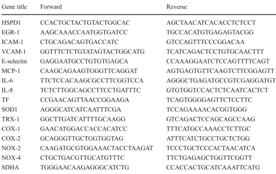
a master mix containing all reagents except for the primers was created; the primers for the gene of interest or a house-keeping gene was added directly to the glass capillary. Primer pairs are given in Table1. Each reaction proceeded for 40 amplification cycles, followed by a melting curve analysis to ensure the specificity of each reaction, controlled by the supplied LightCycler software (version 3.5, Roche). Crossing point and melting curve analyses for each reaction were also performed using the LightCycler software. Western blot analysis
Total protein extracts of C. pneumoniae-infected HUVECs and controls were obtained using standard NP-40 cell lysis buffer (50 mM Tris/HCl, pH 8, 150 mM NaCl, 1 % NP-40). The protein content was determined with Bradford reagent (Bio-Rad, Hercules, CA, USA) following the manufac-turer’s protocol. Equal amounts of protein in loading buffer (200 mM Tris/HCl, pH 6.8, 400 mM DTT, 8 % SDS, 0.4 % bromophenol blue, and 50 % glycerol) were loaded onto 12 % Tris/Glycine SDS-PAGels, purchased from Lonza, and run at 25 mA for 1 h. Proteins were blotted to a nitrocellu-lose membrane (Schleicher & Schuell, Dassel, Germany), and hHSP60 was stained using a mouse anti-human HSP60 antibody (clone II-13, prepared from hybridoma cells in our lab) (Singh and Gupta1992). As a loading control, mouse anti-human α-tubulin antibody (from Santa Cruz Biotech-nology, Santa Cruz, CA, USA; catalogue number: sc-23948) was used. Polyclonal rabbit anti-mouse immunoglobulins, covalently bound to horseradish peroxidase (P0161, Dako, Glostrup, Denmark) as secondary antibody, allowed for de-tection of the signal with the ECL method (Pierce, Rockford, IL, USA).
Table 1 List of the RT-PCR primer pairs
HSPD1 gene coding for hHsp60, EGR-1 early growth response protein 1, ICAM-1 intracellular adhesion molecule 1, VCAM-1 vascular cell adhesion molecule 1, MCP-1 monocyte chemotactic protein 1, IL interleukin, TF tissue factor, SOD1 superoxide dismu-tase 1 (Cu-Zn), TRX-1 thiore-doxin 1, COX cyclooxygenase, NOX nicotinamide adenine dinucleotide phosphate oxidase, SDHA subunit A of the succinate dehydrogenase complex
Gene title Forward Reverse
HSPD1 CCACTGCTACTGTACTGGCAC AGCTAACATCACACCTCTCCT
EGR-1 AAGCAAACCAATGGTGATCC TGCCACATGTGAGAGTACGG
ICAM-1 CTGCAGACAGTGACCATC GTCCAGTTTCCCGGACAA
VCAM-1 GGTTTCTCTGTATAGTACTGGCATG TCATCAGACTCCTGTGCAACTTT
E-selectin GAGGAATGCCTGTGTGAGCA CCAAAGGAATCTCCAGTTTTCAGT
MCP-1 CAAGCAGAAGTGGGTTCAGGAT AGTGAGTGTTCAAGTCTTCGGAGTT
IL-6 TTCTCCACAAGCGCCTTCGGTCCA AGGGCTGAGATGCCGTCGAGGATGTA
IL-8 TCTCTTGGCAGCCTTCCTGATTTC GTGTGGTCCACTCTCAATCACTCT
TF CCGAACAGTTAACCGGAAGA TCAGTGGGGAGTTCTCCTTC
SOD1 AGGGCATCATCAATTTCGA TCCAGAAAACACGGTGGG
TRX-1 GGCTTGATCATTTTGCAAGG GTCAGACTCCAGCAGCCAAG
COX-1 GAACATGGACCACCACATCC TTTCATGCCAAACCTCTTGC
COX-2 GCAGGGTTGCTGGTGGTAG ATTTCATCTGCCTGCTCTGG
NOX-2 CAAGATGCGTGGAAACTACCTAAGAT TCCCTGCTCCCACTAACATCA
NOX-4 CTGCTGACGTTGCATGTTTC TTCTGAGAGCTGGTTCGGTT
Immunofluorescence staining of cells and confocal microscopy
For immunofluorescence analysis, HUVECs, AECs, and VECs were grown in RPMI medium (Lonza) on 18×18 mm glass coverslips (Nunc, Rochester, NY, USA) coated with 10 % gelatine (Sigma-Aldrich, St. Louis, MO, USA). Cells were washed with phosphate-buffered saline (PBS), pH 7.2, fixed, and permeabilized with 99.5 % acetone at−20 °C for 2.5 min for intracellular staining. Subsequently, cells were allowed to dry for 30 min at room temperature (RT), followed by blocking with 0.1 % bovine serum albumin (Sigma-Aldrich) in PBS for 30 min at RT. For surface staining, cells were kept for all steps at 4 °C and fixed with 4 % paraformal-dehyde after incubation with the antibodies. Identical blocking solution and reagents were used for both staining protocols. Staining of eukaryotic HSP60 was performed with either the mouse anti-human HSP60 antibody (clone II-13) or rabbit anti-human HSP60 antibody (sc-13966, Santa Cruz Biotech-nology). Staining of chlamydial HSP60, human intracellular adhesion molecule 1 (ICAM-1), and human vascular cell adhesion molecule 1 (VCAM-1) was performed using mouse anti-chlamydial HSP60 antibody (Stressgen, Enzo Life Sci-ences, Plymouth Meeting, PA, USA), mouse anti-human ICAM-1, and mouse anti-human VCAM-1 antibodies (both e-Bioscience, San Diego, CA, USA). The matching isotype control antibodies were from Dako. After four washing steps with PBS, staining was visualized using either goat mouse IgG Alexa 488- or Alexa 568- and goat anti-rabbit Alexa 568-labeled secondary antibodies (Invitrogen, Carlsbad, CA, USA). Cells were then washed again, nuclei were stained with Hoechst 33342 (Sigma-Aldrich) accord-ing to the manufacturer’s protocol, and the coverslips were mounted with Fluoromount G on to the specimen holder (Nunc, Rochester, NY, USA). Time-resolved 3D stacks were acquired with an SP5 confocal microscope (Leica Microsystems, Wetzlar, Germany) equipped with a fast resonant scanner. We used an HCX PL APO lambda blue ×63 1.4 NA oil immersion objective. Imaging was per-formed with a 476-nm laser line for EGFP, a 561-nm laser line for AlexaFluor568 and mCherry excitation, and 633 nm for AlexaFluor647. Fluorescence emission was detected from 493 to 555 nm (EGFP), 566 to 742 nm (AlexaFluor568, mCherry), and 638 to 750 nm (AlexaFluor647). Images were acquired using the LAS AF acquisition software Version 2.1.
OxyBlot analysis of oxidative protein modification To assess the formation of protein carbonyl groups as a mea-sure of oxidative protein modification, the OxyBlot protein oxidation detection kit (Millipore, S7150) was used according to the manufacturer’s detailed protocol. For OxyBlot analysis,
107cells were washed twice with cold PBS and lysed (150 mM NaCl, 0.1 % Triton X-100, 30 mM Tris, 1 mM PMSF, 10 % glycerol, peptide inhibitors) on ice for 15 min. The cellular debris was removed by centrifugation (10,000×g, 4 °C, 15 min). The supernatant was incubated with 2 μg of the respective antibodies and 40μl of protein A/protein G agarose mix (Oncogene Research Products, Darmstadt, Germany) on a shaker for 2 h at 4 °C. Bound proteins were collected by centrifugation. The pellet was washed four times in lysis buffer, and subsequently suspended in “carbonyl-moiety to dinitrophenylhydrazone (DNP)-derivative converting solu-tion.” After washing in Tris-buffered saline, the pellets were suspended for electrophoretic separation in Western blot load-ing buffer. The separated DNP-containload-ing proteins were detected in Western blots (with DNP-specific antibodies) as described (Bernhard et al.2005).
Statistics
Data are presented as mean ± SD. Statistical analyses were performed using computer software (SPSS 17.0). Variable levels between different treatment groups were compared using ANOVA.
Results
Infection with C. pneumoniae leads to upregulation
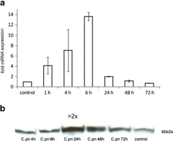
of hHSP60 and its relocalization to the endothelial cell surface Based on our previous finding of increased hHSP60 expres-sion in vascular endothelial cells in the presence of a diverse range of pro-atherogenic stimuli, we examined the effect of chlamydial infection on expression of hHSP60 in endothe-lial cells. As shown in Fig. 1a, chlamydial infection of HUVECs led to a significantly increased expression of hHSP60 mRNA with a maximum of expression at 6 h. As determined by Western blot analysis, hHSP60 levels were markedly increased upon infection with C. pneumoniae (Fig. 1b) with a peak of expression at 24 h (more than twofold upregulation). RT-PCR and Western blot analyses showed increased hHsp60 mRNA and protein expression, respectively, in C. pneumoniae-infected HUVECs, but this approach did not allow any conclusions about the subcellu-lar localization of the HSP60 molecules. Therefore, confocal microscopy of primary adult endothelial cells was per-formed (Fig.2), clearly showing in AECs (Fig.2a) redistri-bution of hHSP60 from the mitochondria to the plasma membrane together with adhesion molecules. Interestingly, neither hHSP60 surface expression nor hHSP60 presence was visible in the autologous VECs (Fig.2b). As shown in Fig.2c, surface expression of hHSP60 was accompanied by pronounced accumulation of intracellular chlamydial
particles (cHSP60 staining) demonstrating successful infec-tion of endothelial cells in our experimental setting. Infection with C. pneumoniae leads to increased expression of chemokines and adhesion molecules in endothelial cells Early atherosclerotic lesions are characterized by the presence of activated T cells and monocyte-derived macrophages. Importantly, lymphocyte and monocyte trafficking is highly dependent on chemoattractant chemokines produced at sites of ongoing inflammation. As outlined in Fig.3, analysis by RT-PCR revealed pronounced upregulation of MCP-1 and IL-8 in endothelial cells, which are known to be the most potent chemokines for macrophage migration. Moreover, expression
of adhesion molecules ICAM-1, VCAM-1, and E-selectin was increased starting at 3 h, with a peak of expression reached at 6 h post-infection. An increased expression of all the investigated adhesion molecules was sustained for up to 24 h post-infection (Fig.3). Additionally, we found the tran-scription factor early growth response gene-1 (Egr-1), a key mediator of inflammation and gene expression after vascular injury, to be markedly upregulated at early time points of infection (Fig.4a).
Infection with C. pneumoniae leads to a pro-coagulative state in endothelial cells
Besides its role in the gradual process of atherosclerotic lesion formation, chlamydial infection has been suggested to play a role in acute vascular events such as acute coronary syndrome. In the present study, infection with C. pneumo-niae led to upregulation of tissue factor (TF) (Fig. 4b). Increased expression of TF would facilitate onset of the external pathway of blood coagulation and, thus, enhance the risk of acute vascular thrombosis.
C. pneumoniae-infected endothelial cells show evidence of increased oxidative stress
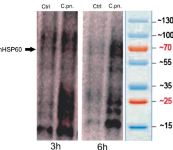
The cellular redox state plays a key role in several cellular fate decisions ranging from regulation of genes and redox-sensitive pathways to triggering cell death mechanisms. To get a deeper insight into the molecular mechanisms involved in the pro-atherosclerotic activities of C. pneumoniae, we examined if the endothelial redox balance was affected by C. pneumoniae infection by measuring the levels of protein carbonyl formation with the OxyBlot-based technique. C. pneumoniae-infected cells showed increased levels of protein carbonyl moieties as compared to noninfected cells (Fig.5). To determine the pathophysiological significance, if any, of this finding, we treated cells with various antioxidants before
Fig. 1 qRT-PCR analysis of human HSP60 (hHSP60) expression in HUVECs infected with C. pneumoniae. Control: succinate dehydroge-nase (SDHA). Mean ± SD, n03. Relative change in expression of the target gene mRNA is shown in relation to untreated samples with SDHA serving as an internal control (a). Western blot analysis of hHSP60 expression in HUVECs infected with C. pneumoniae for the given time points. Shown is one representative experiment out of three (b)
Fig. 2 Confocal microscopy. Human HSP60 (hHsp60) stained at the cell surface of nonpermeabilized cells (a, b). Orthogonal view of a z-stack of donor matched primary endothelial cells (48 h post-C. pneumoniae
infection). ICAM (red), hHSP60 (green). Bar05 μm. AECs (a) or VECs
(b). Chlamydial HSP60 (cHSP60) in C. pneumoniae-infected HUVECs, permeabilized for intracellular staining. Maximum intensity projection of HUVEC (z-stack, 48 h post-C. pneumoniae infection). cHSP60 (green),
infection with C. pneumonia. Figure6ashows that the pres-ence of both the broad spectrum antioxidant N-acetylcysteine and the superoxide anion scavenger tiron (4,5-dihydroxy-1,3-benzene disulfonic acid) significantly attenuated the inflam-matory response of endothelial cells upon C. pneumoniae infection. Taken together, these results strongly suggest that redox-sensitive signaling pathways mediate the upregulation of genes relevant for vascular inflammation.
C. pneumoniae infection causes downregulation of thioredoxin and superoxide dismutase and increased expression of NADPH oxidase
Based on the finding of an increased ROS production in C. pneumoniae-infected cells, we investigated whether chla-mydial infection has an impact on the antioxidative defense systems of endothelial cells. We found the expression of thioredoxin-1 (TRX-1), a major antioxidative defense sys-tem in endothelial cells, and superoxide dismutase-1 (SOD-1) to be strongly downregulated. Concomitantly, the proto-typical endothelial NADPH oxidase-4 (NOX-4) and NADPH oxidase-2 (NOX-2), the two major sources of ROS in the vasculature (Lassègue and Griendling 2010), were upregulated, pointing towards a redox imbalance with-in C. pneumoniae-with-infected cells. Furthermore, our experi-ments showed increased expression of the ROS-producing
enzyme cyclooxygenase-2 (COX-2) but not of COX-1 upon chlamydial infection pointing towards an additional role of COX-2 in the increased cellular ROS level associated with C. pneumoniae infection (Fig.6b).
Discussion
The most striking finding in our study was the observation of upregulation and surface expression of hHSP60 in arterial endothelial cells upon chlamydial infection. Of note, AECs proved to be markedly more susceptible to a chlamydia-mediated stress reaction in terms of surface expression of hHSP60 as compared to VECs from the same donors, which might be explained by the lifelong exposure of AECs to arterial blood pressure and flow conditions. This finding places C. pneumoniae among acquired primary stress fac-tors, especially for AECs, and is in agreement with our notion of a T-cell-mediated immunopathology underlying the atherosclerotic process. Expression of hHSP60 on the surface of endothelial cells might represent a major target for pre-existing hHSP60 auto- and cross-reactive immunity (Wick 2000). The nonphysiological expression of such an intracellular antigen on the cell surface of stressed arterial endothelial cells would lead to a vicious cycle of infiltration of the vascular wall by cross-reactive T cells and destruction
Fig. 3 RT-PCR analysis of adhesion molecules and pro-inflammatory cytokines in HUVECs. Cells were subjected to C. pneumoniae infec-tion for the indicated time periods. Mean ± SD, n03. Relative change
in expression of the target gene mRNA is shown in relation to untreat-ed samples with SDHA serving as an internal control
of stressed endothelial cells (Curry et al. 2000). A study investigating the clonality of such atherosclerotic plaque-derived T cells in cohorts of both anti-C. pneumoniae sero-positive and seronegative patients found plaques of seroneg-ative patients to harbor clones of T cells that recognize hHSP60 epitopes, while in seropositive patients, these T cells were found to cross-react with cHSP60 due to molecular mimicry of certain HSP60 epitopes (Benagiano et al.2005). This observation is in agreement with the finding of our previous cross-sectional studies, namely, that T-cell reactivity to bacterial and human HSP60 is an independent risk factor for incipient atherosclerosis in a cohort of clinically healthy young adults (Knoflach et al.2007; Rossmann et al.2008; Knoflach et al.2009). As HSP60 constitutes a highly con-served molecule, there is significant homology in the protein structure of human and bacterial HSP60 (Young and Elliott 1989). Lifelong reactivation of chlamydial infection in the vasculature (Peters et al.2005) would lead to a chronic im-mune reaction against cHSP60 and would significantly in-crease the risk for immunological cross-reactivity of human
chlamydia-specific T cells against stressed AECs. Besides recruitment of cross-reactive T cells, increased IL-8 and MCP-1 production paralleled by expression of adhesion mol-ecules as demonstrated in the present and other studies points to increased trafficking of monocytes into the vascular wall (Krüll et al.1999; Molestina et al. 1999; Kothe et al.2000; Hogdahl et al. 2008). Besides being a causative agent of systemic and local inflammation, chlamydial particles appear to be potent inducers of a pro-coagulative state of the physi-ologically inert vasculature. Our finding of increased endo-thelial TF expression early after chlamydial infection suggests an increased risk for acute thrombotic events in people sub-jected to chlamydial infection. An in vivo relevance of this finding is suggested by Hoshida et al. (2005) who found serum levels of anti-chlamydial IgM and hHSP60 antibody levels to be significantly higher in acute coronary syndrome patients as opposed to a clinically stable control cohort.
In the present study, several major pro-atherogenic changes observed upon chlamydial infection in HUVECs were found to depend on increased production of ROS, since application of antioxidants markedly ameliorated the endothelial stress response in the presence of C. pneumonia. Transcriptional upregulation of COX-2 as well as NOX-2 and NOX-4 paralleled by downregulation of SOD-1 and TRX-1 demonstrates a shift toward a more oxidative cellular redox state. COX-2 is involved in various inflammatory processes including atherosclerosis, and its inhibition was shown to significantly ameliorate C. pneumoniae-mediated effects on vascular remodeling (Rupp et al. 2004). Thus, chlamydial infection causes a remarkable redox imbalance in the host cell, with a diminished antioxidative defense on
Fig. 5 Endothelial cells infected with C. pneumoniae show evidence of increased oxidative stress. OxyBlot analysis of proteins from unin-fected control (Ctrl) and C. pneumoniae-inunin-fected (C.pn.) HUVECs (3 and 6 h after infection). Representative images of three independent experiments are shown
Fig. 4 RT-PCR analysis of the early growth response gene-1 (Egr-1) in C. pneumoniae-infected HUVECs. Cells were subjected to C. pneumo-niae infection for the indicated time periods. Short but pronounced upregulation of Egr-1 mRNA occurs as an early event at 1 h after
infection. Mean ± SD, n03 (a). RT-PCR analysis of human tissue factor
(TF) in HUVECS infected with C. pneumoniae. Mean ± SD, n03 (b).
Relative change in expression of the target gene mRNA is shown in relation to untreated samples with SDHA serving as an internal control
the one hand and critical enhancement of ROS-levels with upregulated NOX-2 and NOX-4 genes on the other.
In a recent study, it has been shown that LPS induces a superoxide-dependent expression of inflammatory cytokines and prostaglandins via upregulation of NADPH oxidase in a rat model of hypertension (Zhang et al.2010). Besides LPS-mediated signaling events, cHSP60 has been found to directly cause endothelial dysfunction by downregulation of endothe-lial nitric oxide synthase and associated mitochondrial dys-function in human coronary artery endothelial cells, and application of antioxidants was shown to abrogate HSP60-mediated endothelial dysfunction (Chen et al. 2009). Our finding of an increased oxidative state is in agreement with previous studies that could demonstrate increased levels of ROS in smooth muscle cells subsequent to infection with C. pneumoniae that seemed to depend on a functional NADPH oxidase activity (Dechend et al.2003). A contributory role of superoxide anion in this process is suggested by our finding of downregulation of SOD-1 (Fig.6b) as an early event after C. pneumoniae infection and by the markedly reduced endothe-lial stress response to C. pneumoniae infection in the presence of the superoxide radical scavenger tiron (Fig.6a). This find-ing suggests that antioxidant therapy might hold promise as a preventive measure against atherogenesis in the presence of risk factors such as infection with C. pneumoniae.
In summary, the finding of a markedly increased sus-ceptibility of arterial endothelial cells as compared to venous endothelial cells from the same donor for redistri-bution and enrichment of hHSP60 on the cellular surface further lends support to our hypothesis of atherosclerosis being the result of immunological cross-reactivity against eukaryotic hHSP60 under stressful conditions. Our data extend the list of established stress factors for endothelial cells that all converge upon the immunological danger signal hHSP60 being exposed on the cell surface. Com-pared to the data of previous studies on the degree of endothelial cell stressing capacity of canonical risk factors of atherosclerosis as reflected by the abundant expression of hHSP60, C. pneumoniae emerged as the most potent endothelial cell stressor (Amberger et al. 1997; Henderson et al.2008; Wick et al.2008; Grundtman et al. 2011). The cross-recognition of shared epitopes between human and chlamydial HSP60 by autoreactive cellular and humoral im-munity might represent an as yet unappreciated mechanism underlying the atherogenic potential of recurrent infection or reactivation of chlamydial particles in patients who fail to clear the infection. We believe that further in vivo studies are warranted to delineate the association of persistent chlamydial infection with markers of systemic inflammation in order to design novel therapeutic approaches aimed at counteracting
Fig. 6 RT-PCR analysis of adhesion molecules and pro-inflammatory cytokines in C. pneumoniae-infected HUVECs in the presence of antioxidants. Cells were pre-incubated either with the superoxide radical scavenger tiron or N-acetylcysteine (NAC) for 30 min and infected with C. pneumoniae for 6 h. Mean ± SD, n03. (*P<0.05) (a). RT-PCR analysis of ROS-producing enzymes in HUVEC subjected to C. pneumoniae infection. HUVECs infected with C. pneumoniae show transient upregulation of the NADPH oxidase isoforms NOX-2 and NOX-4 at 1 h, while upregulation of COX-2 persists at 6 h, when downregulation of TRX-1 as well as SOD-1 is detected.
Mean ± SD, n03 (b). Relative
change in expression of the target gene mRNA is shown in relation to untreated samples with SDHA serving as an internal control
endothelial cell activation and associated vascular autoim-mune pathogenic mechanisms.
Acknowledgments We thank Rajam Csordas-Iyer for critical reading
and editorial assistance. This work was supported by the European Initia-tive to Fight Chlamydial Infections by Unbiased Genomics (ECIBUG; # 818496 to GW), the Austrian Research Fund (FWF grant # 14741 to GW), and the TOLERAGE Health Research Grant (HEALTH-F4-2008-202156 to GW).
References
Amberger A, Maczek C, Jürgens G, Michaelis D, Schett G, Trieb K, Eberl T, Jindal S, Xu Q, Wick G (1997) Co-expression of ICAM-1, VCAM-ICAM-1, ELAM-1 and Hsp60 in human arterial and venous endothelial cells in response to cytokines and oxidized low-density lipoproteins. Cell Stress Chaperones 2:94–103
Benagiano M, D'Elios MM, Amedei A, Azzurri A, van der Zee R, Ciervo A, Rombolà G, Romagnani S, Cassone A, Prete D (2005) Human 60-kDa heat shock protein is a target autoantigen of T cells derived
from atherosclerotic plaques. J Immunol 174:6509–6517
Bernhard D, Csordas A, Henderson B, Rossmann A, Kind M, Wick G (2005) Cigarette smoke metal-catalyzed protein oxidation leads to vascular endothelial cell contraction by depolymerization of
microtubules. FASEB J 19:1096–1107
Chen C, Chai H, Wang X, Lin PH, Yao Q (2009) Chlamydia heat shock protein 60 decreases expression of endothelial nitric oxide synthase in human and porcine coronary artery endothelial cells.
Cardiovasc Res 83:768–777
Curry AJ, Portig I, Goodall JC, Kirkpatrick PJ, Gaston JS (2000) T lymphocyte lines isolated from atheromatous plaque contain cells capable of responding to Chlamydia antigens. Clin Exp Immunol 121:261–269
Dechend R, Gieffers J, Dietz R, Joerres A, Rupp J, Luft FC, Maas M (2003) Hydroxymethylglutaryl coenzyme A reductase inhibition reduces Chlamydia pneumoniae-induced cell interaction and
ac-tivation. Circulation 108:261–265
George J, Shoenfeld Y, Afek A, Gilburd B, Keren P, Shaish A, Kopolovic J, Wick G, Harats D (1999) Enhanced fatty streak formation in C57BL/6J mice by immunization with heat shock
protein-65. Arterioscler Thromb Vasc Biol 19:505–510
Gieffers J, Füllgraf H, Jahn J, Klinger M, Dalhoff K, Katus HA, Solbach W, Maas M (2001) Chlamydia pneumoniae infection in circulating human monocytes is refractory to antibiotic treatment.
Circulation 103:351–356
Grayston JT (2000) Background and current knowledge of Chlamydia
pneumoniae and atherosclerosis. J Infect Dis 181(Suppl 3):S402–
S410
Grundtman C, Kreutmayer SB, Almanzar G, Wick MC, Wick G (2011) Heat shock protein 60 and immune inflammatory responses in atherosclerosis. Arterioscler Thromb Vasc Biol 31:960–968 Grundtman C, Wick G (2011) The autoimmune concept of
atheroscle-rosis. Curr Opin Lipidol 22:327–334
Hansson GK (2001) Immune mechanisms in atherosclerosis.
Arterios-cler Thromb Vasc Biol 21:1876–1890
Henderson B, Csordas A, Backovic A, Kind M, Bernhard D, Wick G (2008) Cigarette smoke is an endothelial stressor and leads to cell
cycle arrest. Atherosclerosis 201:298–305
Hogdahl M, Soderlund G, Kihlstrom E (2008) Expression of chemo-kines and adhesion molecules in human coronary artery endothe-lial cells infected with Chlamydia (Chlamydophila) pneumoniae.
APMIS 116:1082–1088
Hoshida S, Nishino M, Tanouchi J, Kishimoto T, Yamada Y (2005) Acute Chlamydia pneumoniae infection with heat-shock-protein-60-related response in patients with acute coronary syndrome.
Atherosclerosis 183:109–112
Jackson LA, Campbell LA, Kuo CC, Rodriguez DI, Lee A, Grayston JT (1997) Isolation of Chlamydia pneumoniae from a carotid
endarterectomy specimen. J Infect Dis 176:292–295
Knoflach M, Kiechl S, Mayrl B, Kind M, Gaston JS, van der Zee R, Faggionato A, Mayr A, Willeit J, Wick G (2007) T-cell reactivity against HSP60 relates to early but not advanced atherosclerosis. Atherosclerosis 195:333–338
Knoflach M, Kiechl S, Penz D, Zangerle A, Schmidauer C, Rossmann A, Shingh M, Spallek R, Griesmacher A, Bernhard D, Robacher P, Buchberger W, Draxl W, Willeit J, Wick G (2009) Cardiovas-cular risk factors and atherosclerosis in young women: atheroscle-rosis risk factors in female youngsters (ARFY study). Stroke
40:1063–1069
Kothe H, Dalhoff K, Rupp J, Müller A, Kreuzer J, Maass M, Katus HA (2000) Hydroxymethylglutaryl coenzyme A reductase inhibitors modify the inflammatory response of human macrophages and endothelial cells infected with Chlamydia pneumoniae.
Circula-tion 101:1760–1763
Kreutmayer SB, Messner B, Knoflach M, Henderson B, Niederegger H, Böck G, Van der Zee R, Wick G, Bernhard D (2011) Dynamics of heat shock protein 60 in endothelial cells exposed to cigarette
smoke extract. J Mol Cell Cardiol 51:777–780
Krüll M, Klucken AC, Wuppermann FN, Fuhrmann O, Magerl C, Seybold J, Hippenstiel S, Hagemann JH, Jantos CA, Suttorp N (1999) Signal transduction pathways activated in endothelial cells following infection with Chlamydia pneumoniae. J Immunol 162:4834–4841
Lassègue B, Griendling KK (2010) NADPH oxidases: functions and pathologies in the vasculature. Arterioscler Thromb Vasc Biol
30:653–661
Lewinska A, Wnuk M, Slota E, Bartosz G (2007) Total anti-oxidant capacity of cell culture media. Clin Exp Pharmacol Physiol
34:781–786
Libby P (2002) Inflammation in atherosclerosis. Nature 420:868–874
Mayr M, Metzler B, Kiechl S, Willeit J, Schett G, Xu Q, Wick G (1999a) Endothelial cytotoxicity mediated by serum antibodies to heat shock proteins of Escherichia coli and Chlamydia pneumo-niae: immune reactions to heat shock proteins as a possible link
between infection and atherosclerosis. Circulation 99:1560–1566
Mayr M, Xu Q, Wick G (1999b) Atherogenic effects of chronic infections: the role of heat shock protein 60 in autoimmunity. Isr Med Assoc J 1:272–277
Molestina RE, Miller RD, Ramirez JA, Summersgill JT (1999) Infec-tion of human endothelial cells with Chlamydia pneumoniae stimulates transendothelial migration of neutrophils and
mono-cytes. Infect Immun 67:1323–1330
Peters J, Hess S, Endlich K, Thalmann J, Holzberg D, Kracht M, Schaefer M, Bartling G, Klos A (2005) Silencing or permanent activation: host-cell responses in models of persistent Chlamydia
pneumoniae infection. Cell Microbiol 7:1099–1108
Ross R (1999) Atherosclerosis—an inflammatory disease. N Engl J
Med 340:115–126
Rossmann A, Henderson B, Heidecker B, Seiler R, Fraedrich G, Singh M, Parson W, Keller M, Grubeck-Loebenstein B, Wick G (2008) T-cells from advanced atherosclerotic lesions recognize hHSP60 and have a
restricted T-cell receptor repertoire. Exp Gerontol 43:229–237
Rupp J, Berger M, Reiling N, Gieffers J, Lindschau C, Haller H, Dalhoff K, Maass M (2004) Cox-2 inhibition abrogates Chlamyd-ia pneumonChlamyd-iae-induced PGE2 and MMP-1 expression. Biochem Biophys Res Commun 320:738–744
Saikku P, Leinonen M, Mattila K, Ekman MR, Nieminen MS, Mäkelä PH, Huttunen JK, Valtonen V (1988) Serological evidence of an
association of a novel Chlamydia, TWAR, with chronic coronary
heart disease and acute myocardial infarction. Lancet 2:983–986
Shi Y, Tokunaga O (2004) Chlamydia pneumoniae (C. pneumoniae) infection upregulates atherosclerosis-related gene expression in human umbilical vein endothelial cells (HUVECs). Atherosclero-sis 177:245–253
Singh B, Gupta RS (1992) Expression of human 60-kD heat shock-protein (HSP60 or P1) in Escherichia coli and the development and characterization of corresponding monoclonal antibodies.
DNA Cell Biol 11:489–496
Wick G, Knoflach M, Xu Q (2004) Autoimmune and inflammatory
mechanisms in atherosclerosis. Annu Rev Immunol 22:361–403
Wick G, Perschinka H, Millonig G (2001) Atherosclerosis as an
autoimmune disease: an update. Trends Immunol 22:665–669
Wick G (2000) Atherosclerosis—an autoimmune disease due to an
immune reaction against heat-shock protein 60. Herz 25:87–90
Wick MC, Mayerl C, Backovic A, van der Zee R, Jaschke W, Dietrich H, Wick G (2008) In vivo imaging of the effect of LPS on arterial endothelial cells: molecular imaging of heat shock protein 60
expression. Cell Stress Chaperones 13:275–285
Xu Q, Dietrich H, Steiner HJ, Gown AM, Schoel B, Mikuz G, Kaufmann SH, Wick G (1992) Induction of arteriosclerosis in normocholester-olemic rabbits by immunization with heat shock protein 65. Arte-rioscler Thromb Vasc Biol 12:789–799
Xu Q, Kiechl S, Mayr M, Metzler B, Egger G, Oberhollenzer F, Willeit J, Wick G (1999) Association of serum antibodies to heat-shock
protein 65 with carotid atherosclerosis: clinical significance
de-termined in a follow-up study. Circulation 100:1169–1174
Xu Q, Luef G, Weimann S, Gupta RS, Wolf H, Wick G (1993a) Staining of endothelial cells and macrophages in atherosclerotic lesions with human heat-shock protein-reactive antisera. Arterios-cler Thromb Vasc Biol 13:1763–1769
Xu Q, Schett G, Perschinka H, Mayr M, Egger G, Oberhollenzer F, Willeit J, Kiechl S, Wick G (2000) Serum soluble heat shock protein 60 is elevated in subjects with atherosclerosis in a general
population. Circulation 102:14–20
Xu Q, Schett G, Seitz CS, Hu Y, Gupta RS, Wick G (1994) Surface staining and cytotoxic activity of heat-shock protein 60 antibody
in stressed aortic endothelial cells. Circ Res 75:1078–1085
Xu Q, Willeit J, Marosi M, Kleindienst R, Oberhollenzer F, Kiechl S, Stulnig T, Luef G, Wick G (1993b) Association of serum anti-bodies to heat-shock protein 65 with carotid atherosclerosis.
Lan-cet 341:255–259
Yamashita K, Ouchi K, Shirai M, Gondo T, Nakazawa T, Ito H (1998) Distribution of Chlamydia pneumoniae infection in the
atherscler-otic carotid artery. Stroke 29:773–778
Young RA, Elliott TJ (1989) Stress proteins, infection, and immune surveillance. Cell 59:5–8
Zhang ZH, Yu Y, Wei SG, Felder RB (2010) Centrally administered lipopolysaccharide elicits sympathetic excitation via NAD(P)H oxidase-dependent mitogen-activated protein kinase signaling. J Hypertens 28:806–816