Direct association of Bloom's syndrome gene product with the human mismatch repair protein MLH1
9
0
0
Texte intégral
(2) Nucleic Acids Research, 2001, Vol. 29, No. 21 4379. human homologue of the E.coli MutL protein, known to be involved in MMR and recombination (21–23). MATERIALS AND METHODS Construction of plasmids Various deletion mutants of BLM and hMLH1 were constructed by PCR and restriction digests starting from the vectors pJK1 (24) and pFastBacI-hMLH1 (25) containing the cDNAs of BLM and hMLH1, respectively, and cloned into the YTH vectors pBTM116, pACT2 and pGAD424. All plasmids were verified by DNA sequencing and expression in the YTH strain L40 was checked by western blot analysis using the appropriate antibodies. Full-length and truncated Sgs1 and full-length yMlh1 were amplified by PCR from yeast genomic DNA and confirmed by sequencing. For the in vitro transcription and translation constructs, the different fragments were generated by restriction digests of different YTH constructs and cloned into vectors of the pCite-4 series (Novagen). Sequences of all plasmids and construction schemes are available upon request. Yeast two-hybrid screen The screen was performed essentially as described (26). The yeast strain L40 [MATa trp1 leu2 his3 LYS2::lexA-HIS3 URA3::lexA-lacZ] was sequentially transformed with the bait pBTM116-BLM (amino acids 770–1417) and a random primed human peripheral blood cDNA library cloned into the BglII sites of pACT (Clonetech) using the lithium acetate method. Among over 3 × 10⁶ transformants tested for histidine prototrophy and positive β-galactosidase staining, 13 independent positive clones were found. Co-immunoprecipitation of BLM with anti-hMLH1 antibody Aliquots of 200 µg nuclear extracts of TK6 and HCT116 (prepared as described in Holmes et al.; 27) were incubated for 1 h at 4°C in a total volume of 100 µl of mismatch repair buffer (20 mM Tris–HCl pH 7.6, 40 mM KCl, 5 mM MgCl2, 1 mM PMSF, 1 mM glutathione, 0.1 mM dNTPs, 50 µg/ml BSA, 1.5 mM ATP), supplemented with 10% sucrose and 1× protease inhibitor cocktail, EDTA-free (Roche). The effective salt concentration was adjusted to 110 mM KCl. Aliquots of 2 µg of the monoclonal anti-hMLH1 antibody G168-728 (Pharmingen) were added and the incubation continued for a further 2 h. Samples of 30 µl of Pan Mouse IgG Dynabeads (Dynal) were added to the solution and the incubation was continued for a further 1.5 h before the matrix-bound proteins were isolated following the instructions of the manufacturer. The beads were washed four times with 200 µl of incubation buffer (110 mM KCl) before elution. The eluted proteins were subjected to western blot analysis using the monoclonal antihMLH1 antibody G168-15 (Pharmingen) and the polyclonal anti-BLM antibody IHIC33 (18). Detection was performed using ECL (Amersham Pharmacia Biotech) following the manufacturer’s instructions.. Co-immunoprecipitation of hMLH1 with anti-BLM antibody Aliquots of 200 µg nuclear extracts from BJAB (prepared as described in 28) were incubated for 1 h in the incubation buffer (20 mM HEPES–KOH pH 7.5, 60 mM KCl, 2 mM MgCl2, 0.1% NP-40, protease inhibitors) at 4°C in a total volume of 200 µl. Aliquots of 2 µl of the anti-BLM antibody ab476 (Abcam) and 1 µg contol IgG were added and incubated for a further 2.5 h. Samples of 15 mg equilibrated protein A–Sepharose CL-4B beads (Amersham Pharmacia Biotech) were added and the incubation was continued for a further 1.5 h. The beads were then washed five times with 0.5 ml of incubation buffer prior to elution. The membranes were hybridised with the antibodies mentioned above. Far western analysis This assay was performed essentially as described previously (18). Briefly, 1 µg purified proteins was subjected to SDS–PAGE and transferred to nitrocellulose filters. After renaturation and blocking, the filters were incubated for 60 min in BLM (1 µg/ml) or MutLα (0.5 µg/ml) in TBS (25 mM Tris–HCl pH 7.4, 140 mM NaCl, 2.5 mM KCl) supplemented with 0.25% milk, 0.3% Tween 20, 1 mM DTT and 1 mM PMSF. After extensive washing, conventional western analyses using the indicated antibodies were performed to detect the presence of BLM, hMLH1 and hPMS2. In vitro binding assay Different amounts of recombinant BLM, MutLα and BSA were dotted on a nitrocellulose membrane (MSI NitroBind). After blocking for 1 h at room temperature using TBS supplemented with 5% milk and 0.05% Tween, the membrane was incubated for 3 h at 4°C with different proteins that were 35S-labelled using the TNT T7 quick coupled transcription/ translation system (Promega) in 1 ml of TBS with 0.1% BSA and 0.05% Tween. Aliquots of 20 or 40 µl of the in vitro transcription and translation reactions were used for each incubation. After extensive washes with TBS containing 0.05% Tween, the membranes were dried and exposed either to a PhosphorImager (Molecular Dynamics) or X-ray film (Super RX; Fujifilm). Indirect immunofluorescence analysis These experiments were performed as described (18) with the following changes. The primary antibodies used were the IHIC33 anti-BLM rabbit polyclonal antibody (18) and the antihMLH1 mouse monoclonal antibody G168-728 (Pharmingen), which were both used at a 1:100 dilution. The secondary antibodies were fluorescein isothiocyanate-conjugated anti-rabbit and Cy3 anti-mouse antibodies (both from Sigma) used at 1:200 and 1:800 dilution, respectively. In vitro MMR assay Nuclear extracts were prepared from exponentially growing TK6, HCT116, MRC5-SV40, GM08505 and PSNF5 (SV40immortalized GM08505 BLM) fibroblasts and the two immortalized lymphoblasts GM09960 and GM03403 (Coriell Cell Repository) as described previously (27) with minor modifications. Nuclei were isolated and resuspended in cold extraction buffer (50 mM HEPES–KOH pH 7.5, 10% sucrose, 1 mM PMSF, 0.5 mM DTT, 1 µg/ml leupeptin) in the smallest.
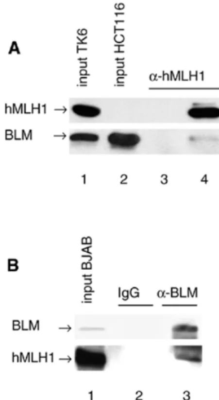
(3) 4380 Nucleic Acids Research, 2001, Vol. 29, No. 21. Figure 1. YTH interactions. (A) Schematic representation of BLM. The two acidic domains (striped), the helicase domain (black), the HRDC domain (stippled) and the two putative nuclear localisation signals (arrow) are indicated. The portion of BLM used as bait in the YTH screen is shown below. (B) Interactions of BLM and hMLH1 in the YTH assay. The L40 yeast strain was co-transformed with plasmids encoding the indicated proteins, and three independent colonies were grown on Trp–Leu–His– selective plates prior to assessment of β-galactosidase activity. hMLH1(198–756) is the prey found in the YTH screen. Bait dependency is shown with a non-cognate protein (S.cerevisiae Rer2p) fused to LexAdbd. (C) YTH interactions of Sgs1p and yMlh1p as well as interspecies interactions. β-Galactosidase filter assay demonstrating the interaction of yMlh1p with a deletion mutant of Sgs1p (amino acids 784–1447) as well as with full-length Sgs1p and BLM and of Sgs1p (amino acids 784–1447) with hMlh1p. Also shown are two negative controls, the empty bait (LexAdbd) vector co-transformed with yMlh1 and and the empty prey (Gal4ad) vector together with the Sgs1 deletion mutant.. Figure 2. BLM and hMLH exist as a complex in human cells. (A) Co-immunoprecipitation of BLM with hMLH1. BLM could be immunoprecipitated with an anti-hMLH1 antibody from 200 µg nuclear extract of TK6 (wt) cells (lane 4), but not from HCT116 (BLM+ hMLH1–) nuclear extract (lane 3). Immunoprecipitated proteins were visualised by western blot analysis with antibodies against hMLH1 (upper) or BLM (lower). (B) Co-immunoprecipitation of hMLH1 with BLM. hMLH1 was immunoprecipitated from 200 µg nuclear extract of BJAB cells with an anti-BLM antibody, but not with IgG. The immunoprecipitated proteins were detected with antibodies against BLM (upper) or hMLH1 (lower).. the BglII site, such that the repair efficiency can be estimated from the amount of phagemid DNA cleaved by BglII. RESULTS. volume possible and 0.031 vol of 5 M NaCl were added. The mixture was then rotated for 1 h at 4°C. Nuclear debris was pelleted at 14 500 g for 20 min at 4°C. The supernatant was dialysed for 2 h at 4°C against dialysis buffer (25 mM HEPES– KOH pH 7.6, 50 mM KCl, 0.1 mM EDTA, 10% sucrose, 0.1% PMSF, 2 mM DTT, 1 µg/ml leupeptin) and clarified by centrifugation at 20 000 g for 15 min at 4°C. The assays were carried out as described (27,29) with some modifications. The reaction mixtures (20 µl) contained mismatch repair buffer, 75 µg nuclear cell extract and 75 ng heteroduplex DNA pGemG·T, containing a G·T mismatch in the BglII restriction site located 369 bp downstream from the nick, that was essentially constructed as described previously (27,29,30). All the reaction mixtures were adjusted to 110 mM KCl, incubated at 37°C for 30 min and the repair reactions terminated by addition of 30 µl of stop solution (25 mM EDTA, 0.67% SDS, 50 µg/ml proteinase K) for 15 min at 37°C. The repair reaction converts the G·T heteroduplex to an A·T homoduplex and thus restores. Two-hybrid screen for BLM-interacting proteins A YTH screen was performed to search for human proteins capable of interacting with the C-terminal 647 amino acids of BLM as a bait among >3 000 000 yeast transformants (Fig. 1A). Thirteen independent clones specifically interacting with the bait were isolated from a random-primed human peripheral blood cDNA library, among which a truncation (amino acids 198–756) of the human MMR protein MLH1 was found. The specificity of this interaction was also confirmed with fulllength hMLH1 (amino acids 1–756) and by switching the hybrid partners: a fusion of hMLH1 to the LexA DNA-binding domain (LexAdbd) interacted with full-length BLM fused to the Gal4 activation domain (Gal4ad) (Fig. 1B). Sgs1p, the S.cerevisiae RecQ homologue, interacts with yMlh1p Since both RecQ helicases and MLH1 have been conserved throughout evolution, and given that RecQ helicase mutants in yeast and humans affect genomic stability, we asked whether.
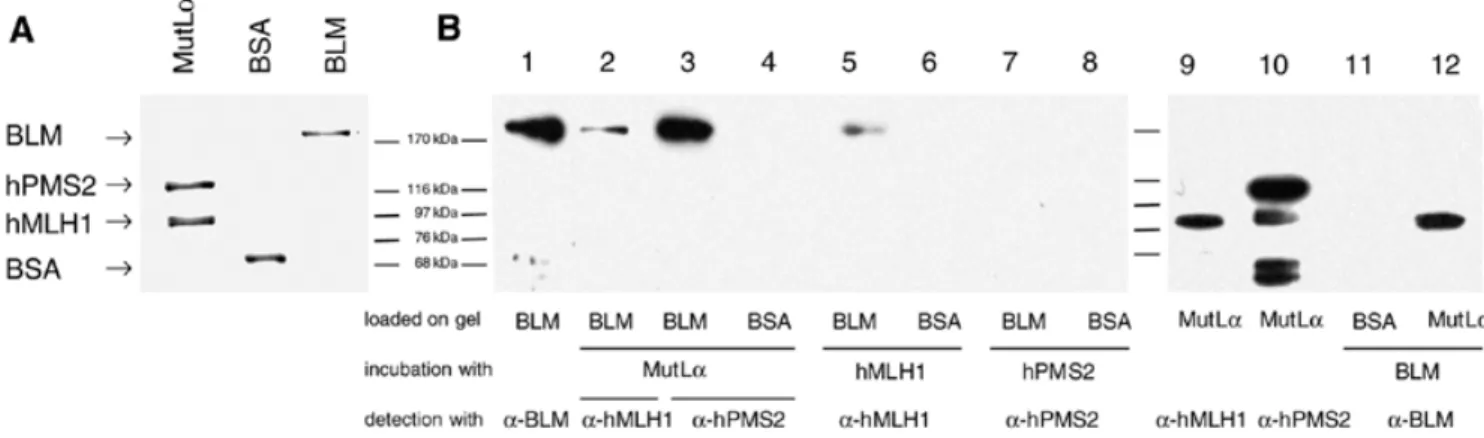
(4) Nucleic Acids Research, 2001, Vol. 29, No. 21 4381. Figure 3. BLM and hMLH interact directly. (A) Purified human MutLα complex (1.5 µg), BSA (1 µg) and BLM (1 µg) were subjected to SDS–PAGE and stained with Coomassie blue. (B) Far western analysis. The proteins were transferred to a nitrocellulose membrane, renatured and incubated with purified MutLα complex (0.5 µg/ml), total Sf9 extracts expressing either hMLH1 or hPMS2, or purified recombinant BLM (1 µg/ml). Western blotting using anti-hMLH1, anti-hPMS2 (Ab-1; Calbiochem) and anti-BLM antibodies was used to detect the presence of the latter proteins on the membrane. The faster running bands in lane 10 are degradation products of hPMS2.. Sgs1p, the S.cerevisiae homologue of BLM, interacts with yeast Mlh1p (yMlh1p) in the two-hybrid system. Although the C-terminal domains of Sgs1p and BLM share little sequence homology, the C-terminal domain (amino acids 784–1447) of Sgs1p was found to specifically interact with yMlh1p. Analogous to the BLM–hMLH1 interaction, the YTH interaction between Sgs1p and yMlh1p was confirmed for full-length yMlh1p and Sgs1p (Fig. 1C). Different studies have shown that expression of the BLM gene can partially complement both the hyper-recombination phenotype (31) and the reduced lifespan of sgs1 mutants (32). We wondered whether there is an interspecies interaction between BLM and MLH1, i.e. whether Sgs1p interacts with hMlh1p and BLM with yMlh1p. As shown in Figure 1C, we were able to detect an interspecies interaction via YTH between Sgs1p (amino acids 784–1447) and hMLH1 as well as between yMlh1 and BLM (Fig. 1C). The result was also confirmed for full-length Sgs1p after switching the hybrid partners (data not shown). BLM and hMLH1 exist as a complex in human cells Given that BLM and hMLH1 interact in the YTH assay, we wanted to test if BLM forms a complex with hMLH1 in human cells in vivo. To this end, co-immunoprecipitation experiments were performed, where an anti-MLH1 monoclonal antibody was used to precipitate its cognate protein from human nuclear cell extracts (Fig. 2A). BLM could be immunoprecipitated with hMLH1 from extracts of the MMR-proficient TK6 cells (lane 4), but not from extracts of hMLH1-deficient HCT116 cells (lane 3). In addition, the inverse co-immunoprecipitation experiment was carried out, in which an anti-BLM polyclonal antibody was used to immunoprecipitate hMLH1 from nuclear extracts of the human BJAB cell line. As seen in the Figure 2B, hMLH1 could be specifically co-immunoprecipitated with anti-BLM (lane 3), but not with the control IgG antibody (lane 2). BLM and hMLH1 interact directly in vitro We next wanted to determine whether the interaction between BLM and hMLH1 was direct, rather than being mediated via. an accessory protein. Far western analysis was therefore performed to determine whether purified recombinant BLM and hMLH1 could interact directly in vitro. To this end, fulllength recombinant BLM protein (24) was immobilised on a nitrocellulose filter, which was then incubated either with purified MutLα, a heterodimer of hMLH1 and hPMS2, or with extracts of Sf9 cells expressing either hMLH1 or hPMS2 (25). The filter was then washed to remove the unbound proteins and the presence of BLM was detected using conventional western blotting with anti-hMLH1 and/or anti-hPMS2 antibodies. As controls, the membrane also contained BSA and BLM alone. Figure 3 shows that BLM protein could be detected with antibodies against both hMLH1 (lane 2) and hPMS2 (lane 3) after incubation with the purified MutLα complex, but only hMLH1 alone (lane 5), and not hPMS2 (lane 7), could bind to BLM. The interaction between hMLH1 and BLM appeared to be specific, because MutLα and hMLH1 failed to bind the control protein, BSA (lanes 4 and 6), which was loaded on the same blot. To re-confirm these data, the reciprocal far western experiment was carried out, wherein human BLM protein was used to probe nitrocellulose-bound hMLH1. In this experiment, the anti-BLM antibody revealed a specific band at ∼80 kDa, the position of migration of hMLH1 (lane 12). This band was due to BLM binding to hMLH1, as its position was identical to the specific hMLH1 band detected when purified MutLα complex was probed with anti-hMLH1 antibody (lane 9), and not to the band specific for PMS2 (lane 10). Again, the interaction between BLM and hMLH1 was specific, because no signal was detected with the control BSA protein (lane 11). We conclude that purified recombinant BLM and hMLH1 interact directly in vitro and that BLM interaction with PMS2 is mediated via hMLH1. Mapping of the BLM and hMLH1 interaction domains To investigate which region of BLM protein was responsible for mediating the interaction with hMLH1, a series of BLM deletion mutants was generated and tested for their ability to interact with full-length hMLH1 in the YTH assay. As negative controls, the empty vector and a fragment of hMLH1.
(5) 4382 Nucleic Acids Research, 2001, Vol. 29, No. 21.
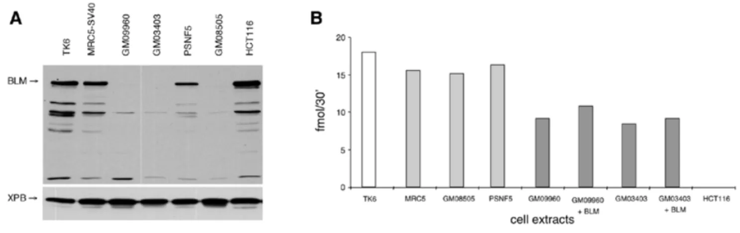
(6) Nucleic Acids Research, 2001, Vol. 29, No. 21 4383. (amino acids 1–388) that did not bind to BLM were included. The results of these experiments indicated the presence of three independent hMLH1-interacting regions in BLM comprising residues 1–131, 448–572 and 1034–1417 (Fig. 4A). A similar approach was used to map the BLM interaction domain of hMLH1. A series of N- and C-terminal deletions of hMLH1 was generated and tested for interaction with fulllength BLM (Fig. 4B). The results clearly indicated that the N-terminus of hMLH1 was dispensable for interaction with BLM, while the C-terminus (amino acids 396–756), a region similar to that involved in the interaction with hMLH3, hPMS1 and hPMS2 (33), was essential. A deletion of as little as 60 amino acids from the C-terminus of hMLH1 was sufficient to destroy the interaction with BLM (Fig. 4B). In addition, the YTH interaction domain mapping of both proteins was further confirmed using an in vitro binding approach, in which one of the interacting proteins was immobilised on a nitrocellulose membrane and probed with several 35S-labelled in vitro transcribed and translated deletion mutants of the other protein. Different in vitro transcribed and translated BLM fragments were incubated with membrane-bound purified recombinant MutLα heterodimer and BSA, included as a negative control (Fig. 4C). The weak binding of the BLM fragments might be due to the low accessibility of hMLH1 due to its heterodimerisation with hPMS2. Nevertheless, all the fragments containing one of the three domains identified in the YTH screen were able to bind to MutLα, but not to BSA, thus confirming the YTH mapping. Moreover, we could further narrow down the C-terminal interacting domain to a region between amino acids 1109 and 1378, as both C-terminal fragments tested clearly interacted with MutLα. In vitro transcribed and translated full-length hMLH1 as well as different deletions thereof were incubated with immobilised purified recombinant BLM and BSA (Fig. 4D). While the N-terminal half of the protein did not interact with BLM, the C-terminal half bound to BLM as strongly as the full-length protein. This interaction was completely abolished when the C-terminal half was further divided. Additionally, in vitro transcribed and translated hMLH1 was shown to bind to immobilised N-terminally truncated purified BLM protein (amino acids 212–1417; data not shown). BLM and hMLH1 co-localise in the nucleus The co-immunoprecipitation of BLM and hMLH1 from human cell extracts, as well as the evidence of a direct interaction between BLM and hMLH1, is consistent with these proteins forming a complex in vivo and in vitro. To provide additional evidence for the existence of this interaction, we wanted to see whether BLM and hMLH1 co-localise within the nucleus of intact human cells. Indirect immunofluorescence of exponentially growing human WI-38/VA-13 cells, using either anti-BLM or anti-hMLH1 antibodies, revealed BLM and hMLH1 to localise. to prominent nuclear foci (Fig. 5). Merging the fluorescent signals for BLM and hMLH1 showed a clear concordance in their localisation, thus strengthening the notion that the two proteins may function in a common biochemical pathway. A similar co-localisation pattern was obtained following aphidicolin treatment of the cells (data not shown). BS cell lines are MMR proficient No DNA helicase activities have so far been found to be associated with the MMR process. To address the question whether BLM is acting together with hMLH1 in MMR, the MMR proficiency of extracts of two lymphoblastoid (GM03403 and GM09960) and one fibroblast (GM08505) cell lines derived from BS patients were analysed. The absence of BLM protein in nuclear extracts of the BS cell lines was confirmed by western blot analysis, using antibodies raised against both the N- and C-termini of BLM (Fig. 6A and data not shown). Extracts derived from the lymphoblastoid TK6 and fibroblastoid MRC5 cells, HCT116 cells, and PSNF5 cells (GM08505 containing the BLM cDNA) were used as controls. As indicated in Figure 6B, all BS-derived nuclear extracts were MMR proficient using a substrate containing a single G·T mismatch and a strand discrimination signal (a nick) upstream (5′) from the mispair. The lower repair efficiency of the nuclear extract derived from the two lymphoblastoid cell lines GM03403 and GM09960, as compared to the MMR-proficient TK6 cells, is due to their high genomic instability and consequently to their reduced viability. Addition of recombinant purified BLM to the GM03403 or GM09960 cell extracts failed to influence the efficiency of MMR (Fig. 6B), which implies that the extracts had an intrinsically lower MMR capacity rather than being MMR deficient due to lack of BLM. Similar results were obtained using cytoplasmic extracts of the BLM cell lines and a template with the nick downstream (3′) from the mismatch (data not shown). DISCUSSION Defects in the BLM gene product result in severe physiological consequences in humans, the most prominent of which is premature death due to cancer. Although a great deal of genetic and biochemical data are available on BLM, the molecular defects resulting in BS remain elusive. In this study, we performed a YTH screen using the C-terminal portion of BLM as bait and a human peripheral blood cDNA library as a source of potential partners. One of the isolated BLM-interacting factors was hMLH1, a protein known to be involved in MMR and recombination. We now present several lines of evidence that BLM interacts directly with hMLH1 via three separate sites on BLM. Deletion of these three sites located in the N-terminal, central and C-terminal domains of BLM, was found to completely abolish the ability of BLM to interact with. Figure 4. (Opposite) Interaction domain mapping of BLM and hMLH1. (A and B) Yeast two-hybrid assays. The sequence boundaries of the deletion mutants tested in a β-galactosidase filter assay are shown with the corresponding amino acid positions indicated on the left. The black bars indicate positive and striped bars negative interactions. (C and D) In vitro binding assays. (C) Aliquots of 0, 0.625, 1.25 and 2.5 pmol recombinant MutLα and BSA were spotted onto a nitrocellulose membrane and probed with 20 (*) or 40 µl (**), respectively, of the reaction mixture containing the indicated in vitro transcribed and translated BLM proteins. The autoradiogram of the gel shows 2 µl of the radiolabelled proteins used in the assay. (D) The same approach as in (C), but with immobilised full-length BLM and BSA on the membrane and the indicated in vitro transcribed and translated hMLH1 deletion mutants as probes..
(7) 4384 Nucleic Acids Research, 2001, Vol. 29, No. 21. Figure 5. Co-localisation of BLM and hMLH1 in the nucleus of WI-38/VA-13 cells. Indirect immunofluorescence of BLM (green) and hMLH1 (red) is shown in WI-38/VA-13 cells. The yellow colour results from overlap of the red and green foci. Nuclear DNA was revealed by staining with Hoechst 33258.. Figure 6. In vitro MMR efficiency of BS cell lines. (A) Western blot showing the absence of BLM protein in the BS cell lines. Aliquots of 25 µg of the indicated nuclear extracts were probed with anti-BLM antibody (IHIC33) and anti-XPB antibody [TFIIH p89 (S-19); Santa Cruz Biotechnology] as control. (B) MMR efficiency of the BLM-negative human fibroblast GM08505 and of the human lymphoblasts GM09960 and GM03403. The MMR-proficient MRC5 SV40 fibroblasts, TK6 lymphoblasts, PSNF5 cells (GM08505 stably transfected with BLM cDNA) and the MMR-deficient hMLH1–/– colon cancer cell line HCT116 were used as controls. The MMR efficiencies of the two BS lymphoblasts complemented with purified recombinant BLM protein are also shown. The repair efficiency is expressed as fmol phagemid DNA cleaved by BglII in 30 min.. hMLH1. In addition, we have shown that the C-terminal 360 residues of hMLH1 are sufficient to interact with BLM. Furthermore, we have shown that the interaction between BLM and hMLH1 is highly conserved throughout evolution, since Sgs1p, the yeast homologue of BLM, interacts with yMlh1p. We have also shown that this interaction occurs between species: Sgs1p interacted with hMLH1 and BLM interacted with yMlh1p. This evolutionary conservation of an. interaction between Sgs1p and yMLH1, together with the interspecies interactions, suggests that these two proteins together perform a fundamentally important role during DNA metabolism. What could be the functional significance of the BLM– hMLH1 interaction? To address this question, we investigated the role of BLM helicase in mismatch repair. Post-replicative mismatch repair is evolutionarily highly conserved, from.
(8) Nucleic Acids Research, 2001, Vol. 29, No. 21 4385. E.coli to humans, and is postulated to consist of three principal steps: mismatch recognition and assembly of the ‘repairosome’, degradation of the error-containing strand, and DNA repair synthesis (34). This implies the existence of other proteins involved in the steps following mismatch recognition. Some of these proteins have already been characterized and their involvement in MMR has been documented. They include DNA polymerase δ (35), PCNA (36), RP-A (37) and exonuclease EXOI (38; for a recent review see 22). However, some members of the MMR repairosome, such as the putative DNA helicase, remain to be identified. Our functional experiments indicate that BLM helicase doesn’t play an essential role in MMR, as three different cell extracts form BS cells were shown to be MMR proficient. However, we cannot exclude the possibility that the helicase function in MMR is redundant and that the lack of only one helicase might result in a qualitatively ‘normal’ phenotype. The elevated sister chromatid exchange and hyperrecombination associated with BS suggest a defect in recombination. BLM helicase (and other RecQ family helicases) have been proposed to play important roles in overcoming structural abnormalities that arise during replication, thus preventing illegitimate recombination events occurring in regions prone to chromosomal rearrangements (14,19). Interestingly, recent findings show that the Msh2, Msh6 and Mlh1–Mlh3 proteins bind not only to mismatched DNA, but also to Holliday junctions in yeast (23,39,40). We therefore suggest that the BLM– hMLH1 complex may act as a potential sensor of recombination and replication fork damage. In the light of the available evidence, we are currently investigating the potential role of BLM and Sgs1p in these processes. NOTE ADDED IN PROOF Langland et al. (41) have also identified an interaction between BLM and hMLH1 protein. ACKNOWLEDGEMENTS We are grateful to Markus Räschle for the gift of the purified MutLα and hMLH1 cDNA, Primo Schär for critical reading of the manuscript, Patrick Dufner for technical help, Walter Schaffner for the gift of BJAB nuclear extracts and Ulrich Hübscher for support. This work was financed by grants from Bonizzi-Theler Stiftung, EMDO Stiftung, Gebert-Rüf Stiftung, Stiftung für Medizinische Forschung, Walter Honegger Stiftung and Swiss National Science Foundation (no. 31-58798.99) to G.P., H.B., G.H.R. and I.S, and Swiss National Science Foundation to C.P. and J.J. S.L.D. and I.D.H. were supported by the Imperial Cancer Research Fund. R.F. is currently supported by a FIS (Fondo Investigaciones Sanitarias) contract. REFERENCES 1. German,J. (1993) Bloom syndrome: a Mendelian prototype of somatic mutational disease. Medicine, 72, 393–406. 2. Ellis,N.A., Groden,J., Ye,T.Z., Straughen,J., Lennon,D.J., Ciocci,S., Proytcheva,M. and German,J. (1995) The Bloom’s syndrome gene product is homologous to RecQ helicases. Cell, 83, 655–666.. 3. Nakayama,K., Irino,N. and Nakayama,H. (1985) The recQ gene of Escherichia coli K12: molecular cloning and isolation of insertion mutants. Mol. Gen. Genet., 200, 266–271. 4. Gangloff,S., McDonald,J.P., Bendixen,C., Arthur,L. and Rothstein,R. (1994) The yeast type I topoisomerase Top3 interacts with Sgs1, a DNA helicase homolog: a potential eukaryotic reverse gyrase. Mol. Cell. Biol., 14, 8391–8398. 5. Stewart,E., Chapman,C.R., Al-Khodairy,F., Carr,A.M. and Enoch,T. (1997) rqh1+, a fission yeast gene related to the Bloom’s and Werner’s syndrome genes, is required for reversible S phase arrest. EMBO J., 16, 2682–2692. 6. Puranam,K.L. and Blackshear,P.J. (1994) Cloning and characterization of RECQL, a potential human homologue of the Escherichia coli DNA helicase RecQ. J. Biol. Chem., 269, 29838–29845. 7. Yu,C.E., Oshima,J., Fu,Y.H., Wijsman,E.M., Hisama,F., Alisch,R., Matthews,S., Nakura,J., Miki,T., Ouais,S., Martin,G.M., Mulligan,J. and Schellenberg,G.D. (1996) Positional cloning of the Werner’s syndrome gene. Science, 272, 258–262. 8. Kitao,S., Shimamoto,A., Goto,M., Miller,R.W., Smithson,W.A., Lindor,N.M. and Furuichi,Y. (1999) Mutations in RECQL4 cause a subset of cases of Rothmund-Thomson syndrome. Nature Genet., 22, 82–84. 9. Kitao,S., Ohsugi,I., Ichikawa,K., Goto,M., Furuichi,Y. and Shimamoto,A. (1998) Cloning of two new human helicase genes of the RecQ family: biological significance of multiple species in higher eukaryotes. Genomics, 54, 443–452. 10. German,J., Crippa,L.P. and Bloom,D. (1974) Bloom’s syndrome. III. Analysis of the chromosome aberration characteristic of this disorder. Chromosoma, 48, 361–366. 11. Watt,P.M., Hickson,I.D., Borts,R.H. and Louis,E.J. (1996) SGS1, a homologue of the Bloom’s and Werner’s syndrome genes, is required for maintenance of genome stability in Saccharomyces cerevisiae. Genetics, 144, 935–945. 12. Harmon,F.G. and Kowalczykowski,S.C. (1998) RecQ helicase, in concert with RecA and SSB proteins, initiates and disrupts DNA recombination. Genes Dev., 12, 1134–1144. 13. Bennett,R.J., Keck,J.L. and Wang,J.C. (1999) Binding specificity determines polarity of DNA unwinding by the Sgs1 protein of S. cerevisiae. J. Mol. Biol., 289, 235–248. 14. Karow,J.K., Constantinou,A., Li,J.L., West,S.C. and Hickson,I.D. (2000) The Bloom’s syndrome gene product promotes branch migration of holliday junctions. Proc. Natl Acad. Sci. USA, 97, 6504–6508. 15. Constantinou,A., Tarsounas,M., Karow,J.K., Brosh,R.M., Bohr,V.A., Hickson,I.D. and West,S.C. (2000) Werner’s syndrome protein (WRN) migrates Holliday junctions and co-localizes with RPA upon replication arrest. EMBO Rep., 1, 80–84. 16. Myung,K., Datta,A., Chen,C. and Kolodner,R.D. (2001) SGS1, the Saccharomyces cerevisiae homologue of BLM and WRN, suppresses genome instability and homologous recombination. Nature Genet., 27, 113–116. 17. Brosh,R.M., Li,J.L., Kenny,M.K., Karow,J.K., Cooper,M.P., Kureekattil,R.P., Hickson,I.D. and Bohr,V.A. (2000) Replication protein A physically interacts with the Bloom’s syndrome protein and stimulates its helicase activity. J. Biol. Chem., 275, 23500–23508. 18. Wu,L., Davies,S.L., North,P.S., Goulaouic,H., Riou,J.F., Turley,H., Gatter,K.C. and Hickson,I.D. (2000) The Bloom’s syndrome gene product interacts with topoisomerase III. J. Biol. Chem., 275, 9636–9644. 19. Wu,L., Davies,S.L., Levitt,N.C. and Hickson,I.D. (2001) Potential role for the BLM helicase in recombinational repair via a conserved interaction with RAD51. J. Biol. Chem., 276, 19375–19381. 20. Wang,Y., Cortez,D., Yazdi,P., Neff,N., Elledge,S.J. and Qin,J. (2000) BASC, a super complex of BRCA1-associated proteins involved in the recognition and repair of aberrant DNA structures. Genes Dev., 14, 927–939. 21. Jiricny,J. (1998) Eukaryotic mismatch repair: an update. Mutat. Res., 409, 107–121. 22. Kolodner,R.D. and Marsischky,G.T. (1999) Eukaryotic DNA mismatch repair. Curr. Opin. Genet. Dev., 9, 89–96. 23. Wang,T.F., Kleckner,N. and Hunter,N. (1999) Functional specificity of MutL homologs in yeast: evidence for three Mlh1-based heterocomplexes with distinct roles during meiosis in recombination and mismatch correction. Proc. Natl Acad. Sci. USA, 96, 13914–13919. 24. Karow,J.K., Chakraverty,R.K. and Hickson,I.D. (1997) The Bloom’s syndrome gene product is a 3′-5′ DNA helicase. J. Biol. Chem., 272, 30611–30614..
(9) 4386 Nucleic Acids Research, 2001, Vol. 29, No. 21. 25. Räschle,M., Marra,G., Nystrom-Lahti,M., Schär,P. and Jiricny,J. (1999) Identification of hMutLβ, a heterodimer of hMLH1 and hPMS1. J. Biol. Chem., 274, 32368–32375. 26. Vojtek,A.B., Hollenberg,S.M. and Cooper,J.A. (1993) Mammalian Ras interacts directly with the serine-threonine kinase Raf. Cell, 74, 205–214. 27. Holmes,J.,Jr, Clark,S. and Modrich,P. (1990) Strand-specific mismatch correction in nuclear extracts of human and Drosophila melanogaster cell lines. Proc. Natl Acad. Sci. USA, 87, 5837–5841. 28. Arnosti,D.N., Merino,A., Reinberg,D. and Schaffner,W. (1993) Oct-2 facilitates functional preinitiation complex assembly and is continuously required at the promoter for multiple rounds of transcription. EMBO J., 12, 157–166. 29. Lahue,R.S., Au,K.G. and Modrich,P. (1989) DNA mismatch correction in a defined system. Science, 245, 160–164. 30. Lu,A.L., Clark,S. and Modrich,P. (1983) Methyl-directed repair of DNA base-pair mismatches in vitro. Proc. Natl Acad. Sci. USA, 80, 4639–4643. 31. Yamagata,K., Kato,J., Shimamoto,A., Goto,M., Furuichi,Y. and Ikeda,H. (1998) Bloom’s and Werner’s syndrome genes suppress hyperrecombination in yeast sgs1 mutant: implication for genomic instability in human diseases. Proc. Natl Acad. Sci. USA, 95, 8733–8738. 32. Heo,S.J., Tatebayashi,K., Ohsugi,I., Shimamoto,A., Furuichi,Y. and Ikeda,H. (1999) Bloom’s syndrome gene suppresses premature ageing caused by Sgs1 deficiency in yeast. Genes Cells, 4, 619–625. 33. Kondo,E., Horii,A. and Fukushige,S. (2001) The interacting domains of three MutL heterodimers in man: hMLH1 interacts with 36 homologous amino acid residues within hMLH3, hPMS1 and hPMS2. Nucleic Acids Res., 29, 1695–1702.. 34. Jiricny,J. (2000) Mediating mismatch repair. Nature Genet., 24, 6–8. 35. Gu,L., Hong,Y., McCulloch,S., Watanabe,H. and Li,G.M. (1998) ATP-dependent interaction of human mismatch repair proteins and dual role of PCNA in mismatch repair. Nucleic Acids Res., 26, 1173–1178. 36. Longley,M.J., Pierce,A.J. and Modrich,P. (1997) DNA polymerase delta is required for human mismatch repair in vitro. J. Biol. Chem., 272, 10917–10921. 37. Lin,Y.L., Shivji,M.K., Chen,C., Kolodner,R., Wood,R.D. and Dutta,A. (1998) The evolutionarily conserved zinc finger motif in the largest subunit of human replication protein A is required for DNA replication and mismatch repair but not for nucleotide excision repair. J. Biol. Chem., 273, 1453–1461. 38. Szankasi,P. and Smith,G.R. (1995) A role for exonuclease I from S. pombe in mutation avoidance and mismatch correction. Science, 267, 1166–1169. 39. Alani,E., Lee,S., Kane,M.F., Griffith,J. and Kolodner,R.D. (1997) Saccharomyces cerevisiae Msh2, a mispaired base recognition protein, also recognizes Holliday junctions in DNA. J. Mol. Biol., 265, 289–301. 40. Marsischky,G.T., Lee,S., Griffith,J. and Kolodner,R.D. (1999) Saccharomyces cerevisiae MSH2/6 complex interacts with Holliday junctions and facilitates their cleavage by phage resolution enzymes. J. Biol. Chem., 274, 7200–7206. 41. Langland,G., Kordich,J., Creaney,J., Goss,K.H., Lillard-Wetherell,K., Bebenek,K., Kunkel,T.A. and Groden,J. (2001) The Bloom’s syndrome protein (BLM) interacts with MLH1 but is not required for DNA mismatch repair. J. Biol. Chem., 276, 30031–30035..
(10)
Figure
Documents relatifs
Title Page Abstract Introduction Conclusions References Tables Figures J I J I Back Close Full Screen / Esc. Print Version
en publiant dans cette édition les résumés de leur carrière, notre revue souhaite ins- crire et perpétuer dans ses pages les accomplissements de ces dix docteurs en méde- cine,
High-flow nasal cannula (HFNC) therapy has emerged as a promising support mode in the pae- diatric intensive care unit (PICU), but no data are available on HFNC used
The group of children older than 4 weeks stayed in the intensive care unit for a mean of 3.2 days (range 1–7 days), depending on the severity of the PM and the underlying
Stefanie HeUweg (ETH Ziirich) presented an LCA on two plant-growth regulators considering various sources of un- certainty. These uncertainties were expressed as
In our methodological study, we aimed to determine whether statistical persistence properties described by DFA exist in time series of accelerometer measurements in upper
Some insect eggs contain additional elicitors that induce highly specific responses in plants that co-evolved with natural enemies (e.g., produc- tion of volatiles and modification
