HAL Id: hal-01535623
https://hal.sorbonne-universite.fr/hal-01535623
Submitted on 13 Jun 2017HAL is a multi-disciplinary open access archive for the deposit and dissemination of sci-entific research documents, whether they are pub-lished or not. The documents may come from teaching and research institutions in France or abroad, or from public or private research centers.
L’archive ouverte pluridisciplinaire HAL, est destinée au dépôt et à la diffusion de documents scientifiques de niveau recherche, publiés ou non, émanant des établissements d’enseignement et de recherche français ou étrangers, des laboratoires publics ou privés.
Lepidic predominant adenocarcinoma and invasive
mucinous adenocarcinoma of the lung exhibit specific
mucin expression in relation with oncogenic drivers
Michaël Duruisseaux, Martine Antoine, Nathalie Rabbe, Anita Rodenas,
Anne Mc Leer-Florin, Roger Lacave, Virginie Poulot, Belinda Duchêne,
Isabelle van Seuningen, Jacques Cadranel, et al.
To cite this version:
Michaël Duruisseaux, Martine Antoine, Nathalie Rabbe, Anita Rodenas, Anne Mc Leer-Florin, et al.. Lepidic predominant adenocarcinoma and invasive mucinous adenocarcinoma of the lung exhibit specific mucin expression in relation with oncogenic drivers. Lung Cancer, Elsevier, 2017, 109, pp.92-100. �10.1016/j.lungcan.2017.05.007�. �hal-01535623�
1
Lepidic predominant adenocarcinoma and invasive mucinous adenocarcinoma of the lung exhibit specific mucin expression in relation with oncogenic drivers
Running title: Mucins and oncogenic drivers in lepidic adenocarcinoma
Duruisseaux Michaëla, Antoine Martinea,b, Rabbe Nathaliea, Rodenas Anitab, Mc
Leer-Florin Annec, Lacave Rogerd, Poulot Virginiea,d, Duchêne Belindae, Van
Seuningen Isabellee, Cadranel Jacquesa,f, and Wislez Mariea,f
a) Sorbonne Universités, UPMC Univ. Paris 06, GRC n°04, Theranoscan, 4 rue de
la Chine, F-75252 Paris
b) AP-HP, GH HUEP, Hôpital Tenon, Service d'Anatomie pathologique, 4 rue de la
Chine, F-75970 Paris
c) Plateforme de Génétique Moléculaire des Tumeurs, Pôle de Biologie et
Pathologie CHU Grenoble et INSERM U 823-Institut A Bonniot-Université J
Fourier, F-Grenoble
d) AP-HP, GH HUEP, Hôpital Tenon, Unité de Génomique des Tumeurs Solides,
Pôle de Biologie Médicale et Pathologie, 4 rue de la Chine, F-75970 Paris
e) Inserm, UMR-S 1172, JPARC, Team « Mucins, differentiation and epithelial
carcinogenesis », Bâtiment G. Biserte, Rue Polonovski, 59045 Lille cedex, France
f) AP-HP, GH HUEP, Hôpital Tenon, Service de Pneumologie, 4 rue de la Chine,
F-75970 Paris
Correspondence to: Marie Wislez, MD, PhD, Service de Pneumologie, Hôpital
Tenon, 4 rue de la Chine, F-75970, Paris; Tel: +33 1 56 01 68 38; Fax :(33) 1 56 01
2 Word count: 2732/3000
3 ABSTRACT
Objectives: To evaluate MUC1, MUC2, MUC5B, MUC5AC, and MUC6 expression
in invasive lepidic predominant adenocarcinoma (LPA) and invasive mucinous
adenocarcinoma (IMA) of the lung, and the impact of oncogenic drivers.
Materials and Methods: MUC1, MUC2, MUC5B, MUC5AC, MUC6, TTF1 and Hnf4α
immunohistochemistry was performed on surgical samples from 52 patients with IMA
(n=25) or LPA (n=27). We searched for EGFR, KRAS, BRAF, and HER2 mutations
and ALK, ROS1, and NRG1 rearrangements.
Results: MUC1, MUC2, MUC5B, MUC5AC, and MUC6 expression was detected in
tumor cells in 77%, 2%, 63%, 36%, and 21% of cases, respectively. MUC1 was
significantly more overexpressed in LPA. MUC5B, MUC5AC, and MUC6 were
typically detected in goblet cells and overexpressed in IMA. Hnf4α-positive IMA
(n=11) were TTF1-negative and typically did not expressed MUC1 and expressed
MUC5AC and MUC6. Hnf4α-negative IMA (n=14) showed a reverse profile of mucins expression, with MUC1 expression and a lack of MUC5AC and MUC6 expression.
EGFR-positive status was significantly associated with LPA, MUC1 expression, and
no MUC5B, MUC5AC, or MUC6 expression. KRAS-positive status was significantly
associated with IMA and MUC5B and MUC5AC expression.
Conclusions: LPA and IMA exhibit specific mucin expression profiles, with MUC1
being associated with LPA, while MUC5B, MUC5AC, and MUC6 were associated
with IMA. Hnf4α expression and EGFR and KRAS mutations may play a role in
mucin expression profiles of these lung adenocarcinoma subtypes.
4
Keywords: mucin, invasive mucinous adenocarcinoma, lepidic predominant
5 1. INTRODUCTION
Lung cancer is the leading cause of cancer mortality worldwide, 85% of cases being
non-small cell lung cancers (NSCLC) [1]. The most frequent NSCLC type is invasive
lung adenocarcinoma (LUAD), further classified into five subtypes: lepidic, acinar,
solid, papillary, or micropapillary [1]. Of these, lepidic predominant adenocarcinomas
(LPA) demonstrate specific characteristics: a unique histological pattern, i.e., “lepidic growth”. A lepidic growth is defined as a proliferation of type II pneumocytes growing along the native alveolar structure. Compared to other adenocarcinoma subtypes,
LPA affects a specific patient population comprising predominantly non-smokers and
females [2].
While LPA is a non-mucinous LUAD, a mucinous variant of LUAD does exist,
designated as invasive mucinous adenocarcinoma (IMA). In IMA, tumor cells show a
goblet and/or columnar cell morphology, and are tall and well-differentiated with
basally-located nuclei, characterized by abundant cytoplasmic mucin. These patients
exhibit a worse prognosis than those with LPA, probably due to the aerogenous
spreading in IMA, accounting for the common multicentric and bilateral lung
involvement [3–13]. Distinguishing between LPA and IMA was an aim of the 2015 World Health Organisation (WHO) classification of lung tumours [1]. LPA typically
expressed TTF1, which is a marker of type II pneumocytes lineage, whereas IMA do
not [14,15]. Interestingly, a large part of IMA are positive for Hnf4α, a nuclear transcription factor important for goblet cell maturation of cells from colonic mucosae
[16]. Hnf4α has been proposed as a diagnostic marker of IMA [14,16]. LPA is associated with epidermal growth factor receptor gene (EGFR) mutations, whereas
6
IMA is linked to Kirsten-Ras gene (KRAS) mutations and oncogenic fusion genes and
never display Epidermal Growth Factor Receptor (EGFR) mutations [15,17–24]. The human mucin (MUC) family has been sub‐classified into secreted and transmembrane mucins [25]. The secreted mucins, termed MUC2, MUC5AC,
MUC5B, and MUC6, are encoded by the cluster of mucin genes at locus 11p15.5
[26]. The encoded mucins form a physical barrier for the epithelial cells that line the
respiratory and digestive tracts. The transmembrane mucins, such as MUC1, display
a single membrane‐ spanning region, contributing to the protective mucous gel, cell-cell interactions and cell-cell signaling [27].
In normal airways and lung, MUC1, MUC2 and MUC5B mucin genes are expressed
in the submucosal glands and MUC1, MUC2, MUC5AC and MUC5B in the surface
epithelium [28]. Deregulated mucin expression is observed in carcinomas, potentially
enhancing tumor cell survival and growth capacities [25]. Depolarized expression of
MUC1 has proven a marker of poor prognosis in lung cancer, its overexpression
favoring the proliferation and survival of lung cancer cells, also interacting with EGFR
to promote proliferation [25,29–33]. Three studies showed an overexpression of MUC5AC and MUC6 in cancer cells from IMA samples [34–38]. The role of MUC5AC, MUC5B, and MUC6 in lung cancer cell biology is not fully understood
[39,40].
In this study, we sought to describe the pattern of MUC1, MUC2, MUC5B, MUC5AC,
and MUC6 protein expression in IMA and LPA, along with their relationship with
7 2. MATERIALS AND METHODS
2.1. Patients
Between January 1992 and December 2012, 52 IMA (n=25) or LPA (n=27) patients
were diagnosed in the respiratory medicine department of Tenon Hospital (AP-HP,
Paris, France) and underwent surgery. All diagnoses were reviewed by a lung
pathologist (MA) based on the 2015 WHO classification [1]. Clinical findings at
diagnosis and follow-up data were recorded. All patients signed an informed consent
form for the research, authorizing analysis of their biological samples. This study was
approved by our hospital’s human research ethics committee.
2.2. EGFR, KRAS, BRAF, and HER2 mutation analyses
For each formalin-fixed paraffin-embedded (FFPE) specimen, a 3μm tissue section
was stained with hematoxylin and eosin (H and E), and the percentage of tumor cells
was assessed by light microscopy. Following DNA isolation (Qiagen, Courtaboeuf,
France) from three 20µm tissue sections, EGFR mutations pG719S, pT790M, and
pL858R (exons 18, 20, and 21, respectively), KRAS mutations pG12S, pG12R,
pG12C, pG12A, pG12V, and pG13D (exon 2), as well as BRAF mutations pV600E
and pV600K (exon 15) were detected by means of allele specific genotyping using
TaqMan® assays (Life Technologies SAS, Saint Aubin, France). EGFR exon 19
deletions, and EGFR and human epidermal growth receptor 2 (HER2) exon 20
insertions were detected by means of fragment analysis after capillary gel
electrophoresis on an ABI 3100® genetic analyser (Appliedbiosystems, Saint Aubin,
France) and size estimation of amplified DNA fragments by Gene Mapper® Software
8 2.3. ALK and ROS1 immunohistochemistry
Immunostainings of the ALK and ROS1 proteins were performed on 3μm tissue
sections by means of a Benchmark Ventana staining module (Roche Diagnostics,
Meylan, France), using either a primary monoclonal ALK antibody (Clone 5A4, Ab
17127; Abcam, Paris, France) or primary monoclonal ROS1 antibody (Clone D4D6,
#3287, Cell Signaling Technology, Danvers, MA, USA), as previously described.
Positive external controls were performed using a LUAD specimen that had
previously been validated for ALK rearrangement by fluorescent in situ hybridization
(FISH) and the ROS1-rearranged cell line HCC78. The staining scores were
categorized as follows: 0:no staining; 1+:faint cytoplasmic staining; 2+:moderate
cytoplasmic staining; 3+:intense granular cytoplasmic staining. If 10% of cells stained
with an intensity of ≥2, the staining was considered positive. Specimens with a
positive staining score were tested for ALK or ROS1 rearrangement by FISH.
2.4. ALK, ROS1, and NRG1 break-apart FISH assay
FISH was performed on unstained 4μm FFPE tumor-tissue sections using an ALK break-apart probe set (Abbott Molecular, Rungis, France) or ZytoLight® SPEC ROS1
Dual Color Break Apart Probe (ZytoVision, Bremerhaven, Germany), along with a
paraffin-pretreated reagent kit (Abbott Molecular), according to the manufacturer’s
instructions. Tumor tissue was considered ALK- or ROS1-FISH positive if >15% of
the cells exhibited split orange and green signals and/or single orange signals for
ALK-FISH and single green signals for ROS1-FISH.
Given that NRG1 fusions have previously been described in tumors without
break-9
apart FISH assays were only performed in pan wild-type samples. An NRG1-specific
fluorescent DNA probe was used, generously provided by ZytoVision (ZytoVision,
Bremerhaven, Germany). Tumor tissues were considered NRG1-FISH positive when
>15% of the nuclei harbored either a split pattern with 3′ and 5′ signals, separated by a distance superior to the diameter of the largest signal, or isolated 3′ (orange) signals.
2.5. Mucin, TTF1 and Hnf4α immunohistochemistry
Immunostaining of MUC1, MUC2, MUC5B, MUC5AC, MUC6, Hnf4α and TTF1
proteins was performed on 3μm tissue sections, processed by means of a BenchMark ULTRA Ventana® staining module (Roche, Tucson, AZ).
For mucins, antigens were retrieved using the CC1 pH 8.8
ethylene-diamine-tetraacetic acid (EDTA) antigen retrieval solution (Ventana Medicals System). The
primary monoclonal antibodies against MUC1 (clone Ma695, 1/400, Novocastra),
MUC2 (clone cp58, 1/50, Novocastra), MUC5B (LUM-5B1 clone, 1/800), MUC5AC
(clone CLH2, 1/50, Novacastra), and MUC6 (clone CLHH5, 1/50, Novacastra) were
incubated for 36 minutes at 37°C. We used the Ultra View DAB detection kit®
(Ventana Medicals System, Roche Group).
For Hnf4α, antigens were retrieved using the DAKO pH9 EDTA antigen retrieval
solution. The primary monoclonal antibodies against Hnf4α (mouse anti-human
Hnf4α, clone H1415, 1/200, Novex Life Technologies) was incubated for 90 minutes at room temperature. We used DAKO mouse Envision+ System-HRP Labelled
Polymer for detection [16]. A positive HNF4a reaction was marked and unequivocal,
10
For TTF1, heat-induced epitope retrieval at pH 6 was used. We used the primary
monoclonal antibodies against TTF1 (clone 8G7G3/1, 1/100, DAKO) and the Ultra
View DAB detection kit® for TTF-1 detection (Ventana Medicals System, Roche
Group). TTF-1 staining was scored as the percentage of positive tumor cells per
slide, from 0 to 100%, and a score of 10% was chosen as the threshold for positivity.
Three investigators (MD, MA, and NM), blinded to the clinicopathological variables,
independently evaluated the immunostaining process for mucins, coming to a strong
consensus. Scores (scale: 0–300) were calculated by multiplying the percentage of positive-stained cells (0-100% by 5% increment) by the staining intensity (1+,2+ or
3+). Tumor samples were considered positive when the immunostaining score was
≥10.
2.6. Statistical analysis
Mucin immunohistochemistry staining scores were expressed as
median±interquartile range (IQR). For quantitative variables, comparisons were made
using Kruskal-Wallis or Mann-Whitney non-parametric tests. For qualitative variables,
the chi-squared test was used for comparisons, and Spearman’s rank correlation
coefficient (rho) for correlation analysis. A p-value <0.05 was considered statistically
significant.
11 3. RESULTS
3.1. Patient and tumor characteristics
The clinical and molecular findings of all 52 patients (IMA: n=25; LPA: n=27) have
been summarized in Table 1. Females accounted for 48% of the cases, non-smokers
for 27%. Compared to LPA, patients with IMA were more frequently non-smokers
(p=0.04) and exhibiting KRAS mutations (p=0.009), none exhibited EGFR mutation
(p=0.024). HER2 mutation was detected in one, a Caucasian female heavy-smoker
with LPA. Oncogenic rearrangements were identified in three patients, all IMA cases:
one ALK rearrangement in a Caucasian male smoker, one ROS1 rearrangement in a
Caucasian female non-smoker, and one NRG1 rearrangement in an Asian female
12
Table 1. Clinical and molecular patient characteristics according to histological subtype
Total cohort (n=52) n (%) n (%) n (%) IMA (n=25) n (%) LPA (n=27) n (%) p* Age (years) 0.918 >65 17 (33) 8 (32) 9 (33) ≤65 35 (67) 17 (68) 18 (67) Gender 0.262 Female 25 (48) 10 (40) 15 (55) Male 27 (52) 15 (60) 12 (45) Race 0.931 Caucasian 46 (88) 21 (84) 25 (92) Asian 2 (4) 1 (4) 1 (4) North-African 4 (8) 3 (12) 1 (4) Smoking status 0.040 Never 14 (27) 10 (40) 4 (15) Former or current 38 (73) 15 (60) 23 (85) TNM 0.243 T1-2 N0 24 (46) 9 (36) 15 (55) T3N0 2 (4) 1 (4) 1 (4) T3N1-2 2 (4) 1 (4) 1 (4) TxN0 24 (46) 14 (56) 10 (37) Driver oncogene EGFR mutation 5 (10) 0 5 (18) 0.024 KRAS mutation 16 (31) 12 (48) 4 (15) 0.009 HER2 mutation 1 (2) 0 1 (4) 0.294 BRAF mutation 0 0 0 __ ALK rearrangement 1 (2) 1 (4) 0 0.294 ROS1 rearrangement 1 (2) 1 (4) 0 0.294 NRG1 rearrangement 1 (2) 1 (4) 0 0.294
*P value for comparison between clinical characteristics of IMA and LPA. Frequency compared using χ2 test. IMA: invasive mucinous adenocarcinoma; LPA: lepidic predominant adenocarcinoma
13
3.2. Mucin expression in tumor cells and normal adjacent lung tissue
MUC1, MUC2, MUC5B, MUC5AC, and MUC6 expression was assessed by means of
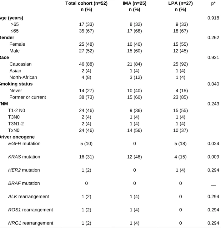
immunohistochemistry. The representative pathological findings can be found in
Figure 1.
MUC1 was expressed in the apical cytoplasmic membrane of tumor cells in 77% of
cases, with a median staining score of 80 (10-155). Hyperplasia of Type II
pneumocytes adjacent to the adenocarcinoma weakly expressed MUC1 (Figure 1C,
1D).
MUC2 was not expressed, except in a single IMA case (2%), with an intense
cytoplasmic staining in almost all tumor cells (not shown). A weak staining was
detected in the mucous cells of submucosal glands and at the basal pole of normal
goblet cells of the respiratory epithelium. MUC2 was not considered for further
analysis.
MUC5B was expressed in the tumor cells’ cytoplasm in 63% of patients, with a median staining score of 35 (0-200). The staining was typically observed in
intra-cytoplasmic vacuoles of goblet cells in IMA cases (Figure 1E). MUC5B was
expressed in normal bronchiolar cells in all patients.
MUC5AC was expressed in the tumor cells’ cytoplasm in 36% of patients, with a median staining score of 0 (0-52.5). MUC5AC was predominantly expressed in goblet
cells in IMA cases (Figure 1G). MUC5AC was weakly expressed in normal goblet
14
MUC6 was expressed in the tumor cells’ cytoplasm in 21% of patients, with a median staining score of 0 (0-0). MUC6 staining was exclusively detected in goblet cells in
15
3.3. Mucin expression according to histological subtype
MUC1, MUC5B, MUC5AC, and MUC6 expression in IMA and LPA patients was
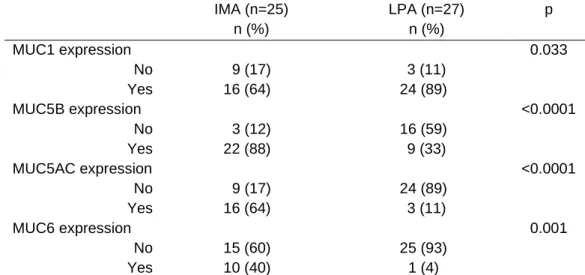
compared (Table 2, Figure 2).
MUC1 was more commonly expressed in LPA samples compared to those of IMA
(p=0.033) (Table 2) and MUC1 staining scores were also significantly higher in LPA
(120 [40-160] vs., 40 [0-110] for IMA; p=0.0225, Figure 2).
MUC5B, MUC5AC, and MUC6 expression was strongly associated with IMA, in
contrast to LPA (Table 2, Figure 2), and detected more frequently (Table 2). MUC5B
staining scores were significantly higher in IMA compared to LPA (160 [60-240] vs., 0
[0-30], respectively; p <0.0001), as were scores for MUC5AC (30 [0-240] vs., 0 [0-0];
p <0.0001) and MUC6 (0 [0-25] vs., 0 [0-0]; p <0.0015).
MUC1 scores negatively correlated with MUC5B 0.501, p <0.0001), MUC5AC
(r=-0.395, p=0.004), and MUC6 scores (r=-0.302, p=0.02) (Supplementary Figure S1).
MUC5B staining scores strongly correlated with MUC5AC (r=-0.665, p <0.0001)
scores, and to a lesser extent with those of MUC6 (r=-0.429, p=0.0015). Finally,
MUC5AC staining scores strongly correlated with MUC6 (r=-0.697, p <0.0001)
scores.
Overall, the IMA samples frequently expressed MUC5B and MUC5AC, and
sometimes MUC6, with MUC1 expression less common. LPA samples widely
16
Table 2. Mucin expression according to histological subtype
* Frequency compared using χ2 test. IMA: invasive mucinous adenocarcinoma; LPA: lepidic predominant adenocarcinoma
IMA (n=25) n (%) LPA (n=27) n (%) p MUC1 expression 0.033 No 9 (17) 3 (11) Yes 16 (64) 24 (89) MUC5B expression <0.0001 No 3 (12) 16 (59) Yes 22 (88) 9 (33) MUC5AC expression <0.0001 No 9 (17) 24 (89) Yes 16 (64) 3 (11) MUC6 expression 0.001 No 15 (60) 25 (93) Yes 10 (40) 1 (4)
17
3.4. Mucin expression according to Hnf4α expression
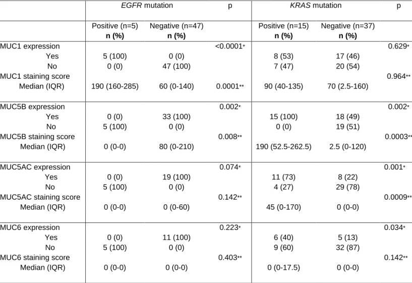
A Hnf4α nuclear staining in the tumor cells was observed in 23% of cases (n=12).
Out of the 25 IMA samples, 11 were Hnf4α-positive and TTF1-negative tumors
(Figure 4A). Among the 14 remaining Hnf4α-negative IMA, 11 cases were weakly
TTF1-positive (10 to 30%) and 3 cases TTF1-negative (Figure 4A). All LPA samples
were Hnf4α-negative and TTF1-positive but one, a weakly TTF1-positive LPA that
exhibited a KRAS mutation (Figure 4A).
Hnf4α-positive tumors strongly expressed MUC5B, MUC5AC and MUC6 compared to Hnf4α-negative tumors (200 300] vs., 5 300]; p=0.0008, 240 300] vs., 0 [0-180]; p<0.0001, 25 [0-60] vs., 0 [0-40]; p<0.0001, for MUC5B, MUC5AC and MUC6
respectively). positive tumors weakly expressed MUC1 compared to
Hnf4α-negative tumors (0 [0-180] vs., 100 [0-300]; p=0.0098).
As depicted in Figure 4, mucins are differentially expressed in Hnf4α-positive IMA, Hnf4α-negative IMA and LPA. Overall, MUC1 was less commonly and less strongly
expressed in Hnf4α-positive IMA compared to Hnf4α-negative IMA and LPA (Figure
4A and B). MUC5B was always and strongly expressed in Hnf4α-positive IMA and, to
a lesser extent, in Hnf4α-negative IMA. MUC5B was uncommonly expressed in LPA
(Figure 4A and C). MUC5AC and MUC6 were very commonly expressed in
Hnf4α-positive IMA, less commonly in Hnf4α-negative IMA and uncommonly in LPA (Figure 4A,D and E).
Overall, the Hnf4α-positive IMA samples exhibited a specific pattern of mucins
18
MUC5AC and MUC6 contrasting with the lack of MUC5AC and MUC6 expression
and MUC1 expression in Hnf4α-negative IMA samples.
3.5. Mucins expression according to oncogenic driver status
Five patients exhibited EGFR mutation. MUC1 was always strongly expressed in
EGFR-positive tumors (190 [160-285]), while MUC5B, MUC5AC, MUC6 and Hnf4α
were never detected (Table 3).
Thirteen patients exhibited KRAS mutation. MUC5B and MUC5AC were more
frequently expressed in positive tumors (p=0.002, vs., p=0.001 for
negative). MUC5B and MUC5AC staining scores were significantly higher in
KRAS-positive tumors (Table 3). Hnf4α was expressed in six KRAS-KRAS-positive tumors.
In the ALK- (n=1), ROS1- (n=1) and NRG1-positive IMA tumors (n=1), MUC5B was
strongly expressed (210, 240 and 240, respectively), MUC 1 was inconstantly
expressed (100, 10 and 0, respectively), and MUC5AC was never expressed as well
as MUC6. These three rearranged-IMA were Hnf4α-negative and partially
TTF1-positive.
19
Table 3. Comparison of mucin expression according to EGFR and KRAS mutation status
* Frequency compared using χ2 test. IMA: invasive mucinous adenocarcinoma; LPA: lepidic predominant adenocarcinoma
EGFR mutation p KRAS mutation p
Positive (n=5) n (%) Negative (n=47) n (%) Positive (n=15) n (%) Negative (n=37) n (%) MUC1 expression <0.0001* 0.629* Yes 5 (100) 0 (0) 8 (53) 17 (46) No 0 (0) 47 (100) 7 (47) 20 (54)
MUC1 staining score 0.964**
Median (IQR) 190 (160-285) 60 (0-140) 0.0001** 90 (40-135) 70 (2.5-160)
MUC5B expression 0.002* 0.002*
Yes 0 (0) 33 (100) 15 (100) 18 (49)
No 5 (100) 0 (0) 0 (0) 19 (51)
MUC5B staining score 0.008** 0.0003**
Median (IQR) 0 (0-0) 80 (0-210) 190 (52.5-262.5) 2.5 (0-120)
MUC5AC expression 0.074* 0.001*
Yes 0 (0) 19 (100) 11 (73) 8 (22)
No 5 (100) 0 (0) 4 (27) 29 (78)
MUC5AC staining score 0.142** 0.0009**
Median (IQR) 0 (0-0) 0 (0-60) 45 (0-170) 0 (0-0)
MUC6 expression 0.223* 0.034*
Yes 0 (0) 11 (100) 6 (40) 5 (13)
No 5 (100) 0 (0) 9 (60) 32 (87)
MUC6 staining score 0.403** 0.142**
20 4. DISCUSSION
Our study assessed the expression profile of MUC1, MUC2, MUC5B, MUC5AC, and
MUC6 at the protein level in 25 IMA and 27 LPA cases. We hereby provide evidence
of there being two opposing patterns of mucin expression that differentiate IMA from
LPA. IMA samples frequently expressed MUC5B and MUC5AC, sometimes MUC6,
and less commonly MUC1. Conversely, LPA samples typically expressed MUC1,
less frequently MUC5B, and expressed neither MUC5AC nor MUC6. IMA exhibited a
distinct mucins expression according to Hnf4α expression, with an expression of MUC1 and TTF1 and a lack of MUC5AC and MUC6 expression in Hnf4α-negative IMA, and a lack of expression of MUC1 and TTF1 and a common expression of
MUC5AC and MUC6 in Hnf4α-positive IMA. We also report a clear relationship between common driver oncogenes and mucin expression. EFGR-mutated tumors
exhibited strong MUC1 expression and no MUC5B, MUC5AC or MUC6 expression.
KRAS-mutated tumors frequently and strongly expressed MUC5B and MUC5AC.
ALK-, ROS1- and NRG1- mutated IMA tumors are Hnf4α-negative and partially
TTF1-positive and strongly expressed MUC5B, inconstantly MUC 1 and do not
express MUC5AC and MUC6.
Our data confirmed the multiple line of evidence suggesting that LPA is a terminal
respiratory unit tumor as MUC1 is typically expressed in normal alveolar pneumocyte.
The origin of IMA is not yet determined. Here, IMA was associated with MUC5B and
MUC5AC expression that are expressed in normal respiratory glands, suggesting a
lineage with glandular cells from respiratory epithelium.
This study demonstrated that IMA is associated with predominantly secreted mucins
21
MUC5AC, and MUC6) [26]. As expected, MUC2, which is the intestinal mucin, was
detected in only one IMA case [25]. Appropriate external and internal positive
controls were used which validated the quality of our assay. Two previous studies
had reported MUC2 protein expression, detected by immunochemistry using the
same primary antibody as that used in our study [35,36]. MUC2 staining was positive
in 17.5% of IMA samples in the 40 patients reported by Tsuta et al. In another study,
conducted by Awaya et al., MUC2 expression was estimated as high (>30% of tumor
cells) in 60% of the 27 LPA cases and in 43% of the seven IMA cases. Loss of MUC2
expression increases the survival and proliferation of intestinal epithelial cells, which
thus supports the belief in its tumor-suppressor capacity in intestinal adenocarcinoma
[41,42]. Our results suggested that MUC2 expression was down-regulated in lepidic
adenocarcinoma, with MUC2 therefore potentially offering a tumor suppressor benefit
in this histological subtype.
MUC1 overexpression was associated with the LPA subtype and EGFR mutation,
found to be mutually exclusive with MUC5B, MUC5AC, and MUC6 expression.
MUC1 overexpression is common in human cancers, particularly lung cancers, and
has not been associated with any specific activating mutations. The MUC1-C subunit
is an oncogene involved in the tumorigenesis of breast cancer in vitro [43]. In
NSCLC, MUC1-C interacts with EGFR, and targeting this subunit suppresses EGFR
activation and the downstream AKT and MEK pathways [31]. A combination
treatment of afatinib, an irreversible inhibitor of EGFR, and GO-203, which targets
MUC1-C, provides a synergistic action that inhibits the growth of NSCLC cells with
22
demonstrated a correlation between MUC1 overexpression and EGFR mutation,
suggesting that this treatment combination could be attractive in EGFR-mutated LPA.
MUC5B, MUC5AC, and MUC6 were overexpressed in the IMA samples, specifically
in goblet cells. We demonstrated that MUC5B and MUC5AC protein expression was
more frequent and strongly expressed in KRAS-positive tumors. IMA are
TTF-1-negative tumours and previous studies showed that the mucinous parts of lung
carcinomas expressing MUC5B are TTF-1 negative [45,46]. A study using targeted
next-generation sequencing identified recurrent loss of function mutations of Nkx2-1
(also known as TTF-1) associated with KRAS mutations in IMA [47]. Interestingly, a
strong expression of MUC5AC and MUC6 has been found in these IMA with Nkx2-1
and KRAS mutations. Furthermore, the first transgenic mouse model of mucinous
adenocarcinomas of the lung has been engineered, combining KRAS mutation with
haploinsufficiency of the Nkx2-1 gene [48,49]. In this model, the tumor cells were
similar to the human goblet tumor cells observed in IMA. A number of mRNAs,
including MUC5AC and MUC5B, were increased in the lungs of
KrasG12D/Nkx2-1+/– mice. This supports the concept that loss of Nkx2-1 and KRAS mutation
promote the goblet cell phenotype via the expression of a specific subset of genes,
including MUC5AC and MUC5B.
In contrast with normal lung, some IMA samples exhibit aberrantly-expressed MUC6.
MUC6 is normally expressed in the pyloric mucus gland, associated with pancreatic
and gastric carcinoma [25]. Interestingly, Hnf4α-positive IMA were always
TTF1-negative, do not expressed MUC1 and were likely to commonly and strongly
expressed MUC5AC and MUC6. This corresponds to the previous observation that
23
to other lung tumors, including aberrant expression of Hnf4α, MUC5B and MUC5AC
mucin-producing gastrointestinal and pancreatic cancers [50]. As previously
suggested, this aberrant expression supports the hypothesis that IMA originates from
a common endodermal precursor cell, with the potential for multicellular
differentiation, including the expression of gastric mucins [34,51,52].
The Hnf4α-negative IMA were mostly TTF1-negative and expressed MUC1 as well as MUC5B but exhibited a lack of MUC5AC and MUC6 expression. These cases
may be not “pure” IMA and may represent mixed cases with IMA and lepidic proliferation. The three oncogenic fusion-driven IMA belong to these group of Hnf4α-negative and TTF1-positive IMA.
5. CONCLUSIONS
We have herein provided evidence of a specific expression profile of mucins in IMA
and LPA. MUC5B and MUC5AC expression may play an important role in the typical
goblet cell phenotype observed in IMA, triggered by KRAS mutation. MUC1 could
24 Acknowledgements
We would like to thank the « Tumorothèque HUEP (AP-HP – GH-HUEP) »,
representing by Pr. Isabelle Brochériou (the East Paris University Hospitals Tumor
Bio-bank), AP-HP, Hôpital Tenon, Service d'Anatomie Pathologique, F-75970, Paris,
for providing us with the samples.
Funding sources
M. Duruisseaux is a doctoral fellow funded by “Fonds de dotation Recherche en
Santé Respiratoire 2010”, “AgiràDom” and “Comité des maladies respiratoires
(COMARES) de l’Isère”. This study also received support from “Subvention 2010 et
2011 Leg Poix - La Chancellerie des Universités de Paris” and “ITMO Cancer 2012
Institut National du Cancer Plan Cancer 2009-2013 « Modèles de tumeurs spontanées chez l’animal pour la recherche translationnelle en cancérologie » ”.
25 Reference List
[1] W.D. Travis, E. Brambilla, M. Noguchi, A.G. Nicholson, K.R. Geisinger, Y. Yatabe, D.G. Beer, C.A. Powell, G.J. Riely, P.E. Van Schil, K. Garg, J.H.M. Austin, H. Asamura, V.W. Rusch, F.R. Hirsch, G. Scagliotti, T. Mitsudomi, R.M. Huber, Y. Ishikawa, J. Jett, M. Sanchez-Cespedes, J.-P. Sculier, T. Takahashi, M. Tsuboi, J. Vansteenkiste, I. Wistuba, P.-C. Yang, D. Aberle, C. Brambilla, D. Flieder, W. Franklin, A. Gazdar, M. Gould, P. Hasleton, D. Henderson, B. Johnson, D. Johnson, K. Kerr, K. Kuriyama, J.S. Lee, V.A. Miller, I. Petersen, V. Roggli, R. Rosell, N. Saijo, E. Thunnissen, M. Tsao, D. Yankelewitz, International association for the study of lung cancer/american thoracic society/european respiratory society international multidisciplinary classification of lung
adenocarcinoma, J. Thorac. Oncol. Off. (2011) 244–285.
[2] D.H. Garfield, J.L. Cadranel, M. Wislez, W.A. Franklin, F.R. Hirsch, The bronchioloalveolar carcinoma and peripheral adenocarcinoma spectrum of diseases, J. Thorac. Oncol. 1 (2006) 344–359. [3] K. Tsuta, M. Kawago, E. Inoue, A. Yoshida, F. Takahashi, H. Sakurai, S.-I. Watanabe, M. Takeuchi, K. Furuta, H. Asamura, H. Tsuda, The utility of the proposed IASLC/ATS/ERS lung
adenocarcinoma subtypes for disease prognosis and correlation of driver gene alterations, Lung Cancer 81 (2013) 371–376.
[4] A. Warth, T. Muley, M. Meister, A. Stenzinger, M. Thomas, P. Schirmacher, P.A. Schnabel, J. Budczies, H. Hoffmann, W. Weichert, The novel histologic International Association for the Study of Lung Cancer/American Thoracic Society/European Respiratory Society classification system of lung adenocarcinoma is a stage-independent predictor of survival, J. Clin. Oncol. 30 (2012) 1438–1446. [5] A. Yoshizawa, N. Motoi, G.J. Riely, C.S. Sima, W.L. Gerald, M.G. Kris, B.J. Park, V.W. Rusch, W.D. Travis, Impact of proposed IASLC/ATS/ERS classification of lung adenocarcinoma: prognostic subgroups and implications for further revision of staging based on analysis of 514 stage I cases, Mod. Pathol. 24 (2011) 653–664.
[6] D.H. Garfield, J. Cadranel, H.L. West, Bronchioloalveolar carcinoma: the case for two diseases, Clin. Lung Cancer. 9 (2008) 24–29.
[7] O.S. Breathnach, D.J. Kwiatkowski, D.M. Finkelstein, J. Godleski, D.J. Sugarbaker, B.E. Johnson, S. Mentzer, Bronchioloalveolar carcinoma of the lung: recurrences and survival in patients with stage I disease, J. Thorac. Cardiovasc. Surg. 121 (2001) 42–47.
[8] J.O. Ebbert, L. Chhatwani, M.C. Aubry, J. Wampfler, S. Stoddard, F. Zhang, G. Qiao, M. de Andrade, P. Yang, Clinical features of bronchioloalveolar carcinoma with new histologic and staging definitions, J. Thorac. Oncol. 5 (2010) 1213–1220.
[9] C. Casali, G. Rossi, A. Marchioni, G. Sartori, F. Maselli, L. Longo, E. Tallarico, U. Morandi, A single institution-based retrospective study of surgically treated bronchioloalveolar adenocarcinoma of the lung: clinicopathologic analysis, molecular features, and possible pitfalls in routine practice, J. Thorac. Oncol. 5 (2010) 830–836.
[10] A. Carretta, P. Ciriaco, G. Melloni, A. Bandiera, L. Libretti, A. Puglisi, M. Giovanardi, P. Zannini, Surgical treatment of multiple primary adenocarcinomas of the lung, Thorac. Cardiovasc. Surg. 57 (2009) 30–34.
26
[11] R.C. Daly, V.F. Trastek, P.C. Pairolero, P.A. Murtaugh, M.S. Huang, M.S. Allen, T.V. Colby, Bronchoalveolar carcinoma: factors affecting survival, Ann. Thorac. Surg. 51 (1991) 368-376-377. [12] P. Volpino, A. Cavallaro, R. Cangemi, F. Chiarotti, A. De Cesare, E. Fiori, M. Bononi, R. Vigliarolo, V. Cangemi, Comparative analysis of clinical features and prognostic factors in resected bronchioloalveolar carcinoma and adenocarcinoma of the lung, Anticancer Res. 23 (2003) 4959–4965. [13] M. Duruisseaux, M. Antoine, N. Rabbe, V. Poulot, J. Fleury-Feith, T. Vieira, A. Lavolé, J. Cadranel, M. Wislez, The impact of intracytoplasmic mucin in lung adenocarcinoma with pneumonic radiological presentation, Lung Cancer. 83 (2014) 334–340.
[14] W.D. Travis, E. Brambilla, A.G. Nicholson, Y. Yatabe, J.H.M. Austin, M.B. Beasley, L.R. Chirieac, S. Dacic, E. Duhig, D.B. Flieder, K. Geisinger, F.R. Hirsch, Y. Ishikawa, K.M. Kerr, M. Noguchi, G. Pelosi, C.A. Powell, M.S. Tsao, I. Wistuba, WHO Panel, The 2015 World Health Organization Classification of Lung Tumors: Impact of Genetic, Clinical and Radiologic Advances Since the 2004 Classification, J. Thorac. Oncol. 10 (2015) 1243–1260.
[15] M. Wislez, M. Antoine, L. Baudrin, V. Poulot, A. Neuville, M. Pradere, E. Longchampt, S. Isaac-Sibille, M.-P. Lebitasy, J. Cadranel, Non-mucinous and mucinous subtypes of adenocarcinoma with bronchioloalveolar carcinoma features differ by biomarker expression and in the response to gefitinib, Lung Cancer. 68 (2010) 185–191.
[16] M. Sugano, T. Nagasaka, E. Sasaki, Y. Murakami, W. Hosoda, T. Hida, T. Mitsudomi, Y. Yatabe, HNF4α as a marker for invasive mucinous adenocarcinoma of the lung, Am. J. Surg. Pathol. 37 (2013) 211–218.
[17] A. Hata, N. Katakami, S. Fujita, R. Kaji, Y. Imai, Y. Takahashi, T. Nishimura, K. Tomii, K. Ishihara, Frequency of EGFR and KRAS mutations in Japanese patients with lung adenocarcinoma with features of the mucinous subtype of bronchioloalveolar carcinoma, J. Thorac. Oncol. 5 (2010) 1197–1200.
[18] K.E. Finberg, L.V. Sequist, V.A. Joshi, A. Muzikansky, J.M. Miller, M. Han, J. Beheshti, L.R. Chirieac, E.J. Mark, A.J. Iafrate, Mucinous differentiation correlates with absence of EGFR mutation and presence of KRAS mutation in lung adenocarcinomas with bronchioloalveolar features, J. Mol. Diagn. 9 (2007) 320–326.
[19] Y. Sakuma, S. Matsukuma, M. Yoshihara, Y. Nakamura, K. Noda, H. Nakayama, Y. Kameda, E. Tsuchiya, Y. Miyagi, Distinctive evaluation of nonmucinous and mucinous subtypes of
bronchioloalveolar carcinomas in EGFR and K-ras gene-mutation analyses for Japanese lung
adenocarcinomas: confirmation of the correlations with histologic subtypes and gene mutations, Am. J. Clin. Pathol. 128 (2007) 100–108.
[20] L. Fernandez-Cuesta, D. Plenker, H. Osada, R. Sun, R. Menon, F. Leenders, S. Ortiz-Cuaran, M. Peifer, M. Bos, J. Daßler, F. Malchers, J. Schöttle, W. Vogel, I. Dahmen, M. Koker, R.T. Ullrich, G.M. Wright, P.A. Russell, Z. Wainer, B. Solomon, E. Brambilla, H. Nagy-Mignotte, D. Moro-Sibilot, C.G. Brambilla, S. Lantuejoul, J. Altmüller, C. Becker, P. Nürnberg, J.M. Heuckmann, E. Stoelben, I. Petersen, J.H. Clement, J. Sänger, L.A. Muscarella, A. la Torre, V.M. Fazio, I. Lahortiga, T. Perera, S. Ogata, M. Parade, D. Brehmer, M. Vingron, L.C. Heukamp, R. Buettner, T. Zander, J. Wolf, S. Perner, S. Ansén, S.A. Haas, Y. Yatabe, R.K. Thomas, CD74-NRG1 fusions in lung adenocarcinoma, Cancer Discov. 4 (2014) 415–422.
27
[21] T. Nakaoku, K. Tsuta, H. Ichikawa, K. Shiraishi, H. Sakamoto, M. Enari, K. Furuta, Y. Shimada, H. Ogiwara, S. Watanabe, H. Nokihara, K. Yasuda, M. Hiramoto, T. Nammo, T. Ishigame, A.J. Schetter, H. Okayama, C.C. Harris, Y.H. Kim, M. Mishima, J. Yokota, T. Yoshida, T. Kohno, Druggable oncogene fusions in invasive mucinous lung adenocarcinoma, Clin. Cancer Res. 20 (2014) 3087–3093.
[22] K. Kadota, Y.-C. Yeh, S.P. D’Angelo, A.L. Moreira, D. Kuk, C.S. Sima, G.J. Riely, M.E. Arcila, M.G. Kris, V.W. Rusch, P.S. Adusumilli, W.D. Travis, Associations between mutations and histologic patterns of mucin in lung adenocarcinoma: invasive mucinous pattern and extracellular mucin are associated with KRAS mutation, Am. J. Surg. Pathol. 38 (2014) 1118–1127.
[23] Y. Yatabe, EGFR mutations and the terminal respiratory unit, Cancer Metastasis Rev. 29 (2010) 23–36.
[24] G. Sartori, A. Cavazza, A. Sgambato, A. Marchioni, F. Barbieri, L. Longo, M. Bavieri, B. Murer, E. Meschiari, S. Tamberi, A. Cadioli, F. Luppi, M. Migaldi, G. Rossi, EGFR and K-ras mutations along the spectrum of pulmonary epithelial tumors of the lung and elaboration of a
combined clinicopathologic and molecular scoring system to predict clinical responsiveness to EGFR inhibitors, Am. J. Clin. Pathol. 131 (2009) 478–489.
[25] D.W. Kufe, Mucins in cancer: function, prognosis and therapy, Nat. Rev. Cancer. 9 (2009) 874–885.
[26] P. Pigny, V. Guyonnet-Duperat, A.S. Hill, W.S. Pratt, S. Galiegue-Zouitina, M.C. d’Hooge, A. Laine, I. Van-Seuningen, P. Degand, J.R. Gum, Y.S. Kim, D.M. Swallow, J.P. Aubert, N. Porchet, Human mucin genes assigned to 11p15.5: identification and organization of a cluster of genes, Genomics. 38 (1996) 340–352.
[27] N. Jonckheere, I. Van Seuningen, The membrane-bound mucins: From cell signalling to transcriptional regulation and expression in epithelial cancers, Biochimie. 92 (2010) 1–11.
[28] M.C. Copin, L. Devisme, M.P. Buisine, C.H. Marquette, A. Wurtz, J.P. Aubert, B. Gosselin, N. Porchet, From normal respiratory mucosa to epidermoid carcinoma: expression of human mucin genes, Int. J. Cancer. 86 (2000) 162–168.
[29] S. Nagai, K. Takenaka, M. Sonobe, E. Ogawa, H. Wada, F. Tanaka, A novel classification of MUC1 expression is correlated with tumor differentiation and postoperative prognosis in non-small cell lung cancer, J. Thorac. Oncol. 1 (2006) 46–51.
[30] Y. Makiguchi, Y. Hinoda, K. Imai, Effect of MUC1 mucin, an anti-adhesion molecule, on tumor cell growth, Jpn. J. Cancer Res. Gann. 87 (1996) 505–511.
[31] S. Ramasamy, S. Duraisamy, S. Barbashov, T. Kawano, S. Kharbanda, D. Kufe, The MUC1 and galectin-3 oncoproteins function in a microRNA-dependent regulatory loop, Mol. Cell. 27 (2007) 992–1004.
[32] F. Guddo, A. Giatromanolaki, C. Patriarca, J. Hilkens, C. Reina, R.M. Alfano, A.M. Vignola, M.I. Koukourakis, M. Gambacorta, G. Pruneri, G. Coggi, G. Bonsignore, Depolarized expression of episialin (EMA, MUC1) in lung adenocarcinoma is associated with tumor progression, Anticancer Res. 18 (1998) 1915–1920.
28
[33] K. Kaira, K. Nakagawa, Y. Ohde, T. Okumura, T. Takahashi, H. Murakami, M. Endo, H. Kondo, T. Nakajima, N. Yamamoto, Depolarized MUC1 expression is closely associated with hypoxic markers and poor outcome in resected non-small cell lung cancer, Int. J. Surg. Pathol. 20 (2012) 223– 232.
[34] M.C. Copin, M.P. Buisine, E. Leteurtre, C.H. Marquette, H. Porte, J.P. Aubert, B. Gosselin, N. Porchet, Mucinous bronchioloalveolar carcinomas display a specific pattern of mucin gene expression among primary lung adenocarcinomas, Hum. Pathol. 32 (2001) 274–281.
[35] K. Tsuta, G. Ishii, J. Nitadori, Y. Murata, T. Kodama, K. Nagai, A. Ochiai, Comparison of the immunophenotypes of signet-ring cell carcinoma, solid adenocarcinoma with mucin production, and mucinous bronchioloalveolar carcinoma of the lung characterized by the presence of cytoplasmic mucin, J. Pathol. 209 (2006) 78–87.
[36] H. Awaya, Y. Takeshima, M. Yamasaki, K. Inai, Expression of MUC1, MUC2, MUC5AC, and MUC6 in atypical adenomatous hyperplasia, bronchioloalveolar carcinoma, adenocarcinoma with mixed subtypes, and mucinous bronchioloalveolar carcinoma of the lung, Am. J. Clin. Pathol. 121 (2004) 644–653.
[37] S. Lantuejoul, A.G. Nicholson, G. Sartori, C. Piolat, C. Danel, E. Brabencova, P. Goldstraw, E. Brambilla, G. Rossi, Mucinous cells in type 1 pulmonary congenital cystic adenomatoid
malformation as mucinous bronchioloalveolar carcinoma precursors, Am. J. Surg. Pathol. 31 (2007) 961–969.
[38] G. Rossi, B. Gasser, G. Sartori, M. Migaldi, M. Costantini, M.C. Mengoli, S. Piccioli, A. Cavazza, F. Rivasi, MUC5AC, cytokeratin 20 and HER2 expression and K-RAS mutations within mucinogenic growth in congenital pulmonary airway malformations, Histopathology. 60 (2012) 1133– 1143.
[39] S. Kim, A.J. Schein, J.A. Nadel, E-cadherin promotes EGFR-mediated cell differentiation and MUC5AC mucin expression in cultured human airway epithelial cells, Am. J. Physiol. Lung Cell. Mol. Physiol. 289 (2005) L1049-1060.
[40] M. Perrais, P. Pigny, M.-C. Copin, J.-P. Aubert, I. Van Seuningen, Induction of MUC2 and MUC5AC mucins by factors of the epidermal growth factor (EGF) family is mediated by EGF receptor/Ras/Raf/extracellular signal-regulated kinase cascade and Sp1, J. Biol. Chem. 277 (2002) 32258–32267.
[41] A. Velcich, W. Yang, J. Heyer, A. Fragale, C. Nicholas, S. Viani, R. Kucherlapati, M. Lipkin, K. Yang, L. Augenlicht, Colorectal cancer in mice genetically deficient in the mucin Muc2, Science. 295 (2002) 1726–1729.
[42] M. Van der Sluis, B.A.E. De Koning, A.C.J.M. De Bruijn, A. Velcich, J.P.P. Meijerink, J.B. Van Goudoever, H.A. Büller, J. Dekker, I. Van Seuningen, I.B. Renes, A.W.C. Einerhand, Muc2-deficient mice spontaneously develop colitis, indicating that MUC2 is critical for colonic protection, Gastroenterology. 131 (2006) 117–129.
[43] J.A. Schroeder, A.A. Masri, M.C. Adriance, J.C. Tessier, K.L. Kotlarczyk, M.C. Thompson, S.J. Gendler, MUC1 overexpression results in mammary gland tumorigenesis and prolonged alveolar differentiation, Oncogene. 23 (2004) 5739–5747.
29
[44] A. Kharbanda, H. Rajabi, C. Jin, J. Tchaicha, E. Kikuchi, K.-K. Wong, D. Kufe, Targeting the oncogenic MUC1-C protein inhibits mutant EGFR-mediated signaling and survival in non-small cell lung cancer cells, Clin. Cancer Res. 20 (2014) 5423–5434.
[45] N. Jonckheere, A. Velghe, M.-P. Ducourouble, M.-C. Copin, I.B. Renes, I. Van Seuningen, The mouse Muc5b mucin gene is transcriptionally regulated by thyroid transcription factor-1 (TTF-1) and GATA-6 transcription factors, FEBS J. 278 (2011) 282–294.
[46] N. Nakamura, E. Miyagi, S. Murata, A. Kawaoi, R. Katoh, Expression of thyroid transcription factor-1 in normal and neoplastic lung tissues, Mod. Pathol. 15 (2002) 1058–1067.
[47] D.H. Hwang, L.M. Sholl, V. Rojas-Rudilla, D.L. Hall, P. Shivdasani, E.P. Garcia, L.E. MacConaill, M. Vivero, J.L. Hornick, F.C. Kuo, N.I. Lindeman, F. Dong, KRAS and NKX2-1 Mutations in Invasive Mucinous Adenocarcinoma of the Lung, J. Thorac. Oncol. 11 (2016) 496–503. [48] Y. Maeda, T. Tsuchiya, H. Hao, D.H. Tompkins, Y. Xu, M.L. Mucenski, L. Du, A.R. Keiser, T. Fukazawa, Y. Naomoto, T. Nagayasu, J.A. Whitsett, Kras(G12D) and Nkx2-1 haploinsufficiency induce mucinous adenocarcinoma of the lung, J. Clin. Invest. 122 (2012) 4388–4400.
[49] E.L. Snyder, H. Watanabe, M. Magendantz, S. Hoersch, T.A. Chen, D.G. Wang, D. Crowley, C.A. Whittaker, M. Meyerson, S. Kimura, T. Jacks, Nkx2-1 represses a latent gastric differentiation program in lung adenocarcinoma, Mol. Cell. 50 (2013) 185–199.
[50] M. Guo, K. Tomoshige, M. Meister, T. Muley, T. Fukazawa, T. Tsuchiya, R. Karns, A. Warth, I.M. Fink-Baldauf, T. Nagayasu, Y. Naomoto, Y. Xu, M.A. Mall, Y. Maeda, Gene signature driving invasive mucinous adenocarcinoma of the lung, EMBO Mol. Med. 9 (2017) 462–481.
[51] T. Honda, H. Ota, K. Ishii, N. Nakamura, K. Kubo, T. Katsuyama, Mucinous
bronchioloalveolar carcinoma with organoid differentiation simulating the pyloric mucosa of the stomach: clinicopathologic, histochemical, and immunohistochemical analysis, Am. J. Clin. Pathol. 109 (1998) 423–430.
[52] A. Hamamoto, Y. Abe, M. Nishi, S. Fujimori, Y. Ohnishi, H. Yamazaki, Y. Oida, N. Miyazaki, K.-I. Inada, Y. Ueyama, M. Iwasaki, H. Inoue, M. Nakamura, Aberrant expression of the gastric mucin MUC6 in human pulmonary adenocarcinoma xenografts, Int. J. Oncol. 26 (2005) 891– 896.
30
Figure Legends
Figure 1. A and B: Hematoxylin and eosin staining in invasive mucinous adenocarcinoma (IMA) and lepidic predominant adenocarcinoma (LPA), respectively. C and D: MUC1 staining in IMA and LPA, respectively. E and F: MUC5B staining in IMA and LPA, respectively. G and H: MUC5AC staining in IMA and LPA, respectively. I and J: MUC6 staining in IMA and LPA, respectively
Figure 2. Comparison of mucin expression in invasive mucinous adenocarcinoma (IMA) and lepidic predominant adenocarcinoma (LPA). A: MUC1 staining score. B: MUC5B staining score. C: MUC5AC staining score. D: MUC6 staining score. Central bars: median; upper and lower bars: 25% and 75% interquartile range (IQR). Mann-Whitney test
Figure 3. Comparison of mucin expression in invasive mucinous adenocarcinoma (IMA) and lepidic predominant adenocarcinoma (LPA) according to Hnf4α expression. A: Proportion of positive staining for TTF1 and mucins in Hnf4α-positive IMA, Hnf4α-negative IMA and LPA. B,C,D and E: Comparison of mucin staining scores in Hnf4α-positive IMA, Hnf4α-negative IMA and LPA. Central bars: median; upper and lower bars: 25% and 75% interquartile range (IQR). Kruskal-Wallis Test.
A
B
C
D
E
F
G
H
I
J
Figure 1A
B
D
C
p=0.0225 p<0.0001 p<0.0001 p<0.0015 Figure 2p=0.0109
B
C
E
D
p<0.0001 p<0.0001 p<0.0001 M UC 1 M UC 5 B M UC 5 A C M UC 6 HNF4𝛼-positive IMA N=11 HNF4𝛼-negative IMA N=14 LPA N=27 Positive TTF1 staining, % 0 79 100Positive MUC1 staining, % 45 79 89
Positive MUC5B staining, % 100 79 41
Positive MUC5AC staining , % 100 36 11
Positive MUC6 staining, % 81 7 4
A B C D E F r = -0.501 p < 0.0001 r = -0.395 p = 0.004 r = -0.302 p = 0.02 r = 0.665 p < 0.0001 r = 0.429 p = 0.0015 r = 0.697 p < 0.0001 Figure S1