HAL Id: inserm-00126155
https://www.hal.inserm.fr/inserm-00126155
Submitted on 2 Sep 2008HAL is a multi-disciplinary open access archive for the deposit and dissemination of sci-entific research documents, whether they are pub-lished or not. The documents may come from teaching and research institutions in France or abroad, or from public or private research centers.
L’archive ouverte pluridisciplinaire HAL, est destinée au dépôt et à la diffusion de documents scientifiques de niveau recherche, publiés ou non, émanant des établissements d’enseignement et de recherche français ou étrangers, des laboratoires publics ou privés.
developing rat brain following chronic treatment with
the antagonist SR 48692.
Isabelle Lépée-Lorgeoux, Catalina Betancur, Frédérique Souazé, William
Rostène, Anne Bérod, Didier Pélaprat
To cite this version:
Isabelle Lépée-Lorgeoux, Catalina Betancur, Frédérique Souazé, William Rostène, Anne Bérod, et al.. Regulation of the neurotensin NT(1) receptor in the developing rat brain following chronic treatment with the antagonist SR 48692.. Journal of Neuroscience Research, Wiley, 2000, 60 (3), pp.362-9. �inserm-00126155�
Regulation of the neurotensin NT
1receptor in the developing rat brain
following chronic treatment with the antagonist SR 48692
Isabelle Lépée-Lorgeoux, Catalina Betancur, Frédérique Souazé, William Rostène, Anne
Bérod* and Didier Pélaprat
INSERM U. 339, Hôpital Saint-Antoine, 184 rue du Faubourg Saint-Antoine, 75571 Paris Cedex 12, France
* Present address: INSERM U. 512, Laboratoire de Neuropharmacologie et Neurochimie, Faculté de Pharmacie, 8 avenue Rockefeller, 69373 Lyon, France
Running title: Regulation of NT1 receptors in the developing rat brain
Correspondence should be addressed to: I. Lépée, INSERM U. 339, Hôpital Saint-Antoine, 184 rue du Faubourg Saint-Antoine, 75571 Paris Cedex 12, France. E-mail: lepee@st-antoine.inserm.fr
Grant information: This work was supported by the Institut National de la Santé et de la Recherche Médicale (INSERM), France.
ABSTRACT
The aim of the present study was to investigate the role of neurotensin in the regulation of N T1 receptors during postnatal development in the rat brain. Characterization of the ontogeny of neurotensin concentration and [125I]neurotensin binding to NT
1 receptors in the brain at different
embryonic and postnatal stages showed that neurotensin was highly expressed at birth, reaching peak levels at postnatal day 5 (P5), and decreasing thereafter. The transient rise in neurotensin levels preceded the maximal expression of NT1 receptors, observed at P10, suggesting that neurotensin may influence the developmental profile of NT1 receptors. Using primary cultures of cerebral cortex neurons from fetal rats, we showed that exposure to the neurotensin agonist JMV 449 (1 nM) decreased (–43%) the amount of NT1 receptor mRNA measured by reverse transcription-PCR, an effect that was abolished by the non-peptide NT1 receptor antagonist SR 48692 (1 µM). However, daily injection of SR 48692 to rat pups from birth for 5, 9 or 15 days, did not modify [125I]neurotensin binding in brain membrane homogenates. Moreover, postnatal
blockade of neurotensin transmission did not alter the density and distribution of NT1 receptors assessed by quantitative autoradiography nor NT1 receptor mRNA expression measured by in situ hybridization in the cerebral cortex, caudate-putamen and midbrain. These results suggest that although NT1 receptor expression can be regulated in vitro by the agonist at an early developmental stage, neurotensin is not a major factor in the establishment of the ontogenetic pattern of these receptors in the rat brain.
KEY WORDS: postnatal development; JMV 449; in situ hybridization; RT-PCR
INTRODUCTION
Neurotensin, a 13 amino-acid neuropeptide, is heterogeneously distributed in the mammalian brain (Alexander et al., 1989).In the central nervous system (CNS), neurotensin acts as a neurotransmitter and neuromodulator of several neuronal systems; in particular, neurotensin has been shown to play an important role in the modulation of dopamine transmission (Lambert et
al., 1995).Neurotensin is also involved in nociception, thermoregulation and modulation of neuroendocrine systems (for reviews, see Nemeroff and Kitabgi, 1992; Rostène and Alexander, 1997).
Two types of neurotensin receptors, NT1 and NT2, have been cloned and shown to belong t o the family of G protein-coupled receptors (Tanaka et al., 1990; Vita et al., 1993; Chalon et al., 1996; Mazella et al., 1996). These two receptors are distinguished by their affinity for neurotensin and their capacity to bind levocabastine, a histamine H1 receptor antagonist (Schotte
et al., 1986). The levocabastine-insensitive, high-affinity neurotensin receptor (NT1) mediates most of the physiological functions ascribed to neurotensin.In contrast, only limited data are available concerning the functional role of the levocabastine-sensitive, lower-affinity neurotensin
receptor (NT2). Using an anti-sense strategy in mice, the N T2 receptor was implicated in the analgesic effects induced by neurotensin (Dubuc et al., 1999). A third neurotensin receptor (NT3) was recently cloned from human brain (Mazella et al., 1998); it does not belong to the superfamily of G protein-coupled receptors, corresponds to the previously cloned gp95/sortilin and could be involved in intracellular trafficking.
Previous studies on the ontogeny of N T1 receptors in the rat brain using autoradiography (Palacios et al., 1988), binding experiments in brain homogenates (Schotte and Laduron, 1987) and in situ hybridization (Sato et al., 1992; Lépée-Lorgeoux et al., 1999), reported marked regional differences in their developmental profile. Thus, the cerebral cortex exhibits a high and transient expression of NT1 receptors during the early postnatal period, which declines to reach adult levels by the third postnatal week, whereas other brain areas, including the midbrain, show a gradual increase in NT1 receptor expression from late gestation to the second week of life. Interestingly, neurotensin is also highly expressed in the brain at birth, followed by a dramatic decrease to adult levels during the second and third weeks of postnatal life (Hara et al., 1982; Sato
et al., 1991; Bennett et al., 1998).
The role of endogenous ligands in establishing the developmental pattern of expression of their corresponding receptors has been suggested for several neurotransmitter systems, including neuropeptides (Kudlacz et al., 1991; Hill et al., 1994; Sircar et al., 1996; Liu et al., 1998). However, the possible involvement of neurotensin in the regulation of N T1 receptors during ontogeny has not been studied. In a recent series of experiments, we used the non-peptide antagonist of NT1 receptors SR 48692 to investigate the role of endogenous neurotensin in the regulation of NT1 receptors in the adult rat brain. We showed that chronic administration of SR 48692 induces an up-regulation of NT1 receptor binding sites and mRNA expression, suggesting a tonic inhibitory control of neurotensin on N T1 receptors in the mature brain (Azzi et al., 1994, 1996; Najimi et al., 1998).
The aim of the present study was to investigate the role of neurotensin in the regulation of N T1 receptors during postnatal development in the rat brain. We first examined concomitantly the ontogeny of neurotensin content and N T1 receptor expression, since previous studies examining separately either parameter rendered difficult their comparison at a given time point. Second, we used primary cultures of embryonic neurons from rat cerebral cortex, the brain region with the highest expression of NT1 receptors during early development, to investigate whether these receptors could be regulated by the agonist at an early developmental stage. The amount of N T1 receptor mRNA transcripts in the cultures was measured by quantitative reverse transcription-polymerase chain reaction (RT-PCR). Finally, we studied the effects of in vivo blockade of neurotensinergic transmission during postnatal development in rat pups injected with the NT1 receptor antagonist SR 48692. [125I]neurotensin binding to N T
1 receptors was examined
using brain homogenates and autoradiography, and NT1 receptor mRNA expression was assessed by in situ hybridization histochemistry.
MATERIALS AND METHODS Animals and treatments
Wistar rats bred in our laboratory were maintained on a 12-hour dark/light cycle and were given food and water ad libitum. To study the ontogeny of neurotensin and N T1 receptor expression in the brain, rats were analyzed at different prenatal and postnatal ages (E15, E20, P0, P5, P10, P15, P21 and P30). The day of the vaginal plug was counted as E1 and the day of birth as P0. All experiments were performed in accordance with the European Communities Council Directive for the care and use of laboratory animals.
In order to examine the role of endogenous neurotensin in the developmental regulation of N T1 receptors, rat pups received a daily subcutaneous (s.c.) injection of the non-peptide NT1 receptor antagonist, SR 48692 (Gully et al., 1993; Sanofi Recherche, Toulouse, France), for different periods of time: P1-P5, P1-P9 or P1-P15 (n = six per group). SR 48692, administered at the dose of 1 mg/kg, was solubilized in Tween 20 and dissolved in sterile 0.9% NaCl solution. This dose of SR 48692 was chosen because it was previously shown to induce an up-regulation of N T1 receptors in the adult rat (Azzi et al., 1996; Najimi et al., 1998). Control pups were injected with vehicle (Tween 20 1% in NaCl 0.9%). In a separate group of animals, prenatal treatment with SR 48692 was started at E15; pregnant dams received a daily injection of the antagonist until the day of birth, after which pups were injected with SR 48692 from P1 to P9. In all experiments, rats were decapitated 24 h after the last injection.
Preparation of fetal brain cortical cultures and treatment with neurotensin agonist and/or antagonist
Cultures were prepared from the cerebral cortex of Wistar rat fetuses at E17 following the procedure previously described for cultures of hypothalamic neurons (Scarcériaux et al., 1996). The cerebral cortex was excised under sterile conditions and microscopic control, and cells were grown in serum-free medium consisting of a mixture of Dulbecco modified Eagle's medium and Ham-F12 (1vol/1vol, Gibco), 15 mM HEPES, NaHCO3 as indicated by the manufacturer, 0.5 U/ml penicillin, 20 µg/ml streptomycin, 5 µg/ml insulin, 100 µg/ml human transferrin, 2.10-8 M
progesterone, 10-12 M 17 β-estradiol, 1 µg/ml arachidonic acid, 0.5 µg/ml docosahexaenoic acid,
3.10-8 M selenium, 10-4 M putrescine and 0.5 mM glutamine. Cells were plated at a density of
400,000 cells per cm2 in six-well plastic culture plates (Costar) previously coated with
poly-D-lysine. Cultures were maintained in a moist atmosphere (95% air, 5% CO2) at 37°C and medium was renewed after 5 days. Cytosine arabinoside (1 µM) was added from the first change onward t o prevent further glial proliferation. After 5 days of culture, neurons were incubated in the presence of the neurotensin agonist JMV 449 (Doulut et al., 1992; Neosystem), 1 nM; the NT1 receptor antagonist SR 48692, 1 µM; or both drugs. SR 48692 was dissolved in dimethyl sulfoxide (DMSO); incubation of cortical neurons in the presence of DMSO (final concentration 0.1%) showed that the solvent alone had no effect on NT1 receptor mRNA expression. After 48 h, the culture medium was removed and the cells were rinsed with ice-cold phosphate-buffered saline. Total RNA
was extracted by the acid guanidium thiocyanate-phenol chloroform method (Chomczynski and Sacchi, 1987), followed by ethanol precipitation.
Quantitative RT-PCR of NT1 receptor mRNA
Internal standard preparation and quantitative RT-PCR were carried out as described (Souazé et
al., 1997). Briefly, a plasmid pNTR18 was constructed by inserting the entire coding sequence of
rat NT1 receptor cDNA (nt: -7 to 1301) into the SmaI/BamHI sites of p T7/T3α18. A 34-nucleotide deletion was created by removing the NCoI-NHeI fragment. A polyA(45) oligonucleotide was then inserted at the SalI/BamHI sites. For RT-PCR quantitation, mixtures of 100 ng of total RNA and a known number of internal control (cRNA) molecules were reverse transcribed in 30 µl of reaction buffer containing 10 mM Tris-HCl, pH 8.3, 50 mM KCl, 1.5 mM MgCl2, 10 mM dithiothreitol, 1 mM of each dNTP, 1 pmol/µl of specific primer, 1 unit/µl RNasin (Promega) and 200 units of Moloney murine leukemia virus reverse transcriptase (Life Technologies). The PCR amplification was performed in an automatic thermal cycler (DNA thermal cycler 480, Perkin Elmer Cetus). The amplification profile included an initial denaturation at 95°C for 7 min, followed by 26 cycles of denaturation at 94°C for 30 s, annealing at 55°C for 1 min and extension at 72°C for 1 min 30 s. The amplification was completed with an additional extension step at 72°C for 10 min. The different PCR products were separated on 5% polyacrylamide gel. Guided by ethidium bromide staining, the bands were excised and the radioactivity counted in a β scintillation counter (Beckman). Radioactivity (counts/min) was plotted against the amount of template (cRNA or target molecule). Results were expressed as molecules of NT1 receptor mRNA per µg of total RNA.
Radioimmunoassay of neurotensin
Brains were homogenized and sonicated in ice-cold 0.1 N HCl. Homogenates were boiled for 10 min and centrifuged at 10,000 g for 15 min at 4°C. Supernatants were separated and frozen at –20°C until use. The neurotensin radioimmunoassay was performed as described previously (Scarcériaux et al., 1995), with a specific antibody directed against neurotensin diluted at 1:15,000 and [125I]neurotensin as the tracer.
Brain membrane preparation and binding assays
Whole brains (minus cerebellum) were homogenized in ice-cold 50 mM Tris-HCl buffer, pH 7.4, centrifuged at 60,000 g for 35 min at 4°C, resuspended in the same buffer and recentrifuged. The pellets were then resuspended in 50 mM Tris-HCl buffer containing 0.1% bovine serum albumin, 0.5 mM 1-10 orthophenantroline, 1 mM EDTA and 4 mg/ml bacitracin, in a final concentration of 10-15 mg protein/ml. Membrane preparations were aliquoted, frozen in liquid nitrogen and stored at –80°C. Binding experiments were performed as described previously (Azzi et al., 1994). Brain membrane homogenates, 0.1 mg protein/ml, were incubated at room temperature in a final volume of 300 µl in 50 mM Tris-HCl buffer containing 0.2% bovine serum albumin, 5 mM MgCl2, 0.5 mM 1-10 orthophenantroline, and 0.1 nM [125I]Tyr
3-neurotensin. All experiments were
conducted in the presence of 1 µM levocabastine (Janssen, Beerse, Belgium), which selectively inhibits neurotensin binding to N T2 receptors (Schotte et al., 1986). Non-specific binding was determined in the presence of 1 µM unlabeled neurotensin (Neosystem Laboratories, Strasbourg, France). After incubation for 30 min, bound and free ligands were separated by centrifugation at 12,000 g for 4 min at 4°C. The bound radioactivity in the pellet was determined with a γ counter. Competition studies were conducted with a single concentration of [125I]neurotensin (0.1 nM) and
increasing concentrations of neurotensin or SR 48692. Data from competition studies were analyzed with a non-linear regression program (Munson and Rodbard, 1980). Ki values were calculated according to the Cheng and Prusoff (1973) equation.
Autoradiography of [125I]neurotensin binding sites
Slide-mounted coronal brain sections (20 µm) were preincubated at 4°C in 50 mM Tris-HCl buffer for 45 min to remove the endogenous ligand and then incubated at 4°C for 60 min in the same buffer used in the binding assays, containing 0.1 nM [125I]neurotensin and 1 µM levocabastine
(Betancur et al., 1998). Additional sections were incubated with 1 µM unlabeled neurotensin, t o determine non-specific binding. After incubation, the sections were washed four times for 2 min each in 40 mM Tris-HCl buffer, pH 7.4, at 4°C, dipped in distilled water and dried. Autoradiograms were generated by apposition of labeled sections to Hyperfilm ßmax (Amersham), for two or five days (6- and 16-day-old rats, respectively).
In situ hybridization histochemistry
In situ hybridization histochemistry of N T1 receptor mRNA was performed as described (Lépée-Lorgeoux et al., 1999), using four oligodeoxynucleotides complementary to nucleotides 128-169, 810-843, 1152-1184, 1375-1411 of the rat NT1 receptor cDNA (Tanaka et al., 1990). The oligonucleotides were 3' end-labeled with α-[3 5S]deoxyadenosine-5'-triphosphate (specific activity:
>1000 Ci/mmol, Amersham), using terminal transferase (Boehringer Mannheim). The mixture containing the labeled probes (0.25 pmol/ml of each oligonucleotide; 107 cpm/ml) was diluted in
hybridization solution (50% vol/vol formamide, 4 x SSC, 1% sarcosyl, 0.1 M potassium phosphate, pH 7.4, 250 µg/ml yeast tRNA, 250 µg/ml herring sperm DNA, 10% dextran sulfate, 1 x Denhardt's solution, 50 µg/ml polyA, 10 mM dithiothreitol), applied onto each slide and overlaid with a coverslip. Hybridization was allowed to proceed for 18 h at 42°C in humidified chambers. Film autoradiograms were obtained by apposition of radiolabeled sections to Hyperfilm ßmax for 20 or 30 days (6- and 16-day-old rats, respectively).
Quantitative analysis of autoradiograms
Quantitative optical density measurements of film autoradiograms were carried out using a computer-based image analysis system (HISTO-RAG, Biocom, Les Ulis, France). Optical densities of non-specific signal for in situ hybridization were determined in the corpus callosum, a brain area which does not contain NT1 receptor mRNA, or in sections incubated with unlabeled neurotensin for [125I]neurotensin autoradiography, and subtracted from all values to obtain
specific values. The analysis was performed bilaterally on six brain sections per animal at each level.
RESULTS
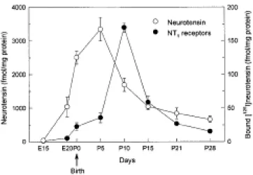
Ontogeny of neurotensin and NT1 receptors in the rat brain
Neurotensin concentration and [125I]neurotensin binding to NT
1 receptors were measured in brain
homogenates at different embryonic and postnatal stages (Figure 1). Neurotensin was first detected at moderate levels at E20. At birth, neurotensin levels were already elevated and further increased to reach peak levels at P5. The concentration of the peptide decreased thereafter, and adult levels were observed at P21. [125I]neurotensin binding to NT
1 receptors was detected at very
low levels at E20, followed by a very high and transient expression during the first postnatal week. NT1 receptor expression peaked at P10 and decreased subsequently, reaching adult levels around P21.
In addition, we studied the binding properties of N T1 receptors in the neonate (five-day-old) rat brain. Experiments were performed in the presence of levocabastine to block the binding of neurotensin to NT2 receptors. Analysis of competition experiments revealed a dissociation constant (Kd) of 0.2 ± 0.05 nM and a maximal bound capacity (Bmax) of 120 ± 20 fmol/mg of protein (mean ± S.E.M. of three independent experiments). The inhibition constant (Ki) of SR 48692 to inhibit neurotensin binding to NT1 receptors was 2.6 ± 0.52 nM. These Kd and Ki values are equivalent to the binding constants observed previously in the adult rat brain (Gully et al., 1993), whereas the number of receptors in the newborn brain is five-times higher than in the adult (Azzi et al., 1996).
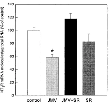
Effect of the neurotensin agonist JMV 449 on NT1 receptor mRNA expression in fetal
brain cortical cultures
Using quantitative RT-PCR, we examined the in vitro effect of a stable neurotensin agonist, JMV 449, on the amount of N T1 receptor mRNA transcripts in fetal brain cortical cultures. Exposure of cortical neurons to JMV 449 (1 nM) for 48 h resulted in a 42% decrease in N T1 receptor mRNA, when compared to control cultures (Figure 2). The effect of JMV 449 was abolished by coadministration of the NT1 receptor antagonist SR 48692 (1 µM). No effect was observed after treatment with SR 48692 alone.
Effect of chronic treatment with the antagonist SR 48692 on the ontogeny of NT1
receptor expression
The contribution of endogenous neurotensin to the ontogeny of N T1 receptors was studied in rat pups exposed to the NT1 receptor antagonist SR 48692 during the third week of pregnancy or during early postnatal life. Daily SR 48692 treatment during pregnancy had no significant effect on litter size, mean body weight, and brain weight of the offspring (data not shown). Furthermore,
there was no difference in the body and brain weight of pups treated postnatally with SR 48692 or vehicle.
Administration of the antagonist (1 mg/kg s.c.) was started at P1 and continued until P5, P 9 or P15, corresponding respectively to the period of increase, the peak or the whole period of high transient expression of NT1 receptors in the brain. No significant difference was found in [125I]neurotensin binding in brain membrane homogenates between SR 48692- and vehicle-treated
rats, whatever the treatment length (Table 1). Treatment with a higher dose of SR 48692, 10 mg/kg, from P1 to P5, also failed to modify neurotensin binding (not shown). In addition, exposure to SR 48692 during the prenatal period, by administration of the antagonist to pregnant dams from gestational day 15 (1 mg/kg i.p.), followed by injection of SR 48692 to pups from P 1 to P9 (1 mg/kg s.c.), was also without effect on the level of [125I]neurotensin binding to N T
1
receptors (Table 1).
Similarly, postnatal treatment with SR 48692 (1 mg/kg s.c.) for five days (not shown) or 15 days (Figure 3C ) did not modify the density of [125I]neurotensin binding sites measured by
quantitative autoradiography in different brain regions, including the cingulate and parietal cortices, caudate-putamen, substantia nigra compact part, and ventral tegmental area. Furthermore, administration of the NT1 receptor antagonist for five days (not shown) or 15 days (Figure 3F) did not modify the expression pattern or the density of N T1 receptor mRNA in any of the brain regions studied.
DISCUSSION
In the present study, we investigated the role of neurotensin in the regulation of N T1 receptors in the developing rat brain. We showed that exposure to the neurotensin agonist JMV 449 induced a down-regulation of NT1 receptor mRNA in primary cultures of fetal rat cerebral cortex neurons, indicating that NT1 receptor gene expression can be regulated by the agonist during the developmental period. However, chronic blockade of endogenous neurotensin by administration of SR 48692 to rat pups did not modify the postnatal expression of N T1 receptor mRNA or binding sites in the brain, suggesting that neurotensin is not essential for the establishment of the ontogenetic pattern of these receptors.
The concomitant study of the ontogeny of brain neurotensin concentration and [125I]neurotensin binding in the rat showed that a marked and transient rise in the levels of
neurotensin (at P5) preceded the increase in the expression of NT1 receptors (at P10). The presence of high levels of neurotensin during the perinatal period is consistent with previous studies showing that neurotensin appears early in development and reaches maximal levels within the first postnatal week, decreasing thereafter (Hara et al., 1982; Bissette et al., 1984; Kiyama et
al., 1992; Sato et al., 1991; Bennett et al., 1998). The peak in [125I]neurotensin receptor binding
observed in brain homogenates during the second postnatal week corresponds mainly to the increased expression of NT1 receptors in the cerebral cortex, the brain region with the highest
expression of these receptors during the perinatal period (Palacios et al., 1988; Sato et al., 1992; Lépée-Lorgeoux et al., 1999). Although only few neurotensin-containing neurons and fibers are present in the cerebral cortex, it has been suggested that neurotensin synthesized in other brain areas and/or present in the cerebrospinal fluid could exert a paracrine action on cortical receptors (Palacios et al., 1988; Sato et al., 1992).
The ability of agonists to regulate the expression of their own receptors in vitro has been described for numerous receptor systems. Previous studies on the regulation of the N T1 receptor in cell cultures showed that exposure to neurotensin results in receptor internalization, receptor down-regulation and regulation at the second messenger level (reviewed in Hermans and Maloteaux, 1998). In order to investigate whether cortical NT1 receptors could be regulated by the agonist at an early developmental stage, we studied the effect of a peptidase-resistant neurotensin agonist, JMV 449, on NT1 receptor mRNA expression in primary cultures of fetal rat cerebral cortex neurons. Exposure of cortical neurons to JMV 449 for 48 h induced a significant decrease in NT1 receptor mRNA, and this effect was abolished by the antagonist SR 48692. These results are in agreement with our previous studies on hypothalamic neuronal cultures, demonstrating that release of endogenous neurotensin induced by dexamethasone and forskolin down-regulates NT1 receptor mRNA (Scarcériaux et al., 1996). These data suggest that N T1 receptors in the developing neocortex and hypothalamus can be regulated by the agonist, despite inherent differences in the maturational profile of N T1 receptors in these two brain regions (Palacios et al., 1988; Sato et al., 1992). In addition, these findings illustrate the ability of SR 48692 to suppress agonist-induced down-regulation of NT1 receptors.
Despite the demonstration of a potential modulation of NT1 receptors by the agonist early in development, blockade of neurotensin transmission during the perinatal period with SR 48692 did not affect the ontogenetic pattern of [125I]neurotensin binding sites in brain homogenates.
Likewise, the density and distribution of N T1 receptor binding sites and mRNA expression was similar in SR 48692- and vehicle-treated pups. These results are in contrast with those obtained previously in the adult rat, showing that chronic administration of SR 48692, at the same dose used in the present study, results in up-regulation of NT1 receptor gene expression in numerous brain regions, including the anterior cingulate and perirhinal cortices, substantia nigra and ventral tegmental area (Azzi et al., 1994, 1996). Thus, whereas endogenous neurotensin appears to exert a tonic inhibitory control on NT1 receptors in the mature CNS, the results of the present study indicate that it does not play a major role in establishing the developmental profile of N T1 receptors during the early postnatal period.
The lack of effect of SR 48692 is not due to undeveloped intracellular pathways of N T1 receptors transiently expressed in the brain of fetal and infant rats, since NT1 receptor gene expression in primary cell cultures prepared from E17 cerebral cortex was down-regulated after agonist-induced receptor activation. In addition, the differential effect of SR 48692 on the regulation of NT1 receptors in the immature and adult rat brain can not be ascribed to different binding properties of these receptors, because NT1 receptors in five-day-old rat brain have the
same affinity for SR 48692 and neurotensin as NT1 receptors present in the adult (Gully et al., 1993; Azzi et al., 1996). Hence, these findings suggest that NT1 receptors expressed during the perinatal period represent functional receptors having the same ligand affinity as NT1 receptors in the adult and may thus be involved in neuronal signaling.
In the neocortex, NT1 receptors are abundantly expressed in the molecular layer, the cortical plate and the developing cortical layer VI during late gestation and the first postnatal week, followed by a dramatic decrease to the low levels seen in the adult at the end of the third postnatal week (Palacios et al., 1988). The transient expression of N T1 receptors in the cerebral cortex could be related to the reorganization of cortical neurons during postnatal development, including neuronal death, which is a prominent feature of the normal development of the neocortex (Ferrer
et al., 1992). Moreover, the formation of the pattern of cortical connectivity, the regression of
particular synapses and the stabilization of others, can act upon the level of gene expression in cortical cells. Therefore, the decline in the amount of cortical N T1 receptors during the maturation of the cerebral cortex could be explained by the programmed cell death of neurons bearing NT1 receptors, or the influence of cortical inputs on the expression of N T1 receptors. This in turn may explain the absence of effect of perinatal SR 48692 treatment on the developmental regulation of NT1 receptors in the cortex.
Interestingly, the time frame of increased expression of neurotensin and N T1 receptors in the cortex coincides with a critical period of cortical development, involving laminar organization, determination of neuronal migration pattern and synapse formation (O'Leary and Koester, 1993). It is possible therefore that neurotensin and NT1 receptor-mediated signaling play a role in the synaptic plasticity associated with the development of the cerebral cortex. In support of this idea, studies in non-neuronal cells have shown that neurotensin exerts mitogenic and trophic effects in normal and cancer cells in the intestine, liver, pancreas, lung and prostate (Evers et al., 1992; Hasegawa et al., 1994; Sehgal et al., 1994). The proliferative effect of neurotensin on intestinal cells is mediated by N T1 receptors (Ehlers et al. 1998). In addition, neurotensin has been shown to stimulate proliferation of astrocytic cell lines (Camby et al., 1996). It is noteworthy that the regionally specific and transient appearance of receptors during brain development has been reported for other neurotransmitters and peptides, including cholecystokinin, substance P, and vasopressin (Parnavelas and Cavanagh, 1988; Pélaprat et al., 1988; Tribollet et al., 1991; Zhang and Harlan, 1994; Taoka et al., 1996), thus supporting the role of neurotransmitters and peptides as growth regulatory signals during maturation of the CNS (Lauder, 1993).
Other brain areas, including the substantia nigra and ventral tegmental area, show a developmental pattern of NT1 receptors radically different from the one observed in the neocortex, characterized by a gradual increase during the postnatal period (Palacios et al., 1988; Sato et al., 1992; Lépée-Lorgeoux et al., 1999). These regions are also characterized by the presence of neurotensin terminals at birth and in the adult (Kiyama et al., 1992), therefore suggesting the possible regulation of N T1 receptors in the developing brain following the
administration of SR 48692, as shown in the adult (Azzi et al., 1994, 1996). However, as mentioned above, chronic blockade of N T1 receptors during fetal and postnatal development did not affect the expression of receptors in the midbrain, suggesting that during this period, peptide release and connection formation are probably not sufficient for the regulation of N T1 receptors by the endogenous ligand.
Early exposure of developing postsynaptic receptors to the appropriate agonist has been suggested to be important for establishing the ontogenetic pattern of several receptor systems. For instance, postnatal treatment with a vasoactive intestinal polypeptide (VIP) antagonist increases expression of VIP receptors in the rat brain (Hill et al., 1994). Furthermore, prenatal or postnatal exposure to GABA(A) or NMDA receptor antagonist selectively decrease the expression of certain receptor subunits in the developing brain (Sircar et al., 1996; Liu et al., 1998). However, lack of agonist involvement in receptor ontogeny has also been described for some other members of the family of G protein-coupled receptors. Thus, prenatal administration of the β-adrenergic antagonist propranolol does not modify the expression of its receptors in the newborn rat brain (Kudlacz et al., 1991; Speiser et al., 1991). Moreover, using the knockout approach, it was recently shown that deletion of the oxytocin gene does not alter the expression of oxytocin receptors in the CNS (Nishimori et al., 1996). Similarly, mice unable to synthesize dopamine after targeted inactivation of the tyrosine hydroxylase gene in dopaminergic neurons show normal development of midbrain dopaminergic neurons and the pattern and intensity of D1 and D2 receptor mRNA expression in the striatum are unaltered (Zhou and Palmiter, 1995). Our results indicate that NT1 receptors represent another instance of receptors for which the agonist, while being involved in receptor regulation in the adult rat brain, does not influence the pattern of expression during ontogeny.
In conclusion, these findings indicate that NT1 receptor regulation in the developing nervous system differs from that seen in adulthood and that endogenous neurotensin is not required for the normal expression of NT1 receptors during neonatal development. The transiently high expression of neurotensin and NT1 receptors during the early postnatal period overlaps with a critical period of development of the brain, suggesting a physiological role for neurotensin and N T1 receptors in the development of the CNS.
ACKNOWLEDGMENTS
We thank Dr. Danielle Gully (Sanofi Recherche) for the generous gift of SR 48692, Sophie Callier for help in the PCR experiments and Anne-Marie Lhiaubet for labeling [125I]neurotensin. I. L. was
supported by a fellowship from the Ministère de l'Enseignement Supérieur et de la Recherche, C. B. by the INSERM and F. S. by the Association pour la Recherche contre le Cancer.
REFERENCES
Alexander MJ, Miller MA, Dorsa DM, Bullock BP, Melloni RH Jr, Dobner PR, Leeman SE. 1989. Distribution of neurotensin/neuromedin N mRNA in rat forebrain: unexpected abundance in hippocampus and subiculum. Proc Natl Acad Sci USA 86:5202-5206.
Azzi M, Boudin H, Mahmudi N, Pélaprat D, Rostène W, Bérod A. 1996. In vivo regulation of neurotensin receptors following long-term pharmacological blockade with a specific receptor antagonist. Mol Brain Res 42:213-221.
Azzi M, Nicot A, Gully D, Kitabgi P, Bérod A, Rostène W. 1994. Increase in neurotensin receptor expression in rat brain induced by chronic treatment with the nonpeptide neurotensin receptor antagonist SR 48692. Neurosci Lett 172:97-100.
Bennett GW, Moss SH, Forster CD, Marsden CA. 1998. Developmental changes in neurotensin and its metabolites in the neonatal rat. Dev Brain Res 111:189-196.
Betancur C, Canton M, Burgos A, Labeeuw B, Gully D, Rostène W, Pélaprat D. 1998. Characterization of binding sites of a new neurotensin receptor antagonist, [3H]SR 142948A, in
the rat brain. Eur J Pharmacol 343:67-77.
Bissette G, Richardson C, Kizer JS, Nemeroff CB. 1984. Ontogeny of brain neurotensin in the rat: a radioimmunoassay study. J Neurochem 43:283-287.
Camby I, Salmon I, Bourdel E, Nagy N, Danguy A, Brotchi J, Pasteels JL, Martinez J, Kiss R. 1996. Neurotensin-mediated effects on astrocytic tumor cell proliferation. Neuropeptides 30:133-139. Chalon P, Vita N, Kaghad M, Guillemot M, Bonnin J, Delpech B, Le Fur G, Ferrara P, Caput D.
1996. Molecular cloning of a levocabastine-sensitive neurotensin binding site. FEBS Lett 386:91-94.
Cheng Y, Prusoff WH. 1973. Relationship between the inhibition constant (Ki) and the
concentration of inhibitor which causes 50 percent inhibition (IC5 0) of an enzymatic reaction. Biochem Pharmacol 22:3099-3108.
Chomczynski P, Sacchi N. 1987. Single-step method of RNA isolation by acid guanidium thiocyanate-phenol-chloroform extraction. Anal Biochem 162:156-159.
Doulut S, Rodriguez M, Lugrin D, Vecchini F, Kitabgi P, Aumelas A, Martinez J. 1992. Reduced peptide bond pseudopeptide analogues of neurotensin. Peptide Res 5:30-38.
Dubuc I, Sarret P, Labbé-Jullié C, Botto JM, Honoré E, Bourdel E, Martinez J, Costentin J, Vincent JP, Kitabgi P, Mazella J. 1999. Identification of the receptor subtype involved in the analgesic effect of neurotensin. J Neurosci 19:1-8.
Ehlers RA 2nd, Bonnor RM, Wang X, Hellmich MR, Evers BM. 1998. Signal transduction mechanisms in neurotensin-mediated cellular regulation. Surgery 124:239-246.
Evers BM, Izukura M, Chung DH, Parekh D, Yoshinaga K, Greeley GH, Uchida T, Towsend CM, Thompson JC. 1992. Neurotensin stimulates growth of colonic mucosa in young and aged rats. Gastroenterology 103:86-91.
Ferrer I, Soriano E, del Rio JA, Alcantara S, Auladell C. 1992. Cell death and removal in the cerebral cortex during development. Prog Neurobiol 39:1-43.
Gully D, Canton M, Boigegrain R, Jeanjean F, Molimard JC, Poncelet M, Gueudet C, Heaulme M, Leyris R, Brouard A, Pélaprat D, Labbé-Jullié C, Mazella J, Soubrié P, Maffrand JP, Rostène W, Kitabgi P, Le Fur G. 1993. Biochemical and pharmacological profile of a potent and selective nonpeptide antagonist of the neurotensin receptor. Proc Natl Acad Sci USA 90:65-69.
Hara Y, Shiosaka S, Senba E, Sakanaka M, Inagaki S, Takagi H, Kawai Y, Takatsuki K, Matsuzaki T, Tohyama M. 1982. Ontogeny of the neurotensin-containing neuron system of the rat: immunohistochemical analysis. I. forebrain and diencephalon. J Comp Neurol 208:177-195.
Hasegawa K, Kar S, Carr BI. 1994. Stimulation of hepatocyte DNA synthesis by neurotensin. J Cell Physiol 158:215-222.
Hermans E, Maloteaux JM. 1998. Mechanisms of regulation of neurotensin receptors. Pharmacol Ther 79:89-104.
Hill JM, Mervis RF, Politi J, McCune SK, Gozes I, Fridkin M, Brenneman DE. 1994. Blockade of VIP during neonatal development induces neuronal damage and increases VIP and VIP receptors in brain. Ann NY Acad Sci 739:211-225.
Kiyama H, Sato M, Emson PC. 1992. Ontogeny of neurotensin immunoreactivity and mRNA in the rat central nervous system. In: Björklund A, Hökfelt T, Tohyama M, editors. Handbook of chemical neuroanatomy, Vol. 10: Ontogeny of neurotransmitters and peptides in the CNS. Amsterdam: Elsevier Science Publishers, p 399-431.
Kudlacz EM, Spencer JR, Slotkin TA. 1991. Postnatal alterations in ß-adrenergic receptor binding in rat brain regions after continuous prenatal exposure to propranolol via maternal infusion. Res Commun Chem Pathol Pharmacol 71:153-161.
Lambert PD, Gross R, Nemeroff CB, Kilts CD, 1995. Anatomy and mechanisms of neurotensin-dopamine interactions in the central nervous system. Ann NY Acad Sci 757:377-389.
Lauder JM. 1993. Neurotransmitters as growth regulatory signals: role of receptors and second messengers. Trends Neurosci 16:233-240.
Lépée-Lorgeoux I, Betancur C, Rostène W, Pélaprat D. 1999. Differential ontogenetic patterns of levocabastine-sensitive neurotensin NT2 receptors and of NT1 receptors in the rat brain revealed by in situ hybridization. Dev Brain Res 113:115-131.
Liu J, Brannen KC, Grayson DR, Morrow AL, Devaud LL, Lauder JM. 1998. Prenatal exposure t o the pesticide dieldrin or the GABA(A) receptor antagonist bicuculline differentially alters expression of GABA(A) receptor subunit mRNAs in fetal rat brainstem. Dev Neurosci 20:83-92. Mazella J, Botto JM, Guillemare E, Coppola T, Sarret P, Vincent JP. 1996. Structure, functional
expression, and cerebral localization of the levocabastine-sensitive neurotensin/neuromedin N receptor from mouse brain. J Neurosci 16:5613-5620.
Mazella J, Zsürger N, Navarro V, Chabry J, Kaghad M, Caput D, Ferrara P, Vita N, Gully D, Maffrand JP, Vincent JP. 1998. The 100-kDa neurotensin receptor is gp95/sortilin, a non-G-protein-coupled receptor. J Biol Chem 273:26273-26276.
Munson PJ, Rodbard D. 1980. LIGAND: A versatile computerized approach for characterization of ligand-binding systems. Anal Biochem 107:220-239.
Najimi M, Souazé F, Méndez M, Hermans E, Berbar T, Rostène W, Forgez P. 1998. Activation of receptor gene transcription is required to maintain cell sensitization after agonist exposure. J Biol Chem 273:21634-21641.
Nemeroff CB, Kitabgi P. 1992. The neurobiology of neurotensin. Ann N Y Acad Sci 668:1-374. Nishimori K, Young LJ, Guo Q, Wang Z, Insel TR, Matzuk MM. 1996. Oxytocin is required for
nursing but is not essential for parturition or reproductive behavior. Proc Natl Acad Sci USA 93:11699-11704.
O'Leary DDM, Koester SE. 1993. Development of projection neuron types, axon pathways, and patterned connections of the mammalian cortex. Neuron 10:991-1006.
Palacios JM, Pazos A, Dietl MM, Schlumpf M, Lichtensteiger W. 1988. The ontogeny of brain neurotensin receptors studied by autoradiography. Neuroscience 25:307-317.
Parnavelas JG, Cavanagh ME. 1988. Transient expression of neurotransmitters in the developing neocortex. Trends Neurosci 11:92-93.
Pélaprat D, Dusart I, Peschanski M. 1988. Postnatal development of cholecystokinin (CCK) binding sites in the rat forebrain and midbrain: an autoradiographic study. Dev Brain Res 44:119-132.
Rostène WH, Alexander MJ. 1997. Neurotensin and neuroendocrine regulation. Front Neuroendocrinol 18:115-173.
Sato M, Kiyama H, Tohyama M. 1992. Different postnatal development of cells expressing mRNA encoding neurotensin receptor. Neuroscience 48:137-149.
Sato M, Kiyama H, Yoshida S, Saika T, Tohyama M. 1991. Postnatal ontogeny of cells expressing prepro-neurotensin/neuromedin N mRNA in the rat forebrain and midbrain: a hybridization histochemical study involving isotope-labeled and enzyme-labeled probes. J Comp Neurol 310:300-315.
Scarcériaux V, Pélaprat D, Lhiaubet AM, Schimpf RM, Tramu G, Rostène W. 1995. Developmental pattern of neurotensin content in rat hypothalamic neurons cultured in serum-free medium: comparison with in vivo data. Dev Brain Res 81:128-130.
Scarcériaux V, Souazé F, Bachelet CM, Forgez P, Bourdel E, Martinez J, Rostène W, Pélaprat D. 1996. Neurotensin receptor down-regulation induced by dexamethasone and forskolin in rat hypothalamic cultures is mediated by endogenous neurotensin. J Neuroendocrinol 8:587-593. Schotte A, Laduron PM. 1987. Different postnatal ontogeny of two [3H]neurotensin binding sites
in rat brain. Brain Res 408:326-328.
Schotte A, Leysen JE, Laduron PM. 1986. Evidence for displaceable non-specific [3H]neurotensin
binding site in rat brain. Naunyn-Schmiedeberg's Arch Pharmacol 333:400-405.
Sehgal I, Powers S, Huntley B, Powis G, Pittelkow M, Maihle NJ. 1994. Neurotensin is an autocrine trophic factor stimulated by androgen withdrawal in human prostate cancer. Proc Natl Acad Sci USA 91:4673-4677.
Sircar R, Follesa P, Ticku MK. 1996. Postnatal phencyclidine treatment differentially regulates N-methyl-D-aspartate receptor subunit mRNA expression in developing rat cerebral cortex. Mol Brain Res 40:214-220.
Souazé F, Rostène W, Forgez P. 1997. Neurotensin agonist induces differential regulation of neurotensin receptor mRNA. J Biol Chem 272:10087-10094.
Speiser Z, Gordon I, Rehavi M, Gitter S. 1991. Behavioral and biochemical studies in rats following prenatal treatment with ß-adrenoreceptor antagonists. Eur J Pharmacol 195:75-83.
Tanaka K, Masu M, Nakanishi S. 1990. Structure and functional expression of the cloned rat neurotensin receptor. Neuron 4:847-854.
Taoka M, Song SY, Kubota M, Minegishi A, Yamakuni T, Konishi S. 1996. Increased level of neurokinin-1 tachykinin receptor gene expression during early postnatal development of rat brain. Neuroscience 74:845-853.
Tribollet E, Goumaz M, Raggenbass M, Dubois-Dauphin M, Dreifuss JJ. 1991. Early appearance and transient expression of vasopressin receptors in the brain of rat fetus and infant. An autoradiographical and electrophysiological study. Dev Brain Res 58:13-24.
Vita N, Laurent P, Lefort S, Chalon P, Dumont X, Kaghad M, Gully D, Le Fur G, Ferrara P, Caput D. 1993. Cloning and expression of a complementary DNA encoding a high affinity human neurotensin receptor. FEBS Lett 317:139-142.
Zhang L, Harlan RE. 1994. Ontogeny of the distribution of tachykinins in rat cerebral cortex: immunocytochemistry and in situ hybridization histochemistry. Dev Brain Res 77:23-36.
Zhou QY, Palmiter RD. 1995. Dopamine-deficient mice are severely hypoactive, adipsic, and aphagic. Cell 83:1197-1209.
Table 1. Effect of chronic treatment with SR 48692 during perinatal development on N T1 receptor binding sites
Bound [125I]neurotensin (fmol/mg protein)
Treatment length Vehicle SR 48692
E15-P9 163.5 ± 7.1 161.1 ± 4.6
P1-P5 85.9 ± 5.2 96.4 ± 16.4
P1-P9 181.8 ± 13.1 190.4 ± 6.6
P1-P15 67.2 ± 3.6 68.4 ± 5.5
Rat pups were injected daily with the neurotensin NT1 receptor antagonist SR 48692 (1 mg/kg s.c.) or vehicle for different lengths of time starting at postnatal day 1 (P1). In another group, exposure to SR 48692 was started during the prenatal period, by daily administration of the antagonist to pregnant females from gestational day 15 (E15). Binding of [125I]neurotensin t o
N T1 receptors was determined in brain membrane homogenates in the presence of levocabastine, to inhibit binding to NT2 receptors. Values represent mean ± S.E.M. (n = six rats per group).
Figure 1. Ontogeny of neurotensin and NT1 receptors in the rat brain. Left axis represents
neurotensin levels, measured by radioimmunoassay, in whole brain homogenates. Right axis shows the developmental pattern of NT1 receptor binding sites, measured by [125I]neurotensin binding in
brain membrane homogenates. Each point represents mean ± S.E.M. of four rats at different developmental stages: gestational day 15 (E15), E20, postnatal day 0 (P0, at birth), P5, P10, P15, P21, and P28.
Figure 2. Regulation of NT1 receptor mRNA in brain cortical cultures after exposure to the
neurotensin agonist JMV 449, and the N T1 receptor antagonist SR 48692. Cortical neurons were treated for 48 h with JMV 449 1 nM, SR 48692 1 µM, or both. N T1 receptor mRNA was
quantified with RT-PCR. Values represent mean ± S.E.M. of four cultures. * p< 0.05 vs. control (Student's t-test).
Figure 3. Effect of chronic administration of the antagonist SR 48692 during postnatal development on NT1 receptor binding sites and mRNA expression in the rat brain. Pups received
a daily injection of SR 48692 (1 mg/kg s.c.) or vehicle from P1 to P15 and were killed 24 h after the last injection. Autoradiograms illustrate the brain areas where the optical density was quantified. A,B,C: [125I]neurotensin binding to NT
1 receptors was assessed by quantitative
autoradiography. D,E,F: NT1 receptor mRNA expression was measured using in situ hybridization.
Bars represent mean ± S.E.M. of six rats. Cg, cingulate cortex; Par, parietal cortex; SNC, substantia nigra, compact part; VTA, ventral tegmental area.