The CDK8 Complex and Proneural Proteins Together
Drive Neurogenesis from a Mesodermal Lineage
The MIT Faculty has made this article openly available.
Please share
how this access benefits you. Your story matters.
Citation
Luo, Shuo, and H. Robert Horvitz. “The CDK8 Complex and
Proneural Proteins Together Drive Neurogenesis from a
Mesodermal Lineage.” Current Biology 27, no. 5 (March 2017): 661–
672.
As Published
http://dx.doi.org/10.1016/J.CUB.2017.01.056
Publisher
Elsevier BV
Version
Author's final manuscript
Citable link
http://hdl.handle.net/1721.1/116544
Terms of Use
Creative Commons Attribution-NonCommercial-NoDerivs License
The CDK8 Complex and Proneural Proteins Together Drive
Neurogenesis from a Mesodermal Lineage
Shuo Luo1 and H. Robert Horvitz1,2,*
1Howard Hughes Medical Institute and Department of Biology, Massachusetts Institute of Technology, Cambridge, MA 02139, USA
SUMMARY
At least some animal species can generate neurons from mesoderm or endoderm, but the underlying mechanisms remain unknown. We screened for C. elegans mutants in which the presumptive mesoderm-derived I4 neuron adopts a muscle-like cell fate. From this screen, we identified HLH-3, the C. elegans homolog of a mammalian proneural protein (Ascl1) used for in vitro neuronal reprogramming, as required for efficient I4 neurogenesis. We discovered that the CDK-8 Mediator kinase module acts together with a second proneural protein, HLH-2, and in parallel to HLH-3 to promote I4 neurogenesis. Genetic analysis revealed that CDK-8 most likely promotes I4 neurogenesis by inhibiting the CDK-7/CYH-1 (CDK7/cyclin H) kinase module of the transcription initiation factor TFIIH. Ectopic expression of HLH-2 and HLH-3 together promoted expression of neuronal features in non-neuronal cells. These findings reveal that the Mediator CDK8 kinase module can promote non-ectodermal neurogenesis and suggest that inhibiting CDK7/cyclin H might similarly promote neurogenesis.
INTRODUCTION
During bilaterian development, mesoderm and endoderm give rise to primarily non-neural tissues, whereas neurons are generated mostly from ectoderm. However, some animals, such as jellyfish and sea urchins, have subsets of neural cells derived from non-ectodermal origins, such as striated muscle and endoderm [1, 2]. It is not known whether the
specification of non-ectodermal neural cells involves molecular mechanisms different from those of ectodermal neural specification. Also, because generating neurons from non-ectodermal cells is an important approach in neuroregenerative medicine, understanding molecular mechanisms underlying such neurogenesis might identify novel factors useful in regenerative medicine.
*Correspondence: [email protected].
2Lead Contact
SUPPLEMENTAL INFORMATION
Supplemental Information includes Supplemental Experimental Procedures and six figures and can be found with this article online at
http://dx.doi.org/10.1016/j.cub.2017.01.056. AUTHOR CONTRIBUTIONS
HHS Public Access
Author manuscript
Curr Biol
. Author manuscript; available in PMC 2018 March 06.Published in final edited form as:
Curr Biol. 2017 March 06; 27(5): 661–672. doi:10.1016/j.cub.2017.01.056.
A
uthor Man
uscr
ipt
A
uthor Man
uscr
ipt
A
uthor Man
uscr
ipt
A
uthor Man
uscr
ipt
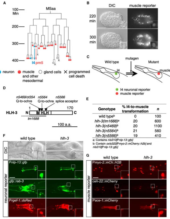
The nervous system of the C. elegans adult hermaphrodite consists of 302 neurons, 294 of which are derived from the AB blastomere, which primarily generates ectodermal cells [3]. By contrast, six pharyngeal neurons are generated from the MS blastomere, which generates mostly mesodermal cells (Figure 1A), and two neurons are generated from the C lineage, which generates both ectoderm and mesoderm. The MS-derived pharyngeal I4 neuron is generated from a progenitor cell that divides to give rise to I4 and a pharyngeal muscle cell [3]. Although pharyngeal muscle cells are sometimes considered myoepithelial, because these cells function as muscles, have molecular features characteristic of muscle cells, and their normal development depends on mesodermal transcription factors, we view them as muscles [4, 5]. We hypothesized that I4 might overcome a mesodermal cell fate to become a neuron.
Here we report the identification of genetic mutants in which the I4 neuron adopts a muscle-like cell fate and show that two conserved genetic pathways, a proneural pathway and a Mediator pathway, act synergistically to promote I4 neurogenesis from mesoderm. We found that HLH-3, the homolog of a mammalian protein (Ascl1) that has been used extensively in neuronal reprogramming [6–8], HLH-2, and the evolutionarily conserved Mediator CDK8 kinase module promote I4 neurogenesis. Overexpression of HLH-2 and HLH-3 together promotes partial neuronal transformation of non-neuronal cells, including body-wall muscle cells. Our findings reveal that the CDK8 kinase module can promote non-ectodermal neurogenesis.
RESULTS
I4 Precursor Cells Transiently Express a Mesodermal Cell-Fate Reporter
We first investigated whether the I4 neuron expresses typical neuronal features by examining the expression of two broadly expressed neuronal reporters, for the small GTPase RAB-3 (gfp::rab-3) [9] and the guanine nucleotide exchange factor homolog RGEF-1
(Prgef-1::dsRed) [10], in I4. We observed that both reporters were expressed in I4 and other
MS-derived neurons (Figure 1F and unpublished data); observations by Stefanakis et al. [11] similarly suggest that MS- and AB-derived neurons share basic neuronal molecular
attributes. To determine whether I4 precursor cells express mesodermal characteristics, we examined the expression of an hlh-1 reporter during embryogenesis. HLH-1 is the C. elegans homolog of the mammalian muscle master regulator MyoD and is expressed exclusively in myogenic lineages (Expression Patterns in Caenorhabditis [EPIC]; http://
epic.gs.washington.edu). We found that I4 progenitor cells and the newly generated presumptive I4 cell were labeled by the HLH-1 reporter (Figure 1B). Our findings indicate that I4 precursor cells are at least to this extent mesodermal.
The I4 Neuron Adopts a Muscle-like Cell Fate in hlh-3 Mutants
To seek mutants in which I4 adopts a muscle cell fate, we used a transgenic strain in which the I4 neuronal cell fate is labeled by the neural peptide reporter Pnlp-13::gfp [12] and
pharyngeal muscle cell fate is labeled by the pharyngeal myosin heavy-chain reporter Pmyo-2::mCherry::H2B [13]. We performed genetic screens and identified mutants that
specifically lost I4 GFP expression (Figures 1C and 1F). Three such mutants carried alleles
A
uthor Man
uscr
ipt
A
uthor Man
uscr
ipt
A
uthor Man
uscr
ipt
A
uthor Man
uscr
ipt
of the gene hlh-3, which encodes a basic-helix-loop-helix (bHLH) transcription factor homologous to the mammalian proneural protein Ascl1/Mash1 (Figures 1D and 1E). Ascl1 is involved in neural development in Drosophila and mammals, and overexpression of Ascl1 with other transcription factors drives reprogramming of various types of mesodermal and endodermal cells into neurons [6, 8, 14–16].
One hlh-3 allele, n5469, contains an early stop codon that truncates the protein before the evolutionarily conserved HLH domain and most likely is a molecular null (Figure 1D). The I4 cell in hlh-3 mutants appears to adopt a muscle-cell-like fate: (1) the nuclear morphology of I4 as visualized using Nomarski optics was transformed from a neuronal speckled morphology to a non-neuronal, fried-egg morphology (Figure 1F); (2) wild-type I4
expressed neuronal markers Pnlp-13::gfp, Prab-3::gfp::rab-3, and Prgef-1::dsRed, whereas none
of these markers was expressed in the mutant presumptive I4 cell (Figure 1F); and (3) the mutant I4 cell expressed pharyngeal muscle reporters Pmyo-2::mCherry:: His2B and
Pceh-22::ceh-22::mCherry [4, 17] (Figure 1G). We found that the acetylcholine esterase
reporter Pace-1::mCherry, which normally labels the I4 sister cell pm5 [18], labeled an extra
pm5 muscle cell in the hlh-3 mutant pharynx (wild-type, n = 6 pm5; hlh-3, n = 7 pm5) (Figure 1G), indicating that the I4 cell in hlh-3 mutants failed to be specified as a neuron and instead adopted the cell fate of its sister pm5 pharyngeal muscle cell. We were able to rescue the I4 defects by expressing a wildtype copy of the hlh-3 gene in hlh-3 mutants (Figure S1A).
HLH-3 Is Mostly Dispensable for Neurogenesis
Of the 20 neurons in the wild-type C. elegans pharynx, only I4 seemed to be affected by the disruption of HLH-3 (Figure S2C and data not shown). To further explore a possible role for HLH-3 in the neurogenesis of neurons other than I4, we scored the number of neurons expressing neurotransmitter reporter transgenes for cholinergic, GABAergic, glutamatergic, dopaminergic, serotonergic, and tyraminergic/octopaminergic neurons in hlh-3 double mutants that also contained hlh-2 or dpy-22 mutations (we used the second mutation to sensitize the strain and potentially increase the magnitude of defects; see below); together, these reporters label about 240 of the 302 neurons in C. elegans (Figure S2A). We found that approximately 10% of wild-type I4 expressed the glutamate transporter transgene
Peat-4::eat-4::mCherry (a fosmid-based translational fusion constructed by [19]) but none of
the other reporters, indicating that I4 might be glutamatergic (Figure S2B). We did not find any significant difference in the number of eat-4-expressing neurons between the wild-type and hlh-3 double-mutant animals, showing that the fates of most glutamatergic neurons were not altered (Figure S2D). (That I4 is transformed to a muscle cell in hlh-3 mutants did not result in a lower count for hlh-3 mutants, because only 10% of wild-type I4s express the reporter.) We similarly observed no major differences in cholinergic, dopaminergic, serotonergic, or tyraminergic/octopaminergic neuron numbers between wild-type and hlh-3 mutant animals (Figures S2E and S2G–S2I). By contrast, we noticed a mild deficit in GABAergic neuron number in hlh-3; hlh-2 double mutants, which had one to five (mean 1.3) fewer GABAergic ventral cord motor neurons than did wild-type animals (Figure S2F). Further analysis indicated that this defect was most likely caused by the hlh-2/3 mutation (data not shown). In mammals, knockout of Ascl1 results in impaired neurogenesis in
A
uthor Man
uscr
ipt
A
uthor Man
uscr
ipt
A
uthor Man
uscr
ipt
A
uthor Man
uscr
ipt
confined neural regions, including the ventral telencephalon, olfactory bulb, and autonomic ganglia, whereas neurogenesis in other brain regions remains grossly normal [15, 20]. We conclude that, like Ascl1, its homolog HLH-3 promotes neurogenesis of I4 and a few GABAergic neurons but is not generally required for neurogenesis.
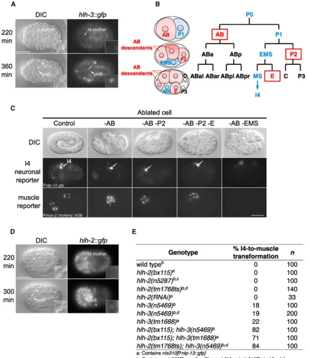
HLH-3 Is Expressed in the Newly Generated I4 Cell and Most Likely Functions Cell Autonomously
We used an HLH-3::GFP fusion protein to examine HLH-3 expression during
embryogenesis. HLH-3::GFP was expressed in the I4 neuron shortly after its mother divided to generate I4; by contrast, the I4 sister, pm5, did not express this protein (Figure 2A). We also observed expression of HLH-3::GFP in multiple AB-derived neural precursors (data not shown). The broad expression of HLH-3::GFP was mostly confined to embryos and was no longer detectable in the I4 neuron in newly hatched L1s (larval stage 1; Figure S1B), suggesting that hlh-3 functions primarily in early embryos to promote I4 specification. To determine whether HLH-3 functions within the I4 lineage or in neighboring cells to promote I4 neurogenesis, we used a laser micro-beam to selectively kill the cells that make direct contact with I4 progenitor cells during embryogenesis (Figure 2B). We asked whether elimination of any neighboring cells impairs I4 neurogenesis. Laser ablation of the founder cells AB, P2, and E, which normally generate neighbors of I4 progenitor cells in early embryos, did not affect I4 GFP reporter expression (Figure 2C). By contrast, killing the I4 progenitor cell ethyl methanesulfonate (EMS) eliminated I4 GFP reporter expression (Figure 2C). These results suggest that HLH-3 most likely functions cell autonomously to drive I4 neurogenesis.
HLH-2, the C. elegans Homolog of Daughterless or Tcf3, Functions Synergistically with HLH-3 to Promote Efficient I4 Neurogenesis
The neurogenesis of I4 is only partially disrupted in the absence of functional HLH-3: about 80% of hlh-3 null mutants (n5469 and tm1688) still generate an I4 neuron (Figures 1D and 1E). Thus, other genes most likely function in addition to hlh-3 to drive I4 neurogenesis. HLH-3 can interact and form heterodimers with another bHLH transcription factor, HLH-2, the C. elegans homolog of the conserved E2A/Tcf3/Daughterless protein [21, 22]. Tcf3 and Daughterless are broadly expressed in developing neural precursors in vertebrates and Drosophila, respectively, and disruption of either protein results in loss of neural tissues and aberrant morphogenesis [22–25]. Using a reporter transgene that expresses an HLH-2::GFP fusion protein [26], we found that HLH-2::GFP was expressed in the I4 neuron shortly after its generation but was absent from its sister cell, pm5 (Figure 2D). Also like HLH-3::GFP, HLH-2::GFP was broadly expressed in early embryos but was not detectable in most neurons, including I4 in newly hatched L1s (Figure S1B), suggesting that hlh-2 most likely functions in early embryos to promote I4 specification. We then asked whether HLH-2 is required for I4 neurogenesis. Both hlh-2(n5287) null mutants and animals treated with hlh-2 RNAi displayed embryonic lethality; nonetheless, we did not observe obvious defects in I4 GFP expression (we note that in both cases maternal HLH-2 was still likely to be present) (Figure 2E; Figure S1C). By contrast, the introduction of an hlh-2 partial loss-of-function allele (bx115 or tm1768) into an hlh-3 null background significantly enhanced I4 misspecification, and in about 80% of hlh-2; hlh-3 double mutants the I4 cell adopted a
A
uthor Man
uscr
ipt
A
uthor Man
uscr
ipt
A
uthor Man
uscr
ipt
A
uthor Man
uscr
ipt
muscle-like cell fate (Figure 2E). Given this genetic enhancement, we conclude that HLH-2 functions to promote I4 neurogenesis at least partly through a genetic pathway that acts in parallel to HLH-3.
Multiple bHLH Proneural Proteins Promote MS Neurogenesis
The C. elegans genome encodes 42 bHLH factors, with multiple bHLH proteins able to form dimers with HLH-2 (Figure S3A) [21]. We tested whether any of the proneural bHLH proteins, including neurogenin NGN-1 and NeuroD CND-1, are required for I4
neurogenesis. Examination of the I4 neuron in either ngn-1 or cnd-1 single mutants or hlh-2; ngn-1 or hlh-2; cnd-1 double mutants using the reporter transgene Pnlp-13::gfp did not reveal
any defects in I4 neurogenesis (Figure S3B). Furthermore, we did not observe defects in I4 neurogenesis in hlh-4, 6, 10, 12, 13, 15, 19, or lin-32 mutant animals using both Nomarski optics and the pan-neuronal reporter transgene Prgef-1::dsRed, suggesting that I4
neurogenesis is specifically dependent on HLH-3 and not on other HLH-2-interacting bHLH proteins (Figures S3E– S3L). However, ngn-1 and cnd-1 mutants were disrupted in the neurogenesis of other MS-derived neurons: around 40% of ngn-1 mutant animals lacked the M1 neuron, and in 25% of cnd-1 mutant animals the I3 neuron adopted a gland cell fate (the cell fate of its sister cell) (Figures S3C and S3D). These results indicate that the generation of mesoderm-derived neurons in C. elegans involves multiple bHLH proneural proteins, with I4 neurogenesis depending specifically on HLH-3 (Figure 3E).
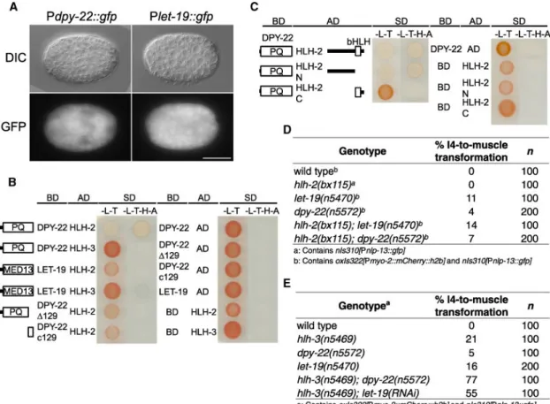
Mediator Subunits Function in the HLH-2 Pathway to Promote I4 Neurogenesis
To search for additional factors that function with HLH-2 and HLH-3 to promote efficient I4 neurogenesis, we examined other mutant isolates from our screens. We found that five mutants carry alleles of dpy-22, and two carry alleles of let-19 (Figure 3A; Figure S4A). Like hlh-3 mutations, mutations in dpy-22 and let-19 disrupted I4 specification, and the I4 cell adopted a pharyngeal muscle cell fate (Figure 3B). dpy-22 and let-19 encode the worm homologs of the evolutionarily conserved Mediator sub-units Med12 and Med13,
respectively. Mediator is a multi-sub-unit complex that bridges DNA-binding proteins (transcription factors/coactivators) with the RNA polymerase II transcription machinery and is involved in many aspects of gene regulation and animal development [27–29]. Med12 disruption in mice and zebrafish results in impaired development of the neural crest and of non-ectodermal tissues, including heart and gut [30–32]. We found that although disruption of LET-19 function in the let-19 mutant n5470 also led to low-frequency (~3%, n = 60) neurogenesis defects of the M1 neuron (Figure 3C), disruption of DPY-22 in all five dpy-22 mutants specifically disrupted I4 neurogenesis (Figure 3D). Because promoter-fusion reporter transgenes for dpy-22 and let-19 revealed broad GFP expression in developing embryos (Figure 4A), we conclude that DPY-22 and LET-19 most likely cooperate with cell-specific factors to drive I4 neurogenesis.
Further analysis revealed that the two let-19 alleles contain missense mutations, and all five dpy-22 alleles contain nonsense mutations that truncate the C-terminal PQ-rich domain (Figure 3A). Because none of these mutations is obviously null, we performed dpy-22 or let-19 RNAi to further reduce gene function in dpy-22 and let-19 mutants, respectively. We did not observe significant enhancement of the I4 misspecification (Figure S4B). In addition,
A
uthor Man
uscr
ipt
A
uthor Man
uscr
ipt
A
uthor Man
uscr
ipt
A
uthor Man
uscr
ipt
we used cell-specific RNAi to express dpy-22 or let-19 siRNA under the nlp-13 promoter (which is active only after I4 is generated; unpublished data) [33]. I4 appeared normal in position and morphology (Figures S4C and S4D), suggesting that Mediator is dispensable for the maintenance of the I4 cell fate after mid-late embryogenesis.
In vertebrates, Med12 interacts with transcription factors through its PQ-rich domain to promote gene expression and tissue development [34, 35]. To determine whether Mediator might promote I4 neurogenesis by interacting with bHLH proneural factors, we performed a yeast two-hybrid assay. We found that the DPY-22 PQ-rich domain selectively interacted with HLH-2 but not HLH-3, whereas the last 129 amino acids truncated in all five dpy-22 alleles were required for this interaction (Figure 4B). Further analysis indicated that the PQ-rich domain interacted with the N-terminal half of HLH-2, the region of a predicted
transactivation domain important for gene expression and neurogenesis [36, 37] (Figure 4C). These findings suggest that Mediator physically interacts with and might function in the same pathway as HLH-2 to promote I4 neurogenesis.
To test this hypothesis, we constructed Mediator and bHLH double mutants. All of the dpy-22 and let-19 single mutants showed a low frequency of I4 misspecification, and dpy-22 or let-19 RNAi in dpy-22 or let-19 mutants, respectively, did not significantly enhance I4 defects (Figures S4A and S4B). Introducing an hlh-2 partial loss-of-function allele into dpy-22 or let-19 mutants also did not enhance I4 misspecification (Figure 4D). By contrast, disruption of dpy-22 or let-19 in an hlh-3 null (n5469) background significantly enhanced I4 misspecification, with 77% and 55% of the I4 cells adopting a muscle cell fate, respectively (let-19 and hlh-3 are tightly linked, and thus let-19 was tested using RNAi) (Figure 4E), indicating that Mediator and HLH-2 act together and in parallel to HLH-3 to promote I4 neurogenesis.
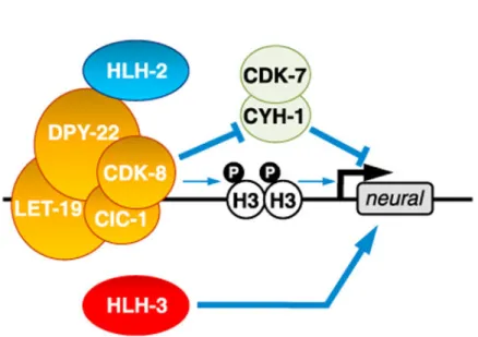
CDK-8 Most Likely Promotes I4 Neurogenesis by Inhibiting CDK-7/CYH-1
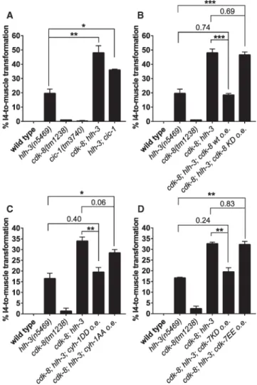
Med12 and Med13 are components of a four-protein Mediator kinase module, with the other two proteins being the cyclin-dependent kinase CDK8 and cyclin C [27, 28]. CDK8 is involved in cell-fate transformation during tumor generation and progression [38, 39]. To investigate whether the C. elegans counterparts of CDK8 and cyclin C are involved in I4 neurogenesis, we examined I4 development in cdk-8(tm1238) (CDK8) and cic-1(tm3740) (cyclin C) mutants. Both mutants contain substantial deletions of coding exons and both are most likely null (Figure S5A). cdk-8 and cic-1 single mutants had only very mild (<1%) defects in I4 neurogenesis (Figure S5B). Introducing a cdk-8 or cic-1 allele into a Mediator (dpy-22) or hlh-2 mutant did not enhance I4 misspecification (Figure S5B). By contrast, disrupting cdk-8 or cic-1 in the hlh-3(n5469) null mutant substantially enhanced I4 misspecification, with 36% of the I4s in hlh-3; cic-1 mutants and 48% of the I4s in cdk-8; hlh-3 mutants adopting a muscle cell fate (Figure 5A). Because introduction of the cdk-8 null allele into cnd-1 or ngn-1 single mutants did not enhance the frequency of I3 or M1 misspecification (Figures S3C, S3D, S5C, and S5D), we conclude that CDK-8 functions together with DPY-22 and HLH-2 and in parallel to HLH-3 to specifically promote I4 neurogenesis.
A
uthor Man
uscr
ipt
A
uthor Man
uscr
ipt
A
uthor Man
uscr
ipt
A
uthor Man
uscr
ipt
The enhanced I4 misspecification of cdk-8; hlh-3 double mutants could be fully rescued with a wild-type, but not a kinase-dead, CDK-8 cDNA, suggesting that the kinase activity of CDK-8 is required for promoting I4 neurogenesis (Figure 5B). We noticed that the
penetrance of I4 misspecification in cdk-8; hlh-3 mutants (~40%) was only about half of that in hlh-3; dpy-22 (~80%) mutants; we speculate that DPY-22 functions only partially through CDK-8 and CIC-1, with other unidentified proteins downstream of DPY-22 acting in parallel to CDK-8 to promote I4 neurogenesis.
We then investigated what molecules act downstream of CDK-8 to promote I4 neurogenesis. Mammalian CDK8 phosphorylates several substrates, including serine 10 of histone 3 (H3S10) [40,41] and the Notch protein [42]. H3S10 phosphorylation promotes the opening of chromatin structure by dissociating heterochromatin protein HP1 from trimethylated H3K9 (H3K9me3) [43–45], and Notch phosphorylation by CDK8 leads to degradation of the Notch intracellular domain [42]. We observed reduced H3S10 phosphorylation in cdk-8; hlh-3 double mutants; this reduction was rescued by expressing a wild-type, but not a kinase-dead, cdk-8 transgene (Figure S5E). However, overexpression of a phosphomimetic form of the independent His3.3 protein HIS-71 (but not of a
replication-dependent His3.1 protein, HIS-9) only partially suppressed the I4 misspecification of cdk-8; hlh-3 mutants (Figure S5F). We did not observe a significant effect of Notch disruption on I4 neurogenesis in wild-type or hlh-3 or cdk-8; hlh-3 mutants (Figures S6E–S6G). These observations suggested that CDK-8 might function primarily through one or more other mechanisms.
Mammalian CDK8 phosphorylates cyclin H on serines 5 and 304 and suppresses CDK7/ cyclin H-activated gene transcription [46]. Serine 5 of cyclin H is completely conserved from C. elegans to mammals, whereas mammalian S304 is probably equivalent to C. elegans S327. We asked whether cyclin H might be a primary mediator of CDK-8 function.
Overexpression of phosphomimetic (S5D S327D; “DD”) CYH-1 cyclin H protein rescued I4 misspecification in the cdk-8; hlh-3 mutant, whereas overexpression of a
non-phosphorylatable (S5A S327A; “AA”) CYH-1 protein did not rescue (Figure 5C), indicating that CDK-8 might promote I4 neurogenesis through inhibiting cyclin H. Because
phosphorylation of cyclin H inhibits CDK7 kinase activity in the general transcription factor complex TFIIH [46], we tested whether mutations that either enhance or reduce CDK7 kinase activity affect the cdk-8; hlh-3 mutant phenotype. We found that overexpression of a kinase-dead version of CDK-7, K34A [47], resulted in full rescue of the cdk-8; hlh-3 mutant phenotype, whereas overexpression of a constitutively active mutant of CDK-7, S157E T163E (“EE”; T loop double mutations) [47], did not have such an effect (Figure 5D). Our findings indicate that CDK-8 most likely promotes I4 neurogenesis by inhibiting CDK-7/ cyclin H function and that H3S10 phosphorylation might also contribute to I4 specification (Figure 7).
Ectopic Expression of HLH-2 and HLH-3 Induces Partial Cell-Fate Transformation of Muscle Cells
To investigate whether the genes we identified are sufficient to induce the cell-fate
transformation of presumptive muscle cells to an I4-like fate, we generated transgenic strains
A
uthor Man
uscr
ipt
A
uthor Man
uscr
ipt
A
uthor Man
uscr
ipt
A
uthor Man
uscr
ipt
that stably express HLH-2, HLH-3, or both using the heat shock promoter Phsp-16.2 [48]. We
found that ectopic expression of HLH-2 and HLH-3 together (but not of either alone) by heat shock induced expression of a pan-neuronal reporter, Prab-3::gfp::rab-3, in several
body-wall muscle cells in 30% of heat-shocked animals (n = 10) (Figure 6A). In an independent experiment, 13% of heat-shocked animals(n = 15) expressed both pan-neuronal reporters Prab-3::gfp::rab-3 and Prgef-1::dsRed in body-wall muscle and hypodermal cells, indicating
that these non-neuronal cells indeed exhibit neuronal characteristics (Figure 6B). The affected muscle cells displayed neuron-like long processes but retained the muscle-like fried-egg morphology of their nuclei (Figures 6A and 6B), suggesting a partial cell-fate transformation. By contrast, overexpression of HLH-2 or HLH-3 alone or of HLH-2 and HLH-3 together did not eliminate myo-2 reporter expression in pharyngeal muscle cells, including pm5 (data not shown), nor did it induce ectopic expression of the muscle reporter Pmyo-3::gfp or the intestinal reporter Pelt-2::gfp (Figures 6C and 6D). Together, these
observations indicate that the combined overexpression of HLH-2 and HLH-3 in L1 larvae drives a partial but specific neuronal cell-fate transformation of non-neuronal (body-wall muscle and hypodermal) cells.
Adult somatic cells in C. elegans might be more refractory to transcription factor-induced cell-fate transformation than cells in younger individuals [49]. We asked whether the ectopic expression of HLH proteins is more efficient in inducing cell-fate transformation in early embryos than in L1 larvae. Overexpression of HLH-2 and HLH-3 in combination or HLH-2 alone (but not HLH-3 alone) during early embryogenesis induced the generation of multiple cells that express the I4 reporter Pnlp-13::gfp, and in the case of HLH-3 the cells developed
long processes, suggesting that HLH-2 is sufficient to induce ectopic I4-like neurogenesis and that HLH-3 might promote neuronal maturation (Figure 6E). We were unable to determine whether the extra I4-like cells were transformed from pm5 pharyngeal muscle cells or from any other mesodermal cells, as the heat-shocked embryos arrested
development. Nevertheless, HLH-2 and HLH-3 overexpression did not eliminate all pharyngeal muscle reporter Pmyo-2::mCherry::H2B expression (Figure 6E), suggesting that
HLH overexpression does not induce transformation of all mesodermal cells. Our findings indicate that the expression of HLH-2 and HLH-3 in early embryos can induce ectopic I4 neurogenesis.
DISCUSSION
Our current understanding of neurogenesis during metazoan development is based primarily on studies of ectoderm-derived neurons; little is known about how non-ectodermal cells can generate neurons in vivo. Previous studies of a natural epithelial (ectodermal)-to-neuron transdifferentiation in C. elegans, Y to PDA, identified the conserved pluripotent factor SOX-2, histone-modifying complexes, and Notch signaling as important components for the reprogramming [50–52]. All of these factors seem to be dispensable for I4 neurogenesis (Figure S6).
A
uthor Man
uscr
ipt
A
uthor Man
uscr
ipt
A
uthor Man
uscr
ipt
A
uthor Man
uscr
ipt
HLH-2 and Mediator Cooperate with HLH-3 to Promote Efficient Neurogenesis from Mesoderm
In this study, we analyzed C. elegans mutants in which the pharyngeal I4 neuron adopts a muscle cell fate and identified the molecular genetic basis of I4 neurogenesis from mesoderm.
We found that HLH-2 and Mediator, both of which are broadly expressed [22] (Figure 4C), cooperate with more restrictedly expressed HLH-3 to drive efficient neurogenesis of the I4 neuron. The kinase activity of the Mediator kinase module subunit CDK-8 is required for efficient I4 neurogenesis, and CDK-8 most likely acts by inhibiting cyclin H CYH-1 and CDK-7. We speculate that CYH-1 and CDK-7 initiate a myogenic program and that phosphorylation of CYH-1 by CDK-8 inhibits muscle differentiation (Figure 7). It is interesting that despite their ability to form dimers [21], HLH-2 and HLH-3 appear to function in parallel to promote I4 neurogenesis. We speculate that in an HLH-3-deficient mutant, HLH-2 forms homodimers to promote neural gene expression in an HLH-3-independent manner. Given the important role of Ascl1 in promoting neuronal
reprogramming of various non-ectodermal cells [53], we hypothesize that HLH-2/Tcf3 and the CDK-8 kinase complex are candidates to enhance Ascl1-mediated mammalian neuronal reprogramming. In addition, because we found that CDK-8 most likely acts by inhibiting CDK-7 and CYH-1 cyclin H, we suggest that small-molecule inhibitors of CDK-7 might similarly promote neurogenesis.
bHLH Proneural Proteins Are Most Likely Required for Neurogenesis from Both Ectodermal and Non-ectodermal Cells
bHLH proneural proteins are evolutionarily conserved transcription factors that consist of multiple families, including achaete-scute (Asc), atonal (Ato), neurogenin, and neuroD, all of which are evolutionarily conserved in their role of promoting neurogenesis [54, 55]. We found that HLH-3/Ascl1 is expressed specifically in I4 and is needed for I4 to express a neuronal cell fate (Figure 2),and the proneural proteins neurogenin NGN-1 and neuroD CND-1 promote neurogenesis of the M1 and I3 neurons from mesoderm, respectively (Figures S3C and S3D). These findings indicate that proneural proteins are important for driving neurogenesis from both ectoderm and mesoderm. Consistent with this notion, the jellyfish atonal-like gene Atl1 is expressed in proliferating neural precursor cells arising from striated muscles during transdifferentiation [56], and mammalian bone marrow stromal cells that can be induced to form neurons express the proneural protein NeuroD [57]. We propose that evolutionarily conserved bHLH proneural proteins are important for
neurogenesis whatever the germ-layer origin of the neuron. Unlike ectodermal neurons, non-ectodermal neurons might also require additional proteins, such as Mediator, to promote highly efficient neurogenesis.
Natural Non-ectodermal Neurogenesis Might Be a General Aspect of Animal Development
At least three animals are known to generate neurons from non-ectodermal origins: the sea urchin, which generates some pharyngeal neurons de novo from endoderm [1]; the jellyfish (hydrozoan medusa), which generates peptidergic neural cells from striated muscle [2]; and C. elegans, which generates neurons from mesodermal MS lineages [3]. Non-ectodermal
A
uthor Man
uscr
ipt
A
uthor Man
uscr
ipt
A
uthor Man
uscr
ipt
A
uthor Man
uscr
ipt
neurogenesis has been observed in these species because they have simple anatomies and are mostly transparent, allowing high-resolution cell-lineage tracing. Vertebrates also have the potential for a germ layer to generate cells that are conventionally thought to be derived from another germ layer. For example, mice generate paraxial mesodermal cells from ectodermal neural plate in a process dependent on the T box transcription factor Tbx6 [58]. Similarly, avian ectodermal neural crest cells can give rise to multiple mesodermal tissues [59]. Conversely, mesoderm-derived bone marrow stromal cells can differentiate into neurons under certain conditions in vitro, although it remains unclear whether this process occurs naturally during mammalian development [60]. Thus, although the generation of neurons from non-ectodermal cells has yet to be reported in more complex animals, advances in methodology for cell-lineage tracing might lead to the identification of such events in the near future.
EXPERIMENTAL PROCEDURES
Mutagenesis Screen
oxIs322; nIs310 L4 larvae were mutagenized with EMS as described previously [61]. See the Supplemental Experimental Procedures for details.
Microscopy
Nomarski differential interference contrast (DIC) and epifluorescence images were obtained using an Axioskop 2 (Zeiss) compound microscope and Open-LAB software (Agilent) and edited using Photoshop CS4 software (Adobe). For tracing embryonic lineages, two- or four-cell-stage embryos were dissected from gravid hermaphrodites and mounted on a slide with a 5% agarose pad. The embryonic lineages were determined by direct observation of cell divisions, and images were taken at appropriate time points. Confocal images were obtained using Zeiss LSM 800 (Figures 6A–6D) and LSM 510 (all other confocal images)
microscopes and processed in Fiji software (NIH) and Photoshop CS4 software (Adobe).
Laser Microsurgery
Laser-ablation experiments were performed as described previously. In brief, two-cell-stage embryos were dissected from gravid hermaphrodites and mounted on a slide with a 2% agarose pad. The embryos were allowed to divide to generate P2 and E cells, and laser ablation [62] of AB, P2, and E was performed. Embryos were recovered, grown at 22°C overnight, and examined using a compound microscope for GFP reporter expression. Full methods are described in the Supplemental Experimental Procedures.
Supplementary Material
Refer to Web version on PubMed Central for supplementary material.
Acknowledgments
We thank E.M. Jorgensen, S. Nakano, M.L. Nonet, K. Oegema, and O. Hobert for transgenic reporter strains and plasmid constructs; R. Droste for determining DNA sequences; N. An for strain management; and D. Denning, D.K. Ma, and T. Hirose for helpful discussions. The Caenorhabditis Genetic Center, which is funded by the NIH
A
uthor Man
uscr
ipt
A
uthor Man
uscr
ipt
A
uthor Man
uscr
ipt
A
uthor Man
uscr
ipt
National Center for Research Resources (NCRR), provided many strains. S. Mitani provided cdk-8(tm1238), cic-1(tm3740), hlh-2(tm1768), and hlh-3(tm1688). S.L. and H.R.H. were supported by funding from the NIH (GM24663 and HD75076). H.R.H. is the David H. Koch Professor of Biology at the Massachusetts Institute of Technology and an Investigator of the Howard Hughes Medical Institute.
References
1. Wei Z, Angerer RC, Angerer LM. Direct development of neurons within foregut endoderm of sea urchin embryos. Proc Natl Acad Sci USA. 2011; 108:9143–9147. [PubMed: 21576476]
2. Seipel K, Schmid V. Evolution of striated muscle: jellyfish and the origin of triploblasty. Dev Biol. 2005; 282:14–26. [PubMed: 15936326]
3. Sulston JE, Schierenberg E, White JG, Thomson JN. The embryonic cell lineage of the nematode
Caenorhabditis elegans. Dev Biol. 1983; 100:64–119. [PubMed: 6684600]
4. Okkema PG, Ha E, Haun C, Chen W, Fire A. The Caenorhabditis elegans NK-2 homeobox gene
ceh-22 activates pharyngeal muscle gene expression in combination with pha-1 and is required for normal pharyngeal development. Development. 1997; 124:3965–3973. [PubMed: 9374394] 5. Haun C, Alexander J, Stainier DY, Okkema PG. Rescue of Caenorhabditis elegans pharyngeal
development by a vertebrate heart specification gene. Proc Natl Acad Sci USA. 1998; 95:5072– 5075. [PubMed: 9560230]
6. Caiazzo M, Dell’;Anno MT, Dvoretskova E, Lazarevic D, Taverna S, Leo D, Sotnikova TD, Menegon A, Roncaglia P, Colciago G, et al. Direct generation of functional dopaminergic neurons from mouse and human fibroblasts. Nature. 2011; 476:224–227. [PubMed: 21725324]
7. Son EY, Ichida JK, Wainger BJ, Toma JS, Rafuse VF, Woolf CJ, Eggan K. Conversion of mouse and human fibroblasts into functional spinal motor neurons. Cell Stem Cell. 2011; 9:205–218. [PubMed: 21852222]
8. Vierbuchen T, Ostermeier A, Pang ZP, Kokubu Y, Südhof TC, Wernig M. Direct conversion of fibroblasts to functional neurons by defined factors. Nature. 2010; 463:1035–1041. [PubMed: 20107439]
9. Mahoney TR, Liu Q, Itoh T, Luo S, Hadwiger G, Vincent R, Wang ZW, Fukuda M, Nonet ML. Regulation of synaptic transmission by RAB-3 and RAB-27 in Caenorhabditis elegans. Mol Biol Cell. 2006; 17:2617–2625. [PubMed: 16571673]
10. Benard C, Tjoe N, Boulin T, Recio J, Hobert O. The small, secreted immunoglobulin protein ZIG-3 maintains axon position in Caenorhabditis elegans. Genetics. 2009; 183:917–927. [PubMed: 19737747]
11. Stefanakis N, Carrera I, Hobert O. Regulatory logic of panneuronal gene expression in C. elegans. Neuron. 2015; 87:733–750. [PubMed: 26291158]
12. Rauthan M, Mörck C, Pilon M. The C. elegans M3 neuron guides the growth cone of its sister cell M2 via the Krüppel-like zinc finger protein MNM-2. Dev Biol. 2007; 311:185–199. [PubMed: 17916347]
13. Frøkjaer-Jensen C, Davis MW, Hopkins CE, Newman BJ, Thummel JM, Olesen SP, Grunnet M, Jorgensen EM. Single-copy insertion of transgenes in Caenorhabditis elegans. Nat Genet. 2008; 40:1375–1383. [PubMed: 18953339]
14. Cabrera CV, Martinez-Arias A, Bate M. The expression of three members of the achaete-scute gene complex correlates with neuroblast segregation in Drosophila. Cell. 1987; 50:425–433. [PubMed: 3607875]
15. Guillemot F, Lo LC, Johnson JE, Auerbach A, Anderson DJ, Joyner AL. Mammalian achaete-scute homolog 1 is required for the early development of olfactory and autonomic neurons. Cell. 1993; 75:463–476. [PubMed: 8221886]
16. Marro S, Pang ZP, Yang N, Tsai MC, Qu K, Chang HY, Südhof TC, Wernig M. Direct lineage conversion of terminally differentiated hepatocytes to functional neurons. Cell Stem Cell. 2011; 9:374–382. [PubMed: 21962918]
17. Okkema PG, Harrison SW, Plunger V, Aryana A, Fire A. Sequence requirements for myosin gene expression and regulation in Caenorhabditis elegans. Genetics. 1993; 135:385–404. [PubMed: 8244003]
A
uthor Man
uscr
ipt
A
uthor Man
uscr
ipt
A
uthor Man
uscr
ipt
A
uthor Man
uscr
ipt
18. Culetto E, Combes D, Fedon Y, Roig A, Toutant JP, Arpagaus M. Structure and promoter activity of the 5′ flanking region of ace-1, the gene encoding acetylcholinesterase of class A in
Caenorhabditis elegans. J Mol Biol. 1999; 290:951–966. [PubMed: 10438595]
19. Serrano-Saiz E, Poole RJ, Felton T, Zhang F, De La Cruz ED, Hobert O. Modular control of glutamatergic neuronal identity in C. elegans by distinct homeodomain proteins. Cell. 2013; 155:659–673. [PubMed: 24243022]
20. Casarosa S, Fode C, Guillemot F. Mash1 regulates neurogenesis in the ventral telencephalon. Development. 1999; 126:525–534. [PubMed: 9876181]
21. Grove CA, De Masi F, Barrasa MI, Newburger DE, Alkema MJ, Bulyk ML, Walhout AJ. A multiparameter network reveals extensive divergence between C. elegans bHLH transcription factors. Cell. 2009; 138:314–327. [PubMed: 19632181]
22. Krause M, Park M, Zhang JM, Yuan J, Harfe B, Xu SQ, Greenwald I, Cole M, Paterson B, Fire A. A C. elegans E/Daughterless bHLH protein marks neuronal but not striated muscle development. Development. 1997; 124:2179–2189. [PubMed: 9187144]
23. Caudy M, Grell EH, Dambly-Chaudière C, Ghysen A, Jan LY, Jan YN. The maternal sex determination gene daughterless has zygotic activity necessary for the formation of peripheral neurons in Drosophila. Genes Dev. 1988; 2:843–852. [PubMed: 3209070]
24. Merrill BJ, Pasolli HA, Polak L, Rendl M, García-García MJ, Anderson KV, Fuchs E. Tcf3: a transcriptional regulator of axis induction in the early embryo. Development. 2004; 131:263–274. [PubMed: 14668413]
25. Kim CH, Oda T, Itoh M, Jiang D, Artinger KB, Chandrasekharappa SC, Driever W, Chitnis AB. Repressor activity of Headless/ Tcf3 is essential for vertebrate head formation. Nature. 2000; 407:913–916. [PubMed: 11057671]
26. Nakano S, Ellis RE, Horvitz HR. Otx-dependent expression of proneural bHLH genes establishes a neuronal bilateral asymmetry in C. elegans. Development. 2010; 137:4017–4027. [PubMed: 21041366]
27. Yin JW, Wang G. The Mediator complex: a master coordinator of transcription and cell lineage development. Development. 2014; 141:977–987. [PubMed: 24550107]
28. Malik S, Roeder RG. The metazoan Mediator co-activator complex as an integrative hub for transcriptional regulation. Nat Rev Genet. 2010; 11:761–772. [PubMed: 20940737] 29. Bourbon HM, Aguilera A, Ansari AZ, Asturias FJ, Berk AJ, Bjorklund S, Blackwell TK,
Borggrefe T, Carey M, Carlson M, et al. A unified nomenclature for protein subunits of Mediator complexes linking transcriptional regulators to RNA polymerase II. Mol Cell. 2004; 14:553–557. [PubMed: 15175151]
30. Hong SK, Haldin CE, Lawson ND, Weinstein BM, Dawid IB, Hukriede NA. The zebrafish kohtalo/trap230 gene is required for the development of the brain, neural crest, and pronephric kidney. Proc Natl Acad Sci USA. 2005; 102:18473–18478. [PubMed: 16344459]
31. Rocha PP, Scholze M, Bleiss W, Schrewe H. Med12 is essential for early mouse development and for canonical Wnt and Wnt/ PCP signaling. Development. 2010; 137:2723–2731. [PubMed: 20630950]
32. Shin CH, Chung WS, Hong SK, Ober EA, Verkade H, Field HA, Huisken J, Stainier DY. Multiple roles for Med12 in vertebrate endoderm development. Dev Biol. 2008; 317:467–479. [PubMed: 18394596]
33. Esposito G, Di Schiavi E, Bergamasco C, Bazzicalupo P. Efficient and cell specific knock-down of gene function in targeted C. elegans neurons. Gene. 2007; 395:170–176. [PubMed: 17459615] 34. Ding N, Zhou H, Esteve PO, Chin HG, Kim S, Xu X, Joseph SM, Friez MJ, Schwartz CE, Pradhan
S, Boyer TG. Mediator links epigenetic silencing of neuronal gene expression with X-linked mental retardation. Mol Cell. 2008; 31:347–359. [PubMed: 18691967]
35. Zhou R, Bonneaud N, Yuan CX, de Santa Barbara P, Boizet B, Tibor S, Scherer G, Roeder RG, Poulat F, Berta P. SOX9 interacts with a component of the human thyroid hormone receptor-associated protein complex. Nucleic Acids Res. 2002; 30:3245–3252. [PubMed: 12136106] 36. Massari ME, Jennings PA, Murre C. The AD1 transactivation domain of E2A contains a highly
conserved helix which is required for its activity in both Saccharomyces cerevisiae and mammalian cells. Mol Cell Biol. 1996; 16:121–129. [PubMed: 8524288]
A
uthor Man
uscr
ipt
A
uthor Man
uscr
ipt
A
uthor Man
uscr
ipt
A
uthor Man
uscr
ipt
37. Zarifi I, Kiparaki M, Koumbanakis KA, Giagtzoglou N, Zacharioudaki E, Alexiadis A, Livadaras I, Delidakis C. Essential roles of Da transactivation domains in neurogenesis and in E(spl)-mediated repression. Mol Cell Biol. 2012; 32:4534–4548. [PubMed: 22949507]
38. Kapoor A, Goldberg MS, Cumberland LK, Ratnakumar K, Segura MF, Emanuel PO, Menendez S, Vardabasso C, Leroy G, Vidal CI, et al. The histone variant macroH2A suppresses melanoma progression through regulation of CDK8. Nature. 2010; 468:1105–1109. [PubMed: 21179167] 39. Firestein R, Bass AJ, Kim SY, Dunn IF, Silver SJ, Guney I, Freed E, Ligon AH, Vena N, Ogino S,
et al. CDK8 is a colorectal cancer oncogene that regulates beta-catenin activity. Nature. 2008; 455:547–551. [PubMed: 18794900]
40. Knuesel MT, Meyer KD, Donner AJ, Espinosa JM, Taatjes DJ. The human CDK8 subcomplex is a histone kinase that requires Med12 for activity and can function independently of Mediator. Mol Cell Biol. 2009; 29:650–661. [PubMed: 19047373]
41. Meyer KD, Donner AJ, Knuesel MT, York AG, Espinosa JM, Taatjes DJ. Cooperative activity of cdk8 and GCN5L within Mediator directs tandem phosphoacetylation of histone H3. EMBO J. 2008; 27:1447–1457. [PubMed: 18418385]
42. Fryer CJ, White JB, Jones KA. Mastermind recruits CycC:CDK8 to phosphorylate the Notch ICD and coordinate activation with turnover. Mol Cell. 2004; 16:509–520. [PubMed: 15546612] 43. Deng H, Bao X, Cai W, Blacketer MJ, Belmont AS, Girton J, Johansen J, Johansen KM. Ectopic
histone H3S10 phosphorylation causes chromatin structure remodeling in Drosophila. Development. 2008; 135:699–705. [PubMed: 18199578]
44. Fischle W, Tseng BS, Dormann HL, Ueberheide BM, Garcia BA, Shabanowitz J, Hunt DF, Funabiki H, Allis CD. Regulation of HP1-chromatin binding by histone H3 methylation and phosphorylation. Nature. 2005; 438:1116–1122. [PubMed: 16222246]
45. Hirota T, Lipp JJ, Toh BH, Peters JM. Histone H3 serine 10 phosphorylation by Aurora B causes HP1 dissociation from heterochromatin. Nature. 2005; 438:1176–1180. [PubMed: 16222244] 46. Akoulitchev S, Chuikov S, Reinberg D. TFIIH is negatively regulated by cdk8-containing Mediator
complexes. Nature. 2000; 407:102–106. [PubMed: 10993082]
47. Garrett S, Barton WA, Knights R, Jin P, Morgan DO, Fisher RP. Reciprocal activation by cyclin-dependent kinases 2 and 7 is directed by substrate specificity determinants outside the T loop. Mol Cell Biol. 2001; 21:88–99. [PubMed: 11113184]
48. Link CD, Cypser JR, Johnson CJ, Johnson TE. Direct observation of stress response in
Caenorhabditis elegans using a reporter transgene. Cell Stress Chaperones. 1999; 4:235–242. [PubMed: 10590837]
49. Tursun B, Patel T, Kratsios P, Hobert O. Direct conversion of C. elegans germ cells into specific neuron types. Science. 2011; 331:304–308. [PubMed: 21148348]
50. Kagias K, Ahier A, Fischer N, Jarriault S. Members of the NODE (Nanog and Oct4-associated deacetylase) complex and SOX-2 promote the initiation of a natural cellular reprogramming event in vivo. Proc Natl Acad Sci USA. 2012; 109:6596–6601. [PubMed: 22493276]
51. Richard JP, Zuryn S, Fischer N, Pavet V, Vaucamps N, Jarriault S. Direct in vivo cellular reprogramming involves transition through discrete, non-pluripotent steps. Development. 2011; 138:1483–1492. [PubMed: 21389048]
52. Zuryn S, Ahier A, Portoso M, White ER, Morin MC, Margueron R, Jarriault S.
Transdifferentiation. Sequential histone-modifying activities determine the robustness of transdifferentiation. Science. 2014; 345:826–829. [PubMed: 25124442]
53. Amamoto R, Arlotta P. Development-inspired reprogramming of the mammalian central nervous system. Science. 2014; 343:1239882. [PubMed: 24482482]
54. Bertrand N, Castro DS, Guillemot F. Proneural genes and the specification of neural cell types. Nat Rev Neurosci. 2002; 3:517–530. [PubMed: 12094208]
55. Huang C, Chan JA, Schuurmans C. Proneural bHLH genes in development and disease. Curr Top Dev Biol. 2014; 110:75–127. [PubMed: 25248474]
56. Seipel K, Yanze N, Schmid V. Developmental and evolutionary aspects of the basic helix-loop-helix transcription factors Atonal-like 1 and Achaete-scute homolog 2 in the jellyfish. Dev Biol. 2004; 269:331–345. [PubMed: 15110704]
A
uthor Man
uscr
ipt
A
uthor Man
uscr
ipt
A
uthor Man
uscr
ipt
A
uthor Man
uscr
ipt
57. Woodbury D, Reynolds K, Black IB. Adult bone marrow stromal stem cells express germline, ectodermal, endodermal, and mesodermal genes prior to neurogenesis. J Neurosci Res. 2002; 69:908–917. [PubMed: 12205683]
58. Takemoto T, Uchikawa M, Yoshida M, Bell DM, Lovell-Badge R, Papaioannou VE, Kondoh H. Tbx6-dependent Sox2 regulation determines neural or mesodermal fate in axial stem cells. Nature. 2011; 470:394–398. [PubMed: 21331042]
59. Le Douarin NM, Creuzet S, Couly G, Dupin E. Neural crest cell plasticity and its limits. Development. 2004; 131:4637–4650. [PubMed: 15358668]
60. Bianco P, Riminucci M, Gronthos S, Robey PG. Bone marrow stromal stem cells: nature, biology, and potential applications. Stem Cells. 2001; 19:180–192. [PubMed: 11359943]
61. Brenner S. The genetics of Caenorhabditis elegans. Genetics. 1974; 77:71–94. [PubMed: 4366476] 62. Avery L, Horvitz HR. A cell that dies during wild-type C. elegans development can function as a
neuron in a ced-3 mutant. Cell. 1987; 51:1071–1078. [PubMed: 3690660]
A
uthor Man
uscr
ipt
A
uthor Man
uscr
ipt
A
uthor Man
uscr
ipt
A
uthor Man
uscr
ipt
Highlights
• The generation of the C. elegans I4 neuron from mesoderm depends on HLH-3/Ascl1
• HLH-2/Tcf3 cooperates with HLH-3/Ascl1 to drive efficient I4 neurogenesis
• The Mediator Cdk8 kinase module acts genetically downstream of HLH-2/ Tcf3
• Cdk8 kinase most likely promotes I4 neurogenesis by inhibiting Cdk7/cyclin H function
A
uthor Man
uscr
ipt
A
uthor Man
uscr
ipt
A
uthor Man
uscr
ipt
A
uthor Man
uscr
ipt
Figure 1. The Mesoderm-Derived I4 Neuron Adopts a Pharyngeal Muscle Cell Fate in hlh-3 Mutants
(A) Diagram of the MSaa embryonic cell lineage, which generates the I4 neuron. Neuronal cells, blue; muscle and other mesodermal cells, red.
(B) A transcriptional reporter for the C. elegans MyoD gene hlh-1 is expressed in the I4 mother cell and I4 during embryogenesis (arrows and insets).
(C) Schematic illustration of the genetic screen for mutants transformed in the I4 cell fate from neural to muscle.
A
uthor Man
uscr
ipt
A
uthor Man
uscr
ipt
A
uthor Man
uscr
ipt
A
uthor Man
uscr
ipt
(D) Schematic showing HLH-3 protein domains and mutations. b, basic domain; HLH, helix-loop-helix.
(E) HLH-3 mutants have partial defects in I4 neurogenesis.
(F) The I4 cell in an hlh-3(n5469) mutant adopts a non-neuronal fried-egg-like (in contrast to a neuronal speckled) nuclear morphology and does not express the I4 reporter Pnlp-13::gfp
or the neuronal reporters Prab-3::gfp::rab-3 and Prgef-1::dsRed2 (boxes and insets).
(G) The I4 cell in hlh-3 mutants expresses a pm5-specific reporter, Pace-1::mCherry, as well
as pharyngeal muscle reporters Pmyo-2::mCherry::H2B and Pceh-22::ceh-22::mCherry, none
of which is expressed in wild-type I4 (boxes and insets). Scale bars, 20 μm. See also Figure S1.
A
uthor Man
uscr
ipt
A
uthor Man
uscr
ipt
A
uthor Man
uscr
ipt
A
uthor Man
uscr
ipt
Figure 2. HLH-3 Functions Cell Autonomously and Synergistically with HLH-2 to Promote I4 Neurogenesis
(A) An HLH-3::GFP fusion protein is expressed in wild-type I4 (arrows and insets), but not in its sister pm5 (arrowheads), shortly after their generation.
(B) Diagram of the first several embryonic cell divisions in wild-type embryos, with I4 and the I4 progenitors shown in blue and the I4-neighboring progenitors boxed and shown in red. (C) Laser ablation of AB, P2, and E does not affect I4 reporter Pnlp-13::gfp expression
(arrows). By contrast, ablation of EMS (which generates I4) eliminates Pnlp-13::gfp
A
uthor Man
uscr
ipt
A
uthor Man
uscr
ipt
A
uthor Man
uscr
ipt
A
uthor Man
uscr
ipt
expression. Number of embryos: –AB, n = 5; –AB–P2, n = 3; –AB–P2–E, n = 3; –AB– EMS, n = 1.
(D) An HLH-2::GFP fusion protein is specifically expressed in wild-type I4 (arrows and insets), but not in its sister pm5 (arrowheads), shortly after their generation.
(E) Even though a null allele of hlh-2, n5287, does not disrupt I4 development (possibly because of a maternal contribution of HLH-2 to arrested homozygotes), introducing weaker hlh-2 alleles into an hlh-3 null mutant (n5469 or tm1688) significantly enhances I4
misspecification.
Scale bars, 20 μm. See also Figures S1–S3.
A
uthor Man
uscr
ipt
A
uthor Man
uscr
ipt
A
uthor Man
uscr
ipt
A
uthor Man
uscr
ipt
Figure 3. Disruption of the Mediator Subunits LET-19 and DPY-22 Leads to I4 Misspecification
(A) Schematics showing DPY-22 and LET-19 protein domains and mutations.
(B) The I4 cell in dpy-22 and let-19 mutants adopts a non-neuronal, fried-egg-like nuclear morphology and expresses the pharyngeal muscle reporter transgene Pmyo-2::mCherry::H2B,
but not the I4 neuronal reporter transgene Pnlp-13::gfp (boxes, arrows, and insets).
(C) let-19 mutants have impaired neurogenesis of the I4 and M1 neurons (red).
(D) dpy-22 mutants that lack the C-terminal PQ-rich domain have impaired neurogenesis of the I4 neuron (red).
(E) Diagram showing the requirement of bHLH and Mediator proteins for the expression of a neuronal cell fate of the MS-derived neurons. Mislocalization: M5, dorsal right (rather than dorsal left); M1, abnormally anterior; I6, abnormally dorsal medial (rather than dorsal left); I4, abnormally ventral, i.e., closer to the M2 neuron.
A
uthor Man
uscr
ipt
A
uthor Man
uscr
ipt
A
uthor Man
uscr
ipt
A
uthor Man
uscr
ipt
Scale bars, 20 μm. See also Figure S4.
A
uthor Man
uscr
ipt
A
uthor Man
uscr
ipt
A
uthor Man
uscr
ipt
A
uthor Man
uscr
ipt
Figure 4. The Mediator Subunits DPY-22 and LET-19 Function in the Same Pathway as HLH-2 and in Parallel to HLH-3 to Promote I4 Neurogenesis
(A) A GFP reporter transgene driven by the dpy-22 or let-19 promoter is expressed ubiquitously in developing embryos.
(B) Yeast two-hybrid assays showing that the DPY-22 PQ-rich domain interacts with HLH-2 (shown by the yeast growth on the –Leu–Trp–His–Ade quadruple-dropout plates). The C-terminal 129 amino acids are required for this interaction. Δ129, last 129 amino acids deleted; c129, only last 129 amino acids; BD, bait vector control; AD, prey vector control; SD, synthetic dropout.
(C) The DPY-22 PQ-rich domain interacts with the N terminus of HLH-2. Abbreviations are as in (B).
(D) Introducing the hlh-2 partial loss-of-function allele bx115 into dpy-22 or let-19 mutants does not enhance I4 misspecification.
(E) Disruption of dpy-22 or let-19 in the hlh-3 null mutant n5469 significantly enhances I4 misspecification. Scale bar, 20 μm.
A
uthor Man
uscr
ipt
A
uthor Man
uscr
ipt
A
uthor Man
uscr
ipt
A
uthor Man
uscr
ipt
Figure 5. CDK-8 Functions Together with Mediator Complex Proteins and in Parallel to HLH-3 to Promote I4 Neurogenesis, Most Likely by Inhibiting CYH-1 and CDK-7
(A) Disruption of cdk-8 or cic-1 in the hlh-3 null mutant n5469 enhances I4 misspecification.
(B) Expressing a wild-type (WT), but not a kinase-dead (KD), copy of cdk-8 cDNA using the dpy-22 promoter rescues I4 misspecification in cdk-8; hlh-3 double mutants.
(C) Overexpression of phosphomimetic CYH-1DD, but not non-phosphorylatable CYH-1AA, using the dpy-22 promoter suppresses I4 defects in cdk-8; hlh-3 mutants. (D) Overexpression of kinase-dead CDK-7KD, but not phosphomimetic CDK-7EE, using the dpy-22 promoter rescues I4 defects in cdk-8; hlh-3 mutants. o.e., overexpression. Mean ± SEM. *p < 0.05, **p < 0.01, ***p < 0.001 by Student’s t test. See also Figures S5 and S6.
A
uthor Man
uscr
ipt
A
uthor Man
uscr
ipt
A
uthor Man
uscr
ipt
A
uthor Man
uscr
ipt
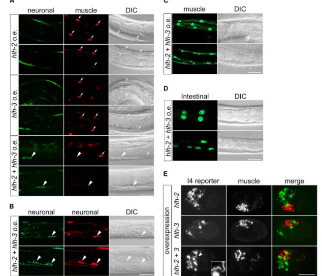
Figure 6. bHLH Overexpression Induces Neuronal Cell-Fate Transformation of Non-neuronal Cells
(A) Confocal images of heat-shocked transgenic animals showing ectopic expression after HLH-2 and HLH-3 coexpression of the neuronal reporter Prab-3::gfp::rab-3 (arrowheads),
but not of the muscle reporter in body-wall muscle nuclei identified by Nomarski and labeled by Pmyo-3::mCherry::H2B in the wildtype (arrows).
(B) HLH-2 and HLH-3 overexpression induces ectopic expression of a second neuronal reporter, Prgef-1::dsRed (red), in addition to Prab-3::gfp::rab-3 (green) in both body-wall
muscle nuclei (upper row, arrowheads) and hypodermal cells (lower row, arrowheads). (C and D) After HLH-2 and HLH-3 co-overexpression, only normal expression patterns are seen for (C) the muscle reporter Pmyo-3::gfp and (D) the intestinal reporter Pelt-2::gfp in
heat-shocked animals.
(E) Co-overexpression of HLH-2 and HLH-3 or HLH-2 alone in early embryos induces formation of multiple cells expressing the I4 reporter but does not eliminate muscle reporter expression. The presence of HLH-3 appears to promote complex neurite formation
(arrowhead and inset). I4 reporter, Pnlp-13::gfp; muscle reporter, Pmyo-2::mCherry::H2B.
Two representative animals are shown for each experiment, except in (E). Scale bars, 20 μm.
A
uthor Man
uscr
ipt
A
uthor Man
uscr
ipt
A
uthor Man
uscr
ipt
A
uthor Man
uscr
ipt
Figure 7. Model
The HLH-2 proneural protein and the CDK-8 Mediator complex kinase module act with the HLH-3 proneural protein to promote I4 neurogenesis. HLH-2 and CDK-8 most likely act by inhibiting the CYH-1/CDK-7 complex and might also act secondarily by phosphorylating serine 10 of histone H3. CYH-1/CDK-7 might negatively regulate I4 neurogenesis by promoting a myogenic program, whereas H3S10 phosphorylation might facilitate neurogenic gene expression.