Publisher’s version / Version de l'éditeur:
Powder Diffraction Journal, 25, 4, pp. 322-328, 2010-12-01
READ THESE TERMS AND CONDITIONS CAREFULLY BEFORE USING THIS WEBSITE.
https://nrc-publications.canada.ca/eng/copyright
Vous avez des questions? Nous pouvons vous aider. Pour communiquer directement avec un auteur, consultez la première page de la revue dans laquelle son article a été publié afin de trouver ses coordonnées. Si vous n’arrivez pas à les repérer, communiquez avec nous à PublicationsArchive-ArchivesPublications@nrc-cnrc.gc.ca.
Questions? Contact the NRC Publications Archive team at
PublicationsArchive-ArchivesPublications@nrc-cnrc.gc.ca. If you wish to email the authors directly, please see the first page of the publication for their contact information.
NRC Publications Archive
Archives des publications du CNRC
This publication could be one of several versions: author’s original, accepted manuscript or the publisher’s version. / La version de cette publication peut être l’une des suivantes : la version prépublication de l’auteur, la version acceptée du manuscrit ou la version de l’éditeur.
For the publisher’s version, please access the DOI link below./ Pour consulter la version de l’éditeur, utilisez le lien DOI ci-dessous.
https://doi.org/10.1154/1.3504496
Access and use of this website and the material on it are subject to the Terms and Conditions set forth at
Crystal structure of the mineral strontiodresserite from laboratory powder diffraction data
Whitfield, P. S.; Mitchell, L. D.; Le Page, Y.; Margeson, J. C.; Roberts, A. C.
https://publications-cnrc.canada.ca/fra/droits
L’accès à ce site Web et l’utilisation de son contenu sont assujettis aux conditions présentées dans le site LISEZ CES CONDITIONS ATTENTIVEMENT AVANT D’UTILISER CE SITE WEB.
NRC Publications Record / Notice d'Archives des publications de CNRC: https://nrc-publications.canada.ca/eng/view/object/?id=04c9e819-8dc9-429a-a02b-6f497bf5198c https://publications-cnrc.canada.ca/fra/voir/objet/?id=04c9e819-8dc9-429a-a02b-6f497bf5198c
http://www.nrc-cnrc.gc.ca/irc
Cryst a l st ruc t ure of t he m ine ra l st ront iodre sse rit e from la bora t ory pow de r diffra c t ion da t a
N R C C - 5 3 5 9 7
W h i t f i e l d , P . S . ; M i t c h e l l , L . D . ; L e P a g e , Y . ; M a r g e s o n , J . ; R o b e r t s , A . C .
D e c e m b e r 2 0 1 0
A version of this document is published in / Une version de ce document se trouve dans:
Powder Diffraction Journal, 25, (4), pp. 322-328, December 01, 2010, DOI:
10.1154/1.3504496
The material in this document is covered by the provisions of the Copyright Act, by Canadian laws, policies, regulations and international agreements. Such provisions serve to identify the information source and, in specific instances, to prohibit reproduction of materials without written permission. For more information visit http://laws.justice.gc.ca/en/showtdm/cs/C-42
Les renseignements dans ce document sont protégés par la Loi sur le droit d'auteur, par les lois, les politiques et les règlements du Canada et des accords internationaux. Ces dispositions permettent d'identifier la source de l'information et, dans certains cas, d'interdire la copie de documents sans permission écrite. Pour obtenir de plus amples renseignements : http://lois.justice.gc.ca/fr/showtdm/cs/C-42
Crystal structure of the mineral strontiodresserite from laboratory powder diffraction data P.S. Whitfield,1,a) L.D. Mitchell,2 Y. Le Page,1 J. Margeson,2 and A.C. Roberts3
1 Institute for Chemical Process and Environmental Technology, National Research Council Canada,
1200 Montreal Road, Ottawa ON, K1A 0R6, CANADA
2 Institute for Research in Construction, National Research Council Canada, 1200 Montreal Road,
Ottawa ON, K1A 0R6, CANADA
3
Geological Survey of Canada, 601 Booth Street, Ottawa ON, K1A 0E8, CANADA
Abstract
The crystal structure of the mineral strontiodresserite, (Sr,Ca)Al2(CO3)2(OH)4·H2O, from the
Francon Quarry, Montreal, Quebec, Canada, has been solved from laboratory powder diffraction data using a combination of charge-flipping and simulated annealing methods. The structure is orthorhombic in space group Pnma with a = 16.0990(7), b = 5.6133(3) and c = 9.1804(4) Å (Z = 4) and the framework of the mineral is isostructural with that of dundasite. The strontium has a coordination number of 9 and the carbonate anions form a bridge between the SrO9 polyhedra
and AlO6 octahedra. The water molecule lies in a channel that runs parallel to the b-axis. An
ordered network of hydrogen atoms could be uniquely determined from crystal-chemical principles in the channels of strontiodresserite. Ab initio DFT energy minimization of the whole structure gave results in full agreement with X-ray refinement results for non-hydrogen atoms. The stability of this model (as well as that of the corresponding model of dundasite) in the proposed Pnma space group was tested by DFT optimization in space group P1 of random small distortions of this structure. This test confirms that both minerals are isostructural, including their hydrogen-bond networks.
I. INTRODUCTION
Strontiodresserite is a hydrated strontium-aluminum carbonate mineral with the formula (Sr,Ca)Al2(CO3)2(OH)4·H2O (Jambor et al, 1977). The type locality is the Francon Quarry, in
Montreal, Quebec, Canada, but strontiodresserite is also reported to occur in France and Norway. It was first described in Jambor et al. (1977), with a Sr:Ca ratio of 4:1. Due to the small grain size, the diffraction data presented by Jambor et al (1977) were limited to powder diffraction data.
Strontiodresserite is compositionally analogous to a small group of minerals which include dundasite PbAl2(CO3)2(OH)4·H2O (Cocco et al, 1972), petterdite PbCr2(CO3)2(OH)4·H2O
(Birch et al, 2000) and dresserite BaAl2(CO3)2(OH)4·H2O (Jambor et al, 1969). However there is
some disagreement in the literature as to whether strontiodresserite is isostructural with dundasite and dresserite. On the basis of the relationship between the elongations for the various directions of the optical axes, Jambor et al (1977) suggested that dundasite, dresserite and strontiodresserite are not isostructural. Roberts (1978) used long exposure X-ray precession single-crystal studies on two fibres of strontiodresserite to determine its space group. The systematic absences were consistent with either Pbnm or Pbn21 and, given the parallels reported by Cocco et al (1972),
Roberts (1978) concluded that strontiodresserite and dundasite are indeed isostructural.
The fine aggregate microstructure of these minerals makes single-crystal structure determination very difficult. The only published crystal structure amongst the group is for dundasite, where the researchers used Weissenberg photographs of a multiple crystal (Cocco et
al, 1972). Despite the problematic sample, Cocco et al (1972) successfully solved the structure
Structure determination from powder diffraction data (SDPD) has developed significantly in recent years. Improvements in global optimization using real-space methods (Shankland and David, 2002), and more recently the application of the tangent formula (Coelho, 2007) to charge-flipping methods (Oszlányi and Süto, 2004) has greatly expanded the scope of SDPD. Together with improvements in data quality facilitated by developments in diffractometer instrumentation, SDPD has become accessible to a greater number of researchers. However, the results of a recent round-robin suggest that it is still not a routine procedure (Le Bail and Cranswick, 2008).
II. EXPERIMENTAL AND DATA ANALYSIS A. Sample preparation
The mineral of interest was presented as a number of 1-mm diameter, white, vitreous hemispheres (Figure 1) formed within vugs of a 10-cm sized piece of bulk silicocarbonatite sill rock from the Francon Quarry in Montreal, Quebec, Canada. Hemispheres were removed from the bulk under an optical microscope and an attempt was made to clean their surface of impurities. Of the six hemispheres obtained, four were gently crushed in ethanol using a mortar and pestle for X-ray diffraction analysis. Once dried, the powder was loaded into a 0.5-mm quartz capillary and the capillary sealed with wax before mounting in a standard brass capillary pin. One of the remaining hemispheres was lightly crushed and mounted onto a scanning electron microscope (SEM) stub using adhesive carbon film. The sample was examined in a Hitachi S4800 field emission SEM. Images and elemental analysis were obtained at 5- and 20-kV accelerating voltage, respectively.
The X-ray data were collected using a Bruker-AXS D8 diffractometer using Cu Kα radiation with a focussing primary mirror and a Vantec PSD detector. Although the diffractometer was a θ−θ instrument, data were collected in the range from 9 to 140 °2θ with a fixed incident angle (Debye-Scherrer geometry) using a 10° detector window. A variable count/step scheme was employed to improve counting statistics at high angle and produce more accurate results in the final Rietveld refinement (Madsen and Hill, 1994). The data collection scheme is given in Table I.
The data ranges were combined into a single file in 'xye' format to retain the counting statistics information. All of the data analysis was done using the Bruker-AXS TOPAS 4.2 software (Bruker-AXS, 2008b). The Least-Squares Indexing method (Coelho, 2003) was used to index the data from the first 25 peak positions determined through single-peak fitting. No background subtraction was necessary (including the capillary) to fit the background satisfactorily using a 15-term Chebyschev polynomial. This contrasts with the slight diffuseness in the powder diffraction data observed by Jambor et al (1977) which they attributed to possible non-stoichiometry in carbonate and water content due to absorption from air.
C. Structure solution and refinement
The ability to extract intensities out to low d-spacings and the presence of a heavy atom (Sr) were positive indications that charge-flipping (Oszlányi and Süto, 2004) might yield some of the atomic positions. The initial structure solution was accordingly attempted with a combination of charge-flipping method and tangent formula (Coelho, 2007). Low-density elimination (Shiono and Woolfson, 1992) was used to sharpen the resulting electron density.
Charge-flipping produced sensible positions for the strontium, aluminum and some oxygen atoms. Use of fixed positions for strontium and aluminum atoms in subsequent simulated annealing runs with data up to 80 °2θ greatly eased the difficulty of extracting a successful solution.
Rigid bodies consisting of regular AlO6 octahedra and CO3 triangles were used in
conjunction with a 'box interaction' penalty function such that at least eight oxygen atoms should be within 2.65 Å of a strontium atom. This model with six oxygen atoms around each aluminum and three around each carbon therefore contained initially 18 oxygen atoms/f.u., compared with 10 or 11 in the chemical formula. Merging of oxygen atoms approaching one another within 0.9 Å was accordingly implemented. Separate isotropic displacement parameters (Biso) were used
for the cations and anions. The oxygen atom for a water molecule was located on a Fourier difference map. The final refinement step involved all variable atom positions as well as individual Biso.
The final refinement used the full data range up to 140 °2θ. The peak profiles were fitted using convolution-based peak profiles in combination with double-Voigt sample size-strain model (Balzar and Ledbetter, 1995). The simple axial divergence correction implemented in TOPAS (Bruker-AXS, 2008a) was used. Individual Biso values were refined for the strontium,
aluminum, carbon and oxygen atoms, except for the equatorial AlO6 oxygen atoms which were
refined with an overall Biso. The Sabine capillary absorption correction (Sabine et al, 1998) was
applied to the intensities. The linear absorption coefficient used in the correction included the contribution from the extraneous fluorite content. The wide 2θ range indicated that the application of the Debye-Scherrer capillary displacement 2θ correction (McCusker et al, 1999) might be useful and was included in the input file as a user-defined macro.
D. Valence summation and location of hydrogen atoms
Bond valence sums were calculated using VaList (Wills, 2008). The bond-valence parameter for the mixed Sr-Ca site (2.088) was determined by applying percentage contribution from the refined site occupancies for Sr and Ca. A value of 0.37 was assumed for B, which is common to the bond-valence entries for Ca and Sr used as end-members. Bond valence parameters for OH were taken from the 2006 version of bond-valence parameters maintained by I.D. Brown (Brown, 2006a). No hydrogen atoms were included in the X-ray refinement of the data. A unique model for H positions in space group Pnma was then derived from crystal-chemical considerations as detailed in III.A.
E. Modeling, DFT energy minimization and structure stability
A crystal structure model combining the X-ray atom coordinates and the above hydrogen atoms was prepared and optimized by ab initio total energy minimization using the Vienna Ab
initio Simulation Package (VASP) (Kresse, 1993; Kresse & Hafner, 1993) within the framework
of Density Functional Theory (DFT). Full occupancy by Sr of the (Sr,Ca) site was assumed. All modeling, VASP input file preparation as well as output file interpretation was performed with Materials Toolkit (Le Page and Rodgers, 2005). The energy minimization scheme detailed in Mercier & Le Page (2008) was followed with a 4×4×4 k-mesh. Distorted models for the structure-stability test were prepared with the NUDGE utility (Le Page & Rodgers, 2006) in Materials Toolkit and optimized with VASP.
III. RESULTS AND DISCUSSION A. The sample and its analysis
The sample under examination had been macroscopically identified as montroyalite, Sr4Al8[(OH),F]26(CO3)3·10-11H2O (Roberts et al, 1986), a mineral also found in the Francon
Quarry that typically occurs as small, white hemispheres (similar to strontiodresserite) and, coincidentally has the same Sr:Al ratio as strontiodresserite. The hemispherical nature of the material in question is apparent in the optical micrograph shown in Figure 1. The identity of the mineral was called into question when the powder diffraction was examined as the data did not resemble that expected from montroyalite (Roberts et al, 1986). The possibility of a dehydration product of montroyalite was considered, but both the position and relative intensities of the reflections closely resembled those reported in the literature for strontiodresserite. Consequently the structure solution process was started on the assumption that it was, in fact, strontiodresserite. A small number of reflections from fluorite were visible and these were fitted as a secondary phase during the complete analysis process. Fluorite is not mentioned as a phase associated with strontiodresserite (Jambor et al, 1977), but is associated with the fluorine-containing montroyalite (Roberts et al, 1986). None of the closely associated minerals mentioned by Jambor et al (1977), such as quartz and dawsonite, were present in the diffraction pattern.
Examination of the material by SEM yielded heavily fractured lath-like needles as shown in Figure 2. Lath-like crystals are consistent both with the observations by Jambor et al (1977) on strontiodresserite and those of Roberts et al (1986) on montroyalite including the 'splintery fracture'. EDX measurements yielded a Sr:Al atomic ratio of approximately 1:2. No signal from fluorine was detected either at 20 kV or when the voltage was lowered to optimize the detection
of lighter elements, which is additional evidence that the sample was strontiodresserite as opposed to montroyalite.
B. Unit-cell analysis
The crystallinity of strontiodresserite was found to be quite good with distinct reflections visible all the way up to the limit of 140 °2θ as can be seen in the insert in Figure 3. Single peak fitting to obtain the first 25 to 30 reflections was straightforward, and 25 of those were used as input in the LSI indexing algorithm. Using a calculated lines to observed lines ratio of 5, the first 70 solutions from the indexing in TOPAS gave the same orthorhombic lattice parameters with a = 16.1, b = 9.2, c = 5.6 Å, giving a very high confidence in the result. The first 4 solutions of the top 5 had the suggested space group Pna21 corresponding to the extinction symbol Pna-. Besides
Pna21, the space group Pnam also belongs to the extinction symbol Pna- (Looijenga-Vos and
Buerger, 2006), yielding effectively the same choice as that presented by Roberts (1978). The top solution had a GOF of 28.1 with a minimal zero point error. A Le Bail full pattern fit (Le Bail et al, 1988) to the data range from 9 to 80 °2θ, including the fluorite secondary phase, yielded good fits in both Pna21 and Pnam (Rwp = 5.0%). Pnam and Pbnm are alternate settings
of the standard space group Pnma (number 62).
C. Structure analysis
The first cycles of charge-flipping yielded the strontium atom positions. Following cycles gave aluminum atom positions. The positions determined from the charge-flipping accounted for the cell-symmetry, and identified the special position of the strontium atoms. Fixing the positions of the strontium and aluminum atoms in the simulated annealing assisted in finding the
other atom positions relatively quickly. The oxygen atom of the water molecule was not subjected to any constraints so took a little longer to locate reliably. The rigid-body octahedra used in the simulated annealing led to excess of oxygen atoms compared to the nominal stoichiometry. The partially merged oxygen atoms accordingly had to be consolidated in single sites for refinement upon removing the rigid-body constraints, leading to the expected stoichiometry.
A 4th-order spherical harmonics preferential orientation correction (Järvinen, 1993) was necessary to finalize the refinement. The application of this correction did not alter the overall structure, but greatly improved the geometry of the carbonate ions and significantly improved the fit. The refined Biso of the Sr appeared to be sensitive to the preferential orientation correction,
and the value increased after the application of the spherical harmonics. Orientation of a powder in a capillary is suggestive of needle or lath-shaped crystallites, which has been reported previously with strontiodresserite (Jambor et al, 1977) and can be seen in Figure 2.
Although a Fourier difference map confirmed the location of the water oxygen atom, no additional water molecules were found and none of the hydrogen atoms could reliably be located by X-rays. As expected, the capillary displacement was minimal (x = +0.03 mm and y = +0.04 mm) but did improve the Rwp residual slightly.
The final refinement was carried out in the standard setting for space group number 62, which is Pnma. The cell and structure can be transformed to the Pbnm cell by the application of a cab matrix transformation for comparison with the previously described structure of dundasite (Cocco et al. 1972). The final refined lattice parameters in Pnma were a = 16.0990(7), b = 5.6133(3) and c = 9.1804(4) Å, with Z = 4. The fit to the experimental data was very good as shown in Figure 3, with a Rwp value of 5.2%. The density of strontiodresserite calculated with
those cell parameters is 2.66 g/cm3, which compares quite well with the measured value of 2.71 g/cm3 (Jambor et al, 1977) given that the refined Ca-content on the Sr site is very slightly lower
at 19.3% than the nominal value of 20%.
D. Structure description
The structure consists of corrugated chains extending along the b-axis. Those chains are made of edge-sharing AlO6 octahedra, as shown in Figure 4. Four coplanar oxygen atoms are
shared in this way in an equatorial plane of each Al atom, while two axial oxygen atoms lie perpendicular to that plane. Next to each of the AlO6 chains, the strontium atom shares edges
with two AlO6 octahedra. In the b-direction the irregular-shaped SrO9 polyhedra alternate with
the carbonate ions which are corner sharing with two axial AlO6 oxygen atoms and share an
oxygen atom with the opposite SrO9. The carbonate ions effectively tie together the two Al/Sr/O
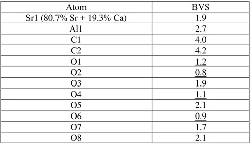
layers in the unit cell. The water molecules are located in channels running along the b-direction and are not coordinated directly with any of the cations. The position of the oxygen atom does suggest the possibility of hydrogen bonding with oxygen atoms in the polyhedral network. Bond valence analysis confirmed that the hydroxyl oxygen atoms are O1, O2, O4 and O6, which are the equatorial oxygen atoms of the AlO6 octahedra. Table II lists the atomic coordinates from
Rietveld refinement of non-hydrogen atoms in space group Pnma. Table III shows the results of bond valence summation using atom coordinates in Table II.
The fact that O9w could be refined to a Biso of 2.0(2) with full oxygen occupancy is an
indication that water molecules are strongly bound with just one scheme of hydrogen bonds and then that an ordered hydrogen-bond scheme most probably exists in strontiodresserite. Examination of experimental O-O approaches for O9w indicates that only three oxygen atoms, O3 (×2) and O6 at respective distances 2.92 and 2.68 Å, can possibly be involved in hydrogen bonding with O9w. The next shortest distance of 3.19 Å is too long to be part of a significant hydrogen bond (see e.g. Fig. 1 in Ferraris & Ivaldi, 1988). From bond valence summation, O6 is known to be an OH group, thus indicating an O6-H6..O9w hydrogen bond. The two short approaches between O9w and O3 are then the two O9w-H9..O3 hydrogen bonds of the water molecule. The remaining OH groups at O1, O2 and O4 are all bonded to two Al3+ ions. Simple electrostatic field considerations then suggest an initial location of corresponding hydroxyl hydrogen atoms on the line joining the midpoint between the two Al3+ ions and the oxygen atom,
away from the aluminum ions. Qualitatively, this hydrogen-bond scheme is unique in space group Pnma. Initial O-H distances were assumed to be 1 Å.
F. DFT energy minimization of strontiodresserite and dundasite
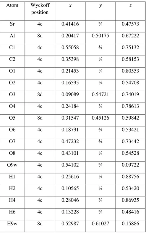
DFT energy minimization by adjustment of atom coordinates of this model in the fixed experimental cell proceeded smoothly, without any large drift of the proposed hydrogen atoms or of the framework atoms, a strong indication of a sensible initial model. Table IV lists all atom coordinates, including hydrogen atoms after DFT optimization with VASP in space group Pnma. Selected bond distances are shown in two columns in Table V for easy comparison of X-ray and
ab initio VASP results. Bond angles from ab initio VASP results pertaining to the hydrogen
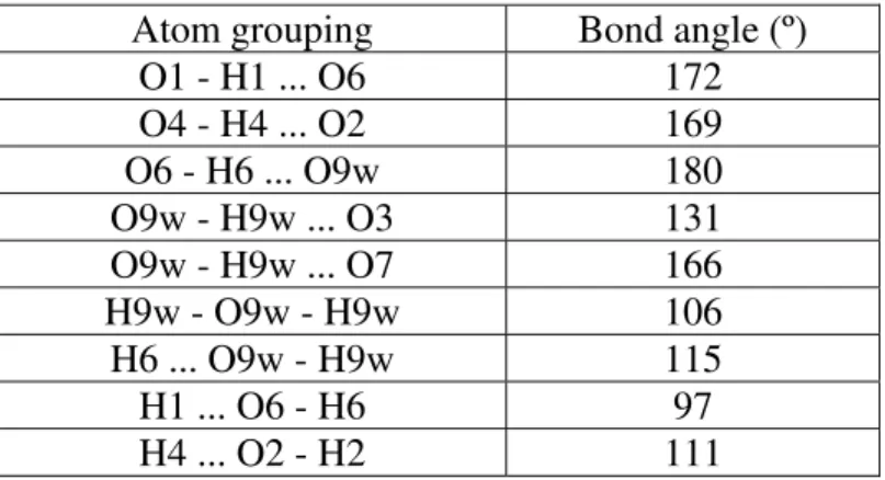
graphically in Figure 5 for easy interpretation of Tables IV, V and VI. There are no large discrepancies from expected values including all optimized O-H distances of about 1 Å. As expected the agreement between experimental and DFT bonds lengths tends to be better with the heavier elements as they are more easily localized with X-ray powder diffraction data. From Table VI, the H-O-H angle in the water molecule as well as H..O-H angles are all reasonably close to tetrahedral angles. The only deviation from an ideal model for a textbook seems to be that the O9w-H9w..O3 angle deviates from 180 degrees. We consider this to be a minor imperfection because the H9w..O3 hydrogen bond is rather weak. H9w is accordingly pointing to an electrostatically negatively charged region on the wall of a wide channel rather than establishing a strong hydrogen bond with O3, which ought to be in straight line. We tested the energy of plausible disordered hydrogen-bond models at O9w, with lower symmetry. All those that did not revert to the above scheme failed to reduce the total energy and yielded atomic coordinates for O9w that differed from those found experimentally.
G. Space-group choice for strontiodresserite and dundasite
The success of the structure solution in a particular space group from diffraction intensities, as well as its DFT optimization in that space group, does not prove that the correct structure is not actually distorted with a lower symmetry. We accordingly tested the ab initio stability of the final tabulated crystal structure of strontiodresserite by creating models of it in
P1, introducing random tiny individual distortions into all atom coordinates and all cell vectors.
Total energy minimization will promptly return this randomly distorted crystal structure to its initial configuration and symmetry if it is stable. In contrast, an unstable model will converge to a new configuration with lower energy and lower symmetry. The test performed with the
NUDGE utility (Le Page & Rodgers, 2006) on the strontiodresserite P1 model confirmed that the
Pnma model of strontiodresserite is stable and lowering the symmetry is not necessary. The
same test, repeated with the literature structure of dundasite combined with hydrogen atoms from strontiodresserite confirmed the structure and symmetry of dundasite as reported by Cocco et al (1972).
H. Complementarity of powder methods, valence summation and DFT optimization
This study illustrates the fact that the combination of laboratory X-ray Rietveld profile analysis with valence summation and ab initio methods can in a number of cases reliably elucidate crystal structures of a complexity that used to require X-ray single-crystal diffraction. As shown here, this possibility opens new perspectives with samples that can be obtained in powder form only. For oxides, bond-valence summation (Brown, 2006b) provides a powerful check of the validity of the crystal structure, but ab initio optimization provides several additional and very stringent checks about the correctness of the structure model. The first such check is that stress calculated ab initio using experimental cell parameters was verified here to be small and uniform. With VASP, this stress is usually calculated as a small negative and essentially isotropic pressure, in part because ab initio calculations are performed at 0 K temperature, but also due to tiny imperfections in the numerical representation of quantum-calculated core electron density. A second test is that quantum-calculated optimized atom coordinates for atoms should actually match the experimentally determined atom positions. This test is particularly powerful in cases like here, where some atoms were located experimentally while others are deduced from crystal-chemical considerations: a wrong assignment of hydrogen atoms would have repercussions on hydroxyl and water oxygen positions. Verification of the match of
coordinates within least squares uncertainties then validates both the framework architecture and the hydrogen-bond scheme deduced from it. The third test is that of the stability of the optimized structure model as explained previously. This test validated here the use of space group Pnma for the structure solution, demonstrating that a lower symmetry was not required.
IV. CONCLUSION
The structure of strontiodresserite has been determined from laboratory powder diffraction data using a combination of charge-flipping and simulated annealing techniques. The crystal structure is orthorhombic and has been described in Pnma, with two chains of AlO6
octahedra along the b-direction linked by irregular SrO9 polyhedra and carbonate ions. The
water molecule lies in a channel that runs parallel to the b-axis. This study confirms the isostructural relationship between strontiodresserite and dundasite proposed by Roberts (1978). The hydroxyl and water oxygens were identified using bond valence sums. A unique model of hydrogen atoms was then deduced from crystal chemistry and optimized by ab initio DFT energy minimization. The stability of the resulting Pnma strontiodresserite structure and that of the corresponding dundasite structure were then demonstrated using ab initio methods. Strontiodresserite and dundasite are isostructural in space group Pnma, basically as described in Cocco et al (1972) albeit in Pbnm, including their hydrogen-bond network established here.
References
Balzar, D. and Ledbetter, H. (1995). "Accurate modelling of size and strain broadening in the Rietveld refinement: The "double-Voigt" approach," Advances in X-Ray Analysis 38, 397-404.
Birch, W. D., Kolitsch, U., Witzke, T., Nasdala, L. and Bottrill, R. S. (2000). "Petterdite, the Cr-dominant analogue of dundasite, a new mineral species from Dundas, Tasmania,
Australia and Callenberg, Saxony, Germany," Can. Mineral. 38, 1467-1476. Brown,I.D. (2006a). bvparm2006.cif. Available from www.ccp14.ac.uk.
Brown, I. D. (2006b). The Chemical Bond in Inorganic Chemistry. The Bond Valence Model, (Oxford: Oxford University Press; IUCr Monographs on Crystallography).
Bruker-AXS (2008a) DIFFRACPlus TOPAS: TOPAS 4.2 Technical Reference. (Computer Software) Bruker-AXS GmbH, Karlsruhe, Germany
Bruker-AXS (2008b) DIFFRACPlus TOPAS: TOPAS 4.2 User Manual. (Computer Software) Bruker-AXS GmbH, Karlsruhe, Germany
Cocco, G., Fanfini, L., Nunzi, A. and Zanazzi, P. F. (1972). "The crystal structure of dundasite," Mineral. Mag. 38, 564-569.
Coelho, A. A. (2003). "Indexing of powder diffraction patterns by iterative use of singular value decomposition," J. Appl. Crystallogr. 36, 86-95.
Coelho, A. A. (2007). "A Charge Flipping algorithm incorporating the tangent formula for solving difficult structures," Acta Crystallogr., Sect A: Found. Crystallogr. 36, 400-406. Ferraris, G. and Ivaldi, G. (1988). "Bond valence vs bond length in O...O hydrogen bonds,"
Jambor, J. L., Fong, D. G. and Sabina, A. P. (1969). "Dresserite, the new barium analogue of dundasite," Can. Mineral. 10, 84-89.
Jambor, J. L., Sabina, A. P., Roberts, A. C. and Sturman, B. D. (1977). "Strontiodresserite, a new Sr-Al carbonate from Montreal Island, Quebec," Can. Mineral. 15, 405-407. Järvinen, M. (1993). "Application of symmetrized harmonics expansion to correction of the
preferred orientation effect," J. Appl. Crystallogr. 24, 525-531. Kresse, G. (1993) Ph.D. Thesis. Technische Universität Wien, Austria.
Kresse, G. and Hafner, J. (1993). “Ab initio molecular dynamics for open-shell transition metals,” Phys. Rev. B 48, 13115-13118.
Le Bail, A. and Cranswick, L. M. D. (2008). SDPDRR-3: Structure determination by powder diffractometry round robin - 3. http://cristal.org/SDPDRR3/results/index.html . Le Bail, A., Duroy, H. and Fourquet, J. L. (1988). "Ab-initio structure determination of
LiSbWO6 by X-ray powder diffraction," Mat. Res. Bull. 23, 447-452.
Le Page, Y. and Rodgers, J. R. (2005). "Quantum software interfaced with crystal structure databases: tools, results and perspectives," J. Appl. Crystallogr. 38, 697-705.
Le Page, Y. & Rodgers, J.R. (2006). “Low energy models from scratch: Application to SiNF,” Comp. Mat. Sci. 37, 537-542.
Looijenga-Vos, A. and Buerger, M. J. (2006). "Space-group determination and diffraction symbols," in International Tables for Crystallography, volume A, edited by Th. Hahn, (International Union for Crystallography, Chester, UK) p. 46
Madsen, I.C. & Hill, R.J. (1994). "Collection and analysis of powder diffraction data with near-constant counting statistics," J. Appl. Crystallogr. 27, 385-392
McCusker, L. B., Von Dreele, R. B., Cox, D. E., Louer, D. and Scardi, P. (1999). "Rietveld refinement guidelines," J. Appl. Crystallogr. 32, 36-50.
Mercier, P. H. J. & Le Page, Y. (2008). “Kaolin polytypes revisited ab initio,” Acta Crystallogr., Section B: Struct. Sci. 64, 131-143.
Oszlányi, G. and Süto, A. (2004). "Ab initio structure solution by charge flipping," Acta Crystallogr., Sect. A: Found. Cryst. 60, 134-141.
Roberts, A.C. (1978). "The space group of strontiodresserite," Scientific and Technical Notes in Current Research, Part B; Geological Survey of Canada Paper 78-1B, 180
Roberts, A. C., Sabina, A. P., Bonardi, M., Jambor, J. L., Ramik, R. A., Sturman, B. D. and Carr, M. J. (1986). "Montroyalite, a new hydrated Sr-Al hydroxycarbonate from the Francon Quarry, Montreal, Quebec," Can. Mineral. 24, 455-459.
Sabine, T. M., Hunter, B. A., Sabine, W. R. and Ball, C. J. (1998). "Analytical expressions for the transmission factor and peak shift in absorbing cylindrical specimens," J. Appl. Crystallogr. 31, 47-51.
Shankland, K. and David, W. I. F. (2002) "Global optimization strategies" in Structure
Determination from Powder Diffraction Data, edited by W.I.F David, K. Shankland, L.B.
McCusker, and Ch. Baerlocher, (Oxford University Press, Oxford, UK) p. 252-285. Shiono, M. and Woolfson, M. M. (1992). "Direct-space methods in phase extension and phase
determination. 1. Low-density elimination," Acta Crystallogr., Sect. A: Found. Crystallogr. 48, 451-456.
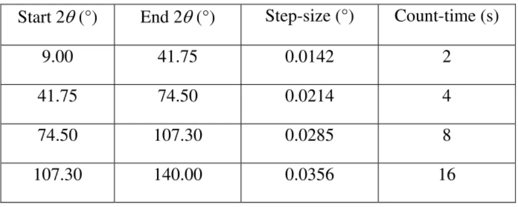
Table I. Variable count-time (VCT) data collection regime.
Start 2θ (°) End 2θ (°) Step-size (°) Count-time (s)
9.00 41.75 0.0142 2
41.75 74.50 0.0214 4
74.50 107.30 0.0285 8
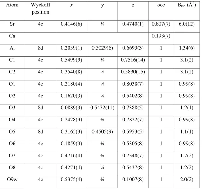
Table II. Refined experimental atomic coordinates for strontiodresserite. Atom Wyckoff position x y z occ Biso (Å2) Sr 4c 0.4146(6) ¾ 0.4740(1) 0.807(7) 6.0(12) Ca 0.193(7) Al 8d 0.2039(1) 0.5029(6) 0.6693(3) 1 1.34(6) C1 4c 0.5499(9) ¾ 0.7516(14) 1 3.1(2) C2 4c 0.3540(8) ¼ 0.5830(15) 1 3.1(2) O1 4c 0.2180(4) ¼ 0.8038(7) 1 0.99(8) O2 4c 0.1620(3) ¼ 0.5402(8) 1 0.99(8) O3 8d 0.0889(3) 0.5472(11) 0.7388(5) 1 1.2(1) O4 4c 0.2428(3) ¾ 0.7822(7) 1 0.99(8) O5 8d 0.3165(3) 0.4505(9) 0.5953(5) 1 1.1(1) O6 4c 0.1859(3) ¾ 0.5305(8) 1 0.99(8) O7 4c 0.4716(4) ¾ 0.7348(7) 1 1.7(2) O8 4c 0.4271(4) ¼ 0.5437(8) 1 1.2(2) O9w 4c 0.5375(4) ¾ 0.1007(8) 1 2.0(2)
Table III. Bond valence sums for the strontiodresserite structure. The underlined oxygen atoms indicate the hydroxide oxygens.
Atom BVS Sr1 (80.7% Sr + 19.3% Ca) 1.9 Al1 2.7 C1 4.0 C2 4.2 O1 1.2 O2 0.8 O3 1.9 O4 1.1 O5 2.1 O6 0.9 O7 1.7 O8 2.1
Table IV. Ab initio optimized atomic coordinates for strontiodresserite. Atom Wyckoff position x y z Sr 4c 0.41416 ¾ 0.47573 Al 8d 0.20417 0.50175 0.67222 C1 4c 0.55058 ¾ 0.75132 C2 4c 0.35398 ¼ 0.58153 O1 4c 0.21453 ¼ 0.80553 O2 4c 0.16595 ¼ 0.54708 O3 8d 0.09089 0.54721 0.74019 O4 4c 0.24184 ¾ 0.78613 O5 8d 0.31547 0.45126 0.59842 O6 4c 0.18791 ¾ 0.53421 O7 4c 0.47232 ¾ 0.73442 O8 4c 0.43101 ¼ 0.54528 O9w 4c 0.54102 ¾ 0.09722 H1 4c 0.25616 ¼ 0.88756 H2 4c 0.10565 ¼ 0.53420 H4 4c 0.28046 ¾ 0.86935 H6 4c 0.13228 ¾ 0.48416 H9w 8d 0.52987 0.61027 0.15886
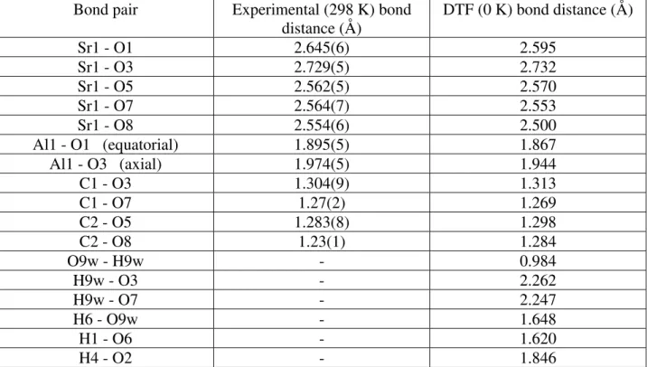
Table V. Selected bond distances derived from the refinement of the experimental data and the DFT-optimized structure.
Bond pair Experimental (298 K) bond
distance (Å) DTF (0 K) bond distance (Å) Sr1 - O1 2.645(6) 2.595 Sr1 - O3 2.729(5) 2.732 Sr1 - O5 2.562(5) 2.570 Sr1 - O7 2.564(7) 2.553 Sr1 - O8 2.554(6) 2.500 Al1 - O1 (equatorial) 1.895(5) 1.867 Al1 - O3 (axial) 1.974(5) 1.944 C1 - O3 1.304(9) 1.313 C1 - O7 1.27(2) 1.269 C2 - O5 1.283(8) 1.298 C2 - O8 1.23(1) 1.284 O9w - H9w - 0.984 H9w - O3 - 2.262 H9w - O7 - 2.247 H6 - O9w - 1.648 H1 - O6 - 1.620 H4 - O2 - 1.846
Table VI. Selected bond angles from the DTF-optimized structure.
Atom grouping Bond angle (º)
O1 - H1 ... O6 172 O4 - H4 ... O2 169 O6 - H6 ... O9w 180 O9w - H9w ... O3 131 O9w - H9w ... O7 166 H9w - O9w - H9w 106 H6 ... O9w - H9w 115 H1 ... O6 - H6 97 H4 ... O2 - H2 111
Figure captions
Figure 1. Optical image of strontiodresserite hemispheres embedded in the bulk silicocarbonatite sill rock. The hemispheres are approximately 1mm in diameter.
Figure 2. A SEM micrograph of a crushed strontiodresserite hemisphere.
Figure 3. Rietveld difference plot for the refinement of the strontiodresserite structure. The inset shows a blowup of the region between 100 and 140 °2θ.
Figure 4. Polyhedral representation of the experimental strontiodresserite crystal structure. Figure 5. The hydrogen bonding network in strontiodresserite.