0095-1137/05/$08.00⫹0 doi:10.1128/JCM.43.12.6098–6107.2005
Copyright © 2005, American Society for Microbiology. All Rights Reserved.
Association of Virulence Genotype with Phylogenetic Background in
Comparison to Different Seropathotypes of Shiga Toxin-Producing
Escherichia coli Isolates
Jean Pierre Girardeau,
1* Alessandra Dalmasso,
1† Yolande Bertin,
1Christian Ducrot,
2Se
´verine Bord,
2Vale
´rie Livrelli,
3Christine Vernozy-Rozand,
4and Christine Martin
1Unite´ de Microbiologie, Centre de Recherche INRA de Clermont-Ferrand-Theix, 63122 Saint-Genes Champanelle, France1; Groupe de Recherche Pathoge´nie Bacte´rienne Intestinale, Faculte´ de Pharmacie, Universite´ d’Auvergne Clermont 1,
63001 Clermont-Ferrand Cedex, France3; Unite´ d’Epide´miologie Animale, INRA, Centre de Recherche INRA de Clermont-Ferrand-Theix, 63122 Saint Gene`s Champanelle, France2; and Unite´ de Microbiologie
Alimentaire et Pre´visionnelle, Ecole Nationale Ve´te´rinaire de Lyon, Marcy l’Etoile, France4 Received 26 May 2005/Returned for modification 1 August 2005/Accepted 7 September 2005
The distribution of virulent factors (VFs) in 287 Shiga toxin-producing Escherichia coli (STEC) strains that were classified according to Karmali et al. into five seropathotypes (M. A. Karmali, M. Mascarenhas, S. Shen, K. Ziebell, S. Johnson, R. Reid-Smith, J. Isaac-Renton, C. Clark, K. Rahn, and J. B. Kaper, J. Clin. Microbiol. 41:4930–4940, 2003) was investigated. The associations of VFs with phylogenetic background were assessed among the strains in comparison with the different seropathotypes. The phylogenetic analysis showed that STEC strains segregated mainly in phylogenetic group B1 (70%) and revealed the substantial prevalence (19%) of STEC belonging to phylogenetic group A (designated STEC-A). The presence of virulent clonal groups in seropathotypes that are associated with disease and their absence from seropathotypes that are not associated with disease support the concept of seropathotype classification. Although certain VFs (eae, stx2-EDL933, stx2-vha, and stx2-vhb) were concentrated in seropathotypes associated with disease, others (astA, HPI, stx1c, and
stx2-NV206) were concentrated in seropathotypes that are not associated with disease. Taken together with the
observation that the STEC-A group was exclusively composed of strains lacking eae recovered from seropatho-types that are not associated with disease, the “atypical” virulence pattern suggests that STEC-A strains comprise a distinct category of STEC strains. A practical benefit of our phylogenetic analysis of STEC strains is that phylogenetic group A status appears to be highly predictive of “nonvirulent” seropathotypes.
Shiga toxin-producing Escherichia coli (STEC) causes a spectrum of human illness, including hemorrhagic colitis (HC) and hemolytic-uremic syndrome (HUS) (21, 24, 29, 30). STEC isolates that cause human infections belong to a large number of O:H serotypes. E. coli O157:H7 is the most prevalent sero-type associated with large outbreaks and sporadic cases of HC and HUS in many countries (24, 29, 30). The O157:H7 strains harbor a large pathogenicity island (PAI) termed the locus for enterocyte effacement (LEE) encoding the different determi-nants necessary for the development of the characteristic attaching-and-effacing lesion on enterocytes (12, 20, 29). LEE-positive serotypes are commonly referred to as entero-hemorrhagic E. coli (EHEC) (19, 22). The LEE seems to confer enhanced virulence since LEE-positive STEC are more commonly associated with outbreaks and HUS than LEE-neg-ative serotypes (5, 19). However, LEE-negLEE-neg-ative STEC strains are also associated with severe human disease (17, 29), and some serotypes of LEE-positive STEC isolated from cattle have never been associated with human disease (50). These observations suggest that other unknown factors, possibly PAIs
or genomic islands, may enhance the virulence potential of STEC strains (19, 24, 31).
Shiga toxins (Stx1 and Stx2) are the most critical virulence factors responsible for the principal manifestations of HUS and HC (11, 26, 30, 41). STEC isolates producing Stx2 are more commonly associated with severe disease (e.g., HUS) compared to isolates producing Stx1 alone or Stx1 and Stx2 (5, 27, 41, 42). Although only three stx1alleles were described, stx2
comprises at least 11 distinct subtypes (8, 30). Recently, we described in bovine STEC strains a new stx2 subtype (stx
2-NV206) showing a high cytotoxicity for Vero cells (2). In
addi-tion, several other virulence factors, including adhesins and plasmid-encoded virulence factors, contribute to the pathoge-nicity of STEC strains (12, 19, 24). Potential virulence genes such as the enteroaggregative E. coli heat-stable enterotoxin (EAST1) gene (astA) associated with diarrheogenic E. coli strains (3, 45, 51, 52) and PAIs such as the high pathogenicity island (HPI) of Yersinia spp. were also detected in STEC strains (2, 20).
A large variety of STEC serotypes have been implicated in disease. However, certain STEC serotypes recovered from an-imals and food have never been associated with severe human disease. For a better understanding of the apparent differences in virulence between groups of STEC serotypes, STEC strains were classified into five seropathotypes (A to E) by Karmali et al. (22), according to incidence and association with HUS and outbreaks. Recent studies have demonstrated that the
deter-* Corresponding author. Mailing address: Institut National de cherches Agronomiques, Laboratoire de Microbiologie, Centre de Re-cherche INRA de Clermont Ferrand Theix, 63122 Saint-Gene`s Cham-panelle, France. Phone: (33) 4 73 62 42 42. Fax: (33) 4 73 63 45 81. E-mail: girard@clermont.inra.fr.
† Present address: Facolta` di Medicina Ve´te´rinaria Universita` degli Studi di Torino, Turin, Italy.
mination of the seropathotype distribution of virulent factors (VFs) allows identification of DNA targets for selective detec-tion of strains that present a risk to public health (22, 43). Such an approach has highlighted the association between the genomic island EDL933 OI-122 and seropathotypes linked to epidemic and/or severe disease (22).
To study their evolutionary relationship, different authors (7, 13, 37, 49) have studied the clonal relationship of STEC strains. Based on multilocus enzyme electrophoresis analysis and multilocus sequence typing, Whittam and coworkers have studied the clonal relationships of STEC strains (STEC Ref-erence Center [http://www.shigatox.net/stec/index.html]). Four clonal groups have been identified: EHEC 1, EHEC 2, STEC 1, and STEC 2 (dendrograms showing these clonal groups may be viewed at the STEC Reference Center Web site).
A strategy for investigating the evolutionary origins of pathogenic E. coli is to determine the phylogenetic distribution of the virulence determinants (14, 33, 37). Phylogenetic anal-yses have shown that most E. coli strains belonged to four main phylogenetic groups, A, B1, B2, and D (25, 37). Whereas most commensal and diarrheogenic strains belong to groups A and B1, extraintestinal E. coli strains belong mainly to group B2 and group D (14). Recent phylogenetic studies indicated that STEC and EHEC strains fell into phylogenetic groups A, B1, and D (4, 13, 14). However, there was a paucity of information regarding the phylogenetic distribution of the virulence factors of STEC strains.
In the present study, the distribution of selected VFs (stx1
and stx2 subtypes, eae, astA, and HPI) in a collection of 287
well-defined STEC isolates was determined and analyzed. The first aim of the present study was to analyze the distribution of VFs among isolates classified by seropathotypes. The second aim was to examine the phylogenetic structure of the different seropathotypes. Finally, we sought to analyze the association between VFs and the genetic background of the strains based on classification by phylogenetic and clonal groups.
MATERIALS AND METHODS
Bacterial strains.A total of 287 STEC isolates classified into four different sets were used in the present study. The first set includes 172 bovine isolates which are part of a well-characterized bacterial collection obtained during a 1-year prospective study in the same geographic area in France (6, 36). The bovine strains belonged to 74 serotypes. The second set comprised 50 STEC strains originating from food samples collected in France (36). The third set is com-posed of 11 environmental isolates collected in France from dairy cattle herd manure, a wastewater treatment plant, and pig farm manure (46). The fourth set, including 54 STEC strains of diverse geographic origins isolated from human disease, was provided by the STEC Center, National Food Safety and Toxicology Center, Michigan State University. Of the 287 STEC isolates, 274 belong to non-O157 serotypes. They were found to be positive for the presence of stx1 variants (n⫽ 141), stx2variants (n⫽ 243), or both (n ⫽ 88) by PCR and Southern hybridization (1, 2, 36, 46). Each strain was isolated from different patients, animals, or foods and from environmental samples. Reference strains for geno-typic detection were as previously reported (2, 34, 46).
Seropathotype classification.Based on their clinical and epidemiological fea-tures, STEC strains are classified into the five seropathotypes described by Karmali et al. (22). Seropathotypes A and B (29 strains) included strains with serotypes known to be associated with HUS and outbreaks. Seropathotype C included 96 isolates associated with sporadic HUS but not with outbreaks. Se-ropathotype D included 70 isolates that were assigned according to the criteria of low incidence in humans and no association with HUS. The 92 isolates included in seropathotype E belong to serotypes that have not been found in humans. Assignment of O:H serotypes to seropathotype groups was based on published reports (17, 29, 50) and on three internet databases (available at
http://www.microbionet.com.au/vtec2u.htm, http://www.who.int/emcdocuments /zoonoses/docs/whocsraph988.html, and http://www.lugo.usc.es/ecoli).
Nomenclature of Shiga toxin types.In the literature, different designations have been used for the same genes, causing confusion. The stx1abbreviation was used interchangeably to designate all stx1-related genes or to designate the original stx1subtype found in the strain EDL933. Similarly, the term stx2was used to designate all stx2-related genes or only the stx2subtype found in the EDL933 strain. In the present study, to avoid confusion, the term stx1 variants was attributed to all stx1-related genes and stx2variants refers to all stx2-related genes. The terms stx1and stx2were reserved for the original subtypes found in the E. coli EDL933 reference strain. Toxins of Shiga toxin type 2d include the genetically closely related toxins (stx2d-Ount, stx2d-OX3a, and stx2d-O111). Shiga toxin type 2c is reserved for the mucus-activatable toxins (stx2-vhaand stx2-vhb) described by Melton-Celsa et al. (28), and toxins of type 2e include the porcine edema disease-associated toxins.
Subtyping of stx1genes.In the present study, we developed a restriction fragment length polymorphism (RFLP)-PCR system to discriminate the three
stx1variants (stx1, stx1c, and stx1d). The three stx1gene variants were first ampli-fied by using the VT1-A (ACACTGGATGATCTCAGTGG) and VT1-B (CTG AATCCCCCTCCATTATG) oligonucleotide primers at an annealing tempera-ture of 55°C to obtain a 603-bp amplified PCR product. The amplicons were then digested with the restriction endonucleases BglI, HaeI, and RsaI as recom-mended by the manufacturer (Roche Applied Science). The amplified product from STEC possessing stx1, stx1c, and stx1dgenerated fragments of 215 and 387 bp with BglI, 220 and 382 bp with RsaI, and 415 and 187 bp with HaeI, respec-tively. The PCR products were separately digested with each restriction endo-nuclease and incubated at 37°C for 4 h. Agarose gel (1.4%) electrophoresis was used to separate the restricted fragments. The restriction enzymes were selected by using the NEBcutter V2.0 program (http://tools.neb.com/NEBcutter2/index .php). To validate the RFLP-PCR procedure, the results obtained were con-firmed by PCR amplification as previously described using the VT1AvarF/ VT1AvarR and Lin-up/1OX3primer pairs specific to the stx1dand stx1cvariants, respectively (9, 23).
Subtyping of stx2genes.An RFLP-PCR system using the VT2c-VT2d primer pair was used to distinguish stx2, stx2-vha, stx2-vhb, and stx2-NV206subtypes (2, 35, 44). The VT2 cm-VT2f primer pair was used to detect stx2d(34). Using the VTea-VTeb primer pair, the detection of the stx2evariant was performed as described previously (18, 48).
Detection of the genes encoding intimin and enteroaggregative heat-stable enterotoxin 1 and marker genes for the HPI of Yersinia spp.The intimin gene (eae) included in the LEE pathogenicity island was detected by PCR amplifica-tion as previously described (2, 36, 46). The astA gene encoding the EAST1 enteroaggregative E. coli heat-stable enterotoxin 1 was detected by Southern hybridization with a DNA probe amplified using the east11a/east11b primer pair (39, 51). The probe was labeled with alkali-labile DIG-dUTP (PCR DIG Probe Synthesis Kit; Roche Diagnostics). The hybridization was performed at 42°C. Chemiluminescence detection with CDP-Star (NEN) was done by exposure of membranes to Hyper film ECL (Amersham Pharmacia Biotech). PCR detection of the marker genes irp1, irp2, and fyuA specific to the HPI of Yersinia was performed as described previously (40).
Phylogenetic group determination.The main phylogenetic groups (A, B1, B2, and D) of the E. coli strains were determined by triplex PCR amplification as described by Clermont et al. (10).
Statistical analysis.Statistical analyses were performed with SAS for Unix Windows (version 8.01; SAS Institute, Cary, N.C.). Comparison of the preva-lence for a particular characteristic in different populations was evaluated with the chi-square test and odds ratios (ORs) with 95% confidence intervals (95%CI) were determined. The threshold for statistical significance was P values ofⱕ0.01.
RESULTS
Strain characterization.Serotypes, sources of isolation, and virulence genotypes of the studied strains, sorted by seropatho-type, are shown in Table 1. The 287 STEC strains included in the five reported seropathotypes belonged to 107 different O:H serotypes. All O157:H7 and O157:NM strains (13 isolates) were included in seropathotype A. The seropathotype B com-prised 16 strains belonging to serotypes O26:H11, O26:NM, O103:H2, O111:NM, and O111:H2. The 96 strains in pathotype C belonged to 24 serotypes. The best-known
sero-TABLE 1. Seropathotypes, serotypes, sources, genotypes, and phylogenetic status of the studied strains Seropathotype Serotype No. of isolates No. of isolates containing the indicated virulence determinant(s) Phylogeny eae astA HPI stx 1 variants stx 2 variants stx 2 stx 2-vha stx 2-vhb stx 2-NV206 stx 2d stx 1 variants ⫹ stx 2 variants stx 2 ⫹ stx 2-vhb Phylogenetic group Clonal group A (13) O157:H7 12 12 1 0 8 1 2 9 4 0 0 0 8 0 D EHEC1 O157:NM 1 1 0 0 0 1 1 0 0 0 0 0 0 D EHEC1 B (16) O26:H11 5 5 0 5 4 2 2 0 0 0 0 2 0 B 1 EHEC2 O26:H? 1 1 0 1 1 0 0 0 0 0 0 0 0 B 1 EHEC2 O103:H2 6 6 0 0 5 1 0 1 0 0 1 1 0 B 1 STEC2 O111:NM 3 3 1 2 3 3 3 0 0 0 0 3 0 B 1 EHEC2 O111:H2 1 1 1 1 0 1 1 0 0 0 0 0 0 B 1 EHEC2 C (96) ON:NM 3 2 0 0 0 3 1 3 2 0 1 0 1 B 1 EHEC-B1 ON:H2 4 0 2 0 2 2 2 1 4 0 0 2 2 B 1 STEC-B1 OR:H16 1 0 0 0 0 1 1 0 0 0 1 0 0 B 1 STEC-B1 OR:H25 2 2 0 0 0 2 0 2 0 0 2 0 0 B 1 EHEC-B1 O5:NM 2 2 0 0 2 0 0 0 2 0 0 0 0 B 1 EHEC-B1 O8:H2 1 0 0 0 0 1 1 0 1 0 0 0 1 B 1 STEC-B1 O8:H19 3 0 0 1 3 3 3 0 1 0 0 3 1 B 1 STEC-B1 O18ac:H? 1 0 0 0 1 0 0 0 0 0 0 0 0 D STEC-D O22:H8 5 0 0 0 1 4 1 1 2 1 1 1 1 B 1 STEC-B1 O84:NM 2 1 0 0 2 1 0 0 1 0 0 1 0 B 1 EHEC-B1 O91:H? 2 0 0 0 1 2 0 0 1 0 1 1 0 B 1 STEC-B1 O91:H10 10 0 0 1 0 8 3 6 5 0 0 0 2 B1 STEC-B1 O91:H21 11 0 0 0 4 11 5 4 10 0 0 4 4 B1 STEC1 O98:NM 1 1 0 0 1 0 0 0 0 0 0 0 0 B 1 EHEC-B1 O105:H18 2 0 0 0 2 2 2 0 2 0 0 2 2 D STEC-D O105:H? 1 0 0 0 1 1 1 0 0 0 0 1 0 A STEC-A O112ac:H? 1 0 0 0 0 1 0 1 0 0 0 0 0 B 1 STEC-B1 O113:H21 16 0 0 0 2 16 6 8 13 0 0 2 5 B1 STEC1 O128ab:H2 1 0 0 1 1 1 0 0 1 0 1 1 0 B 1 STEC-B1 O157:H26 1 1 0 1 1 1 1 0 1 0 0 1 0 B 1 EHEC-B1 O163:H19 1 0 0 0 0 1 1 0 1 0 0 0 0 B 1 STEC-B1 O174:H2 13 0 0 0 1 3 5 0 0 0 0 0 1 1 0 B1 STEC-B1 O174:H21 10 0 1 1 0 10 2 2 7 0 0 0 0 B 1 STEC1 O174:NM 2 0 0 0 0 1 0 1 0 0 0 0 0 B 1 STEC-B1 D (70) OR:NM 3 1 0 1 2 2 0 0 0 0 1 2 0 B 1 EHEC-B1 OR:H21 1 0 0 0 0 1 1 0 1 0 1 0 1 B 1 STEC1 ON:H8 3 0 0 0 1 0 0 0 0 0 0 0 0 B 1 STEC-B1 ON:H21 5 0 0 0 2 4 2 2 2 0 0 2 1 B 1 STEC1 O6:H? 1 0 0 0 0 1 1 0 0 0 0 0 0 B 1 STEC-B1 O8:H9 1 0 0 1 1 0 0 0 0 0 0 0 0 A STEC-A O22:NM 2 0 0 0 0 2 0 0 0 2 1 0 0 B 1 STEC-B1 O22:H16 6 0 0 1 2 6 0 0 2 1 4 2 0 A STEC-A O39:H? 1 0 0 0 0 1 0 0 1 0 0 0 0 B 1 STEC-B1 O39:H8 1 0 0 0 0 1 1 0 1 0 0 0 1 B 1 STEC-B1 O49:NM 1 0 0 1 1 0 0 0 0 0 0 0 0 A STEC-A O49:H? 1 1 0 0 1 1 0 1 1 0 0 0 0 B 1 EHEC-B1 O75:H8 1 0 0 1 1 1 0 0 0 1 1 1 0 B 1 STEC-B1 O76:H19 2 1 0 1 2 2 0 0 0 0 2 2 0 B 1 EHEC-B1 O77:H18 1 0 0 0 0 1 1 0 0 0 0 0 0 D STEC-D
O83:K1:H? 1 0 0 1 1 0 0 0 0 0 0 0 0 B 2 ECOR B2 O110:H? 2 0 2 0 2 0 0 0 0 0 0 0 0 A STEC-A O112ac:H19 2 0 0 0 1 2 0 1 1 0 0 1 0 B 1 STEC-B1 O113:H4 10 0 0 1 0 10 0 1 2 7 1 0 0 A STEC-A O113:H? 3 0 2 0 1 2 1 0 0 3 1 1 0 A STEC-A O113:H28 1 0 1 0 1 0 0 0 0 0 0 0 0 A STEC-A O116:H21 1 0 0 0 0 1 1 0 0 0 0 0 0 B 1 STEC-B1 O117:H7 1 0 0 0 1 1 0 1 0 0 0 1 0 B 1 STEC-B1 O120:H? 1 0 0 0 1 1 1 0 0 0 0 1 0 B 1 STEC-B1 O127:H? 1 0 0 0 1 0 0 0 0 0 0 0 0 A STEC-A O141:NM 1 1 0 0 0 1 1 0 0 0 0 0 0 B 1 EHEC-B1 O150:NM 1 0 1 0 1 0 0 0 0 0 0 0 0 B 1 STEC-B1 O166:H28 1 0 0 0 0 1 0 0 0 0 1 0 0 B 2 EPEC1 O171:H2 7 0 0 0 0 7 1 3 5 0 2 0 1 B 1 STEC-B1 O172:H? 3 0 0 0 2 3 0 3 0 0 0 2 0 B 1 STEC-B1 O174:H? 1 0 0 0 0 1 0 1 1 0 1 0 0 B 1 STEC-B1 O175:H16 3 0 0 0 0 3 0 3 0 0 0 0 0 A STEC-A E (92) ON:H27 1 0 1 0 0 1 1 0 0 0 0 0 0 A STEC-A ON:H38 1 0 0 0 0 1 0 0 0 0 0 0 0 A STEC-A ON:H42 1 1 0 0 1 1 0 0 1 0 0 1 0 B 1 EHEC-B1 ON:K84:H19 1 0 0 0 0 1 1 0 1 0 0 0 1 B 1 STEC-B1 O1:H18 1 0 0 0 0 1 1 0 1 0 0 0 1 D STEC-D O1:H20 1 0 0 0 1 0 0 0 0 0 0 0 0 D STEC-D O2:H27 1 0 1 0 0 1 1 0 0 0 1 0 0 A STEC-A O2:H45 1 0 1 0 1 0 0 0 0 1 0 0 0 A STEC-A O3:H? 2 0 2 1 2 0 0 0 0 0 0 0 0 A STEC-A O6:H? 3 0 0 3 1 3 1 0 1 3 0 1 1 A STEC-A O6:H10 8 0 3 7 1 7 1 0 0 6 0 1 0 A STEC-A O6:H49 1 0 0 0 1 1 1 0 0 0 0 1 0 B 1 STEC-B1 O15:H16 2 0 2 0 0 2 1 0 0 1 0 0 0 A STEC-A O15:H45 2 0 1 0 1 2 1 0 1 0 0 1 1 D STEC-D O15:H21 1 0 0 0 0 1 1 0 1 0 1 0 0 B 1 STEC-B1 O20:H16 1 0 1 0 0 1 0 0 0 0 0 0 0 B 1 STEC-B1 O23:H? 1 0 0 0 1 1 0 0 1 0 0 1 0 D STEC-D O23:H15 2 0 0 0 2 2 0 0 2 0 0 2 0 D STEC-D O25:H21 1 0 1 0 1 0 0 0 0 0 0 0 0 A STEC-A O39:NM 1 0 0 0 0 1 0 0 1 0 0 0 0 B 1 STEC-B1 O46:H38 7 0 0 0 7 7 6 0 1 0 0 7 0 B 1 STEC-B1 O54:H2 2 0 0 0 2 1 0 0 0 0 1 1 0 B 1 STEC-B1 O70:H? 1 0 0 0 1 0 0 0 0 1 0 0 0 A STEC-A O74:H? 2 0 1 0 1 2 1 0 1 0 0 1 1 B 1 STEC-B1 O76:H? 1 0 0 0 1 0 0 0 0 0 0 0 0 B 1 STEC-B1 O87:H16 1 0 0 0 0 1 0 0 0 0 1 0 0 A STEC-A O96:H19 2 0 0 0 0 2 2 0 0 0 0 0 0 B 1 STEC-B1 O102:H21 1 0 0 0 0 1 0 0 0 0 0 0 0 B 1 STEC-B1 O103:H14 1 0 0 0 1 0 0 0 0 1 0 0 0 A STEC-A O109:NM 4 0 3 4 4 1 0 0 0 0 0 1 0 A STEC-A O113:NM 3 0 1 0 0 3 0 0 0 3 0 0 0 A STEC-A O116:H28 1 0 1 0 1 0 0 0 0 0 0 0 0 B 1 STEC-B1 O117:H? 4 0 0 1 2 3 1 1 0 0 0 1 0 B 1 STEC-B1 O130:H43 1 0 0 0 1 1 1 0 1 0 0 1 1 D STEC-D O132:H18 2 0 0 0 0 2 0 0 0 0 0 0 0 D STEC-D Continued on following page
types that conform to the features of seropathotype C are O91:H21, O113:H21, and O174:H21, but serotypes O8:H19, O22:H8, O91:H10, O105:H18, and O174:H2, which were as-sociated with HUS, were also included in seropathotype C. The 70 strains in seropathotype D belonged to 32 serotypes. The most commonly isolated serotypes included in seropatho-type D are O22:H16, O113:H4, and O171:H2. The 92 strains in seropathotype E belonged to 47 serotypes. The most com-monly isolated serotypes included in seropathotype E are O6: H10, O46:H38, O172:H21, and OX178:H19.
Distribution of studied virulent determinants among the different seropathotypes. The studied virulent determinants differed with respect to their distribution among the different seropathotypes (Table 2). Compared to seropathotypes that are not associated with disease (D and E, combined), sero-pathotypes that are associated with disease (A, B, and C, com-bined) exhibited a significant higher prevalence of various VFs analyzed (specifically, eae, stx2, stx2-vhaand stx2-vhb). Inversely, astA, stx1c, and stx2-NV206 were significantly more prevalent
among seropathotypes D and E.
Of the 287 STEC strains, only 44 (15%) had the intimin gene (eae) considered as a stable marker of the LEE. The preva-lence of eae was significantly higher (P⬍ 0.0001) in types A, B, and C (linked to severe disease) than in seropatho-types D and E (no link with disease) with an OR of 11.6, revealing eae as a strong predictor of seropathotypes associ-ated with severe disease.
The astA gene encoding the EAST1 enteroaggregative E.
coli heat-stable enterotoxin-1 was detected in 35 isolates
(12%). A substantial part (40%) of the astA gene was recov-ered from strains which possessed only the stx1subtype. The
prevalence of astA was significantly higher in seropathotypes D and E (not linked to severe disease) than in seropathotypes A, B, and C (linked with disease). The HPI also exhibited a significant nonrandom distribution among the seropathotypes. However, the difference in the prevalence of HPI between seropathotypes associated or not with disease was not signifi-cant.
The differences in the prevalence of stx1 variants or stx2
variants (alone or combined) between seropathotypes associ-ated or not with disease were not significant. Subtyping re-vealed the wide distribution of the stx1 among the five
sero-pathotypes. Consistent with previous studies indicating that isolates that possess stx1cwere recovered either from
asymp-tomatic patients or from healthy sheep (23), stx1c was more
frequent in seropathotypes D and E, which are not associated with disease. The stx1dsubtype that was commonly recovered
from sheep was not encountered in the present study. Analysis of the stx2genotype showed a significant
nonran-dom distribution of the different subtypes among the five se-ropathotypes. The differences in the prevalence of stx2, stx2-vha,
and stx2-vhb between seropathotypes associated with disease
(A, B, and C, combined) and seropathotypes not associated with disease (D and E, combined) was significant, favoring seropathotypes that have been associated with disease. Anal-ysis of the distribution of stx2-vhbin relation to the serotypes
indicates that stx2-vhb was restricted to eae-negative strains.
Interestingly, among these strains, the prevalence of stx2-vhb
was more than twice higher (56% versus 22%) among isolates in seropathotype C than among isolates in seropathotypes D
TABLE 1— Continued Seropathotype Serotype No. of isolates No. of isolates containing the indicated virulence determinant(s) Phylogeny eae astA HPI stx 1 variants stx 2 variants stx 2 stx 2-vha stx 2-vhb stx 2-NV206 stx 2d stx 1 variants ⫹ stx 2 variants stx 2 ⫹ stx 2-vhb Phylogenetic group Clonal group O136:H12 3 0 3 0 3 0 0 0 0 0 0 0 0 A STEC-A O140:H32 2 0 0 0 2 0 0 0 0 0 0 0 0 A STEC-A O150:H8 1 0 0 0 1 0 0 0 0 0 0 0 0 B 1 STEC-B1 O159:H28 1 0 0 0 1 0 0 0 0 0 0 0 0 A STEC-A O168:H8 1 0 0 0 0 1 0 0 1 0 0 0 0 B 1 STEC-B1 O172:H16 1 0 0 0 0 1 0 0 1 0 0 0 0 B 1 STEC-B1 O172:H21 6 0 0 0 5 6 0 6 0 0 0 5 0 B 1 STEC-B1 O174:H43 1 0 0 0 1 0 0 0 0 0 0 0 0 B 1 STEC-B1 O174:H49 1 0 1 0 1 1 0 0 0 0 0 1 0 B 1 STEC-B1 OX177:NM 1 1 0 0 0 1 0 1 1 0 1 0 0 B 1 EHEC-B1 OX178:H19 2 0 0 1 2 2 1 0 1 1 0 2 1 A STEC-A OX178:H19 6 0 0 0 4 6 3 0 3 0 0 4 0 B 1 STEC-B1
plus E (combined). This finding reveals stx2-vhbas a significant
predictor of the “virulent” status among the eae-negative strains (P⫽ 0.0002; OR ⫽ 4.2 [95%CI ⫽ 2.3 to 7.3]). Similarly, a significant higher prevalence of stx2-vha was found in
sero-pathotype C. Strains of several serotypes predominantly found in seropathotype C (particularly O91:H10, O91:H21, O113: H21, and O174:H2), possessed two toxin type 2 variants
(stx2-vhb/stx2or stx2-vhb/stx2-vha).
The stx2dsubtype was infrequently detected among isolates
included in this report (10%). Consistent with previous re-ported findings suggesting that the Stx2d-producing strains might be less pathogenic for humans (34, 53), the stx2dsubtype
was more frequent in seropathotype D (low incidence in hu-mans and no association with severe disease) and rare (only one isolate) in seropathotypes A and B (associated with HUS and outbreaks). The stx2e subtype associated with porcine
edema disease (18) was only detected on four isolates (from seropathotypes D and E).
Interestingly, the newly identified subtype stx2-NV206 was
detected among 11% of the studied strains. All but one
stx2-NV206-positive strain carried only one stx gene. Analysis
of the distribution of stx2-NV206indicates that the prevalence
of stx2-NV206differed considerably among the different
sero-pathotypes. The difference in the prevalence of stx2-NV206
be-tween seropathotypes D and E (not linked with disease) and seropathotypes A, B, and C (linked to severe disease) was highly significant, revealing stx2-NV206as a significant predictor
of seropathotypes not associated with severe disease (P ⬍ 0.0001; OR⫽ 12.1 [95%CI ⫽ 2.7 to 50.6]).
Phylogenetic distribution of the STEC strains.Phylogenetic analyses revealed that the studied STEC strains segregated mainly in phylogenetic group B1 (201 of 287 [70%]). Of the remaining strains, 53 (19%) and 29 (10%) segregated in phy-logenetic groups A and D, respectively. As expected from previous studies (13, 14), phylogenetic group B2, which is
pre-dominant among extraintestinal strains, was rarely found in STEC (two isolates).
Among the phylogenetic group B1 strains, only 15% (31 of 201 isolates) were eae positive. All of the non-O157 strains with
eae fell into this phylogenetic group. According to other studies
(13, 16, 22), the O103:H2 strains (six isolates) were classified as STEC 2 and the strains of serotypes O26:H11, O26:NM, O111: H2, and O111:NM (ten isolates) were classified as EHEC 2. The 15 remaining eae-positive strains belonging to phyloge-netic group B1 (of 12 different serotypes such as OR:H25, O5:NM, O49:NM, O84:NM, O98:NM, and O157:H26) were designated as EHEC-B1. Of the 170 eae-negative isolates of phylogenetic group B1, 41 isolates of serotypes ON:H21, O91: H21, O113:H21, and O174:H21 were classified according to Whittam and coworkers as STEC 1. The remaining 129 isolates lacking eae (of 57 different serotypes) were designated STEC-B1 (major serotypes were OR:H5, OR:H8, O22:H8, O22:H16, O46:H38, O74:H42, O91:H10, O171:H2, O172:H21, and O174:H2).
Of the phylogenetic group D (29 isolates), 13 strains with eae of serotypes O157:H7 and O157:NM were classified as EHEC 1. All of the 16 remaining group D strains lacked eae. These strains of 11 different serotypes (such as O1:H18, O1:H20, O15:H45, O23:H15, O77:H18, O105:H18, O130:H43, and O132:H18) were designated STEC-D.
All of the 53 isolates that belonged to the phylogenetic group A were found to lack eae. These strains, designated STEC-A, belonged to 25 different serotypes but only 6 ac-counted for 60% of the strains (O6:H10, O15:H16, O109:NM, O113:H4, O136:NM, and OX178:H19).
Phylogenetic origin in relation to seropathotypes.The dif-ferent seropathotypes were clearly distinguished by the phylo-genetic origin of their constituting isolates (Table 3). Most of the isolates in seropathotypes A, B, and C (associated with human disease) belonged to a major phylogenetic group
TABLE 2. Seropathotype distribution of virulence factors and stx subtypesa
Virulence genotype Total (n⫽ 287)
No. (%) of isolates Pa OR (95%CI)b A (n⫽ 13) B (n⫽ 16) C (n⫽ 96) D (n⫽ 70) E (n⫽ 92) eae 44 (15) 13 (100) 16 (100) 9 (9) 4 (6) 2 (2) ⬍0.0001 11.6 (4.6–28.1) astA 35 (12) 1 (8) 2 (12) 3 (3) 6 (9) 23 (25) ⬍0.001 0.23 (0.09–0.5) HPI 39 (13) 0 9 (56) 5 (5) 8 (12) 17 (19) stx1variants 136 (47) 8 (61) 13 (81) 36 (38) 25 (31) 54 (59) Only stx1variants 49 (17) 0 9 (56) 7 (7) 10 (14) 23 (25) stx1 117 (41) 8 (61) 13 (81) 31 (32) 16 (23) 49 (53) stx1c 18 (6) 0 0 3 (3) 7 (10) 8 (10) 0.01 0.24 (0.07–0.8) stx2variants 236 (82) 13 (100) 7 (43) 87 (90) 60 (85) 69 (76) Only stx2variants 151 (53) 5 (38) 3 (19) 58 (60) 48 (68) 37 (40) stx1variatns⫹ stx2 variants 86 (30) 8 (61) 5 (31) 29 (30) 12 (17) 32 (35) stx2 96 (33) 10 (77) 6 (37) 38 (39) 16 (23) 26 (28) 0.002 2.17 (1.2–3.6) stx2-vha 58 (20) 4 (31) 1 (6) 29 (30) 16 (23) 8 (9) 0.01 2.15 (1.2–3.8) stx2-vhb 91 (32) 0 0 54 (56) 17 (24) 20 (21) 0.0002 2.61 (1.5–4.2) stx2-nv206 32 (11) 0 0 1 (1) 14 (20) 17 (19) ⬍0.0001 0.03 (0.004–0.2) stx2d 29 (10) 0 1 (6) 7 (7) 16 (23) 5 (5) stx2e 4 (1) 0 0 0 3 (4) 1 (1) stx2/stx2-vhb 30 (10) 0 0 19 (20) 4 (6) 7 (7) stx2-vha/stx2-vhb 21 (7) 0 0 16 (17) 4 (6) 1 (1) 0.002 4.61 (1.6–12.9)
aP values (from chi-square test) are shown only if P was⬍0.01.
bSeropathotypes A, B, and C, combined (associated with disease) are compared to seropathotypes D and E, combined (not associated with disease) and taken as
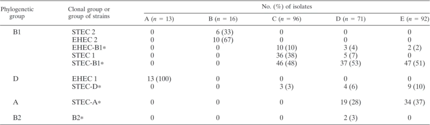
(group D for seropathotype A and group B1 for seropatho-types B and C). In contrast, isolates in seropathoseropatho-types D and E (not associated with disease) appeared to be genetically more heterogeneous. In seropathotype D, 63% of the isolates be-longed to group B1, 28% bebe-longed to group A, 6% bebe-longed to group D, and 3% belonged to group B2. Nonhuman isolates in seropathotype E belonged either to phylogenetic group B1 (53%) or to phylogenetic group A (37%), and a small propor-tion (10%) belonged to phylogenetic group D. Virulent clonal groups (STEC 1, STEC 2, EHEC 1, and EHEC 2) were pre-dominantly distributed among seropathotypes associated with disease (A, B, and C). In contrast, “atypical” groups (e.g., STEC-A and STEC-D) were concentrated within seropatho-types that are not associated with disease (D and E).
Of particular interest was the clear association observed between phylogenetic group A and the “non virulent” sero-pathotypes (D and E) (P⬍ 0.0001; OR ⫽ 63.2 [95%CI ⫽ 8.6 to 464]). In view of this association, the phylogenetic group A
status appears highly predictive of the “nonvirulent” sero-pathotypes.
Phylogenetic distribution of VFs.To investigate the relation-ships between genetic background and virulence genes, we assessed the phylogenetic distribution of the VFs. The distri-bution of VFs in the different phylogenetic and clonal groups is shown in Table 4. Compared to phylogenetic groups D and B1 (combined), the phylogenetic group A exhibited a signifi-cant higher prevalence of various VFs analyzed (specifically,
astA, HPI, stx1c, and stx2-NV206). In contrast, eae, stx2, stx2-vha,
and stx2-vhbwere significantly more prevalent among
phyloge-netic groups D and B1 than in phylogephyloge-netic group A. The difference in the prevalence of eae between phyloge-netic group D and group B1 (P⫽ 0.004) and group A (P ⬍ 0.001) was highly significant, favoring isolates in phylogenetic group D. The difference in the prevalence of HPI and astA genes between phylogenetic group A on the one hand and phylogenetic groups B1 and D on the other was highly
signif-TABLE 3. Phylogenetic distribution of isolates among the different seropathotypes
Phylogenetic group Clonal group or group of strains No. (%) of isolates A (n⫽ 13) B (n⫽ 16) C (n⫽ 96) D (n⫽ 71) E (n⫽ 92) B1 STEC 2 0 6 (33) 0 0 0 EHEC 2 0 10 (67) 0 0 0 EHEC-B1ⴱ 0 0 10 (10) 3 (4) 2 (2) STEC 1 0 0 36 (38) 5 (7) 0 STEC-B1ⴱ 0 0 46 (48) 37 (53) 47 (51) D EHEC 1 13 (100) 0 0 0 0 STEC-Dⴱ 0 0 3 (3) 4 (6) 9 (10) A STEC-Aⴱ 0 0 0 19 (28) 34 (37) B2 B2ⴱ 0 0 0 2 (3) 0 aⴱ, group of strains.
TABLE 4. Phylogenetic distribution of virulence factors and stx subtypes
Virulence genotype No. (%) of isolates Pa OR (95%CI)b D B1 A Overall (n⫽ 29) EHEC 1 (n⫽ 13) STEC-D (n⫽ 16) Overall (n⫽ 201) STEC 1 (n⫽ 41) STEC-B1 (n⫽ 129) EHEC 2, STEC 2, EHEC-B1 (n⫽ 31) STEC-A (n⫽ 53) eae 13 (42) 13 (100) 0 31 (15) 0 0 31 (100) 1 (2) ⬍0.01 0.08 (0.01–0.59) astA 2 (6) 1 (7) 1 (5) 12 (6) 1 (2) 8 (6) 3 (10) 21 (40) 0.01 2.64 (1.4–4.9) HPI 1 (3) 0 1 (5) 18 (9) 1 (2) 7 (5) 10 (34) 22 (41) ⬍0.0001 7.46 (3.6–15.3) stx1variants 19 (61) 8 (61) 11 (61) 93 (46) 8 (20) 64 (47) 21 (71) 23 (42) Only stx1variants 2 (6) 0 2 (11) 28 (14) 1 (2) 12 (9) 15 (52) 18 (34) 0.001 3.04 (1.5–5.9) stx1 18 (58) 8 (61) 10 (56) 82 (41) 7 (17) 56 (43) 19 (61) 16 (30) stx1c 1 (3) 0 1 (5) 7 (3) 1 (2) 6 (4) 0 7 (13) 0.01 4.08 (1.4–11.8) stx2variants 29 (93) 13 (100) 16 (89) 172 (85) 39 (95) 118 (87) 15 (48) 37 (70) Only stx2variants 12 (38) 5 (38) 7 (39) 108 (54) 33 (80) 66 (51) 9 (29) 31 (58) stx1⫹ stx2variants 17 (55) 8 (61) 9 (50) 65 (32) 7 (17) 52 (38) 6 (19) 5 (9) ⬍0.001 0.18 (0.07–044) stx2 16 (52) 11 (85) 5 (27) 73 (36) 14 (34) 51 (37) 8 (26) 7 (13) 0.001 0.23 (0.1–0.53) stx2-vha 4 (13) 4 (31) 0 53 (26) 16 (39) 31 (23) 6 (19) 2 (4) 0.001 0.12 (0.3–0.51) stx2-vhb 5 (16) 0 5 (29) 83 (41) 32 (78) 49 (36) 2 (6) 5 (9) 0.001 0.16 (0.6–0.42) stx2-nv206 0 0 0 5 (2) 0 5 (4) 0 23 (43) ⬍0.0001 32.6 (11.6–92) stx2d 1 (3) 0 1 (5) 24 (12) 3 (7) 15 (11) 6 (19) 4 (7) stx2/stx2-vhb 0 0 0 29 (14) 12 (29) 17 (13) 0 1 (2) stx2-vha/stx2-vhb 0 0 0 20 (10) 11 (27) 7 (5) 2 (6) 1 (2)
aP values (from chi-square test) are shown only if P was⬍0.01.
icant for HPI (P ⬍ 0.0001) and astA (P ⫽ 0.01), favoring phylogenetic group A (STEC-A). Consistent with vertical in-heritance of HPI within lineages, all strains belonging to sero-types O6:H10, O26:H11, and O109:NM possessed the HPI determinants. In contrast, and consistent with horizontal trans-fer within lineages, the distribution pattern of astA showed variability among strains belonging to the same serotype. The substantial prevalence (35%) of HPI within the clonal group EHEC 2 was due to the constant presence of HPI in strains of serotypes O26:H11, O26:NM, O111:H2, and O111:NM.
Distribution of the stx genes was analyzed in relation to the phylogenetic origin of the isolate (Table 4). The difference in the prevalences of stx1 or stx2variants between the different
phylogenetic groups was not significant. It is well documented that isolates producing Stx1 are more rarely associated with serious disease than isolates producing Stx2 alone. Consistent with this finding, a substantial prevalence (34%) of strains possessing only the stx1variants was observed in phylogenetic
group A. Among these strains a substantial part (28%) possess the stx1c subtype which is predominantly recovered from
healthy animals, asymptomatic infection or uncomplicated di-arrhea in humans (53).
The distribution pattern of the different stx2 subtypes
fered considerably between the phylogenetic groups. The dif-ference in the prevalence of stx2between phylogenetic groups
D and B1 (P⫽ 0.01) and between groups D and A (P ⬍ 0.001) was highly significant, favoring isolates in phylogenetic group D (concentrated into the clonal group EHEC 1). The differ-ence in the prevaldiffer-ence of stx2-NV206 between phylogenetic
group A and the phylogenetic groups B1 and D (combined) was also highly significant (P⬍ 0.0001; OR ⫽ 32.6 [95%CI ⫽ 11.6 to 92]), revealing a close association between stx2-nv206and
phylogenetic group A. Indeed, among the phylogenetic group A, the predominant stx2genotype was the stx2-nv206genotype,
accounting for 62% of the stx2-positive isolates.
There was a significant difference in the prevalence of
stx2-vha between phylogenetic groups B1 and A (P ⬍ 0.001)
favoring isolates in phylogenetic group B1. The distribution patterns of stx2-vha in group B1 suggest a vertical inheritance
within certain lineages (O91:H10, O91:H21, O113:H21, and O172:H21). A similar phylogenetic distribution was observed with stx2-vhb, with a significant difference in the prevalence of
stx2-vhbbetween phylogenetic group B1 and D (P⫽ 0.03; OR
⫽ 2.4 [95%CI ⫽ 0.9 to 5.8]) and between groups B1 and A (P ⬍ 0.001; OR ⫽ 7.3 [95%CI ⫽ 2.7 to 18.4) also favoring isolates in phylogenetic group B1. The distribution patterns of stx2-vhb
in group B1 suggest both vertical inheritance and horizontal transfer within lineages. Consistent with horizontal transfer, a strain-to-strain distribution of stx2-vhb was observed in
sero-types O22:H16, O91:H10, and O174:H2. In contrast and con-sistent with vertical inheritance, stx2-vhb was detected in all
strains belonging to serotypes O91:H21, O113:H21, and O174: H21, which are the archetypal strains of the clonal group STEC 1. The combination of stx2and stx2-vhbwas restricted to isolates
in phylogenetic group B1 (the most frequent serotypes are O74:H42, O91:H21, and O113:H21).
When the 170 strains lacking eae in the phylogenetic group B1 were stratified dichotomously as serotypes associated with disease (seropathotype C [81 isolates]) versus serotypes not associated with disease (seropathotypes D plus E [89 isolates]),
the only significant difference in the prevalence of VF between the two groups of strains was observed with stx2-vhb. The
prev-alence of stx2-vhb within group B1 was nearly twice as high
(62% versus 32%) among isolates in the seropathotype C than among isolates in “nonvirulent” seropathotypes (D plus E, combined). The finding that an apparent association exists between stx2-vhb and seropathotype C revealed stx2-vhb as a
significant predictor of the “virulent” status among group B1 isolates lacking eae (P⬍ 0.0001; OR ⫽ 6.70 [95%CI ⫽ 3.6 to 12.0]). When only STEC 1 strains in seropathotype C were compared to isolates in seropathotypes D and E, stx2-vhb
ap-peared to be an even stronger predictor of “virulent” status (P ⫽ 0.0004; OR ⫽ 8.2 [95%CI ⫽ 3.4 to 19.4]).
DISCUSSION
To our knowledge, our study is the first to provide an over-view of characteristics of STEC strains classified in different seropathotypes by combining the prevalence of several known virulence determinants and their classification into the major phylogenetic groups of the E. coli species. Consistent with previous studies on EPEC and STEC strains (13, 14), our phylogenetic analysis shows that STEC strains segregate mainly in phylogenetic group B1 and confirms the rarity of the phylogenetic group B2. Of the remaining strains, phylogenetic groups A and D represent, respectively, 20 and 10% of the collection.
We provide here novel insights into the phylogenetic struc-ture of the different seropathotypes. The presence in sero-pathotypes that are associated with disease (A, B, and C) of well-known virulent clonal groups (STEC 1, STEC 2, EHEC 1, and EHEC 2) and their concomitant replacement by “atypical” groups (e.g., STEC-A and STEC-D) in seropathotypes that are not associated with disease (D and E) support the concept of the seropathotype classification proposed by Karmali et al. (22).
From the whole data analysis, we show that there is a link between seropathotype classification, prevalence of various VFs, and phylogeny. The astA gene which is widely distributed among diarrheogenic E. coli strains in humans and animals may represent an additional virulent determinant of STEC strains (15, 38). Consistent with previous studies on human STEC infections (45), our study confirms the presence of the
astA gene among strains carrying eae associated with HUS and
outbreaks (O26:H11, O111:H2, and O157:H7). However, the
astA gene was mostly (92%) recovered from strains lacking eae
that belonged predominantly (70%) to phylogenetic group A (STEC-A). Of these strains, over half possessed only the stx1
variants and were distributed in seropathotypes not associated with disease (D and E). Whether STEC-A strains harboring only the stx1variants and astA are true pathogens needs further
elucidation.
Consistent with previous findings involving other STEC iso-lates (20), our data show the presence of the Yersinia HPI among both human and animal STEC isolates and confirm the absence of HPI from O157:H7 and O157:NM strains. In the present study a majority (61%) of the HPI-positive strains belong to seropathotypes that are not associated with disease (D and E). However, HPI was detected in all members of the clonal group EHEC 2 (O26:H11, O26:NM, O111:NM, and
O111:H2) indicating that the HPI is a common component of the genome of these strains and suggesting a high degree of stability of HPI in the genome of certain lineages. To explain the presence of HPI among STEC strains, Karch et al. (20) formed the hypothesis that HPI in STEC is a form of fitness island rather than a PAI. Similarly to the distribution of the
astA gene, HPI was predominantly recovered from strains
lack-ing eae distributed in seropathotypes D and E. Although the significance of these findings remains to be established, the present study reveals that astA and HPI were significantly predictive, among eae-negative isolates, of the “nonvirulent” seropathotypes.
There is considerable epidemiological evidence indicating that STEC isolates producing Stx2 alone are more commonly associated with serious disease than isolates producing Stx1 only or both Stx1 and Stx2 (27, 32, 41, 42). Many isolates produce two or more Stx2 variants, but the relative contribu-tion of each variant to the pathogenesis is not known. In the present study, the 287 STEC isolates were subjected to stx2
subtyping. The stx2 subtype was prominent and was widely
distributed among the different seropathotypes. Consistent with previous reports involving bovine non-O157 STEC (2, 8),
stx2-vhbwas also frequently detected, and the combination stx2/
stx2-vhbwas the most frequent among STEC possessing more
than one stx2subtype. Melton-Celsa et al. (28) demonstrated
that the Stx2c variants (Stx2-vhaand Stx2-vhb) are activatable by
intestinal mucus which causes a marked increase in toxicity and may compensate for a lack of other virulence components such as the LEE. Our data showing the confinement of stx2-vhbto eae-negative isolates support this hypothesis. In contrast to stx2, the prevalence of stx2-vhbdiffered considerably among the
different seropathotypes. Indeed, the prevalence of stx2-vhbwas
twice as high among isolates associated with HUS (seropatho-type C) than among isolates that are not associated with dis-ease (seropathotypes D plus E), revealing stx2-vhbas a
signifi-cant predictor of “virulent” status. In addition, our phylogenetic analysis indicates that stx2-vhbsegregates mainly
within phylogenetic group B1 and is concentrated in the clonal group STEC 1. Moreover, the distribution pattern of stx2-vhb
suggests a vertical inheritance of stx2-vhbin serotypes ON:H21,
O91:H21, O113:H21, and O174:H21, which are the best known serotypes of the clonal group STEC 1. These findings indicate that stx2-vhbis a common component of the genome of certain
lineages in STEC 1 and suggest an important role of the stx2-vhb
subtype in the pathogenesis of these lineages. Consistent with this hypothesis, a major role of the Stx2-activatable variants
(Stx2-vhaand Stx2-vhb) was recently demonstrated in the
patho-genesis of the eae-negative strain B2F1 (O91:H21) in neonatal pigs (11). In that study, while the absence of intimin and plasmid-associated virulence determinants had little impact on the pathogenesis of STEC disease during infection, an appar-ent association between the severity of lesions in pigs and the production of the Stx2-activatable toxin was observed.
Our results demonstrate a close association between
stx2-NV206and STEC-A and reveals stx2-NV206as a strong
pre-dictor of the “nonvirulent” seropathotypes E and D. Consid-ering the high cytotoxic activity of the Stx2-NV206variant
ob-served in vitro toward Vero cells (2), the substantial prevalence (42%) of stx2-NV206in STEC-A was unexpected. The observed
link between stx2-NV206and STEC-A may result either from the
preferential association of a given phage with a particular ge-netic background or from a coevolution of the bacteria and phage chromosomes (47). Although the role of stx2-NV206in
pathogenesis of STEC disease remains to be established, the strong association observed between stx2-NV206 and STEC-A
supports the concept that arrival, retention, and/or expression of virulence factors require a particular genetic background (14).
One limitation of the present study results from the collec-tion, which included multiple representatives of certain geno-types from different sources. This has resulted, for example, in a disproportionate representation of certain clones that inflate the apparent prevalence of VFs associated with these clones (for example, O113:H4, O113:H21, and O174:H2). As sug-gested by Johnson et al. (16), to protect against this bias, we used a more-stringent-than-usual criterion for statistical signif-icance (Pⱕ 0.01).
One of the major findings of the present study is the dem-onstration of a striking phylogenetic distribution of various VFs. Specifically, when group A was compared to other phy-logenetic groups combined, significant differences in preva-lence favoring group A were seen for stx1c, stx2-NV206, astA, and
HPI. Taken together with the observation that STEC-A com-prises exclusively strains from seropathotypes that are not as-sociated with disease, their “atypical” virulence pattern sug-gests that STEC-A isolates comprise a distinct category of STEC strains. The ability to identify STEC-A isolates is of clinical and epidemiological significance because these isolates are not commonly associated with human disease. Because of the geographically circumscribed study population, further ep-idemiological investigations on other STEC populations are needed to generalize these results.
REFERENCES
1. Bertin, Y., K. Boukhors, V. Livrelli, and C. Martin. 2004. Localization of the insertion site and pathotype determination of the locus of enterocyte efface-ment of Shiga toxin-producing Escherichia coli strains. Appl. Environ. Mi-crobiol. 70:61–68.
2. Bertin, Y., K. Boukhors, N. Pradel, V. Livrelli, and C. Martin. 2001. stx2 subtyping of Shiga toxin-producing Escherichia coli isolated from cattle in France: detection of a new Stx2 subtype and correlation with additional virulence factors. J. Clin. Microbiol. 39:3060–3065.
3. Bertin, Y., C. Martin, J. P. Girardeau, P. Pohl, and M. Contrepois. 1998. Association of genes encoding P fimbriae, CS31A antigen and EAST 1 toxin among CNF1-producing Escherichia coli strains from cattle with septicemia and diarrhea. FEMS Microbiol. Lett. 162:235–239.
4. Bidet, P., P. Mariani-Kurkdjian, F. Grimont, N. Brahimi, C. Courroux, P.
Grimont, and E. Bingen.2005. Characterization of Escherichia coli O157:H7 isolates causing hemolytic uraemic syndrome in France. J. Med. Microbiol.
54:71–75.
5. Boerlin, P., S. A. McEwen, F. Boerlin-Petzold, J. B. Wilson, R. P. Johnson,
and C. L. Gyles.1999. Associations between virulence factors of Shiga toxin-producing Escherichia coli and disease in humans. J. Clin. Microbiol.
37:497–503.
6. Bonnet, R., B. Souweine, G. Gauthier, C. Rich, V. Livrelli, J. Sirot, B. Joly,
and C. Forestier.1998. Non-O157 Stx2-producing Escherichia coli strains associated with sporadic cases of haemolytic-uremic syndrome in adults. J. Clin. Microbiol. 36:1777–1780.
7. Boyd, E. F., and D. L. Hartl. 1998. Chromosomal regions specific to patho-genic isolates of Escherichia coli have a phylogenetically clustered distribu-tion. J. Bacteriol. 180:1159–1165.
8. Brett, K. N., M. A. Hornitzky, K. A. Bettelheim, M. J. Walker, and S. P.
Djordjevic.2003. Bovine non-O157 Shiga toxin 2-containing Escherichia coli isolates commonly possess stx-EDL933and/or stx2-vhbsubtypes. J. Clin. Micro-biol. 41:2716–2722.
9. Bu¨rk, C., R. Dietrich, G. Ac¸ar, M. Moravek, M. Bu¨lte, and E. Ma¨rtlbauer.
2003. Identification and characterization of a new variant of Shiga toxin 1 in
Escherichia coli ONT:H19 of bovine origin. J. Clin. Microbiol. 41:2106–2112.
deter-mination of the Escherichia coli phylogenetic group. Appl. Environ. Micro-biol. 66:4555–4558.
11. Dean-Nystrom, E. A., A. R. Melton-Celsa, J. F. Pohlenz, H. W. Moon, and
A. D. O’Brien.2003. Comparative pathogenicity of Escherichia coli O157 and intimin-negative non-O157 Shiga toxin-producing E. coli strains in neonatal pigs. Infect. Immun. 71:6526–6533.
12. Donnenberg, M. S., S. Tzipori, M. L. McKee, A. D. O’Brien, J. Alroy, and
J. B. Kaper.1993. The role of the eae gene of enterohemorrhagic Escherichia
coli in intimate attachment in vitro and in a porcine model. J. Clin. Investig. 92:1418–1424.
13. Donnenberg, M. S., and T. S. Whittam. 2001. Pathogenesis and evolution of virulence in enteropathogenic and enterohemorrhagic Escherichia coli. J. Clin. Investig. 107:539–548.
14. Escobar-Pa´ramo, P., O. Clermont, A. B. Blanc-Potard, H. Bui, C. Le Bou-guenec, and E. Denamur.2004. A specific genetic background is required for acquisition and expression of virulence factors in Escherichia coli. Mol. Biol. Evol. 21:1085–1094.
15. Girardeau, J. P., L. Lalioui, A. M. Said, C. De Champs, and C. Le
Bougue-nec.2003. Extended virulence genotype of pathogenic Escherichia coli iso-lates carrying the afa-8 operon: evidence of similarities between isoiso-lates from humans and animals with extraintestinal infections. J. Clin. Microbiol. 41: 218–226.
16. Johnson, J. R., P. Delavari, M. Kuskowski, and A. L. Stell. 2001. Phyloge-netic distribution of extraintestinal virulence-associated traits in Escherichia
coli. J. Infect. Dis. 183:78–88.
17. Johnson, R., R. C. Clarke, J. B. Wilson, S. C. Read, K. Rahn, S. A. Renwick,
K. A. Sandhu, D. Alves, M. A. Karmali, H. Lior, S. A. McEwen, J. S. Spika, and C. L. Gyles.1996. Growing concerns and recent outbreaks involving non-O157:H7 serotypes of verotoxigenic Escherichia coli. J. Food Prot. 59: 1112–1122.
18. Johnson, W. M., D. R. Pollard, H. Lior, S. D. Tyler, and K. R. Rozee. 1990. Differentiation of genes coding for Escherichia coli verotoxin 2 and the verotoxin associated with porcine edema disease (VTe) by the polymerase chain reaction. J. Clin. Microbiol. 28:2351–2353.
19. Kaper, J. B., J. L. Mellies, and J. Nataro. 1999. Pathogenicity islands and other mobile genetic elements of diarrheagenic Escherichia coli, p. 33–58. In J. B. Kaper and J. Hacker (ed.), Pathogenicity islands and other mobile virulence elements. ASM Press, Washington, D.C.
20. Karch, H., S. Schubert, D. Zhang, W. Zhang, H. Schmidt, T. Olschlager, and
J. Hacker.1999. A genomic island, termed high-pathogenicity island, is present in certain non-O157 Shiga toxin-producing Escherichia coli clonal lineages. Infect. Immun. 67:5994–6001.
21. Karmali, M. A. 1989. Infection by verocytotoxin-producing Escherichia coli. Clin. Microbiol. Rev. 2:15–38.
22. Karmali, M. A., M. Mascarenhas, S. Shen, K. Ziebell, S. Johnson, R.
Reid-Smith, J. Isaac-Renton, C. Clark, K. Rahn, and J. B. Kaper.2003. Associ-ation of genomic O island 122 of Escherichia coli EDL 933 with verocyto-toxin-producing Escherichia coli seropathotypes that are linked to epidemic and/or serious disease. J. Clin. Microbiol. 41:4930–4940.
23. Koch, C., S. Hertwig, R. Lurz, B. Appel, and L. Beutin. 2001. Isolation of a lysogenic bacteriophage carrying the stx1OX3gene, which is closely associated with Shiga toxin-producing Escherichia coli strains from sheep and humans. J. Clin. Microbiol. 39:3992–3998.
24. Law, D. 2000. Virulence factors of Escherichia coli O157 and other Shiga toxin-producing E. coli. J. Appl. Microbiol. 88:729–745.
25. Lecointre, G., J. Rachdi, P. Darlu, and E. Denamur. 1998. Escherichia coli molecular phylogeny using the incongruence length difference test. Mol. Biol. Evol. 15:1685–1695.
26. Lindgren, S. W., A. R. Melton, and A. D. O’Brien. 1993. Virulence of enterohemorrhagic Escherichia coli O91:H21 clinical isolates in an orally infected mouse model. Infect. Immun. 61:3832–3842.
27. Louise, C. B., and T. G. Obrig. 1995. Specific interaction of Escherichia coli O157:H7-derived Shiga-like toxin II with human renal endothelial cells. J. Infect. Dis. 172:1397–1401.
28. Melton-Celsa, A. R., S. C. Darnell, and A. D. O’Brien. 1996. Activation of Shiga-like toxins by mouse and human intestinal mucus correlates with virulence of enterohemorrhagic Escherichia coli O91:H21 isolates in orally infected, streptomycin-treated mice. Infect. Immun. 64:1569–1576. 29. Nataro, J. P., and J. B. Kaper. 1998. Diarrheagenic Escherichia coli. Clin.
Microbiol. Rev. 11:142–201.
30. Paton, J. C., and A. W. Paton. 1998. Pathogenesis and diagnosis of Shiga toxin-producing Escherichia coli infections. Clin. Microbiol. Rev. 11:450–479. 31. Paton, A. W., and J. C. Paton. 1998. Detection and characterization of Shiga toxigenic Escherichia coli by using multiplex PCR assays for stx1, stx2, eaeA, enterohemorrhagic E. coli hlyA, rfbO111, and rfbO157. J. Clin. Microbiol.
36:598–602.
32. Paton, A. W., M. C. Woodrow, R. M. Doyle, J. A. Lanser, and J. C. Paton. 1999. Molecular characterization of a Shiga toxigenic Escherichia coli O113:
H21 strain lacking eae responsible for a cluster of cases of hemolytic-uremic syndrome. J. Clin. Microbiol. 37:3357–3361.
33. Picard, B., J. S. Garcia, S. Gouriou, P. Duriez, N. Brahimi, E. Bingen, J.
Elion, and E. Denamur.1999. The link between phylogeny and virulence in
Escherichia coli extraintestinal infection. Infect. Immun. 67:546–553.
34. Pierard, D., G. Muyldermans, L. Moriau, D. Stevens, and S. Lauwers. 1998. Identification of new verocytotoxin type 2 variant B-subunit genes in human and animal Escherichia coli isolates. J. Clin. Microbiol. 36:3317–3322. 35. Pollard, D. R., W. M. Johnson, H. Lior, S. D. Tyler, and K. R. Rozee. 1990.
Rapid and specific detection of verotoxin genes in Escherichia coli by poly-merase chain reaction. J. Clin. Microbiol. 28:540–545.
36. Pradel, N., V. Livrelli, C. DeChamps, J.-B. Palcoux, A. Reynaud, F. Scheutz,
J. Sirot, B. Joly, and C. Forestier.2000. Prevalence and characterization of Shiga toxin-producing Escherichia coli isolated from cattle, food, and chil-dren during a one-year prospective study in France. J. Clin. Microbiol.
38:1023–1031.
37. Reid, S. D., C. J. Herbelin, A. C. Bumbaugh, R. K. Selander, and T. S.
Whittam.2000. Parallel evolution of virulence in pathogenic Escherichia coli. Nature 406:64–67.
38. Savarino, S. J., P. Fox, D. Yikang, and J. P. Nataro. 1994. Identification and characterization of a gene cluster mediating enteroaggregative Escherichia
coli aggregative adherence fimbria I biogenesis. J. Bacteriol. 156:4949–4957.
39. Savarino, S. J., A. Fasano, D. C. Robertson, and M. M. Levine. 1991. Enteroaggregative Escherichia coli elaborate a heat-stable enterotoxin de-monstrable in an in vitro rabbit intestinal model. J. Clin. Investig. 87:1450– 1455.
40. Schubert, S., A. Rakin, H. Karch, E. Carniel, and J. Heesemann. 1998. Prevalence of the “high-pathogenicity island” of Yersinia species among
Escherichia coli strains that are pathogenic to humans. Infect. Immun. 66:
480–485.
41. Siegler, R. L., T. G. Obrig, T. J. Pysher, V. L. Tesh, N. D. Denkers, and F. B.
Taylor.2003. Response to Shiga toxin 1 and 2 in a baboon model of hemo-lytic-uremic syndrome. Pediatr. Nephrol. 18:92–96.
42. Tesh, V. L., J. A. Burris, J. W. Owens, V. M. Gordon, E. A. Wadolkowski,
A. D. O’Brien, and J. E. Samuel.1993. Comparison of the relative toxicities of Shiga-like toxins type I and type II for mice. Infect. Immun. 61:3392–3402. 43. Toma, C., E. Martinez Espinosa, T. Song, E. Miliwebsky, I. Chinen, S. Iyoda,
M. Iwanaga, and M. Rivas.2004. Distribution of putative adhesins in dif-ferent seropathotypes of Shiga toxin-producing Escherichia coli. J. Clin. Microbiol. 42:4937–4946.
44. Tyler, S. D., W. M. Johnson, H. Lior, G. Wang, and K. R. Rozee. 1991. Identification of verotoxin type 2 variant B subunit genes in Escherichia coli by the polymerase chain reaction and restriction fragment length polymor-phism analysis. J. Clin. Microbiol. 29:1339–1343.
45. Vaz, T. M. I., K. Irino, M. A. M. F. Kato, A. M. G. Dias, T. A. T. Gomes,
M. I. C. Medeiros, M. M. M. Rocha, and B. E. C. Guth.2004. Virulence properties and characteristics of Shiga toxin-producing Escherichia coli in Sa˜o Paulo, Brazil, from 1976 through 1999. J. Clin. Microbiol. 42:903–905. 46. Vernozy-Rozand, C. V., M. P. Montet, F. Lequerrec, E. Serillon, B. Tilly, C.
Bavai, S. Ray-Gueniot, J. Bouvet, C. Mazuy-Cruchaudet, and Y. Richard.
2002. Prevalence of verotoxin Escherichia coli (VTEC) in slurry, farmyard manure and sewage sludge in France. J. Appl. Microbiol. 93:473–478. 47. Wagner, P. L., D. W. K. Acheson, and M. K. Waldor. 1999. Isogenic lysogens
of diverse Shiga toxin 2-encoding bacteriophages produce markedly different amounts of Shiga toxin. Infect. Immun. 67:6710–6714.
48. Weinstein, D. L., M. P. Jackson, J. E. Samuel, R. K. Holmes, and A. D.
O’Brien.1988. Cloning and sequencing of a Shiga-like toxin type II variant from Escherichia coli strain responsible for edema disease of swine. J. Bac-teriol. 170:4223–4230.
49. Whittam, T. S., M. L. Wolfe, I. K. Wachsmuth, F. Orskov, I. Orskov, and
R. A. Wilson.1993. Clonal relationships among Escherichia coli strains that cause hemorrhagic colitis and infantile diarrhea. Infect. Immun. 61:1619– 1629.
50. World Health Organization. 1999. Zoonotic non-O157 Shiga toxin-produc-ing Escherichia coli (STEC): report of a W.H.O. Scientific Worktoxin-produc-ing Group Meeting, Berlin, Germany, 22 to 23 June 1998, p. 1–30. World Health Organization, Geneva, Switzerland.
51. Yamamoto, T., and P. Echeverria. 1996. Detection of the enteroaggregative
Escherichia coli heat-stable enterotoxin 1 gene sequences in enterotoxigenic E. coli strains pathogenic for humans. Infect. Immun. 64:1441–1445.
52. Yatsuyanagi, J., S. Saito, Y. Miyajima, K. Amano, and K. Enomoto. 2003. Characterization of atypical enteropathogenic Escherichia coli strains har-boring the astA gene that were associated with a waterborne outbreak of diarrhea in Japan. J. Clin. Microbiol. 41:2033–2039.
53. Zweifel, C., J. E. Blanco, M. Blanco, J. Blanco, and R. Stephan. 2004. Serotypes and virulence genes of ovine non-O157 Shiga toxin-producing