HAL Id: hal-03016005
https://hal.archives-ouvertes.fr/hal-03016005
Submitted on 30 Nov 2020HAL is a multi-disciplinary open access archive for the deposit and dissemination of sci-entific research documents, whether they are pub-lished or not. The documents may come from teaching and research institutions in France or abroad, or from public or private research centers.
L’archive ouverte pluridisciplinaire HAL, est destinée au dépôt et à la diffusion de documents scientifiques de niveau recherche, publiés ou non, émanant des établissements d’enseignement et de recherche français ou étrangers, des laboratoires publics ou privés.
CD5 signalosome coordinates antagonist TCR signals to
control the generation of Treg cells induced by foreign
antigens
Gaëtan Blaize, Hélène Daniels-Treffandier, Meryem Aloulou, Nelly Rouquié,
Cui Yang, Marlène Marcellin, Mylène Gador, Mehdi Benamar, Mariette
Ducatez, Ki-Duk Song, et al.
To cite this version:
Gaëtan Blaize, Hélène Daniels-Treffandier, Meryem Aloulou, Nelly Rouquié, Cui Yang, et al.. CD5 signalosome coordinates antagonist TCR signals to control the generation of Treg cells induced by foreign antigens. Proceedings of the National Academy of Sciences of the United States of America , National Academy of Sciences, 2020, 117 (23), pp.12969-12979. �10.1073/pnas.1917182117�. �hal-03016005�
1
CD5 signalosome coordinates antagonist TCR signals to control the
1
generation of T reg cells induced by foreign antigens
2 3 4
Gaëtan Blaize1, Hélène Daniels-Treffandier1,3*, Meryem Aloulou1*, Nelly Rouquié1, Cui
5
Yang1, Marlène Marcellin2, Mylène Gador1, Mehdi Benamar1, Mariette Ducatez3, Ki-duk
6
Song4, Odile Burlet-Schiltz2, Abdelhadi Saoudi1, Paul E. Love4, Nicolas Fazilleau1, Anne
7
Gonzalez de Peredo2, Renaud Lesourne1
8 9
1Centre de Physiopathologie de Toulouse Purpan, Université de Toulouse, Centre National de
10
la Recherche Scientifique, Institut National de la Santé et de la Recherche Médicale, UPS, 11
31024, Toulouse, France. 12
2Institut de Pharmacologie et de Biologie Structurale, IPBS, Université de Toulouse, CNRS,
13
UPS, Toulouse, France. 14
3IHAP, Université de Toulouse, ENVT, INRA, UMR 1225, 31076 Toulouse, France.
15
4Section on Hematopoiesis and Lymphocyte Biology, Eunice Kennedy Shriver National
16
Institute of Child Health and Human Development, National Institutes of Health, Bethesda, 17
MD 20892, USA. 18
* Equally contributed to this work 19
20
Correspondence should be addressed to R.L. 21 Email: renaud.lesourne@inserm.fr 22 23 24 25
2 Abstract
26 27
CD5 is characterized as an inhibitory co-receptor with important regulatory role during T cell 28
development. The molecular mechanism by which CD5 operates has been puzzling and its 29
function in mature T cells suggests promoting rather than repressing effects on immune 30
responses. Here, we combined quantitative mass spectrometry and genetic studies to analyze 31
the components and the activity of the CD5 signaling machinery in primary T cells. We found 32
that TCR engagement induces the selective phosphorylation of CD5 tyrosine 429, which 33
serves as a docking site for proteins with adaptor functions (c-Cbl, CIN85, CRKL), 34
connecting CD5 to positive (PI3K) and negative (UBASH3A, SHIP1) regulators of TCR 35
signaling. c-CBL acts as a coordinator in this complex enabling CD5 to synchronize positive 36
and negative feedbacks on TCR signaling through the other components. Disruption of CD5 37
signalosome in mutant mice reveals that it modulates TCR signal outputs to selectively 38
repress the transactivation of Foxp3 and limit the inopportune induction of peripherally-39
induced regulatory T cells during immune responses against foreign antigen. Our findings 40
bring new insights into the paradigm of co-receptor signaling, suggesting that, in addition to 41
providing dualistic enhancing or dampening inputs, co-receptors can engage concomitant 42
stimulatory and inhibitory signaling events which act together to promote specific functional 43 outcomes. 44 45 Keywords 46 47
T cells / Signaling / Co-receptors 48
3 Significance statement
49 50
T-cell co-receptors are often described as molecular switches that broadly regulate T-cell 51
activation by enhancing or repressing TCR signaling according to the immunological context. 52
However, many co-receptors act more selectively by instructing or restricting the generation 53
of specific T-cell subsets. The molecular mechanisms by which these subtle regulations occur 54
remain incompletely defined. In this study, we show that CD5 co-receptors engage a 55
multimeric signaling complex which synchronize positive and negative feedback on TCR 56
signaling to limit the induction of inopportune regulatory T cells during immune response. 57
Our findings suggest that rather than exclusively acting as stimulators or inhibitors of TCR 58
signaling, co-receptors may coordinate antagonist TCR signals which act together to promote 59
specific T-cell responses. 60 61 62 63 64 65 66 67 68
4 Introduction
69 70
T cells have the ability to develop a wide variety of cellular responses following the 71
stimulation of a single receptor, namely the T-cell antigen receptor (TCR). The recognition 72
by TCRs of self or foreign peptides bound to the major histocompatibility complex (pMHC) 73
triggers multiple signaling pathways, which lead to the activation of specific effector proteins 74
involved in the transmission of distinct signaling responses. The relative intensity and the 75
persistency by which signals are transmitted in each pathway play a critical role in specifying 76
and driving specific T-cell responses. Because different pathways may have either synergistic 77
or antagonist effects on these responses, their coordination in time and space (signaling 78
patterns) is also critical to shape T-cell effector profiles and determine specific outcomes. 79
80
Signals transmitted by the TCR can be regulated by co-receptors that are engaged 81
differentially based on their relative expression on the T-cell surface and on the availability of 82
their cognate ligands in the extracellular environment. Initial work on co-receptor signaling 83
led to the classification of these proteins into two main functional categories depending on 84
their overall effect on T-cell activity. Stimulatory co-receptors, such as CD28, ICOS or OX40, 85
which promote naïve T-cell activation and amplify effector T-cell responses, and inhibitory 86
co-receptors, such as CTLA-4, PD-1 or BTLA which prevent the potential activation of T 87
cells by self-antigens and contribute to terminate or tune down effector T-cell responses 88
following antigen clearance. More recent investigations indicate that many co-receptors act 89
more selectively on specific signaling pathways and contribute to shape the effector profile of 90
T cells according to the immunological context (1-3). Although the mechanisms by which co-91
receptors positively or negatively regulate T-cell activity have been well documented (1, 4-7), 92
5
the molecular processes by which they convey signals to selectively modulate T-cell 93
responses remain poorly understood. 94
95
CD5 is a type 1 transmembrane cell surface glycoprotein that is essentially expressed in T 96
cells. Initial characterization of Cd5-/- mice indicated an inhibitory function for this receptor
97
on TCR signaling (8). Later work showed that CD5 surface levels on thymocytes are 98
correlated to the strength of TCR signaling that is dictated during positive selection by the 99
affinity of the TCR for self-pMHC (9). The increased expression of CD5 in thymocytes that 100
express TCRs with greater self-reactivity dampens TCR signals, possibly enabling some 101
thymocytes that would otherwise be negatively selected to avoid activation-induced cell death 102
and instead complete their maturation and be exported to peripheral lymphoid organs (10). 103
Through this mechanism CD5 would enable the selection of T cells with higher self-104
reactivity, which are presumed to be more effective responders to foreign antigens (11). 105
106
Whereas the role of CD5 during thymic selection has been well characterized, its function in 107
peripheral T cells is less clear. The expression level of CD5 remains correlated with the 108
affinity of TCR for self-pMHC in peripheral T cells (11), suggesting that CD5 could be 109
important for the maintenance of self-tolerance by dampening homeostatic TCR signals that 110
could otherwise cause activation and autoimmunity. However, Cd5-/- mice do not exhibit signs
111
of spontaneous autoimmune or inflammatory pathology and show, on the contrary, a reduced 112
susceptibility to active experimental autoimmune encephalomyelitis (12) and inflammatory 113
bowel disease (13). This suggests that in the absence of CD5 compensatory mechanisms 114
might prevent the expansion and the full activation of T cells expressing TCRs with relatively 115
high affinity to self-pMHC. Notably, previous studies showed that the numbers and 116
6
suppressive function of regulatory T cells (Treg) are increased in CD5 deficient mice (13, 14). 117
However, more recent findings indicate that CD5 plays an instructive role in the generation of 118
peripherally-induced Treg cells (iTreg) in response to tolerizing antigens (15), suggesting that 119
CD5 could have different influences on this T-cell subset according to the immunological 120
context. 121
122
Although several ligands have been reported for CD5 (16-18), it was shown that its 123
extracellular domain is not required for negative regulation of TCR signaling in thymocytes 124
(19), indicating that CD5 is engaged in a feedback loop that tunes down TCR signals 125
following TCRs engagement. Accordingly, CD5 is constitutively associated with the TCR 126
subunits at the cell surface (20) and contains several phospho-tyrosine-binding sites that are 127
phosphorylated by SRC kinases following TCR engagement (21). Although many CD5-128
interacting partners have been reported, the relative importance of these interactions remains 129
unclear because most of these binding partners were identified in independent studies or 130
within distinct cellular models through approaches that do not always enable global 131
comparisons of protein-protein interactions. Interestingly, whereas some of these proteins are 132
well characterized inhibitors of TCR signaling (22, 23), others are known to be positive 133
effectors (24-27), suggesting that CD5-mediated feedback on TCR signaling might be more 134
complex than what was initially presumed. 135
136
In this study, we combined quantitative mass spectrometry and mouse genetics to analyze the 137
composition, the mode of assembly and the molecular function of the CD5 transduction 138
machinery in primary T cells. We found that CD5 coordinates the recruitment of a signaling 139
complex composed of proteins with adapter functions (c-CBL, CIN85 and CRKL) that 140
7
connect CD5 to positive (PI3K) and negative (UBASH3A and SHIP1) regulators of TCR 141
signaling. The recruitment of this complex is entirely dependent on the Y429 of CD5 which is 142
predominantly phosphorylated following TCR engagement and serves as a docking site for c-143
CBL. Disruption of Y429 phosphorylation site in primary T cells shows that this signaling 144
complex promotes on one hand AKT-mediated inhibition of FOXO1 and represses on the 145
other hand ERK kinase activity to selectively dampen the transactivation of Foxp3 gene 146
expression. Analysis of antigen-specific Treg cells in CD5-Y429F mutant mice show that 147
CD5 signaling selectively represses the generation of these cells presumably to promote the 148
development of optimal immune responses. 149
8 Results
150 151
Mass-spectrometry analysis of the CD5 interactome 152
153
To investigate the molecular mechanism by which CD5 regulates TCR signaling, we 154
performed an MS-based analysis of CD5-containing complexes in thymocytes. CD5 was 155
immunoprecipitated from wild-type (WT) or Cd5−/− thymocytes that were treated with 156
pervanadate for 1 minute to induce widespread activation of protein tyrosine kinases. CD5 157
protein complexes were eluted, and the components of the different purified complexes were 158
characterized by nanoflow liquid chromatography combined with tandem MS. To 159
discriminate CD5-binding molecules from the background of contaminant proteins, a 160
thorough quantitative comparison based on MS intensity values was performed for each 161
identified protein between samples immunoprecipitated from WT versus Cd5−/− thymocytes.
162
Candidate proteins were selected based on their significant enrichment in WT samples (fold 163
change >2 and Student t-test p-value<0.001, n=8 replicate experiments, see Materials and 164
Methods for details). On this basis, we identified 11 proteins as potential interacting partners 165
of CD5 in thymocytes (Fig. 1A, SI Appendix, Fig. S1A and Dataset S1). Among these, six 166
proteins were previously identified as regulators of TCR-mediated signaling (c-CBL, 167
UBASH3A/STS-2, CIN85/SH3KBP1, SHIP1, CRKL and PI3K), two proteins are known 168
components of the AP2 complex which is involved in clathrin-mediated internalization of 169
CD5 (AP2a1, AAK1)(28) and three proteins have diverse reported functions not directly 170
associated with TCR signaling (IGH, TRIM21, CYB5). The interactions of CD5 with c-CBL, 171
UBASH3A, PI3K, CRKL and AP2 were reported previously in independent studies (22, 24, 172
25, 28, 29) but not its interaction with SHIP1 and CIN85. Among these proteins, three are 173
cytosolic adaptors (CRKL, CIN85 and AP2) and five are effector molecules with enzymatic 174
9
function (c-CBL, UBASH3A, SHIP1, AAK1 and PI3K). The association of these proteins 175
with CD5 was still detected after 10 minutes of stimulation and no additional interactors were 176
identified at this later time of stimulation (SI Appendix, Fig. S1B and Dataset S2). 177
To estimate the relative abundance of these signaling partners in the immunoprecipitated 178
samples, we used the intensity-based absolute quantification (iBAQ) metric, which 179
corresponds to the sum of all of the peptide intensities divided by the number of theoretically 180
observable tryptic peptides of a protein. Analysis of these interactions (with normalized iBAQ 181
intensities) shows that UBASH3A (IBAQ=604 x 104) and c-CBL (IBAQ=560 x 104) are more
182
abundantly recruited to CD5 than CIN85 (IBAQ=147 x 104), CRKL (IBAQ=143 x 104),
183
SHIP1 (IBAQ=55 x 104) and PI3K (IBAQ=18 x 104), suggesting an essential role for c-Cbl
184
and UBASH3A in CD5-mediated regulation of T-cell activation (SI Appendix, Fig. S1A). 185
Analysis by Western blot showed that CD5 interacts with c-CBL, UBASH3A, SHIP1, CIN85 186
and PI3K both in thymocytes and in peripheral CD4+ T cells, suggesting that CD5 engages 187
similar signaling processes upon T cell development and primary T cell responses (Fig. 1B). 188
Further analysis showed that c-CBL, UBASH3A, SHIP1, CRKL, CIN85 and PI3K interacted 189
poorly with CD5 in resting cells but were recruited to CD5 upon TCR+CD4 cross-linking 190
(Fig. 1C and Dataset S3), suggesting that CD5 contributes to TCR signaling through the 191
activity of these proteins. Note that RAS-GAP (22), SHP-1 (23), CBL-b (30) and CK2 (27), 192
which were previously reported to interact with CD5 in thymocytes or in T-cell lines, were 193
either undetected (Ras-GAP), detected at the same level in immunoprecipitated samples from 194
WT and Cd5-/- controls (SHP-1), or inconsistently detected across biological replicates
195
without a statistically significant enrichment ratio in immunoprecipitated samples versus 196
controls (CBL-b and CK2). Thus, these proteins were not selected in the list of major CD5-197
interacting proteins (Dataset S1). 198
10 200
Tyrosine 429 of CD5 is essential for assembly of the CD5 signalosome 201
202
We next investigated the mechanism by which CD5 recruits these proteins following TCR 203
stimulation. Previous studies performed on cell lines identified three potential tyrosine-204
phosphorylation sites on the intracytoplasmic domain of human CD5: tyrosine 429 (pY429), 205
tyrosine 441 (pY441) and tyrosine 463 (pY463) (31, 32). To address which of these tyrosines 206
is preferentially phosphorylated following TCR engagement in primary thymocytes, we 207
analyzed the relative MS intensity of CD5 phosphorylated peptides encompassing these three 208
modification sites compared to their respective unmodified forms. It must be noted that this 209
percentage does not strictly measure the phosphorylation stoichiometry of each site (as 210
phosphorylated and unphosphorylated forms may have different ionization efficiencies in the 211
mass spectrometer). We nevertheless used this metric to qualitatively illustrate the 212
phosphorylation occupancy before and after stimulation. Whereas the phosphorylation of 213
these residues was barely detectable in unstimulated thymocytes, we found that CD5 was 214
nearly exclusively phosphorylated on Y429 after TCR cross-linking with CD4 upon different 215
times of stimulation (Fig. 2A, Dataset S3 and S4). All three tyrosine residues of CD5 were 216
phosphorylated following pervanadate treatment, suggesting that Y441 and Y463 might be 217
phosphorylated independently of TCR engagement upon additional co-stimulatory signals 218
(Fig. 2A). 219
220
To determine the role of Y429 in the context of CD5 signaling, we expressed a wild-type 221
(CD5tgWt) or a mutated form of CD5, containing a tyrosine to phenylalanine substitution at
222
position 429 (CD5tgY429F), as transgenes under the control of the T-cell specific hCD2
223
promoter/enhancer and crossed both transgenes into the Cd5-/- background (Cd5-/-; CD5tgWt
11
or Cd5-/-; CD5tgY429F, hereafter designated CD5tgWt or CD5tgY429F, respectively). We verified
225
that theWT and Y429F CD5 transgenes were similarly expressed in thymocyte subsets and in
226
peripheral CD4+ and CD8+ T cells (SI Appendix, Fig. S2A). The surface levels of CD5
227
remained unchanged following stimulation with anti-CD3 antibodies and were comparable in 228
CD5tgwt and CD5tgY429F thymocytes (Fig S2B). Moreover, the proportions and numbers of
229
thymocytes and peripheral T cells in each subset were similar in CD5tgWt, CD5tgY429F and
230
Cd5-/- mice (SI Appendix, Fig. S2C and S2D). Percentages of FOXP3+ T cells in the thymus
231
and the spleen (SI Appendix, Fig. S2E) and surface levels of activation/memory markers such 232
as CD44, CD69 and PD-1 (SI Appendix, Fig. S2F and S2G) were also comparable between 233
these three lines. To investigate the impact of this mutation on the formation of CD5 signaling 234
complexes, we compared the interactome of the wild-type and the mutated form of CD5 in 235
thymocytes stimulated with pervanadate. We verified that c-CBL, UBASH3A, PI3K, CRKL, 236
SHIP1 and CIN85 interacts with WT CD5 in CD5tgWt thymocytes similarly to what we
237
observed in the C57BL/6 background (Fig. 2B and Dataset S5). IGH, TRIM21, CYB5 were 238
not detected in this interactome suggesting that they represent low affinity or non-specific 239
interactions. Notably, the mutation of tyrosine Y429 disrupted the association of c-CBL, 240
UBASH3A, PI3K, CRKL, SHIP1 and CIN85 with CD5 following pervanadate treatment 241
(Fig. 2B and Dataset S5) and TCR cross-linking (Fig. 2C), despite comparable 242
phosphorylation of tyrosine Y463 of CD5Wt and CD5Y429F (Fig. 2B and Dataset S5).
243
Quantitative analysis of the LFQ intensity metrics showed that the amount of each protein 244
partner detected in the CD5tgY429F interactome was drastically decreased compared to
245
CD5tgWt, and nearly comparable to those detected in Cd5-/- thymocytes (SI Appendix, Fig.
246
S1C), suggesting that the mutation of Y429 almost entirely disrupted the assembly of the CD5 247
signalosome. No additional proteins were detected in the interactome of CD5 when tyrosine 248
429 was mutated (Fig. 2B). 249
12 250
251
c-CBL harbors a connective function that links CD5 to other regulators of TCR 252
signaling 253
254
We next investigated the mechanism by which CD5 interacting proteins are recruited by 255
tyrosine phosphorylation of a single residue (Y429) on the cytoplasmic tail of CD5. Y429 is 256
included in a D(N/D)XpY motif predicted to be a potential binding site for the 257
phosphotyrosine binding (PTB) domain of c-CBL (33). In addition, UBASH3A, SHIP1, 258
CRKL, PI3K and CIN85 were also previously characterized as direct or indirect c-CBL-259
interacting proteins, suggesting that c-CBL might be important to recruit these molecules to 260
CD5 (34-36). To investigate this possibility, we immunoprecipitated CD5 in wild-type, c-Cbl
-261
/- and Cd5-/- thymocytes and evaluated if UBASH3A, SHIP1, CRKL, PI3K and CIN85 were
262
co-immunoprecipitated with CD5 by mass spectrometry (Fig. 2D and Dataset S3) and 263
Western blot (Fig. 2E). The amount (Fig. 2D and 2E) and the phosphorylation level (Fig. 2E) 264
of immunoprecipitated CD5 were slightly increased in c-Cbl-/- thymocytes compared to that in
265
wild-type cells. Nevertheless, we found that the amounts of UBASH3A, SHIP1, CRKL, PI3K 266
and CIN85 that co-immunoprecipitated with CD5 were strongly reduced in c-Cbl
-/-267
thymocytes compared to those in wild-type thymocytes and were similar to those observed in 268
Cd5-/- thymocytes, suggesting that c-CBL is required to recruit these molecules to CD5. In
269
addition, we found that CD5 was not required for the phosphorylation of c-CBL (Fig S3A) or 270
for its interaction with PI3K, SHIP1 and CIN85 (Fig S3B), suggesting that CD5 is important 271
for the recruitment of these molecules at the membrane but not for their assembly with c-272
CBL. 273
13
CD5 integrates both co-stimulatory and co-inhibitory signaling 275
276
To elucidate the mechanism by which the CD5 regulates TCR signals, we next focused our 277
study on c-CBL, which has well characterized inhibitory functions on TCR signaling (37). It 278
has been proposed that c-CBL, an E3 ubiquitin-protein ligase, negatively regulates TCR 279
signals by targeting TCRs and other components of the TCR signaling machinery to 280
lysosomal or proteasomal degradation through ubiquitination. Consequently, TCR surface 281
expression is increased in c-Cbl-/- CD4+CD8+ thymocytes (referred to as double-positive (DP)
282
thymocytes) compared to wild-type (c-Cbl+/+) DP thymocytes (Fig S3C). Contrasting with
283
this observation, we found that CD5 deficiency did not affect TCR surface expression in DP 284
thymocytes, suggesting that CD5 is not required for c-CBL-mediated TCR turnover. 285
Supporting this conclusion, the degradation of the TCR -chain, which is induced following 286
TCR cross-linking, was reduced in c-Cbl-/- thymocytes but not in Cd5-/- thymocytes (SI
287
Appendix, Fig. S3D and S3E). Expression of LCK, which was previously identified as a target
288
of c-CBL in T cells (38), was slightly enhanced in c-Cbl-/- thymocytes but was also unaffected
289
in Cd5-/- thymocytes (Fig S3F). Finally, we found that PI3K, SHIP1 and CIN85 are expressed
290
similarly in WT, c-Cbl-/- and Cd5-/- thymocytes suggesting that c-CBL does not drive the
291
degradation of the other CD5-interacting proteins (Fig S3F). 292
We next hypothesized that CD5 could use c-CBL, CIN85 and CRKL adapter functions to 293
recruit positive (PI3K) and negative (UBASH3A, SHIP1) regulators of TCR signaling that 294
would act more selectively to regulate specific signaling pathways. Recent studies show that 295
CIN85 negatively regulates the phosphorylation of ZAP-70, SLP-76 and ERK by recruiting 296
UBASH3A into TCR microclusters (39). CIN85 also interacts with SHIP1, which also exerts 297
an inhibitory effect on ZAP-70 activity through the proteins Dok-1 and Dok-2 (40) that 298
presumably act through CSK to inhibit SRC kinases activity (41, 42). This suggested that 299
14
SHIP1 and UBASH3A could act together to repress ZAP-70 activity. In addition, SHIP1 300
negatively regulates calcium responses (43) and the phosphorylation of Tec kinases (44, 45) 301
which play an essential role in PLC1 and ERK kinases activation. PI3K may interact directly 302
with c-Cbl (46) or indirectly through CRKL (47). The co-recruitment of SHIP1 and PI3K by 303
CD5 was surprising since these two proteins were reported to have antagonistic effect on 304
AKT activity (40, 48). To analyze whether these effector proteins are involved in CD5 305
signaling function, we stimulated thymocytes or peripheral CD4+ T cells from CD5tgWt,
306
CD5tgY429F and Cd5-/- mice with anti-CD3 and anti-CD4 antibodies and compared the
307
intracellular increase of calcium and the phosphorylation of several of their downstream 308
signaling targets. The calcium response was increased in CD5tgY429F DP thymocytes
309
compared to that in CD5tgWt DP thymocytes (Fig. 3A). Accordingly, Western blot analysis
310
showed that the phosphorylation of ERK and SLP-76 was increased in CD5tgY429F
311
thymocytes and peripheral CD4+ T cells similarly to what was observed in Cd5-/- cells (Fig.
312
3B and 3C). Although we could not detect significant ZAP-70 phosphorylation in CD5tgWt
313
thymocytes with the stimulation conditions used in these experiments, we found that TCR-314
stimulation induced phosphorylation of ZAP-70 was increased in both CD5tgY429F and Cd5-/-
315
CD4+ T cells compared to that in CD5tgWt CD4+ T cells (Fig. 3C). By contrast, the
316
phosphorylation of AKT was strikingly reduced in thymocytes and peripheral CD4+ T cells
317
from CD5tgY429F and Cd5-/- mice compared to that in similar T-cell subsets from CD5tgWt
318
mice (Fig. 3B and 3C). In comparison, the phosphorylation of LCK and P38 were similar in 319
thymocytes and CD4+ T cells from the three different lines of mice (Fig. 3B and 3C). Finally,
320
we found that the phosphorylation of CD5-interacting proteins such as SHIP1 and PI3K were 321
not significantly impaired in CD5tgY429F CD4+ T cells despite a consistent decreased of Akt
322
phosphorylation in those cells, suggesting that CD5 is important to relocate those proteins 323
close to their molecular targets rather than directly controlling their activity (Fig. 3D). 324
15
Altogether these results suggest that CD5 acts through c-CBL-binding proteins to exert both 325
enhancing and inhibitory effects on specific TCR generated signals. 326
327
328
CD5-mediated signaling negatively regulates CD4+ T-cell activation and restrains the
329
generation of regulatory T cells induced by foreign antigens 330
331
We next analyzed the functional consequences of these CD5-mediated signaling modulations 332
on thymocyte and naïve CD4+ T cell activation. We found that expression of Nur77, a
333
quantitative sensor of TCR signal strength (49), was enhanced in CD5tgY429F and Cd5-/- DP
334
and CD4-SP thymocytes as compared to that in CD5tgWt thymocytes following TCR
335
crosslinking (Fig. 4A). TCR stimulation-induced surface expression of CD25 on CD4+ T cells
336
and the proportions of CD4+ T cells expressing CD69 were also similarly augmented in
337
CD5tgY429F and Cd5-/- T cells as compared to those in CD5tgWt thymocytes (Fig 4B and 4C).
338
The up-regulation of CD25 surface expression was also increased in CD5tgY429F CD4+ T cells
339
when cells were co-stimulated with anti-CD28 antibodies, indicating that CD28 signaling 340
does not compensate for CD5 signaling deficiency (Fig. 4B). Finally, we found that 341
CD5tgY429F and Cd5-/- CD4+ T cells proliferated more than CD5tgwt CD4+ T cells in response
342
to anti-CD3 stimulation alone (Fig. 4D) or with anti-CD4 antibodies (Fig. 4E), suggesting that 343
CD5 negatively regulates peripheral CD4+ T-cell activation independently of CD4
co-344
stimulation. 345
16
The analysis of TCR signaling suggested that, rather than repressing broadly TCR signals, 347
CD5 acts more selectively by promoting AKT and repressing ERK kinases and calcium 348
response activities through a dedicated set of effector molecules. We thus suspected that CD5 349
signaling could exert a more selective effect on T cell responses in addition to its role in 350
controlling the threshold for T-cell activation. Among the many reported effects of AKT in 351
CD4+ T cells, it is well known that strong AKT-mediated signals reduce their ability to
352
differentiate into FOXP3+ regulatory T cells in presence of TGF- (50). One proposed
353
mechanism to explain this effect is that AKT stimulates the cytoplasmic retention of FOXO1 354
through the phosphorylation of two residues (Thr24 and Ser256) inhibiting the translocation 355
of FOXO1 to the nucleus and the subsequent transactivation of Foxp3 gene (51). The 356
activation of ERK kinases was shown, in similar experimental settings, to have positive effect 357
on Foxp3 transactivation in vitro (52) suggesting that CD5 could coordinate these signaling 358
pathways to selectively repress the induction of Treg cells. 359
360
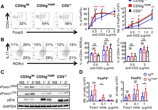
To address this possibility, we first compared FOXP3 expression in CD5tgWt, CD5tgY429F and
361
Cd5-/- CD4+ T cells stimulated with different doses of anti-CD3 antibodies in presence of
362
TGF-. We found that the percentages of CD5tgY429F and Cd5-/- CD4+ T cells expressing
363
FOXP3 were enhanced, compared to that in CD5tgWt CD4+ T cells (Fig. 5A), upon weak but
364
not high TCR stimulations, suggesting that CD5 may contribute to enhance the threshold at 365
which the development of iTreg is engaged. By comparison, we found that CD5tgWt,
366
CD5tgY429F and Cd5-/- CD4+ T cells differentiated similarly into RORt+IL-17+ cells following
367
TCR stimulation under Th17 polarizing conditions (Fig. 5B). Numbers of CD5tgY429F and
368
Cd5-/- FOXP3+ cells were enhanced independently of the concentration of anti-CD3
369
antibodies (Fig. 5A), suggesting that CD5 operates both by controlling the ability of CD4+ T
17
cells to differentiate into iTreg and by repressing the proliferation/survival of conventional T 371
cells prior or during their engagement into the Treg lineage. Accordingly, the percentages of 372
cells expressing FOXP3 were higher in non-divided (CTVhi) CD5tgY429F CD4+ T cells
373
compared to the same cell populations from CD5tgWt CD4+ T cells (SI Appendix, Fig. S4).
374
Confirming the positive effect of CD5 on AKT activity, we found that the phosphorylation of 375
FOXO1 on Thr24 and Ser256 was impaired in CD5tgY429F and Cd5-/- CD4+ T cells (Fig. 5C).
376
The phosphorylation of ERK, which is also correlated with efficient generation of induced 377
Treg cells (52), was enhanced in CD5tgY429F and Cd5-/- CD4+ T cells compared to that in
378
CD5tgWt CD4+ T cells, showing further that CD5 delivers both stimulatory and
co-379
inhibitory signals to modulate a specific T-cell response, namely the induction of Foxp3 and 380
the generation of iTreg cells. Finally, the numbers of FOXP3+ cells were similar in CD5tgWt
381
and CD5tgY429F CD4+ T cells when cells were incubated with high concentrations of FOXO1
382
inhibitors in in vitro assays, suggesting that the increased activity of ERK observed in 383
CD5tgY429F CD4+ T cells was not sufficient alone to enhance the generation of Treg cells
384
(Fig. 5D). In comparison, the numbers of CD5tgY429F CD4+FOXP3- T cells, reflecting T-cell
385
expansion, remained increased compared to those in CD5tgWt CD4+FOXP3- T cells when
386
cells were treated with similar doses of FOXO1 inhibitors (Fig. 5D). Altogether these results 387
suggested that CD5 signaling shape TCR signals to selectively repress FOXP3 expression. 388
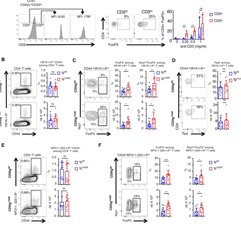
389
The surface level of CD5 correlates with TCR signal intensity, which is dictated by the 390
affinity of the TCR for self-ligands (9). To determine whether quantitative variations of CD5 391
surface expression within normal physiological ranges influences the generation of induced 392
FOXP3+ cells, we next sorted naïve CD62Lhi CD25- CD4+ T cells expressing either high or
393
low surface levels of CD5 and compared their ability to differentiate into FOXP3+ cells in
394
Treg-cell polarizing conditions. We found that CD5loCD4+ T cells differentiate more
18
efficiently into FOXP3+ cells than CD5hiCD4+ T cells under identical TCR stimulation
396
conditions, suggesting that physiologically high levels of CD5 on naïve CD4+ T cells might
397
reduce the ability of these cells to differentiate into Treg cells upon antigenic recognition (Fig. 398
6A). Contrasting with those results, previous studies suggested that CD5 promotes, rather 399
than represses, the differentiation of peripherally-induced Treg cells when those cells are 400
generated in a tolerogenic context such as the gut mucosa (15). Accordingly, we found that 401
the percentages of FOXP3+CD4+ T cells and of FOXP3+NeuropilinlowCD4+ T cells, which are
402
essentially composed of iTreg cells (53), were decreased in the Peyer patches of CD5-/- and 403
CD5tgY429F mice as compared to those in CD5tgWt mice (Fig. S5). We thus hypothesized that
404
CD5 signalosome might be influenced by the environment in which T cells are localized and 405
that CD5 may operate differently in a non-tolerogenic environment when CD4+ T cells
406
encounter foreign antigens or pathogens. To examine this possibility, we first immunized 407
CD5tgWt and CD5tgY429F mice with a peptide variant (EAWGALANKAVDKA, called 1W1K
408
peptide hereafter) of the I‐E alpha chain immunodominant peptide 52‐68 in the presence IFA, 409
which was shown to favor the polarization of iTreg cells (54). To follow antigen-induced 410
FOXP3+ T cells, we stained cells from the draining lymph nodes with 1W1K-pMHCII
411
tetramer and analyzed FOXP3 expression in tetramer+CD4+ T cells. We found that the
412
percentages of CD44+tetramer+CD4+ T cells were similar in CD5tgWt and CD5tgY429F mice,
413
indicating that CD5 signaling does not significantly impact the clonal expansion of antigen-414
specific CD4+ T cells in this experimental setting (Fig. 6B). We observed that the percentages
415
and numbers of tetramer+FOXP3+CD4+ T cells were increased in CD5tgY429F mice compared
416
to those in CD5tgWt mice (Fig. 6C). In comparison, the percentages and numbers of
417
tetramer+T-BET+CD4+ T cells were similar in CD5tgWt and CD5tgY429F mice (Fig. 6D).
418
Proportions and numbers of tetramer+FOXP3+NeuropilinlowCD4+ T cells were also higher in
419
CD5tgY429F mice compared to those in CD5tgWt mice, suggesting that CD5 signaling restrains
19
the generation of iTreg cells following immunization with foreign antigens. To confirm this 421
observation in a more pathophysiological model, we next analyzed whether CD5 signaling 422
could repress the generation of Treg cells following mice infection with the respiratory virus 423
influenza A, which was shown to drive important antigen-specific Treg-cell responses (55). 424
Mice were infected intranasally and pathogen-specific Treg were analyzed with NP311-325-425
IAb tetramer in the draining lymph nodes 5 days following infection. We found that the
426
percentages of virus-specific CD4+ T cells were comparable in CD5tgWt and CD5tgY429F mice,
427
indicating that CD5 signaling does not influence the overall expansion of antigen-specific 428
CD4+ T cell following infection (Fig. 6E). The proportions and numbers of virus-specific 429
FOXP3+CD4+ T cells and of FOXP3+NeuropilinlowCD4+ T cells were increased in CD5tgY429F
430
mice as compared to those in CD5tgWt, showing that CD5 represses the development of those
431
cells during pathogenic infection (Fig. 6F). 432
20 Discussion
433
434
In this study, we identified a multimeric signaling complex which acts through CD5 to limit 435
the induction of regulatory T cells. This complex is composed of proteins with adaptor 436
functions (c-CBL, CIN85, CRKL) that connect CD5 to distinct effector proteins (UBASH3A, 437
SHIP1, PI3K) which acts cooperatively by repressing ERK and promoting AKT activity to 438
inhibit the transactivation of Foxp3. This signalosome is recruited to CD5 Y429 following 439
TCR engagement, suggesting that it is part of a feedback loop that is differentially engaged 440
according to the strength of TCR signals that gradually regulates CD5 expression at the cell 441
surface. It is generally admitted that stimulation of CD4+ T cells with high dose of strong
442
agonist exerts a repressive effect on the generation of iTreg cells, presumably because it 443
promotes strong AKT-mediated signals that prevent the transactivation of Foxp3 (50). A 444
molecular model to explain this effect was that strong, but not weak, TCR signals repress the 445
expression of the PtdIns(3,4,5)P3 phosphatase and tensin homologue (PTEN), which is an 446
inhibitor of the AKT signaling pathway (56). Our study also suggests that CD4+ T cells could
447
finely tune AKT signaling by titrating the surface expression of CD5 to reduce the generation 448
of iTreg cells upon TCRs stimulation with relatively high dose of high affinity ligands. It was 449
shown that CD5hiCD4+ T cells respond better than CD5loCD4+ T cells to diverse foreign
450
antigens (11). This was explained by the enrichment, in the CD5hi population, of CD4+ T cells
451
with greater self-reactivity, which exhibit enhanced TCR signaling potential (57). An 452
additional explanation inferred from our study is that CD5 could act by reducing the 453
emergence of inopportune antigen-specific Treg cells that might occur during the recognition 454
of foreign antigens and which may lead to the development of ineffective immune responses. 455
This interpretation could also bring an explanation to the reduced development of active 456
autoimmunity observed in CD5 deficient mice models (12).
21 458
The modes of interaction of c-CBL with UBASH3A, SHIP, CIN85, CRKL and PI3K have 459
been extensively studied particularly in the context of EGF receptor (EGFR) signaling for 460
which a similar molecular machinery as the one described in this study for CD5 has been 461
described (36, 58, 59). PI3K can bind directly to c-CBL through a phospho-tyrosine binding-462
site located at the C-terminal end of c-Cbl (Y731) (60), or indirectly, through CRKL, which 463
binds to two phospho-tyrosine binding sites located in the same region (Y700 and Y774) (61). 464
CIN85 binds directly with SHIP1 and was shown to compete with UBASH3A for binding to 465
c-CBL, suggesting that two distinct CD5 signaling subsets might co-exist in T cells (62). 466
Previous studies have shown that CIN85/c-CBL complexes drive EGFR internalization and 467
degradation in lysosomal compartments whereas UBASH3A/c-CBL complexes prevent these 468
processes and sustain EGFR-mediated signals (36, 58). Thus, these distinct complexes could 469
contribute to regulate the turnover of CD5 in addition to their regulatory function on TCR 470
signaling. Although c-CBL was shown to mediate CD5 ubiquitylation resulting in its 471
degradation in lysosomes following TCR/CD5 co-cross-linking (63), we found that the 472
substitution of tyrosine 429 by phenylalanine does not significantly modify CD5 surface 473
expression either before or after TCR engagement, suggesting that the direct engagement of 474
CD5 by external ligands might be required for its degradation. 475
476
Several proteins previously identified as CD5-interacting proteins, such as Ras-Gap (22), 477
SHP-1 (23), Vav1 (25) and ZAP70 (64), were not detected as CD5 interactors in our mass 478
spectrometry analysis. Although initial studies suggested that SHP-1 interacts with CD5 in 479
thymocytes (65) and Jurkat cells (23), several studies since then have failed to reproduce this 480
interaction (22, 66). Moreover, both the phosphatase activity associated to CD5 481
immunoprecipitates and the reduction in positive selection conferred by CD5 overexpression 482
22
were shown to be unaffected by SHP-1 deficiency, suggesting that CD5 operates 483
independently of this phosphatase (67). CK2 (26) and Cbl-b (30), also previously described as 484
CD5-binding proteins, were recruited to CD5 but not as robustly as other interactors, 485
suggesting that CD5 could engage these proteins but in other molecular or cellular contexts 486
than the one used in this study. It was shown that CD5 enhances Th17 responses when cross-487
linked to the TCR but not when the TCR is engaged alone (68, 69), suggesting that CD5 488
could recruit distinct signaling effectors whether or not it is engaged by extracellular ligands. 489
Accordingly, the deletion of a CK2 binding site on CD5 impairs the polarization of CD4+ T
490
cells into Th17 cells following stimulation with antigen-presenting cells but not following 491
anti-CD3 antibodies (69). 492
493
We show that CD5 exerts a combined effect on TCR signaling, reducing ZAP-70 and ERK 494
activity, presumably through the joint action of UBASH3A and SHIP1, which are known 495
regulators of these signaling molecules (40, 44, 45, 70, 71), and enhancing AKT activity, 496
likely through PI3K, which facilitates the retention of FOXO1 in the cytoplasm and prevents 497
the consequent transactivation of Foxp3 (51). This signaling complex does not detectably 498
modulate the expression of RORt or T-BET in activated CD4+ T cells in vitro and in vivo
499
respectively, indicative of a selective effect of CD5 on the generation of iTreg cells. 500
Remarkably, studies using SHIP1 and PI3K deficient murine models identified repressive 501
functions for both proteins on the generation of induced Treg cells (72, 73). Whether 502
UBASH3A is also directly involved in the control of this population has not yet been studied. 503
However, a recent investigation performed in human T cells has shown that it represses NFB 504
signaling (74), a key pathway for the generation of induced Treg cells (75). Thus, CD5 could 505
act as a scaffold that selectively engages specific signaling effectors involved in the 506
23
generation of peripheral Treg cells to selectively control the expansion of this population 507
during immune responses. 508
509
Previous studies have shown that CD5 deficiency on the BALB/c but not on the C57BL/6 510
genetic background leads to increase numbers of thymic Treg cells, suggesting that CD5 may 511
also repress the generation of these cells according to the molecular context in which CD5 512
operates in the thymus (13, 14). CD5 is highly expressed at the surface of thymic Treg cells, 513
indicating that the ability of CD5 to negatively regulate FoxP3 expression in the thymus could 514
also be overcome by additional signals (such as high TCR affinity for self-ligand) that impose 515
FoxP3 expression despite the opposite force exerted by CD5. We thus speculate that CD5 516
mediated inhibition may be sufficient to block Foxp3 expression and Treg lineage 517
commitment in thymocytes that express TCRs that bind with intermediate affinity to self-518
ligands preventing their commitment to the Treg lineage. Also, co-receptors such as GITR, 519
OX40 and TNFR2 are highly expressed by thymic Treg progenitors and were also shown to 520
trigger co-stimulatory signals that induce thymic Treg differentiation (76). By contrast, these 521
co-receptors are poorly expressed by naïve peripheral CD4 T cells suggesting that the 522
negative effect of CD5 on FoxP3 expression may become predominant in this subset. 523
524
A recent study reported an effect of CD5 opposite to that shown in our study on the 525
generation of peripherally-induced Treg cells in a mouse model in which tolerance to EAE is 526
induced by direct delivery of encephalitogenic peptides to antigen-presenting dendritic cells in 527
the absence of adjuvant (15). Those authors found that CD5 prevents the inhibition of Treg-528
cell induction potentially mediated by effector cell cytokines such as IL-6, IL-4 and IFN. 529
Although they do not provide a clear signaling mechanism by which CD5 operates here, they 530
show that CD5 promotes the generation of Treg cells by inhibiting IL-6-mediated AKT 531
24
signaling leading to mTOR activation. One potential explanation for the apparent discrepancy 532
between those results and our current findings is that CD5 might exert opposite effects on 533
PI3K/ AKT signaling in tolerized and non-tolerized T cells. A recent proteomic study showed 534
that CD5 interacts with both c-CBL and CBL-b in peripheral CD4+ T cells (30), suggesting
535
that distinct pools of CD5, connected either to a c-CBL or CBL-b signalosome, could be 536
preferentially assembled under specific stimulation conditions. Whereas c-CBL has known 537
positive effects on PI3K/AKT signaling (46), CBL-b blocks this pathway by inducing PI3K 538
ubiquitylation (77) and by blocking Nedd4-mediated ubiquitylation of PTEN (78). Because 539
CBL-b expression is up-regulated in CD4+ T cells after tolerizing signals(79) and degraded in
540
CD4+ T cells that have received appropriate co-stimulatory signals (80), we speculate that
541
CD5 signaling pools may vary in size or composition, tuning up or down the PI3K/AKT 542
signaling axis according to the immunological context in which cells are stimulated. 543
544
Altogether the findings of our study suggest that CD5 engages a polymorphic signaling 545
machinery that can transduce both stimulatory and inhibitory signals to selectively control 546
specific T-cell responses depending on the immunological context. Our results also provide 547
new insights into the paradigm of co-receptor signaling suggesting that, in addition to 548
providing classical enhancing or dampening inputs, co-receptors coordinate TCR signals that 549
may have antagonist effects to promote specific functional outcomes, such as the generation 550
of iTreg cells. 551
25 Materials and Methods
553 554
Full details of materials and methods, including mice, antibodies, cell stimulation, 555
immunoprecipitations, mass spectrometry analysis, calcium flux, Western blot, immunization 556
and influenza virus infection, statistical analysis are provided in the SI Appendix. 557
558
Data availability statement 559
The data generated or analyzed during this study are included in the published article and its 560
supplementary information appendix or dataset files. The mass spectrometry proteomics data 561
have been deposited to the ProteomeXchange Consortium via the PRIDE partner repository 562
with the dataset identifier PXD017343. Raw data from figures 1A/S1A, S1B, 1C/2A/2F, 563
2B/S1C can be respectively found in Datasets S1, S2, S3 and S5. 564
26 Acknowledgements
565 566
We thank L. Dupré for critical reading of the manuscript. We also thank F.-E. L’Faqihi-567
Olive, V. Duplan-Eche, and A.-L. Iscache for technical assistance at the flow-cytometry 568
facility of INSERM U1043, the personnel of the US006 ANEXPLO/CREFRE animal facility 569
for expert animal care, and L. Guennec for administrative assistance. Funding: This work was 570
supported by INSERM and Sanofi (Avenir grant to R.L.); the Association pour la Recherche 571
sur le Cancer (ARC); the Intramural Research Program of the Eunice Kennedy Shriver, 572
National Institute of Child Health and Human Development; a Marie Curie International 573
Reintegration Grant (R.L.); the French Ministry of Higher Education and Research (PhD 574
fellowship for G.B.); the Région Midi-Pyrénées, European funds (Fonds Européens de 575
Développement Régional, FEDER), Toulouse Métropole, and the French Ministry of 576
Research with the ‘Investissement d’Avenir Infrastructures Nationales en Biologie et Santé 577
program’ (ProFI, Proteomics French Infrastructure project, ANR-10-INBS-08, to O.B.S.). 578
579
The authors declare no competing interest. 580
27 References
581
1. Chen L & Flies DB (2013) Molecular mechanisms of T cell co-stimulation and co-inhibition.
582
Nature reviews. Immunology 13(4):227-242.
583
2. Coquet JM, Rausch L, & Borst J (2015) The importance of co-stimulation in the orchestration
584
of T helper cell differentiation. Immunol Cell Biol 93(9):780-788.
585
3. Anderson AC, Joller N, & Kuchroo VK (2016) Lag-3, Tim-3, and TIGIT: Co-inhibitory
586
Receptors with Specialized Functions in Immune Regulation. Immunity 44(5):989-1004.
587
4. Acuto O & Michel F (2003) CD28-mediated co-stimulation: a quantitative support for TCR
588
signalling. Nat Rev Immunol 3(12):939-951.
589
5. Parry RV, Riley JL, & Ward SG (2007) Signalling to suit function: tailoring phosphoinositide
590
3-kinase during T-cell activation. Trends Immunol 28(4):161-168.
591
6. Rudd CE, Taylor A, & Schneider H (2009) CD28 and CTLA-4 coreceptor expression and
592
signal transduction. Immunol Rev 229(1):12-26.
593
7. Walker LSK (2017) PD-1 and CTLA4: Two checkpoints, one pathway? Sci Immunol 2(11).
594
8. Tarakhovsky A, et al. (1995) A role for CD5 in TCR-mediated signal transduction and
595
thymocyte selection. Science 269(5223):535-537.
596
9. Azzam HS, et al. (1998) CD5 expression is developmentally regulated by T cell receptor
597
(TCR) signals and TCR avidity. The Journal of experimental medicine 188(12):2301-2311.
598
10. Azzam HS, et al. (2001) Fine tuning of TCR signaling by CD5. J Immunol 166(9):5464-5472.
599
11. Mandl JN, Monteiro JP, Vrisekoop N, & Germain RN (2013) T cell-positive selection uses
600
self-ligand binding strength to optimize repertoire recognition of foreign antigens. Immunity
601
38(2):263-274.
602
12. Axtell RC, Webb MS, Barnum SR, & Raman C (2004) Cutting edge: critical role for CD5 in
603
experimental autoimmune encephalomyelitis: inhibition of engagement reverses disease in
604
mice. J Immunol 173(5):2928-2932.
605
13. Dasu T, et al. (2008) CD5 plays an inhibitory role in the suppressive function of murine
606
CD4(+) CD25(+) T(reg) cells. Immunology letters 119(1-2):103-113.
607
14. Ordonez-Rueda D, et al. (2009) Increased numbers of thymic and peripheral CD4+
608
CD25+Foxp3+ cells in the absence of CD5 signaling. European journal of immunology
609
39(8):2233-2247.
610
15. Henderson JG, Opejin A, Jones A, Gross C, & Hawiger D (2015) CD5 instructs extrathymic
611
regulatory T cell development in response to self and tolerizing antigens. Immunity
42(3):471-612
483.
28
16. Van de Velde H, von Hoegen I, Luo W, Parnes JR, & Thielemans K (1991) The B-cell surface
614
protein CD72/Lyb-2 is the ligand for CD5. Nature 351(6328):662-665.
615
17. Vera J, et al. (2009) The CD5 ectodomain interacts with conserved fungal cell wall
616
components and protects from zymosan-induced septic shock-like syndrome. Proc Natl Acad
617
Sci U S A 106(5):1506-1511.
618
18. Zhang C, et al. (2016) CD5 Binds to Interleukin-6 and Induces a Feed-Forward Loop with the
619
Transcription Factor STAT3 in B Cells to Promote Cancer. Immunity 44(4):913-923.
620
19. Bhandoola A, et al. (2002) CD5-mediated inhibition of TCR signaling during intrathymic
621
selection and development does not require the CD5 extracellular domain. Eur J Immunol
622
32(6):1811-1817.
623
20. Osman N, Lazarovits AI, & Crumpton MJ (1993) Physical association of CD5 and the T cell
624
receptor/CD3 antigen complex on the surface of human T lymphocytes. Eur J Immunol
625
23(5):1173-1176.
626
21. Burgess KE, Yamamoto M, Prasad KV, & Rudd CE (1992) CD5 acts as a tyrosine kinase
627
substrate within a receptor complex comprising T-cell receptor zeta chain/CD3 and
protein-628
tyrosine kinases p56lck and p59fyn. Proc Natl Acad Sci U S A 89(19):9311-9315.
629
22. Dennehy KM, Broszeit R, Ferris WF, & Beyers AD (1998) Thymocyte activation induces the
630
association of the proto-oncoprotein c-cbl and ras GTPase-activating protein with CD5.
631
European journal of immunology 28(5):1617-1625.
632
23. Perez-Villar JJ, et al. (1999) CD5 negatively regulates the T-cell antigen receptor signal
633
transduction pathway: involvement of SH2-containing phosphotyrosine phosphatase SHP-1.
634
Molecular and cellular biology 19(4):2903-2912.
635
24. Dennehy KM, et al. (1997) Thymocyte activation induces the association of
636
phosphatidylinositol 3-kinase and pp120 with CD5. Eur J Immunol 27(3):679-686.
637
25. Gringhuis SI, de Leij LF, Coffer PJ, & Vellenga E (1998) Signaling through CD5 activates a
638
pathway involving phosphatidylinositol 3-kinase, Vav, and Rac1 in human mature T
639
lymphocytes. Molecular and cellular biology 18(3):1725-1735.
640
26. Raman C & Kimberly RP (1998) Differential CD5-dependent regulation of CD5-associated
641
CK2 activity in mature and immature T cells: implication on TCR/CD3-mediated activation. J
642
Immunol 161(11):5817-5820.
643
27. Raman C, Kuo A, Deshane J, Litchfield DW, & Kimberly RP (1998) Regulation of casein
644
kinase 2 by direct interaction with cell surface receptor CD5. J Biol Chem
273(30):19183-645
19189.
646
28. Lu X, et al. (2002) AP2 adaptor complex-dependent internalization of CD5: differential
647
regulation in T and B cells. J Immunol 168(11):5612-5620.
29
29. Voisinne G, et al. (2019) Quantitative interactomics in primary T cells unveils TCR signal
649
diversification extent and dynamics. Nature immunology 20(11):1530-1541.
650
30. Voisinne G, et al. (2016) Co-recruitment analysis of the CBL and CBLB signalosomes in
651
primary T cells identifies CD5 as a key regulator of TCR-induced ubiquitylation. Mol Syst
652
Biol 12(7):876.
653
31. Dennehy KM, et al. (2001) Determination of the tyrosine phosphorylation sites in the T cell
654
transmembrane glycoprotein CD5. Int Immunol 13(2):149-156.
655
32. Vila JM, et al. (2001) Residues Y429 and Y463 of the human CD5 are targeted by protein
656
tyrosine kinases. Eur J Immunol 31(4):1191-1198.
657
33. Lupher ML, Jr., Songyang Z, Shoelson SE, Cantley LC, & Band H (1997) The Cbl
658
phosphotyrosine-binding domain selects a D(N/D)XpY motif and binds to the Tyr292
659
negative regulatory phosphorylation site of ZAP-70. J Biol Chem 272(52):33140-33144.
660
34. van Leeuwen JE, Paik PK, & Samelson LE (1999) Activation of nuclear factor of activated T
661
cells-(NFAT) and activating protein 1 (AP-1) by oncogenic 70Z Cbl requires an intact
662
phosphotyrosine binding domain but not Crk(L) or p85 phosphatidylinositol 3-kinase
663
association. J Biol Chem 274(8):5153-5162.
664
35. Take H, et al. (2000) Cloning and characterization of a novel adaptor protein, CIN85, that
665
interacts with c-Cbl. Biochem Biophys Res Commun 268(2):321-328.
666
36. Kowanetz K, et al. (2004) Suppressors of T-cell receptor signaling Sts-1 and Sts-2 bind to Cbl
667
and inhibit endocytosis of receptor tyrosine kinases. J Biol Chem 279(31):32786-32795.
668
37. Naramura M, Kole HK, Hu RJ, & Gu H (1998) Altered thymic positive selection and
669
intracellular signals in Cbl-deficient mice. Proc Natl Acad Sci U S A 95(26):15547-15552.
670
38. Rao N, et al. (2002) Negative regulation of Lck by Cbl ubiquitin ligase. Proc Natl Acad Sci U
671
S A 99(6):3794-3799.
672
39. Kong MS, et al. (2019) Inhibition of T cell activation and function by the adaptor protein
673
CIN85. Science signaling 12(567).
674
40. Dong S, et al. (2006) T cell receptor for antigen induces linker for activation of T
cell-675
dependent activation of a negative signaling complex involving Dok-2, SHIP-1, and Grb-2. J
676
Exp Med 203(11):2509-2518.
677
41. Van Slyke P, et al. (2005) Dok-R mediates attenuation of epidermal growth factor-dependent
678
mitogen-activated protein kinase and Akt activation through processive recruitment of c-Src
679
and Csk. Mol Cell Biol 25(9):3831-3841.
680
42. Zhao M, Janas JA, Niki M, Pandolfi PP, & Van Aelst L (2006) Dok-1 independently
681
attenuates Ras/mitogen-activated protein kinase and Src/c-myc pathways to inhibit
platelet-682
derived growth factor-induced mitogenesis. Mol Cell Biol 26(7):2479-2489.
30
43. Liu Q, et al. (1998) The inositol polyphosphate 5-phosphatase ship is a crucial negative
684
regulator of B cell antigen receptor signaling. The Journal of experimental medicine
685
188(7):1333-1342.
686
44. Tomlinson MG, Heath VL, Turck CW, Watson SP, & Weiss A (2004) SHIP family inositol
687
phosphatases interact with and negatively regulate the Tec tyrosine kinase. J Biol Chem
688
279(53):55089-55096.
689
45. Scharenberg AM, et al. (1998) Phosphatidylinositol-3,4,5-trisphosphate (PtdIns-3,4,5-P3)/Tec
690
kinase-dependent calcium signaling pathway: a target for SHIP-mediated inhibitory signals.
691
EMBO J 17(7):1961-1972.
692
46. Thien CB, et al. (2010) c-Cbl promotes T cell receptor-induced thymocyte apoptosis by
693
activating the phosphatidylinositol 3-kinase/Akt pathway. The Journal of biological chemistry
694
285(14):10969-10981.
695
47. Sattler M, et al. (1997) Steel factor induces tyrosine phosphorylation of CRKL and binding of
696
CRKL to a complex containing c-kit, phosphatidylinositol 3-kinase, and p120(CBL). The
697
Journal of biological chemistry 272(15):10248-10253.
698
48. Deane JA, et al. (2007) T-cell function is partially maintained in the absence of class IA
699
phosphoinositide 3-kinase signaling. Blood 109(7):2894-2902.
700
49. Moran AE, et al. (2011) T cell receptor signal strength in Treg and iNKT cell development
701
demonstrated by a novel fluorescent reporter mouse. The Journal of experimental medicine
702
208(6):1279-1289.
703
50. Li MO & Rudensky AY (2016) T cell receptor signalling in the control of regulatory T cell
704
differentiation and function. Nature reviews. Immunology 16(4):220-233.
705
51. Fabre S, et al. (2005) Stable activation of phosphatidylinositol 3-kinase in the T cell
706
immunological synapse stimulates Akt signaling to FoxO1 nuclear exclusion and cell growth
707
control. J Immunol 174(7):4161-4171.
708
52. Lu L, et al. (2010) Role of SMAD and non-SMAD signals in the development of Th17 and
709
regulatory T cells. J Immunol 184(8):4295-4306.
710
53. Weiss JM, et al. (2012) Neuropilin 1 is expressed on thymus-derived natural regulatory T
711
cells, but not mucosa-generated induced Foxp3+ T reg cells. The Journal of experimental
712
medicine 209(10):1723-1742, S1721.
713
54. Korn T, et al. (2008) IL-6 controls Th17 immunity in vivo by inhibiting the conversion of
714
conventional T cells into Foxp3+ regulatory T cells. Proc Natl Acad Sci U S A
105(47):18460-715
18465.
716
55. Betts RJ, et al. (2012) Influenza A virus infection results in a robust, antigen-responsive, and
717
widely disseminated Foxp3+ regulatory T cell response. Journal of virology 86(5):2817-2825.