Université de Sherbrooke
Crosstalk between TAF6δ and Notch Signalling Pathways in Cancer Cell Lines
Par
Edith Milena Alvarado Cuevas
Programme de microbiologie et d´infectiologie
Mémoire présenté à la Faculté de médecine et des sciences de la santé en vue de l’obtention du grade de maitre ès sciences (M. Sc.)
en Microbiologie
Sherbrooke, Québec, Canada Mai, 2017
Membres du jury d’évaluation
Brendan BELL PhD, Département de Microbiologie et d’infectiologie Benoit CHABOT PhD, Département de Microbiologie et d’infectiologie
Marie-Josée BOUCHER PhD, Département de Biologie cellulaire
Dedicated to God Guadalupe Virgin Chucho Priest My Lovely Parents and Family et
Don't give up, please don't give way, Even if the cold burns, Even if fear bites, Even if the sun sets, And the wind goes silent, There is still fire in your soul There is still life in your dreams. Because every day is a new beginning, Because this is the hour and the best moment. By Mario Benedetti
Thank you GOD for everything in my life. The good and bad. Some were blessings and some were Lessons! ^^
Some people has an American dream but in my case, I had a Canadian dream. Coming to Canada, the “Land of Endless Opportunities” and study here was my wildest, incredible and exciting dream. I made it.
Jefesito, the success I've had in my master is due to your support and encouragement. I appreciate you so much and value everything that I have learned from you and thank you for the tremendous opportunity. I am so very grateful to you, jefesita and linda. All of you that worked so very hard to help me achieve a step towards completing my goals. Thank you all for your support, advice, words of encouragement that mean a lot to me and for guiding me professionally and personally!!!
I would like to take this opportunity also to thank all the members of my team (Sonrisita, Mik, Kitty, Chica, Alex crazy, Jeremy, Leo) for their involvement and dedication.
I am truly blessed to be able to share knowledge, work and adventures together (All my Beautiful Bell Lab). Muchasss Graciasss. Los quiero mucho.
So thank you very much for this pleasant experience and please accept my good wishes for your continued success. \(^o^)/
Marie-Josée Boucher, thanks for being in my comité d’encadrement. Thanks for your guidance and advice helped brighten my master and professional life.
Thanks to Dr Benoit Chabot for all the sharing moments with your lab, your amazing food and for accepting to be part of my jury and correct my thesis.
Thanks to Dr Steve Jean for generously sharing his expertise in the use of the CellProfiler software.
Thanks to Dr Arndt Benecke, our collaborator in France, for microarray analysis and thanks to his entire lab.
I would like to thank every member of the Microbiology and Infectology department, the faculty, staff and students. I enjoyed the activities and collaborative academic atmosphere in my program. I also thank to our dear secretaries: Carole Picard, Chantale Simard, Suzanne Topping, Isabelle Bérubé and Sophie Mailloux who have been working hard in our program.
Thanks to my parents, my brother batman, and my sister turtle, my aunties and all my family for always listening to me, supporting me, and encouraging me. I want you to know how much I love and appreciate you. You´re the best y los Amo.
Merci mon chéri, tu es l’Etoile de ma vie, qui illumine mes nuits, qui remplit mon cœur. D’un grand bonheur. Je t’aime pour toujours Mon plus bel amour. Mais, si t’aimer est une erreur alors je ferai cette erreur dans toute ma vie. ^^ Te Amo Mucho et aussi merci pour la belle-famille parce que avoir un endroit où aller est un foyer. Avoir quelqu’un à aimer est une famille. Mais avoir les deux à la fois est une bénédiction.
Soulmates are people who bring out the best in you. They are not perfect but are always perfect for you. Thank you very much for being my soulmate Mi Cosita Bonita (Jen2). Muchas gracias for being perfect for me.
Thanks Monstrou, you know, good friends are like stars. You don’t always see them but you know they’re always there!!. Gracias por tar ahí.
Thanks La Mía, asere ke bola!!^^. ¡Sabías que detrás de toda mujer, hay una amiga loca que la anima constantemente! Gracias por ser mi motivación ^^
Thanks to Leandro Fequino for helping me out in all my computer problems, including save all my results and my thesis. Muchas Gracias por salvar mi maestría!!
Thanks also to all my Canadians, French’s, Cubans, Algerians, Mexicans and Colombians friends; because it does not matter where I go, they are always there. Mil gracias por hacerme parte de sus vidas!
The friendships and professional relationships that have come because of involvement in science research are some of the greatest gifts of my professional career. When I will finish, it will be with a grateful heart, so deeply enriched by all of the colleagues, friends, mentors, my boyfriend and my boss, all who have so kindly shared so much with me.
The future belongs to those who believe in the beauty of their dreams….Eleanor Roosevelt
Thanks a lot to CANADA and Université de Sherbrooke for the greatest opportunity in my life and thanks for my scholarship, tranzyme pharma bursary and the funding agencies (IRSC/CIHR, CRSNG/NSERC).
R
ÉSUMÉ
Interaction entre les voies de signalisation TAF6δ et Notch dans des lignées de cellules Cancéreuses
Par
Edith Milena Alvarado Cuevas
Programme de microbiologie et d´infectiologie
Mémoire présenté à la Faculté de médecine et des sciences de la santé en vue de l’obtention du diplôme de maître ès sciences (M.Sc.) en microbiologie et d´infectiologie, Faculté de
médecine et des sciences de la santé, Université de Sherbrooke, Sherbrooke, Québec, Canada, J1H 5N4
La voie de signalisation de Notch contrôle de multiples processus cellulaires, telle la différenciation, la prolifération cellulaire et l’apoptose. Son activation repose sur la liaison du récepteur Notch par son ligand. Par la suite, le domaine intracellulaire actif de Notch (NIC) est relâché après son clivage médié par la γ-sécrétase. Cela permet au NIC d’être transporté au noyau où celui-ci lie la protéine CSL et active la transcription de ses gènes cibles, comme Hes1. TAF6 est une sous-unité du facteur de transcription général TFIID qui joue un rôle important dans la régulation de la transcription effectuée par l’ARN polymérase II. L’isoforme TAF6δ peut induire l’apoptose et aussi l’expression des gènes cibles de Notch. Cette étude a pour objectif d’explorer l’interaction croisée entre les voies de signalisation de Notch et de TAF6δ et leur impact sur l’apoptose. Pour valider l’impact de l’expression de TAF6δ sur la voie de signalisation de Notch, nous avons effectué une analyse par micropuce. L’expression de TAF6δ médiée par la transfection de SSOs (oligonucléotides Splice-Switching) a révélé une induction γ-sécrétase dépendante de gènes cibles de Notch dans les cellules HeLa. La cytométrie de flux a en outre montré que l'apoptose TAF6δ-dépendante est réduite par un traitement avec des inhibiteurs de gamma-sécrétase. L'analyse par immunofluorescence a révélé que TAF6δ induit la translocation de NIC-2 au noyau. Enfin, une analyse par qPCR a montré que l'expression du gène cible Notch est augmentée dans plusieurs lignées de cellules cancéreuses en réponse à l’induction TAF6δ. Nos données montrent, que la voie de signalisation de Notch est activée par TAF6δ dans plusieurs modèles de cancer et que l’interaction entre ces deux voies contribue à l'apoptose dans un modèle de cancer du col de l'utérus.
S
UMMARY
Crosstalk between TAF6δ and Notch Signalling Pathways in Cancer Cell Lines By
Edith Milena Alvarado Cuevas
Department of Microbiology and infectiology
Thesis presented at the Faculty of medicine and health sciences to obtain the Master degree diploma of Sciences (M.Sc.) in Microbiology and infectiology, Faculty of medicine and
health sciences, Université de Sherbrooke, Sherbrooke, Québec, Canada, J1H 5N4 Background: The Notch pathway controls multiple cellular processes, such as differentiation, cell proliferation and apoptosis. Its activation is based on the ligand binding to a Notch receptor after which, the Notch intracellular active domain (NIC) is released through cleavage mediated by γ-secretase. Upon cleavage, NIC translocates to the nucleus, where it binds CSL (CBF1/Su (H)/Lag-1) and activates the transcription of its target genes such as Hes1. TAF6 is a subunit of the TFIID basal transcription complex that plays an important role in the regulation of RNA polymerase II transcription. TAF6δ is a specialized isoform of TAF6 that can induce apoptosis and induces the expression of Notch target genes. This study aims to explore the potential crosstalk between TAF6δ and Notch signalling pathways and its impact on apoptosis.
Results: To validate the impact of TAF6δ expression on the Notch pathway, we performed microarray analysis. TAF6δ induction, mediated through transfection of SSOs (Splice-Switching oligonucleotides), revealed a γ-secretase–dependent induction of Notch target genes in HeLa cells. Flow cytometry analysis further showed that TAF6δ-dependent apoptosis is reduced by treatment with γ-secretase inhibitors. Immunofluorescence analysis revealed that TAF6δ induced translocation of NIC-2 to the nucleus. Finally, qPCR showed that Notch target gene expression is increased in several cancer cell lines in response to TAF6δ induction.
Conclusion: Our data show that the Notch pathway is activated by TAF6δ in several models of cancer, and that this association contributes to apoptosis in cervical cancer.
T
ABLE OF CONTENTS
Résumé ... ivi
Summary ... ivii
Table of contents ... viii
List of figures ... xi
List of tables ... xii
List of abbreviations ... xiii
1 INTRODUCTION ... 1
1.1 Programmed cell death ... 2
1.2 Apoptosis ... 3 1.2.1 Definition……..……….3 1.2.2 Morphologies of apoptosis……….. 4 1.2.3 Mechanisms of apoptosis……… 5 1.2.3.1 The Caspases ... 5 1.2.3.2 Extrinsic pathway……… 7 1.2.3.3 Intrinsic pathway………. 9
1.2.4 Regulatory mechanism of apoptosis……….…… 10
1.2.4.1 The BCL-2 family……….…….10
1.3 The Notch Signalling Pathway……….. 13
1.3.1 Molecular biology of Notch Receptors……….. 14
1.3.2 Molecular biology of Notch Ligands………. 16
1.3.3 Activation mechanism of Notch signalling pathway……….. 17
1.3.4 Notch targets genes……… 19
1.3.5 Notch in tumorigenesis………... 20
1.3.6 The role of Notch pathway in cervical cancer……… 20
1.4 Transcription…...………. 22
1.4.1 Transcription by RNA Pol II……..……….. 23
1.4.1.1 Initiation………....23
1.4.1.1.1 Generalities of the basal transcription factors……….… 24
1.4.1.1.2 Mechanism of RNA Pol II initiation………..………... 25
1.4.1.1.4 Mechanism of RNA Pol II termination………. 28
1.5 TFIID………...……….………….30
1.5.1 TAF6……… 32
1.5.2 TAF6δ……….. 35
2 HYPOTHESIS ... 37
3 MATERIAL AND METHODS..……….………39
3.1 Cell culture ... ...40
3.1.1Transfections with SSOs (Splice Switching Oligonucleotides)……….….40
3.2 Blocking Notch pathway by GSI treatment…….………..…..41
3.2.1 Activation of Notch Pathway through EGTA treatment………41
3.3 Antibodies………... 42
3.4 Extraction of RNA………...42
3.4.1 RT-PCR (Reverse transcription polymerase chain reaction)……….43
3.4.2 PCR (Polymerase chain reaction)………..43
3.4.3 qPCR (Real time PCR)………...44
3.5 Western Blot………....45
3.6 Immunofluorescence………... 46
3.7 Apoptosis assays………..46
3.8 Microarray Analysis of Gene Expression………...47
3.9 Statistical analyses………...47
4 RESULTS ... 49
4.1 To determine the impact of Notch pathway inhibition on the TAF6δ-driven transcriptome changes in HeLa cells………..50
4.1.1 Notch signalling impacts TAF6δ-driven transcriptome changes…..………50
4.1.2 Effect of TAF6δ induction on genes of the Notch pathway at mRNA levels…….53
4.1.3 Effect of GSI on Notch pathway at mRNA levels after TAF6δ induction...55
4.2 Determine if TAF6δ affect Notch target genes expression in other cancer cell lines………...56
4.2.1 Effect of TAF6δ induction on Notch pathway genes in pancreatic and breast cancer cell lines………..56
4.3 Test whether there is activation of the Notch pathway in response to TAF6δ in HeLa cells ………...57
4.3.2 Effect of GSI and EGTA on NIC-1 levels..………...59
4.3.3 Effect of SSOs and EGTA on NIC-1 levels..……….60
4.3.4 Effect of TAF6δ induction on NIC-2 levels….…….………61
4.3.5 Effect of TAF6δ induction on cleaved NIC-2…….………..63
4.4 Determine the effect of inhibiting the Notch pathway on TAF6δ- induced apoptosis in HeLa cells………..67
4.4.1 Effect of GSI on apoptosis………67
4.4.2 Effect of GSI on Cisplatin induced apoptosis………68
4.4.3 Down-regulation of Notch signalling by GSI treatment reduces apoptotic effects of TAF6δ………...69
5 DISCUSSION AND CONCLUSION ... 70
6 ATTACHMENTS ... 81
Attachment 1 Licenses for use of figures and tables from publications…………...81
Attachment 2 Table 5. Notch Signalling Pathway Target Genes………...82
Figure 1. Apoptotic morphological changes………...4
Figure 2. Classification of Caspases (Cysteine Aspartate acid proteases)………...5
Figure 3. Activation of procaspase-3 by cleavage………...6
Figure 4. Receptor-mediated caspase activation at the DISC………...7
Figure 5. Intrinsic and extrinsic pathways of apoptosis………...…8
Figure 6. Intrinsic apoptosis pathway……… …...10
Figure 7. BCL-2 family of proteins………...11
Figure 8. Drosophila with Notches………...14
Figure 9. Structure of the four human Notch receptors……….15
Figure 10. Domain organization of mammalian Notch ligands……….16
Figure 11. The canonical Notch signalling pathway………...……..18
Figure 12. Comparative view of the repression and activation complexes………19
Figure 13. Architecture of the basal PIC………...23
Figure 14. The transcription cycle……….30
Figure 15. Recognition of core promoter elements by TFIID and TFIIB………32
Figure 16.TAF6 alternative splicing modified interaction with his dimer partner TAF9………...………. 33
Figure 17. Isoforms of TAF6………34
Figure 18. A model for the TAF6δ pathway……….35
Figure 19. A fraction of TAF6-regulated transcription depends on Notch signalling……51
Figure 20. TAF6-regulated transcription of Notch target genes……….53
Figure 21. Effect of TAF6δ induction on Notch target genes at mRNA levels in HeLa cells………... 54
Figure 22. Effect of GSI on Notch target genes mRNA levels after TAF6δ induction in HeLa cells……….……….. 55
Figure 23. Effect of TAF6δ induction on Notch target genes at mRNA levels in different cancer cell lines………..………....57
Figure 24. Effect of GSI on Hes1 protein levels after TAF6δ induction in HeLa cells……59
Figure 25. Effect of GSI and EGTA on the intracellular active domain of Notch1 (NIC-1) in HeLa cells………..60
Figure 26. Effect of SSOs and EGTA on the intracellular active domain of Notch1 (NIC-1) in HeLa cells………..………61
Figure 27. TAF6δ expression increases level of active Notch2 (NIC-2) in HeLa cells……….……….. 63
Figure 28. TAF6δ-expression increases level of active nuclear Notch2 (NIC-2) in HeLa cells………... 66
Figure 29. GSI does not affect apoptosis in HeLa Cells ………. 67
Figure 30. HeLa Cells in the presence of GSI and two doses of Cisplatin apoptosis-inductor……… .68
Figure 31. GSI reduces apoptotic effects of TAF6δ induction in HeLa Cells…….……… 69
Figure 32. First model for crosstalk between the TAF6δ and Notch pathways……… 79
L
IST OF TABLES
Table 1. Complexes involved in RNA Pol II PIC assembly………..………….27 Table2. Nomenclature of TAFs involved in RNA polymerase II-mediated transcription………...………31 Table 3. Cancer cell lines used in this study……….40 Table 4. Primer sequences, annealing temperatures and amplicon sizes for qPCR analysis used in this study………...44 Table 5. Notch Signalling Pathway Target Genes……….82
ADAM AIF ANK Apaf1 APS APH1 Bad Bak Bax B-ALL Bcl 2 Bcl XL BH Bid Bik Bim bHLH Bmf Bok BSA Caspase CC cDNA c-MYC CO2 Co-R CPEs CPF CS CSL CStF CTD DAPT DCE DD DED Deltex DIABLO DISC Dll
A desintegrin and metalloproteinase Apoptosis-Inducing Factor
Ankyrin repeat domain
Apoptosis Protease Activating Factor 1 Ammonium Persulfate
Anterior pharynx-defective 1
BCL2-associated agonist o f cell death BCL2-antagonist/killer 1
BCL2-associated X
B- cell acute lymphoblastic leukemia B-cell CLL/lymphoma 2
B-cell lymphoma-extra large Bcl-2 Homology
BH3 Interacting Death Domain Agonist BCL2-interacting killer
B-cell lymphoma 2 Interacting Mediator o f cell death Basic helix-loop-helix
Bcl2 modifying factor BCL2-related ovarian killer Bovine Serum Albumin
Cysteinyl Aspartate Specific Protease Cervical Cancer
Complementary Deoxyribonucleic acid
V-Myc Avian Myelocytomatosis Viral Oncogene Homolog Carbon Dioxide
Corepressor’s
Core promoter elements
Cleavage and Polyadenylation Specific Factor Calf Serum
CBF1/Su(H)/Lag-1
Cleavage Stimulation Factor Carboxyl-terminal Domain
N-[N-(3, 5-Difluorophenacetyl)-L-alanyl]-S-phenylglycine t-butyl ester
Downstream Core Element Death Domains
Death Effector Domains Deltex E3 Ubiquitin Ligase
Direct IAP-binding protein with low PI Death Inducing Signalling Complex Delta-like
DMEM DMSO DNA DNTP’s DOS DPE DSIF DSL DTT EDTA EGF EGTA ER EtBr FADD FACS FBS GATA GO GSI GTFs HD HEPES Hes1 Herp HEY1 HFD HRP HPVs HR-HPVs IAP Inr IMS IMM JAG KCl KH2PO4 Lfng LNR MAML1 Mcl-1 Mg MgCl2 µL mL µM mM
Dulbecco’s Modified Eagle Medium Dimethyl sulfoxide
Deoxyribonucleic acid
Deoxyribonucleotide triphosphates Delta and OSM-11-like proteins Downstream core Promoter Element DRB-sensitivity-inducing factor Delta/Serrate/LAG-2
Dithiothreitol
Ethylenediaminetetraacetic acid Epidermal Growth Factor
Ethylene glycol-bis(2-aminoethylether)-N, N, N’, N’-tetraacetic acid Endoplasmic Reticulum
Ethidium Bromide
Fas Associated via Death Domain Fluorescence-activated cell sorting Fetal Bovine Serum
GATA binding protein Gene Ontology
γ-secretase inhibitor
General Transcription Factors Heterodimerization Domain
4-(2-hydroxyethyl)-1-piperazineethanesulfonic acid Hairy/Enhancer of split 1
Hes-related repressor protein
Hairy/enhancer of split with motif YRPW 1 Histone Fold Domain
Horseradish peroxydase Human Papillomaviruses
High-risk human papillomaviruses Inhibitor of Apoptosis Protein Initiator
Mitochondrial Intermembrane Space Inner Mitochondrial Membrane Jagged
Potassium Chloride
Potassium Dihydrogen Phosphate Lunatic Fringe
Cysteine-rich LNR repeats Mastermind-like
Myeloid Cell Leukemia sequence 1 Microgramme Magnesium chloride Microlitre Milliliter Micromolar Millimolar
MOMP MPT mRNA MTE Na2HPO4 NaCl NELF NCR NCT NIC NLS Noxa nM NRARP NRR OMM Omi/HtrA2 Opti-MEM PARP1 pb pRB PBS PCD PCR qPCR PEN2/PSENEN PEST PFA PIC PT P-TEFb Pre-Tα PUMA P300/CBP RAM RNA RNA pol rRNA rpm RT RT-PCR SHARP SDS SDS-PAGE Smac SMRT SS
Mitochondrial Outer Membrane Permeabilization Mitochondrial Permeability Transition
Messenger Ribonucleic acid Motif Ten Element
Sodium phosphate dibasic, Disodium hydrogen phosphate Sodium Chloride
Negative Elongation Factor Cysteine response region Nicastrin
Notch intracellular active domain Nuclear Localizing Signals NADPH oxidase activator 1 Nanomolar
NOTCH-Regulated Ankyrin Repeat Protein Negative Regulatory Region
Outer Mitochondria Membrane Mitochondrial Serine Protease Minimal Essential Medium Poly(ADP-Ribose) Polymerase 1 Base pair
Retinoblastoma protein Phosphate Buffered Saline Programmed Cell Death Polymerase Chain Reaction
Real-time Polymerase Chain Reaction Presenilin Enhancer 2
proline (P), glutamine (E), serine (S) and threonine (T) residues Paraformaldehyde
Preinitiation Complex Pore Transition
Positive Transcription Elongation Factor-b Pre-T-cell receptor alpha chain
p53 Upregulated Modulator of Apoptosis CREB-binding protein
RBP-J kappa-associated module Ribonucleic acid
RNA polymerase
Ribosomal Ribonucleic acid Revolutions per minute Reverse transcription
Reverse transcription polymerase chain reaction SMRT/HDAC1 histone deacetylase 1
Sodium Dodecyl sulfate
sodium dodecyl sulfate polyacrylamide gel electrophoresis Second Mitochondria-derived Activator of Caspase
Silencing Mediator of Retinoid and Thyroid receptors Splice Sites
SSOs TAD TAFs TBE tBid TBP TEMED TFIID TFs TNF TM tRNA TSS …
Splice Switching Oligonucleotides Transactivation Domain
TBP-associated factors Tris/Borate/EDTA
Truncated BH3 Interacting Death Domain Agonist TATA binding protein
Tetramethylethylenediamine Transcription factor IID Transcription Factors Tumor Necrosis Factor Transmembrane domain Transfer Ribonucleic acid Transcription Start Site …
1.1 PROGRAMMED CELL DEATH
The term “Programmed” was defined as the “exact instance in which physiological cell death occurs” (Bruce Alberts 2002). Programmed cell death (PCD) is important in different biological phenomena such as ageing, development and pathology. Therefore, in multicellular organisms the number of cells is highly regulated by controlling not only the proportion of proliferation but also the proportion of cell death, such as the loss of cells during aging and development, thus establishing a balance (Lockshin and Beaulaton 1974, Bruce Alberts 2002). Since PCD was originally discovered, several studies reported different mechanisms of cell death. Specifically, three types of cell death have been classified:
Type I: Apoptosis, a physiological process that kill useless cells during development, but does not generate an inflammatory reaction because the cells do not release the cellular components within the surrounding tissue, are phagocytosed quickly and do not produce anti-inflammatory cytokines (Rode 2005, Elmore 2007).
Type II: Autophagy, a catabolic process that responds to extracellular or intracellular stress and sequestrates cytosolic structures, organelles and aggregates of proteins in a membrane vesicle called the autophagosome. Autophagosomes are degraded by lysosomes (Coates et al. 2010, Ouyang et al. 2012, Fuchs and Steller 2015).
Type III: Necrosis, a pathological process that is distinguished by the swelling of cells and organelles, which lead to damage of the plasma membrane and release of intracellular contents. Due to the rupture of the cells and the release of their contents, necrosis results in an intense inflammatory response (Choudhury et al. 2012, Fuchs and Steller 2015).
1.2 APOPTOSIS
1.2.1 Definition
The term apoptosis was first introduced by Kerr, Wyllie and Currie (Kerr et al. 1972) and is derived from the Greek language “απόπτωσις”, meaning leaves falling off trees or petals dropping off flowers (Hongmei 2012). Apoptosis represents a normal physiological mechanism that allows the removal of damaged or excessive cells to balance cell division with cell death during development. Apoptosis, also known as physiological cell death, cell suicide, cell deletion and programmed cell death (PCD) plays an important role during the physiological processes of multicellular organisms, especially during embryogenesis and metamorphosis (Gewies 2003, Choudhury et al. 2012). One of the main functions of PCD is to maintain a balance in the development and maintenance of multicellular biological systems that depends on sophisticated interconnections between the cells that form the organism. Apoptosis is also tightly regulated during development when many cells are overproduced and subsequently undergo PCD. Developmental processes require a balance between the number of cells generated by proliferation and the number of cells that are killed by cell death, contributing to the tissue-specific regulatory mechanisms underlying the formation of many organs (Zhang and Herman 2002, Gewies 2003, Dlamini et al. 2004). A relevant example is the role played by apoptosis in the regulation of the immune system. Lymphocytes T matured in the thymus are responsible for destroying infected cells in the body but before they enter the bloodstream, they have to be tested to validate their reactivity against foreign antigens, but not self-antigens. Therefore, some inefficient and self-reactive lymphocytes T are subjected to cell death controls at many points during their lifespan to maintain peripheral homeostasis and prevent autoimmunity (Figure 1) (Rathmell and Thompson 2002, Dash 2015).
Figure 1. Apoptotic morphological changes. T-cells undergoing apoptosis in vitro and in the thymus after activation. Morphological appearance of the dying cell observed by electron micrographs. (A) Corresponding to a blebbing cell and (B) corresponding to nuclear condensation. (From Zimmermann, Bonzon et al. 2001).
The aberrant regulation of apoptosis is implicated in the emergence of numerous diseases, including neurodegenerative disorders (Alzheimer, Huntington and Parkinson), cardiovascular diseases (Myocardial, Stroke) and hematologic diseases (Aplasia anaemia), where there is excessive apoptosis. In contrast, insufficient apoptosis, outpaced by proliferation, can lead to cancer, restenosis, and autoimmunity (Kiechle and Zhang 2002, Reed 2002, Rajesh P. Rastogi 2009, Coates et al. 2010).
1.2.2 Morphologies of apoptosis
Apoptosis can be induced by several stimuli from outside or within the cell, such as strong DNA damage, chemical drugs or irradiation, as well as lack of survival pathways or increased death signals (Gewies 2003). The morphological hallmarks of the dying cell have been identified by light and electron microscopy, which includes membrane blebbing, chromatin condensation, nuclear DNA fragmentation, cell rounding concomitant with loss of adhesion to neighbouring cells, and cell shrinkage (Fuchs and Steller 2015). Morphological changes in cell shrinkage are visible by light microscopy, such as small size, tightly packed organelles and condensed cytoplasm. Electron microscopy has also been used to detect subcellular changes at higher resolution (Elmore 2007). Cell fragments are compacted into
bound apoptotic bodies containing organelles, cytosol and condensed chromatin that are rapidly phagocytosed by macrophages and neighbouring cells such as neoplastic and parenchymal cells. Ultimately, apoptotic bodies are degraded in phagolysosomes of phagocytic cells (Van Cruchten and Van Den Broeck 2002, Elmore 2007). All the morphological changes in apoptotic cells are caused by a number of molecular and biochemical events that includes the involvement of proteolytic enzymes that permit the cleavage of DNA into oligonucleosomal fragments and the cleavage of a multitude of protein-specific substrates, which often establish the integrity and shape of the cytoplasm or organelles (Saraste and Pulkki 2000).
1.2.3 Mechanisms of apoptosis
The mechanism of apoptosis is a specialized cascade of consecutive molecular events that have been categorized into two broad pathways: the extrinsic pathway (death receptor pathway) and the intrinsic pathway (mitochondrial pathway). Both pathways ultimately converge to activate effector caspases that are essential for the orchestrated sequence of biochemical events during programmed cell death (Choudhury et al. 2012).
1.2.3.1 The caspases
Caspases (cysteine aspartate specific proteases) are a family of cysteine proteases, which are expressed as inactive proenzymes (zymogens), also known as procaspases. Their structures can be classified into three domains: A N-terminal regulatory prodomain, the catalytic center containing the active site cysteine within a conserved pentapeptide sequence QACXG and a small C-terminal subunit (Figure 2).
Figure 2. Classification of Caspases (Cysteine Aspartate acid proteases). Apoptotic caspases can be divided into two classes: initiator and executioner caspases. (From Tait and Green 2010).
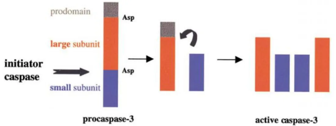
The procaspases are activated by proteolytic cleavage at specific aspartate residues (Zimmermann et al. 2001, Choudhury et al. 2012). Upon maturation, the procaspases are proteolytically processed (aspartate cleavage site) between the large and small subunit, resulting in a small and large subunit that allows the formation of a heterotetramer composed of two small and two large subunits forming an active caspase (Figure 3) (Gewies 2003).
Figure 3. Activation of procaspase-3 by cleavage. Schematic processing of caspases during activation. (From Zimmermann, Bonzon et al. 2001).
In mammals, based on structure and function, caspases have been subdivided into two main categories (Figure 2). The first category is the initiator caspases, which include caspases 1, 2, 4, 5, 8, 9, 10, 11 and 12, which contain a long amino- terminal prodomain that plays a regulatory role in activating downstream effector caspases. The second category is the executioner caspases, also called effector caspases, which include caspases 3, 6, 7 and 14 contain a small prodomain and are responsible for cleavage of different cellular substrates. Caspases can be further classified into two subclasses. The first subclass is inflammatory caspases, which includes caspases 1, 4, and 5 that are involved in cytokine activation and the second subclass is other cellular caspases, including caspases 11, 12, 13 and 14, whose roles are less well established (Rajesh P. Rastogi 2009, Choudhury et al. 2012, Dipak D. Ghatage 2012, Fuchs and Steller 2015).
1.2.3.2 Extrinsic pathway
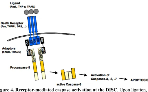
The extrinsic signalling pathway can be activated by ligation of death receptors (Fuchs and Steller 2015). These death receptors are part of the tumor necrosis factor receptor (TNF) gene superfamily (which includes, FasR, TNFR1 and DR4) and are localized on the surface of the cell then became active through binding specific ligands such as Fas ligand (also called CD95L), TNF alpha and TRAIL (also known as Apo2L). Subsequently, a conformational change exposes the death domain that allows the recruitment of adapter proteins (such as FADD or TRADD dependent on the active receptor), forming the Death Inducing Signalling complex (DISC). DISC can associate with procaspase 8, resulting in autocatalytic activation of procaspase 8. Active caspase 8 can then induce the initiation of apoptosis by cleavage and activation of effector caspases (caspases 3, 7 and 6) (Figure 4) (Gewies 2003, Elmore 2007, Choudhury et al. 2012).
Figure 4. Receptor-mediated caspase activation at the DISC. Upon ligation, the trimeric death receptor recruits adaptor molecules via its cytoplasmic death domains (DD). Besides possessing DDs, the adaptors additionally contain death effector domains (DED) which recruit procaspase-8 to the receptor complex, which now is called the death-inducing signalling complex (DISC). Procaspase-8 is activated by autoproteolytic cleavage and forms the active caspase-Procaspase-8. The initiator caspase-8 cleaves and thereby activates effector caspases for the execution of apoptosis. (From Gewies 2003).
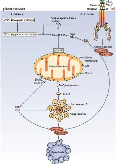
The crosstalk between extrinsic and intrinsic pathways can occur through caspase 8. Once caspase 8 is active, it can induce apoptosis through two parallel cascades. Firstly, it can be directly cleave and activate effector-caspases in a regular cascade. Secondly, it can mediate the cleavage of a pro-apoptotic protein Bcl-2; Bid (a BH3 domain-only protein). Truncated Bid (tBid) can then be translocated to mitochondria to induce mitochondrial outer membrane permeabilization (MOMP). MOMP causes the release of cytochrome c, which can subsequently activate caspase 9 and effector caspase 3 (Figure 5) (Tait and Green 2010, Choudhury et al. 2012).
Figure 5. Intrinsic and extrinsic pathways of apoptosis. a) Intrinsic apoptotic stimuli, such as DNA damage or endoplasmic reticulum (ER) stress. b) The extrinsic apoptotic pathway is initiated by the ligation of death receptors with their cognate ligands. Crosstalk between the extrinsic and intrinsic pathways occurs through caspase 8 cleavage and activation of the only protein BH3-interacting domain death agonist (BID), the product of which (truncated BID;
tBID) is required in some cell types for death receptor-induced apoptosis. (From Tait and Green 2010).
1.2.3.3 Intrinsic pathway
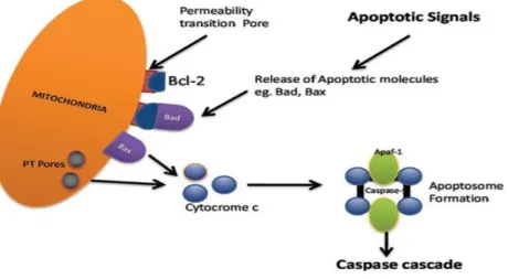
The intrinsic signalling pathway is activated by different signals coming from inside of the cell, such as strong DNA damage, radiation, toxins, hypoxia, hyperthermia, viral infections, and free radicals (Elmore 2007, Fuchs and Steller 2015). Mitochondria play an important role in this pathway, as they are crucial to induce the caspase-cascade activation and contain pro-apoptotic proteins in the intermembrane mitochondria space (IMS), between the inner and outer membranes of the mitochondria (IMM and OMM, respectively) (Tait and Green 2010, Choudhury et al. 2012). All of the above-mentioned stimuli cause changes in the mitochondrial outer membrane permeability (MOMP). MOMP generates an opening of the mitochondrial permeability transition pore (MPT), also called PT pore resulting in a loss of the mitochondrial transmembrane potential, and stimulating the release of the sequestered pro-apoptotic proteins Apoptosis Inducing Factor (AIF), Smac/DIABLO and cytochrome C in the cytosol (Elmore 2007). Once the mitochondria release cytochrome c, it binds the apoptotic protease-activating factor 1 (APAF1) which allows the association of procaspase 9. When cytochrome c is associated with APAF1, a conformational change leads to oligomerization and formation of a multiprotein complex termed apoptosome. The apoptosome induces cleavage and activation of caspase 3 and caspase 7, resulting in induction of apoptosis (Tait and Green 2010, Choudhury et al. 2012). MOMP is a tightly regulated process controlled by interactions between pro- and anti-apoptotic members of the B-cell lymphoma 2 family (BCL-2) (Figure 6) (Tait and Green 2010).
Figure 6. Intrinsic apoptosis pathway. Cytochrome c is a protein released from the mitochondria that binds to Apaf-1 and procaspase-9, in order to form the apoptosome, that then actives caspase-9 and effector caspases. (From Dipak D. Ghatage 2012).
1.2.4 Regulatory mechanisms of apoptosis
The control and regulation of apoptotic mitochondrial events are mediated by members of the Bcl-2 family proteins (Cory and Adams 2002). Tumor suppressor protein, p53 is an important pro-apoptotic factor that has a specific role in regulating the Bcl-2 family by activating the transcription of positive regulators such as DR-5 and Bax (Elmore 2007, Ouyang et al. 2012). Moreover, the PT pore formed during MOMP is due to the action of pro-apoptotic members of the Bcl-2 family, which in turn are activated by apoptotic signals such as cell stress, free radical damage or growth factor deprivation (Choudhury et al. 2012).
1.2.4.1 The BCL-2 family
The Bcl-2 family proteins were discovered as an oncogene in follicular B-cell lymphoma that inhibited cell death. This finding demonstrated for the first time that the promotion tumorigenesis is not only based on uncontrolled cell growth, but also depends on the ability to block apoptosis (Zimmermann et al. 2001, Gewies 2003). The Bcl-2 family has emerged as a critical regulator of apoptosis whose increased expression may lead to cancer and resistance to chemotherapy (Ouyang et al. 2012). The Bcl-2 family regulates apoptosis by controlling the mitochondria membrane permeability (MOMP), and members of the Bcl-2
family may possess pro-apoptotic or anti-apoptotic activity (Elmore 2007, Choudhury et al. 2012).
The Bcl-2 family is composed of 25 pro-apoptotic and anti-apoptotic members containing one or more Bcl-2 homology (BH) domains. These proteins are divided according to their function into two main categories. The first category is anti-apoptotic proteins presenting four BCL-2 homology domains including BCL-2, MCL-1, A1/Bfl-1, Bcl-B/Bcl2L10 and BCL-xL (BCL extra-large). Anti-apoptotic Bcl-2 family members prevent apoptosis by inhibiting their pro-apoptotic partners via protein-protein interactions (Zimmermann et al. 2001, Goldar et al. 2015). The second category is the pro-apoptotic Bcl-2 family proteins, which are sub-classified into two groups according to their structure. Group A, proteins having multiple BH domains (effector proteins) including: BAX, BAK, and BOK (BCL-2 related ovarian killer) and group B, proteins having only the BH3 domain, including: BID, BIM, PUMA, NOXA, BIK, BAD, HRK, and BMF (Figure 7) (Goldar et al. 2015).
Figure 7. BCL-2 family of proteins. The B cell lymphoma 2 (BCL-2) family of proteins is divided into three groups based on their BCL-2 homology (BH) domain organization. Anti-apoptotic BCL-2 proteins and Pro-apoptotic BCL-2 proteins can be sub-divided into effectors (the proteins that actually cause mitochondrial outer membrane permeabilization (MOMP)) or BH3 only (the proteins that relay the apoptotic signal to the effectors). (From Tait and Green 2010).
Following death stimuli, pro-apoptotic proteins can undergo post-translational modifications such as dephosphorylation and subsequent cleavage that allow their activation and translocation to the mitochondria to initiate apoptosis (Ouyang et al. 2012). Therefore, pro-apoptotic BH3-only proteins act as a sensor of these pro-apoptotic signals that permit the
activation of multidomain proteins like Bax and Bak which subsequently perform a pore (PT) on the outer membrane of mitochondria that allow the release of cytochrome c and other mitochondrial proteins. These proteins are apoptosis-inducing factor (AIF), endonuclease G, Smac/DIABLO (second mitochondria-derived activator of caspase/direct IAP-binding protein with low PI) and the serine protease Omi/HtrA2 (Braun et al. 2013, Goldar et al. 2015). Once these mitochondrial proteins are released into the cytosol, they can induce the caspase-cascade activation through repression of the inhibitor of Apoptosis proteins (IAP) in the case of Smac/DIABLO and Omi/HtrA2 (Gustafsson and Gottlieb 2008, Vande Walle et al. 2008). In addition, AIF and endonuclease G may also cause cell death in a caspase-dependent manner or in a caspase-incaspase-dependent manner based on the cellular context (Arnoult et al. 2003, Prabhu et al. 2013).
Another important pro-apoptotic BH3-only protein is Bim, well known as an efficient killer that can potentially induce cell death. Bim is essential for initiating intrinsic apoptosis pathway by apoptotic signals such as cytokine deprivation. These signals allow Bim to interact with all the anti-apoptotic Bcl-2 proteins (Mcl-1, Bcl-2, Bcl-xL, Bcl-w, and Bfl-1) and act in association with other partners like Noxa that selectively binds Mcl-1 and A1. Thus, a combination of selective binders and broader binders (Bim, Puma and tBid) promote apoptosis (Adams and Cory 2007, Sionov et al. 2015).
In summary, apoptosis can be induced by numerous apoptotic stimuli through the intrinsic pathway by intracellular signals (eg. DNA damage), or through the extrinsic pathway by extracellular signals (eg. death ligands). The intrinsic pathway can be activated by death receptors that subsequently form the DISC (Death Inducing Signalling Complex) leading to the activation of caspase 8 that can cleave and activate caspase-effectors (Caspases 3, 6 and 7) for induction of apoptosis. Simultaneously, caspase 8 can promote the activation of the intrinsic pathway through cleavage of a pro-apoptotic protein Bcl-2; Bid (tBid) which is subsequently translocated to the mitochondria to activate MOMP (strictly regulated by interactions between pro- and anti-apoptotic members of the BCL-2 family). Activation of MOMP in turn causes the release of cytochrome c into the cytosol to form the apoptosome in association with APAF1 that activates caspase 9 and effector-caspases.
Other cell signalling pathways have been reported to impinge on the core apoptotic machinery to modulate apoptosis. In relation to my research project, it has been shown that the Notch signalling pathway has an impact on cell death decisions (Zweidler-McKay et al. 2005, Robert-Moreno et al. 2007). An example of a mechanistic link between the Notch pathway and apoptosis is the known Notch target gene, Hes1. Once activated by the Notch pathway, it can regulate apoptotic signals by interacting with the PARP1 protein, causing the permeabilization of the outer membrane mitochondria (MOMP) and the release of AIF (apoptosis inducing factor), resulting in activation of the caspase-cascade and subsequent cell death (Cande et al. 2002, Kannan et al. 2011, Prabhu et al. 2013). Another example of Notch signalling to promote apoptosis includes Notch signalling reducing the transcription of the pro-apoptotic Bcl-2 family member, Bcl-xL, thereby enhancing apoptosis (Robert-Moreno et al. 2007). The regulation of Notch receptors can also modulate apoptosis by inducing pro-apoptotic BH3-only proteins, Bim and Noxa (Nickoloff et al. 2005, Konishi et al. 2010).
1.3 THE NOTCH SIGNALLING PATHWAY
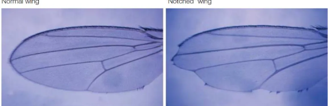
More than 100 years ago, John S. Dexter discovered in Drosophila an irregular notched shape in the wings (Figure 8) (Dexter 1914). Later, in 1917, Morgan identified the alleles responsible for the notched wing phenotype for which he received the Nobel Prize in 1933 (Morgan 1917). Decades later, the gene was cloned in 1985 by Spyros Artavanis-Tsakonas and Michael Young, and the sequence was shown to be a cell surface receptor (Wharton et al. 1985). Furthermore, Artavanis-Tsakonas and Young characterized Notch as a regulator of cell-fate decisions (Artavanis-Tsakonas et al. 1999). Notch signalling has been shown to control several key cellular processes such as cell proliferation, differentiation, and apoptosis (Yao et al. 2007, Melino et al. 2008, Schwanbeck et al. 2011, Li et al. 2014).
Figure 8. Drosophila with Notches. Wing blade of a wild-type Drosophila melanogaster (left), and of a mutant with a partial loss of the NOTCH gene (right). The notches, which are absent in the wild type, but clearly visible at the border of the wing blade, have given the name to the implicated gene (Notch). (From Radtke and Raj 2003).
1.3.1 Molecular biology of Notch Receptors
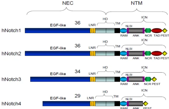
The Notch family of genes is conserved evolutionarily among the species. In mammals, this pathway involves a group of four receptors called Notch (Notch1-4), while Drosophila melanogaster has one and Caenorhabditis elegans has two. Each receptor is a single-pass type I transmembrane protein and is composed of two domains (Figure 9) (Kopan and Ilagan 2009, Pancewicz and Nicot 2011):
The extracellular domain: contain different repeats that share homology with epidermal growth factor (EGF). Notch1 and Notch2 have 36 EGF repeats and it has been reported that repeat 11 and 12 are important for ligand binding. Notch 3 and Notch 4 contain 34 and 29 repeats, respectively. The number of EGF repeats and their ability to bind to calcium ions play an important role in the affinity for Notch ligands (Kopan and Ilagan 2009, Ntziachristos et al. 2014).
The intracellular domain has several domains: RBPJ-k association module (RAM domain) and seven ankyrin repeats (ANK domain). Both domains are important for interacting with co-activators and forming the ternary complex with the mammalian CBF1/Drosophila Suppressor of Hairless/C. elegans LAG-1 protein (CSL; also known as RBPJ-k). Proline/glutamic acid/serine/threonine-rich, PEST domain
provides stability and is responsible for targeting the Notch intracellular domain (NIC) for degradation upon sel10 ubiquitin ligase recognition. Two nuclear localization sequences (NLS). The transcription transactivation domain (TAD domain), that is responsible for the activation of transcription, is strong in Notch1, weak in Notch2 and absent in Notch 3 and Notch 4 (Radtke and Raj 2003, Kato 2011).
Notch receptors also have a heterodimerization domain (HD) and a negative regulatory region (NRR) that prevents activation of the receptor in absence of the ligand (Kopan and Ilagan 2009, Wang 2011).
Figure 9. Structure of the four human Notch receptors. NEC: extracellular domain; NTM: transmembrane domain; EGF: epidermal growth factor; HD: heterodimerization domain; NIC: Notch intracellular domain; LNR: cysteine-rich LNR repeats; TM: transmembrane domain; RAM: RBPjk-association module; NLS: nuclear localizing signals; ANK: ankyrin repeat domain; NCR: cysteine response region; TAD: transactivation domain; PEST: region rich in proline (P), glutamine (E), serine (S) and threonine (T) residues. (From Pancewicz and Nicot 2011).
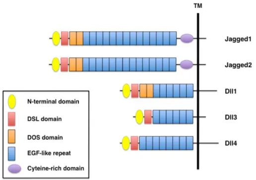
1.3.2 Molecular biology of Notch Ligands
Notch ligands are also transmembrane type I cell surface proteins. In mammalians, five Notch ligands have been reported which are classified into two families: Jagged family (JAG1-2) and Delta-like family (Delta-like 1, 3 or 4), based on the structural homology of the two Drosophila ligands, Serrate and Delta, respectively (D'Souza et al. 2008). Canonical Notch ligands possess a DSL domain (Delta/Serrate/LAG-2) and multiple EGF-like repeats but only the Jagged family and Dll-1 contain a DOS domain (Delta and OSM-11-like proteins). Both DSL and DOS domains are very important for receptor binding. In addition, members of the Jagged family have a cysteine-rich domain that, along with the DOS domain contribute to the structural diversity between the ligands (Figure 10) (Kume 2009).
Figure 10. Domain organization of mammalian Notch ligands. All Notch ligands have an N-terminal domain, a DSL (Delta/Serrate/LAG-2) domain and EGF-like repeats. Jagged1 and Jagged2 contain a cysteine rich domain, whereas Jagged1, Jagged2, and Dll1 have two DOS (Delta and OSM-11-like proteins) domains located immediately following the DSL domain. (From Kume 2009).
1.3.3 Activation mechanism of Notch signalling pathway
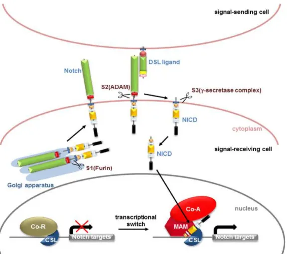
Initially, Notch receptors are synthesized as a single precursor in the Trans-Golgi, where they become a non-covalently linked heterodimer as a consequence of cleavage by a furin-like convertase at the S1 site (Schwanbeck et al. 2011, Ntziachristos et al. 2014). Subsequently, the receptor becomes glycosylated by O-fucosyltransferase and Fringe Family N-acetylglucosaminidyl transferases. Following cleavage at S1 and glycosylation, the matured heterodimer Notch receptor is translocated to the cell surface (Kato 2011, Previs et al. 2015). Once on the cell surface, the activation of the Notch receptor depends on its interaction with one of its five canonical Notch ligands (JAG1, JAG2 and Delta-like 1, 3 or 4) (Kopan and Ilagan 2009, Wang 2011) in a neighbor cell. The interaction of Notch with its ligands initiates the signalling cascade by induction of a conformational change that allows proteolytic cleavage by the ADAM17 metalloprotease/TNFα converting enzyme (TACE) at the S2 site and subsequently endocytosis of the extracellular Notch domain in the signal-sending cell (Fortini 2009, Schwanbeck et al. 2011). Next, release of the intracellular active domain (NIC) is triggered by a third sequential proteolytic cleavage mediated by presenilin, the catalytic subunit of the γ-secretase complex, at the S3 site (Schroeter et al. 1998, De Strooper et al. 1999, Okochi et al. 2002). The γ-secretase complex is composed of 4 subunits: Presenilin 1/2, nicastrin (NCT), presenilin enhancer 2 (PEN2) and anterior pharynx-defective 1 (APH1)) (Schroeter et al. 1998, De Strooper et al. 1999, Okochi et al. 2002, Ranganathan et al. 2011). Different compounds known as γ-secretase inhibitors (GSIs), classified into two types, can target γ -secretase cleavage at S3 pharmacologically. The two types are transition and non-transition state inhibitors. Treatment with GSI blocks the release of NIC from the plasma membrane, blocking the activation of Notch signalling (Ranganathan et al. 2011, Olsauskas-Kuprys et al. 2013). Once released, NIC is translocated to the nucleus where it acts as a transcriptional activator that interacts with the DNA-binding protein CSL and recruits co-activators such as mastermind-like (MAML1) and p300/CBP (CREB-binding protein) thereby regulating the transcription of their Notch target genes (Figure 11) (Kovall 2008, Andersson et al. 2011, Bray 2016).
Figure 11. The canonical Notch signalling pathway. Notch receptor is glycosylated and cleaved by Furin at site 1 (S1). The interaction between Notch receptors and ligands on neighbouring cells results in the conformational change of receptor and the site 2 (S2) is cleaved by ADAM metalloproteases. Then, γ-secretase complex-mediated cleavage at site 3 (S3) releases the Notch intracellular domain (NIC). NIC then translocates into the nucleus and binds to DNA binding protein CBF1/Su (H)/Lag-1 (CSL). Transcriptional co-activator Mastermind (MAM) recognizes the NIC/CSL complex. Ternary complex formation causes the release of repressor’s (Co-R) and recruit additional co-activators (Co-A) to activate transcription of target genes. (From Kato 2011).
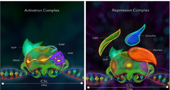
In the absence of NIC, CSL acts as a transcriptional repressor through interactions with corepressors (Co-R). The Co-R including SMRT (silencing mediator of retinoid and thyroid receptors), SKIP (Ski-interacting protein), CtBP (C-terminal binding protein), Groucho/TLE (Transducin-like enhancer of split), CIR (CBF1-interacting corepressor) and SHARP (SMRT/HDAC1 (histone deacetylase 1) associated repressor protein (Figure 12) (Zhou et al. 2000, Lai 2002, Jennings and Ish-Horowicz 2008, Fortini 2009).
Figure 12. Comparative view of the repression and activation complexes. Drosophila melanogaster. The CSL transcription factor acts as a bridging protein between the DNA and a complex of proteins intended to modify chromatin topology in a specific locus. CSL: CBF1/Drosophila Suppressor of Hairless/C. elegans LAG-1 protein; RAM: RBPjk-association module of NIC; ANK: ankyrin repeat domain of NIC; NIC: Notch intracellular domain; MAM: Mastermind; SKIP: Ski-interacting protein; CtBP: C-terminal binding protein; Groucho/TLE: Transducin-like enhancer of split. (From Contreras-Cornejo, Saucedo-Correa et al. 2016).
1.3.4 Notch Target Genes
The transcription of the Notch target genes depends on three features: ligand-receptor interactions, cell type (which also include several genes that participate in different cellular processes like metabolism, differentiation and regulation of the cell cycle) and the activity of the transcriptional complex, CSL-NIC (Ntziachristos et al. 2014, Contreras-Cornejo et al. 2016).
The CSL-NIC complex activates the expression of Notch target genes including transcriptional factors, Hes (Hairy in mammals and E (spl) in Drosophila) and Herp (Hes-related repressor protein) (also known as Hey/Hesr/HRT/CHF/gridlock). Another Notch target gene includes: Cyclin D1, p21, NF-κB, pre-Tα (pre-T-cell receptor alpha chain), GATA3, NRARP, c-Myc and Deltex1 (Iso et al. 2003, Yin et al. 2010). The most well studied Notch target gene, and well known as a primary Notch effector, is HES/E (spl) which is part
of the basic helix-loop-helix (bHLH) family and binds DNA sequences as a dimer. This family of transcription factors is very important as effectors of the Notch pathway because they participate in the development of various organs (heart, skeletal muscles, pancreas) and cell types (Iso et al. 2003).
1.3.5 Notch in tumorigenesis
The contribution of the Notch signalling pathway to tumorigenesis is complex. Depending on the context, the Notch pathway has been shown to have an oncogenic or tumor suppressor effect (Ranganathan et al. 2011, Ntziachristos et al. 2014, Previs et al. 2015). The Notch signalling cascade shows an oncogenic effect in some cancers such as ovarian, prostate, nasopharyngeal, T-cell acute lymphoblastic leukemia (T-ALL), breast and sarcoma (Engin et al. 2009, Efferson et al. 2010, Wang et al. 2010, Wang et al. 2010, Chen et al. 2011, Hernandez Tejada et al. 2014). In apparent contrast, the Notch pathway also has a tumor suppressor effect on other cancers such as skin, endometrial, cervical, B-cell acute lymphoblastic leukemia (B-ALL) and lung (Sriuranpong et al. 2001, Nicolas et al. 2003, Yao et al. 2007, Dotto 2008, Kannan et al. 2011, Jonusiene et al. 2013). The role of the Notch pathway in cervical cancer remains ambiguous, as there is evidence that it can act as a tumor suppressor or as an oncogene (Maliekal et al. 2008).
1.3.6 The role of Notch pathway in cervical cancer
Cervical cancer (CC) is the fourth most common cancer in women and it is the seventh most common cancer worldwide, causing nearly 8% of all women deaths from cancer in 2012 (Jemal et al. 2011, Cancer 2012, Ferlay et al. 2015). The main risk factor for the development of cervical cancer is high-risk human papillomaviruses (HR-HPVs) infection (zur Hausen 2002). About 70% of all invasive cervical cancers are associated with the oncogenic HR-type 16 HPV (HPV16) and 18 HPV (HPV18). Fifty percent of HPV-positive cervical tumors carry HPV16, whereas HPV18 is present in approximately 10-20% (zur Hausen 1996, Khan et al. 2005, Smith et al. 2007, Guan et al. 2012, Goodman 2015). The development of cervical cancer develops through several steps that include. Firstly, HPV infects basal cells in the
cervical epithelium. Secondly, the HPV DNA is integrated into the genome of the host cell. Thirdly, there is viral persistence (more than two years). Fourthly, there is progression to the neoplastic phenotype (classified 1 to 3, taking 3 to 5 years) and fifthly, invasive carcinoma develops (range 10 to 30 years) (zur Hausen 2002, Maglennon and Doorbar 2012, Steenbergen et al. 2014, Goodman 2015).
Under normal conditions, the ectocervix is covered by a squamous epithelium and the endocervical canal is covered by a columnar epithelium. The basal layer of the endocervical epithelium contains precursor cells that have the ability to differentiate into squamous or columnar cells (Crum 2000, Allenspach et al. 2002). This region, known as the cervical transformation zone, is the site most susceptible to HPV infection, as well as the site for initiation of neoplastic transformation (Reid 1983, Doorbar et al. 2012, Lopez et al. 2012). HPVs expresses two oncoproteins, E6 and E7, which are essential for oncogenesis. The concerted action of E6 and E7 disrupts normal mechanisms of cell cycle regulation. In particular, E6 targets the p53 tumor suppressor protein, accelerating its degradation by the proteasome (Scheffner et al. 1990). The E7 oncoprotein functionally inactivates the pRb tumor suppressor by targeting to the proteasome for degradation (Dyson et al. 1989). pRb’s interaction with the E2F transcription factors inhibit their transcriptional activity of genes required for S-phase progression. The E7-induced degradation of pRb results in the release of E2F, allowing the expression of S-phase genes and progression of the cell cycle (Münger et al. 2001, Moody and Laimins 2010, Ghittoni et al. 2015). Although E6 and E7 are necessary for the induction and maintenance of the transformed phenotype, they are not sufficient to induce the development of cervical cancer. It has been shown that at least one other cellular/genetic alteration is necessary for the development of cancer. Accumulation of evidence suggests that aberrant Notch signalling is a cellular event that can play a role in cervical carcinogenesis (Zagouras et al. 1995, Daniel et al. 1997, Rangarajan et al. 2001, Talora et al. 2002, Lathion et al. 2003, Weijzen et al. 2003, Talora et al. 2005, Wang et al. 2007).
In human cervical cancer, it was discovered that Notch expression is reduced in invasive and metastatic cells, suggesting that down-modulation of Notch pathway is required in the
tumorigenesis process (Talora et al. 2002, Sakamoto et al. 2012). In cervical cancer cells, it has been reported that the activation of Notch pathway results in inhibition of tumor growth through the induction of apoptosis and cell cycle arrest (Yao et al. 2007). In particular, in HeLa cells, the activation of the Notch pathway has been shown that decrease cell proliferation and induces apoptosis (Wang et al. 2007).
To conclude, apoptosis can be induced by the activation of the Notch signalling pathway following binding of a ligand, which results in cleavage of the Notch receptor by the γ-secretase complex, releasing the active intracellular domain of Notch (NIC) and induction of Hes1 expression (Yao et al. 2007, Kannan et al. 2011, Wang 2011). Interestingly, the pro-apoptotic transcription factor TAF6 (Bell et al. 2001) in HeLa cervical carcinoma cells can increase mRNA expression levels of Notch target genes and also pro-apoptotic genes as it was shown by microarray assays using Splice-Switching Oligonucleotides (SSOs) (Wilhelm et al. 2008, Wilhelm et al. 2010). Indeed, HeLa cells were shown to undergo apoptosis when TAF6 expression is triggered through SSOs (Wilhelm et al. 2010). The TAF6 pathway therefore represents a possible new therapeutic target for treating human cervical cancer cells.
1.4 TRANSCRIPTION
The regulation of transcription is a very important step in the control of cell identity, differentiation, growth and development (Grunberg and Hahn 2013). The transcription machinery initially recognizes double-stranded deoxyribonucleic acid (DNA), but only one strand serves as a template for transcription. The transcription process begins when a specific enzyme known as RNA polymerase (RNA pol) binds to the DNA strand template to initiate the production of complementary RNA (ribonucleic acid). When the RNA polymerases are active on DNA they form a complex with different factors that allow the transcription of a specific gene (Clancy 2008). These complex sets of factors are referred as general transcription factors (GTFs), which have several functions such as promoter recognition, Pol recruitment, interaction with regulatory factors, DNA unwinding and transcription start site (TSS) recognition (Hahn 2004, Thomas and Chiang 2006).
In eukaryotes, there are three different classes of RNA polymerases: RNA pol I transcribes genes encoding 18S and 28S ribosomal RNAs (rRNAs) within the nucleoli. RNA pol III transcribe genes for 5S rRNA and transfer RNAs that play a role in the translation process and RNA pol II transcribing the messenger RNAs, which serve as templates for protein production, both localized at the nucleoplasm (Thomas and Chiang 2006, Clancy 2008). Since my work focused on the transcription factor TAF6, the focus here will be the mechanisms of transcriptional regulation by RNA pol II.
1.4.1 Transcription by RNA Pol II
Transcription by RNA pol II depends on a cascade of events, which are classified into three steps: initiation, which include the binding of activators to enhancers and the formation of the pre-initiation complex (PIC), elongation by RNA pol II and termination (Kandiah et al. 2014).
1.4.1.1 Initiation
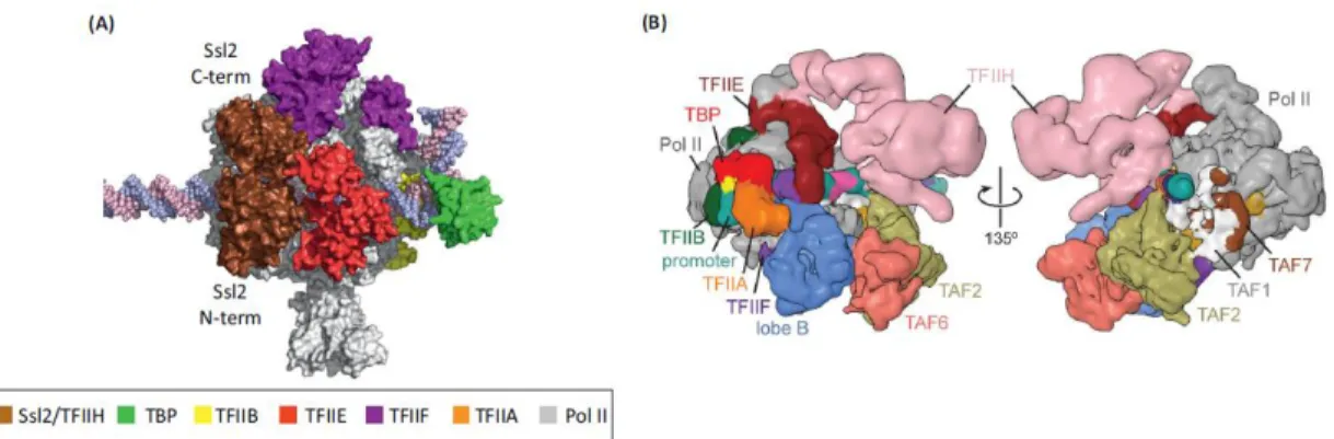
The initiation of transcription by RNAPII requires basal transcription factors known as TFIIA, TFIIB, TFIID, TFIIE, TFIIF, and TFIIH (Figure 13).
Figure 13. Architecture of the basal PIC. (A) PIC model based on crystal structures and biochemical mapping. Note that the two Ssl2 domains (labeled Ssl2 C-term and Ssl2 N-term) of TFIIE, encircling promoter DNA at positions – 2 to +6 with respect to the human TSS at +1. From (Grunberg and Hahn 2013). (B) Model of the TFIID-based PIC, Cryo-EM reconstruction of the human TAF-less PIC, with fitted atomic models. (From Louder, He et al. 2016).
1.4.1.1.1 Generalities of the basal transcription factors
TFIIA participates in transcription by stabilizing the binding between TBP (TATA-binding protein) and the TATA box by regulating the dimerization of the TBP or TFIID that accelerate DNA binding. TFIIA also plays an important role in transcriptional activation of the TATA-less promoters and RNA pol III (Hoiby et al. 2007).
TFIIB is a factor that is associated with TBP and RNA pol II that has four functional domains: N-terminus B-ribbon, reader, linker and core (contain two cyclin-like repeats), and all of them interact with RNA pol II. B-ribbon strongly involves RNA pol II, B-reader and linker form a hairpin, termed the B-finger in the RNA pol II and B-core, in addition to bind TBP-DNA. Thus, the conformation of TFIIB within PIC plays a crucial role in transcription activation, promoter recognition and start site selection (Reese 2003, Grunberg and Hahn 2013).
TFIID is a multi-subunit complex of mega-Dalton-sized that is thought to nucleate PIC formation on a core promoter by binding to the TATA box through its TBP subunit. TFIID also interacts with nucleosomes covalently modified and has been associated with enzymatic activities (post-translational histone modifications and transcription factors). This complex assumes a horseshoe-shaped structure containing three lobes (A, B and C). In the usual conformation, the lobe A is engaged to lobe C but the binding of TFIIA induces a conformational change that reorganizes the shape and, consequently, lobe A dislocates from lobe C to lobe B (Thomas and Chiang 2006, Grunberg and Hahn 2013, Kandiah et al. 2014).
TFIIE, contain two TFIIEα and TFIIEβ subunits. TFIIEα containing an N-terminal WH and central Zn-ribbon, which are essential, and TFIIEβ containing tandem WH domains that are conserved. TFIIE function as a stabilizer of the non-template DNA strand and also interact directly with TFIIH because TFIIH is associated with the PIC just after TFIIE binds (Grunberg and Hahn 2013).
TFIIF enters the PIC together with RNA pol II and contains two conserved polypeptides (Rap74/30), each containing an N-terminal dimerization domain and a C-terminal winged helix (WH). The dimerization domain binds to the lobe domain of RNA Pol II and the WH domain is bound to the protrusion of RNA Pol II (Grunberg and Hahn 2013).
TFIIH, is a large multi-subunit complex containing ten subunits, three of them contain ATP-dependent enzymatic activities, CDK7, the Pol II CTD kinase, Rad3/XPD, a DNA helicase and Ssl2/XPB (DNA translocase). The helicase subunits and kinase enzymatic activities are required for the initiation, elongation and promoter escape steps in RNA Pol II transcription. In addition to its role in DNA unwinding, TFIIH also has a role in the phosphorylation of the RNA pol II (Reese 2003, Grunberg and Hahn 2013).
1.4.1.1.2 Mechanism of RNA Pol II initiation
These accessory factors were defined as general transcription factors (GTFs) using the following nomenclature: TF represents the Transcription Factor, the Roman numeral II indicates the transcription driven by pol II, and the “letter” corresponds to the chromatographic fraction from which the specific GTF was isolated (Thomas and Chiang 2006). The first step in the general mechanism is the recognition of the core promoter through PIC recognition of the different DNA elements located in the promoter region, known as a core promoter elements (CPEs) (Goodrich and Tjian 2010, Shandilya and Roberts 2012). These sequences are located upstream or downstream of the TSS on the target gene (Shandilya and Roberts 2012). One of the most studied CPE is the TATA box (TATAAA consensus sequence between 25 to 35 bases) upstream of the initiation site. However, not all Pol II promoters contain TATA sequences. TATA-containing promoters, which are actually the minority, account for 20-30% of the promoters in eukaryotes. In contrast, TATA-less promoters that generally direct the transcription of housekeeping genes and possess a heterogeneous TSS (Clancy 2008, Goodrich and Tjian 2010, Grunberg and Hahn 2013). The DNA sequence of the TATA box is recognized by TBP as part of the TFIID complex. TBP
possesses two domains: a highly conserved C terminus (TBPcore) that binds the TATA box and a divergent N terminus that is dispensable for viability. Once, TBP binds to the TATA sequence and induces a bend in the DNA that serves as a platform for the assembly of all GTFs and subsequent formation of PIC (Reese 2003, Shandilya and Roberts 2012). Consequently, the nucleation of PIC formation through the interactions of the TBP promoter is highly regulated (see below).
Positive regulation involves gene-specific activator proteins (activators) that increase the binding of TBP to promoters. The activator can induce multiple genes and multiple activators can also regulate a single gene. These activators are composed of an activation domain that allows association with different transcription factors and a promoter-targeting DNA-binding domain (Bhaumik 2011, Shandilya and Roberts 2012).
Negative regulation involves the repression of the TBP-DNA binding activity through negative factors such as Mot1/BTAF1 and NC2 (Sikorski and Buratowski 2009, Shandilya and Roberts 2012).
During PIC formation, once TBP (subunit of TFIID) recognizes and binds to the TATA box, a sequential binding of TFIIA stabilizes the interactions of the TFIID-core promoter. TFIIB interacts with TBP and DNA promoter, and is assembled to the RNA pol II-TFIIF complex. However, transcription cannot begin until TFIIB, TFIIF and RNA pol II orient the DNA template, select the TSS and then TFIIE and TFIIH will be recruited into the PIC (table 1). TFIIH possesses a helicase activity and is therefore capable of catalyzing ATP-dependent melting of the promoter and making a transition from initiation of transcription to elongation (Reese 2003, Kandiah et al. 2014). Another important element for the transition is based on the length of the transcript that has to be about 25nt in order to stable transition complex that allows the elongation process. Otherwise, transcripts with less that 5nt result in an unstable transcription complex and therefore abortive initiation (Liu et al. 2011).
Protein Complex Functions
RNA pol II
12 Subunits; catalyzes transcription of all mRNAs and a subset of noncoding RNAs including snoRNAs
and miRNAs TFIIA
2–3 subunits; functions to counteract repressive negative cofactors like NC2; acts as a coactivator by
interacting with activators and components of the basal initiation machinery
TFIIB
Single subunit; stabilizes TFIID-Promoter binding; helps in recruitment of TFIIF/Pol II to the promoter;
directs accurate start site selection
TFIID
14 subunits including TBP and TBP Associated Factors (TAFs); nucleates PIC assembly either through TBP binding to TATA sequences or TAF
binding to other promoter sequences; coactivator activity through direct interaction of TAFs and gene
specific activators TFIIE
2 subunits; helps recruit TFIIH to promoters; stimulates helicase and kinase activities of TFIIH; binds ssDNA and is essential for
promoter melting TFIIF
2–3 subunits; tightly associates with RNA Pol II; enhances affinity of RNA Pol II for
TBP-TFIIB-promoter complex; necessary for recruitment of TFIIE/TFIIH to the PIC; helps in start site selection and promoter escape; enhances elongation efficiency
TFIIH
10 subunits; ATPase/helicase necessary for promoter opening and promoter clearance; helicase activity for
transcription coupled DNA repair; kinase activity required for phosphorylation of RNA Pol II CTD; facilitates transition from initiation to elongation.
Table 1. Complexes involved in RNA Pol II PIC assembly. (From Sikorski and Buratowski 2009).