DOI 10.1007/s11548-008-0275-9 O R I G I NA L A RT I C L E
Generation of three-dimensional prototype models based
on cone beam computed tomography
J. Th. Lambrecht · D. C. Berndt · R. Schumacher · M. Zehnder
Received: 28 May 2008 / Accepted: 23 September 2008 / Published online: 15 November 2008 © CARS 2008
Abstract
Purpose The purpose of this study was to generate three-dimensional models based on digital volumetric data that can be used in basic and advanced education.
Methods Four sets of digital volumetric data were establi-shed by cone beam computed tomography (CBCT) (Accui-tomo, J. Morita, Kyoto, Japan). Datasets were exported as Dicom formats and imported into Mimics and Magic soft-ware programs to separate the different tissues such as nerve, tooth and bone. These data were transferred to a Polyjet 3D Printing machine (Eden 330, Object, Israel) to generate the models.
Results Three-dimensional prototype models of certain limited anatomical structures as acquired volumetrically were fabricated.
Conclusions Generating three-dimensional models based on CBCT datasets is possible. Automated routine fabrication of these models, with the given infrastructure, is too time-consuming and therefore too expensive.
Keywords Cone beam computed tomography· Digital volume tomography· 3D models · Rapid prototyping Introduction
The technology of three-dimensional prototype modeling based on CT or MRI data was introduced into oral and J. Th. Lambrecht (
B
)· D. C. Berndt · M. ZehnderDepartment of Oral Surgery, University Hospital for Oral Surgery, Oral Radiology and Oral Medicine, University of Basel,
Hebelstraße 3, 4056 Basel, Switzerland e-mail: [email protected] R. Schumacher
School of Life Sciences, Institute for Medical and Analytical Technologies, University of Applied Sciences Northwestern Switzerland, Muttenz, Switzerland
maxillofacial radiology and surgery over two decades ago Brix and Lambrecht [1] after Marsh and Vannier [2] had reconstructed the “third dimension” of sequences of high-resolution axial CT scans with algorithms. The models were used to plan and simulate maxillo-craniofacial surgery in three dimensions [3] and could be milled from a variety of materials. Laser-hardened acrylic resin had been shown to be a useful alternative [4,5].
Cone beam computed tomography (CBCT) was introdu-ced as a high-resolution imaging prointrodu-cedure in oral and maxil-lofacial radiology. Cone beam CT technology has a two-dimensional sensor and uses a cone-shaped X-ray beam in place of a fan-shaped beam (CT). Volume data can be acqui-red in a single 360◦rotation of beam and sensor around the skull [6,7]. The characteristics of CBCT are lower entrance doses and higher resolution in the axial direction compared with conventional CT [8–10].
Nowadays individual medical models are routinely gene-rated by rapid prototyping. Physical models are produced by selectively solidifying UV-sensitive liquid resin using a laser beam. The technology was first introduced in mechanical engineering and has also attracted a great deal of attention in clinical dentistry [11,12]. The aim of this study was to pro-duce three-dimensional individual patient prototype models out of CBCT datasets that could eventually be used in basic and advanced education.
Materials and methods Generating data
Different datasets of four patients were established by 3D CBCT (Accuitomo, J. Morita, Kyoto, Japan). One dataset is used as an example in this study. The imaging volume was
176 Int J CARS (2009) 4:175–180 a cylinder measuring 40 mm (diameter)× 30 mm (height)
at the X-ray rotational center. Images were taken under the exposure conditions of 75–80 kV and 4–6 mA, which are the standard parameters and were changed for different subjects. The cone beam technique involves a single 360◦ scan in which the X-ray source and a reciprocating area detector synchronously move around the patient’s head. The top of the cone is the source of radiation and the bottom of the cone is the sensor, an image intensifier with a CCD camera. Within 17 s the region of interest is scanned in a total of 512 frames of two-dimensional images; these were recorded as an “.avi” format file.
i-Dixel
The Accuitomo software (i-Dixel images) calculates a volu-metric dataset from 512 single digital images. Out of this dataset the single slices are calculated from within a volume of 3× 4 cm by selecting them in Dicom with a voxel volume of 0.125 mm× 0.125mm × 0.125mm.
Mimics
The dataset is imported with a slice thickness of 0.5 mm into the planning software MIMICS (Materialise’s Interac-tive Medical Image Control System, Materialise, Belgium). MIMICS is a link between the scanner data (CT, MRT, CBCT) and a simple virtual 3D representation.
To work with the data, a so-called “mask” is generated. The threshold is necessary to create a first separation of the single anatomical structures (tooth, bone, nerve). A mask is generated for each structure and makes it possible to proceed within the work flow. The masks can be edited slice by slice by simply adding or removing voxels manually. Bone, teeth and soft tissue were separated using the above-mentioned functions. The masks can be exported as an STL data repre-sentation, which is a simple triangulated 3D surface dataset (Table1).
Magics
The acquired 3D data can be imported as an STL data-set. Since the major part in the previous process was edited manually, the resulting surface of the anatomical structures was relatively rough. For this reason, a remesh was perfor-med. The triangulated surface was smoothened and the edges of the triangles were assimilated. Using booling operations, figures without overlaps can be generated. These smoothened data were reimported into MIMICS as an STL dataset.
Table 1 Chart showing the workflow
1. CBCT Generation of 3-d X-ray data
2. i-Dixel Visualisation and selection of Datasets
3. MIMICS Addition of 2-d layers to 3-d Datasets
4. Magics Surfacesmoothing, Booling ops, Storing
5. Polyjet 3D-Printer
Dicom
Polyjet 3D printing
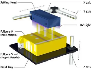
Three-dimensional models are produced by the Polyjet method (Objet EDEN 250). Prototypes with high resolution and excellent surface quality can be produced layer by layer. Figure1shows the Polyjet rapid prototype fabrication sys-tem. The high resolution and good surfaces (without “stare steps”) are acquired because of the layer thickness of 0.016 mm.
UV-sensitive resins are added using printheads in extre-mely thin layers. Adding UV hardens the photopolymer. In the same process, supporting material is added that has a gel character and can be removed by water jet at the end of the construction process.
Fig. 1 The polyjet rapid prototype fabrication system showing the
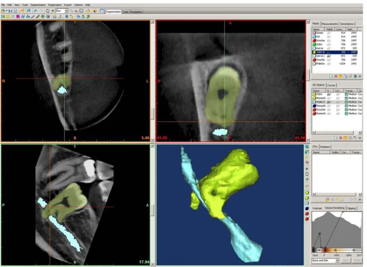
Fig. 2 The MIMICS software platform showing the 2D axial (upper
left) coronal (upper right) and sagittal (lower left) dimensions with a
mask for the gray values of the of the nervus alveolaris inferior (light
blue). The 3D visualization of the three rendered masks shows the
impacted wisdom tooth in close anatomical relation to the alveolar nerve (lower right)
Results
The MIMICS software uses segmentation algorithms to obtain a 3D visualization at the end. The data which were acquired by i-Dixel can be read into MIMICS and are ready to be viewed in axial, sagittal and coronal dimensions.
The “reslice project” function makes it possible for the user to change the direction of the slice axes. A correct direc-tion of axis simplifies later use of the data. The “threshold” function enables the user to tell the computer the gray value within the grayscale from which the program should detect and colour the voxels (Fig.2).
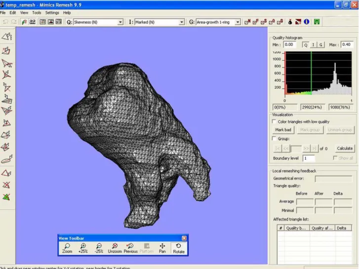
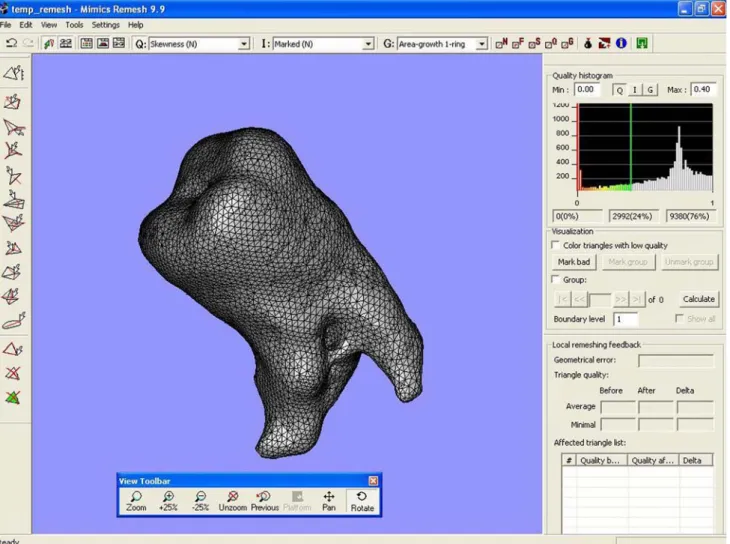
The Magics software enables the user to smoothen the manually worked out MIMICS data (Figs.3,4).
Each generated structure of tooth, nerve and bone was separately produced as a model by the Polyjet method (Figs.5,6).
Thus fabrication of three-dimensional models based on CBCT data is possible. Featuring separate representation of
hard and soft tissue enables the user to realize topographic relations of anatomic structures.
Discussion
Conventional CT has been the appropriate technology of choice for acquiring datasets in order to fabricate three-dimensional models [13]. The disadvantages are space requi-rements, relatively high doses and high costs [14].
By comparison, CBCT, which is nowadays available for dento- maxillo-facial radiological units, requires less space, lower radiation doses, lower costs and offers a higher reso-lution than CT images [15–18]. The latter is due to the voxel size of the system used. Of course, voxel size is also crucial for accuracy. Studies on accuracy were done with various CBCT machines for implants [19] and human skulls and cadavers for the purpose of cephalometric measurements
178 Int J CARS (2009) 4:175–180
Fig. 3 Magics representation of a rough surface wisdom tooth
[20,21]. With regard to CBCT accuracy with Accuitomo, there is one report by Loubele et al. [22] yielding submillime-ter accuracy for linear measurements on an ex vivo specimen. These results provide the basis for creating haptic models with adequate accuracy.
Artefact reduction is relevant to creating acceptable models [23]. Contrary to CT, CBCT does not provide read-outs in Hounsfield Units (HU). The HU is the standard scale for measuring conventional CT values and was the basic plat-form for acquiring 3D models where defined tissues could be extracted because of their different HU. Mah and McDavic [24] suggest exporting CBCT data as a raw DICOM data-set and transferring the datadata-set to UTHSCSA Image Tool for analysis. Another method described showed that the atte-nuation coefficient of the test material in the phantom was determined with less than one per cent error. This sounds promising for the future and should be the subject of further research.
A grayscale is so far available in our set-up. With the MIMICS software program the aim was to extract certain gray values which were representative of a specific tissue, for example to discriminate soft tissue (nerve) from hard tissue (bone). To get the real 2D profile of the different tissues it was necessary to check every slice manually. Thus the separation of tissues layer by layer became very time-consuming. In the final stage individual patient datasets could be visualized. The computer-generated results provided a sufficient basis for rapid prototyping [25].
The use of three-dimensional reconstructions to evaluate the anatomic relationship of the mandibular canal and impac-ted mandibular third molars has recently been demonstraimpac-ted by Friedland et al. [26].
The technique used in the present study goes one step further and seems to be highly suitable for creating haptic models to demonstrate complex anatomic situations, ins-tructing students and planning operations. This technique
Fig. 4 Magics representation after smoothing of the data set shown in Fig.3
Fig. 5 3D models of a segmental piece of the mandible and the wisdom
tooth according to the CBCT dataset
Fig. 6 3D models of the wisdom tooth in close anatomical relationship
to the nervus alveolaris inferior according to the CBCT dataset shown in a mechanical composition for demonstration
180 Int J CARS (2009) 4:175–180 could also be used in performing certain dento-alveolar
ope-rations. The disadvantage for the time being is a lack of cost-effectiveness because of the time-consuming procedure. These problems have to be solved before series for basic and advanced education can be produced.
Conclusion
To the best of the authors’ knowledge, this is the first report on generating three-dimensional models based on CBCT data-sets. It may become a useful technique for demonstration, planning and teaching, but other options for processing the datasets and producing models will need to be studied. The ultimate aim will be to use these models as a matter of rou-tine in basic and advanced education, being comparable to conventional CT/MRI-based models.
References
1. Brix F, Lambrecht JTh (1987) Individuelle Schädelmodell-herstellung auf der Grundlage computertomographischer Informationen. In: Schwenzer N, Pfeifer G (eds) Bildgebendes Untersuchungsverfahren in der Mund-, Kiefer- und Gesichts-Chirurgie, Band XXXII. Thieme Verlag Stuttgart, New York, pp 74–77
2. Marsh JL, Vannier MW (1993) The “third” dimension in craniofa-cial surgery. Plast Reconstr Surg 71:759–767
3. Lambrecht JT, Brix F (1990) Individual skull model fabrication for craniofacial surgery. Cleft Palate J 27:382–385
4. Lambrecht JTh, Hammer B, Jacob AL, Schiel H, Hunziker M, Kreusch T, Kliegis U (1995) Individual model fabrication in maxil-lofacial radiology. Dentomaxillofac Radiol 24:147–157
5. Petzold R, Zeilhofer HF, Kalender WA (1999) Rapid prototyping technology in medicine-basics and applications. Comput Med Ima-ging Graph 23:277–284
6. Toyofuku F, Konishi K, Kanda S (1986) Representation of arbi-trarily curved sections of dentomaxillofacial region by the X-ray video CT. Oral Radiol 2:9–13
7. Bianchi SD, Lojacono A (1998) 2D and 3D images generated by cone beam computed tomography (CBCT) for dentomaxillofacial investigations. In: Lembke HU, Vannier M, Inamuson K, Forman A (eds) CARS computer assited radiology and surgery. Elsevier, Amsterdam pp 792–797
8. Honda K, Hashimoto K, Arai Y (1998) Clinical experience with ortho-CT for giagnosis of the temporomandibular joint disorders. Dentomaxillofac Radiol 27(Suppl 1):39
9. Iwai K, Arai Y, Nishizawa K, Tammisalo E, Hashimoto K, Shinoda K (1998) Estimation of radiation doses from ortho cubic super high resolution CT. Dentomaxillofac Radiol 27(Suppl 1):39 10. Arai Y, Tammisalo E, Iwai K, Hashimoto K, Shinoda K (1999)
Developement of a compact computed tomographic apparatus for dental use. Dentomaxillofac Radiol 28:245–248
11. Lee SJ, Jung IY, Lee CY, Choi SY, Kum KY (2001) Clinical appli-cation of computer-aided rapid prototyping for tooth transplanta-tion. Dental Traumatol 17:114–119
12. Choi JY, Choi JH, Kim NK, Kim Y, Lee JK, Kim MK, Lee JH, Kim MJ (2002) Analysis of errors in medical rapid prototyping models. Int J Oral Maxillofac Surg 31:23–32
13. Lambrecht JTh (1995) 3D-modeling technology in oral and maxil-lofacial surgery. Hauser, München
14. Vannier MW, Hildebolt CF, Conover G, Knapp RH, Crothers NY, Wang G (1997) Three-dimensional dental imaging by spiral CT. Oral Surg Oral Med Oral Pathol 84:561–570
15. Lemkamp M, Filippi A, Berndt D, Lambrecht JTh (2006) Diagnostische Möglichkeiten der digitalen Volumentomographie. Schweiz Monatsschr Zahnmed 116:645–653
16. Scarfe W, Farman A, Surkovic P (2006) Clinical application of cone-beam computed tomography in dental practice. J Can Dent Assoc 72:75–80
17. Arai Y, Honda K, Iwai K, Shinoda K (2001) Practical model “3DX” of limited cone-beam X-ray CT for dental use. Int Congr Ser 1230:713–718
18. Mozzo P, Procaci C, Tacconi A, Tinazzi Martini P, Gergamo Andreis I (1998) A new volumetric CT machine for dental imaging based in cone-beam technique: preliminiary results. Eur Radiol 8:1558–1564
19. Naitoh M, Kubota Y, Katsumata A, Ohsaki C, Ariji E (2006) Dimensional accuracy of a binder jet model produced from com-puterized tomography data for dental implants. J Oral Implantol 32:273–276
20. Periago DR, Scarfe WC, Moshiri M, Scheetz JP, Silveira AM, Farman AG (2008) Linear accuracy and reliability of cone beam CT derived 3-dimensional images constructed using an orthodontic volumetric rendering program. Angle Orthod 78:387–395 21. Veyre-Goulet S, Fortin T, Thierry A (2008) Accuracy of linear
measurement provided by cone beam computed tomography to assess bone quantitiy in the posterior maxilla: a human caldaver study. Clin Implant Dent Relat Res (Epub ahead of print, accessed April 1 2008)
22. Loubele M, Van Assche N, Carpentier K, Maes F, Jacobs R, van Steenberghe D, Suetens P (2008) Comparative localized linear accuracy of small-field cone-beam CT and multislice CT for alveo-lar bone measurements. Oral Surg Oral Med Oral Pathol Oral Radiol Endod 105:512–518
23. Schulze D, Heiland M, Blake F, Rother U, Schmelzle R (2005) Evaluation of quality reformatted images from two cone-beam computed tomographic systems. J Craniomaxillofac Surg 33:19– 23
24. Mah P, McDavid WD (2008) Conversion of CBCT gray levels to HOUNSFIELD units. Oral Surg Oral Med Oral Pathol. In: Selected abstracts from the 58th annual scientific session of the American academy of oral and maxillofacial radiology, Chicago, November 28–December 2, 2007; online accessed April 2008
25. Markl M, Schumacher R, Kuffer J, Bley TA, Henning J (2005) Rapid vessel prototyping: vascular modlin using 3t magnsetic reso-nance angiography and rapid prototyping technology. MAGMA 18:288–292
26. Friedland B, Donoff B, Dodson TB (2008) The use of 3-dimensional reconstructions to evaluate the anatomic relation-ship of the mandibular canal and impacted mandibular third molars. J Oral Maxillofac Surg 66:1678–1685