OATAO is an open access repository that collects the work of Toulouse
researchers and makes it freely available over the web where possible
Any correspondence concerning this service should be sent
to the repository administrator:
[email protected]
This is an author’s version published in: http://oatao.univ-toulouse.fr/24693
To cite this version: Sfeir, Rose and Michetti, Jérôme and Chebaro,
Bilal and Diemer, Franck and Basarab, Adrian and Kouamé, Denis
Dental root canal segmentation from super-resolved 3D cone beam
computed tomography data. (2017) In: IEEE Nuclear Science
Symposium and Medical Imaging Conference (2017), 21 October 2017 -
28 October 2017 (Atlanta, United States)
Official URL:
https://doi.org/10.1109/NSSMIC.2017.8533054
Dental root canal segmentation from super-resolved
3D cone beam computed tomography data
Rose Sfeir,
1,2J´erˆome Michetti,
1,3Bilal Chebaro,
2Frank Diemer,
3,4Adrian Basarab
1and Denis Kouam´e
1Abstract—This paper aims at evaluating the potential of super-resolution (SR) image processing to enhance the super-resolution of Cone Beam Computed Tomography (CBCT) images and to further improve the root canal segmentation in endodontics. First we perform SR based on a linear model, then, we apply an automated segmentation procedure to native and super-resolved CBCT volumes in order to extract the root canal structure. Seven intact extracted teeth have been used to evaluate the potential of SR CBCT in detecting the dental root canal. For all the considered teeth, the SR CBCT volumes provided a smaller error compared to the native CBCT data.
Index Terms—Super-resolution, inverse problems, cone beam computed tomography, Micro computed tomography, endodon-tics, image segmentation.
I. INTRODUCTION
Endodontics is the dental speciality aiming at preserving the health of the dental pulp cavity formed by the pulp chamber and root canal. The treatment success mainly depends on a good knowledge of the root canal anatomy. Micro computed tomography (µCT) is the standard technique for imaging and investigating the root canal anatomy [1], providing high spatial resolution and high contrast 3D images. µCT has however several issues limiting its clinical use: limited field of view only allowing extracted tooth imaging, high irradiation doses, limited access and long acquisition time (a few hours per tooth). Cone Beam Computed Tomography (CBCT) is an interesting alternative to µCT, that allows to image the dento-maxillo-facial structures using a conic X-ray beam moving around the patient head. In contrast to µCT, CBCT has a larger field of view, a reduced acquisition time and lower irradiation doses, making it suitable for clinical applications. However, CBCT suffers from its insufficient spatial resolution, that pre-vents it, for the moment, from exploring the root canal system quantitatively. The objective of this paper is to evaluate the potential of super-resolution (SR) image processing to enhance the resolution of CBCT images and to further improve the root canal segmentation. A reconstruction-based SR method using total variation regularization is evaluated on seven extracted teeth. The root canal segmentations from native and super-resolved CBCT data are compared to the one extracted from µCT volumes, considered as ground truth in this work. The results show the interest of SR in improving the root canal segmentation on CBCT data.
1University of Toulouse, IRIT, CNRS UMR 5505, Universit´e Paul Sabatier, Toulouse, France
2Lebanese University, Lab. LaRIFA, Hadath, Lebanon
3Facult´e de Chirurgie Dentaire, Universit´e Paul Sabatier, Toulouse, 31059 Cedex 9, France
4Cl´ement Ader Institute, Toulouse, France
II. CBCT SUPER-RESOLVED IMAGING A. Direct model
In this study, we apply SR image processing to 3D recon-structed CBCT volumes. As mentioned previously, the spatial resolution of CBCT remains limited for specific applications such as the quantitative analysis of dental root canal. In this paper, we model the loss of spatial resolution by an image-degradation process consisting of both blurring and a down-sampling operators and also an additive white Gaussian noise. Note that although Poisson noise is generally considered for tomographic projections, the assumption of Gaussian noise on reconstructed volumes has been adopted by several existing studies (e.g., [2]). Using the standard lexicographic order let us denote yk the vectorized version of one slice from the native low-resolution (LR) CBCT volume y. The direct model for a given slice k is
yk= SHxk+ nk, (1) k = 1, 2, ...M, where M is the number of slices of the volume y, yk∈ RNl×1 is the kth LR CBCT slice, xk∈ RNh×1 is the kth high-resolution (HR) slice to be recovered, with Nh= d2Nl (d is an integer representing the SR factor), and nk is an additive Gaussian noise. S ∈ RNl×Nh is the decimation matrix. Left multiplying by S turns to decimating by a factor of d in each spatial direction. H ∈ RNh×Nh is a block circulant with circulant blocks matrix that stands for the 2D deconvolution operator with the system point-spread-function (PSF). Note that in this study the spatially invariant PSF was considered known and was fixed to a 2D Gaussian window [2].
B. Super-resolution model inversion
Given the ill-posedness of SR, regularization is necessary in order to stabilize the solution. Based on the piecewise constant nature of CBCT slices, we employed the total variation in this paper. For a given slice, the SR model inversion follows the optimization problem hereafter.
min xk 1 2kyk− SHxkk 2 2+ τ kxkkTV, (2) where kxkkTV= q kDhxkk 2 + kDvxkk 2 is the total variation penalization term, with Dh∈ RNh×Nh and Dv∈ RNh×Nh standing for horizontal and vertical numerical derivative operators. τ is a regularization hyperparameter that balances between data fidelity and regularization. To solve (2), we use a recently proposed SR method shown in our previous studies to be more accurate and computationally efficient than several existing methods [3]. This method uses an alternative
TABLE I
VOLUMES OF ROOT CANAL SEGMENTATIONS FROMµCT,CBCT AND SUPER-RESOLVED CBCT. THE ERRORS ARE RELATIVE TO THEµCT,CONSIDERED AS THE GROUND TRUTH IN THIS PAPER.
Tooth Left Left Right Right Right Right Left mandibular maxillary maxillary maxillary maxillary maxillary mandibular first premolar first premolar first premolar lateral incisor central incisor second molar first molar µCT (mm3 , ground truth) 7 504 18.609 16.462 5.974 5.702 35 576 5.089
CBCT (mm3 ) 7.757 19.123 17 505 6.920 6.143 36.980 5.867 Relative error of CBCT 3% 3% 6 3% 16% 8% 4% 15% CBCT SR (mm3 ) 7.604 18.941 16.254 5.948 6.006 35.908 5.146 Relative error of CBCT SR 1% 2% -1 3% -0.4% 5% 1% 1%
direction method of multipliers (ADMM)-based algorithm. The SR reconstruction was processed independently slice by slice, resulting into a super-resolved CBCT volume further used as input for the root canal segmentation process. C. Root canal segmentation
An automated segmentation procedure has been applied to µCT, native CBCT and super-resolved CBCT volumes in order to extract the root canal structure. Due to the nature of the resulting images, a simple procedure is used to perform the segmentation. The segmentation approach used in this paper is a slightly modified version of the thresholding method in [4]. It takes into account the three main anatomical structures (enamel, dentine, pulp) having different densities and resulting in noisy homogeneous regions. The main steps of the seg-mentation procedure are: global thresholding using standard Otsu’s threshold, computation of a local threshold map and elementary morphological operations for apical closure. Al-gorithm 1 resumes the main flowchart of the proposed root canal segmentation from super-resolved CBCT data.
Algorithm 1 Root canal segmentation from super-resolved CBCT data
1:Input : y, d, H, τ Reconstruction of super-resolved CBCT volume 2:For each LR slice
3:Reconstruct the HR slice by solving (2)
4:Output 1 : super-resolved CBCT volume Super-resolved CBCT volume segmentation 5:Otsu’s global thresholding computation
6:Canny edge detection
7:Local thresholding map computation 8:Binary dental image computation 9:Morphological operations
10: Output 2 : Binary volume highlighting the root canal structure. III. RESULTS
Seven intact extracted teeth (anonymous donations for re-search) have been used to evaluate the potential of super-resolved CBCT in endodontics. They were firstly scanned with a CBCT system available in dental offices (CS 8100 3D, Carestream Health, Trophy, France), resulting in volumes with a resolution of 75 µm (isotropic voxel). The same teeth were further scanned with a Quantum FX µCT scanner, providing volumes with a voxel resolution of 40 µm. The SR factor d has been set to 2, such as the super-resolved CBCT volumes have roughly the same voxel resolution as the µCT data. The PSF was tuned manually to a 5 × 5 Gaussian window with the standard deviation equal to 3. The regularization parameter τ has been also manually tuned and kept to the same value for all the CBCT volumes and for all the slices. The segmentation parameters have been independently tuned for CBCT and super-resolved CBCT (CBCT SR) in order to minimize the error compared to µCT. Table I shows the
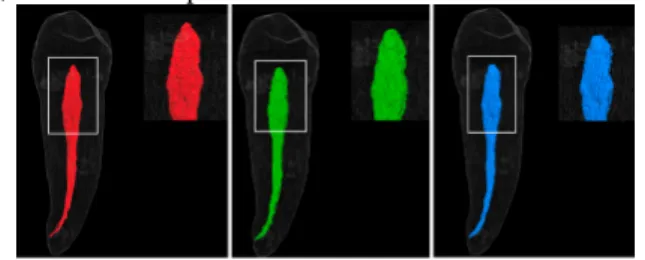
results for all the samples in terms of segmented root canal volume. Given the high-resolution and contrast of µCT data, the resulting segmented root canals were considered as the ground truth. We may remark that for all the considered teeth, the CBCT SR volumes provided a smaller error compared to the native CBCT data. An illustrative result is shown in Fig. 1 that highlights the root canal anatomy segmentations from µCT, CBCT and super-resolved CBCT.
Fig. 1. Premolar root canal segmentation results from (a) high resolution µCT, (b) low resolution CBCT and (c) the estimated super-resolved CBCT data volumes.
IV. CONCLUSION
This paper evaluated the potential of SR in dental CBCT, applied to the segmentation of the root canal anatomy. Using total variation and a recently proposed ADMM-based SR approach, the algorithm has been applied slice by slice in order to increase the spatial resolution of CBCT volumes. Root canal anatomy was further extracted automatically from native and super-resolved CBCT data, and compared to the one obtained from µCT. The interest of super-resolution has been confirmed through experiments on seven different teeth. To the best of our knowledge, this is the first attempt of using SR in root canal segmentation. This work opens several research perspectives that will be addressed in further studies: 3D deconvolution, blind approaches and experiments on dedicated phantoms [5] or non-extracted teeth.
REFERENCES
[1] W. Scarfe, A. Farman, and P. Sukovic, “Clinical applications of cone beam computed tomography in dental practice,” J Can Dent Assoc, vol. 72, no. 1, pp. 75–80, 2006.
[2] A. Toma, B. Sixou, and F. Peyrin, “Iterative choice of the optimal regularization parameter in tv image restoration,” Inverse Problems and Imaging, vol. 9, no. 4, pp. 1171–1179, 2015.
[3] N. Zhao, Q. Wei, A. Basarab, N. Dobigeon, D. Kouam´e, and J.-Y. Tourneret, “Fast single image super-resolution using a new analytical solution for l2-l2problems,” IEEE Trans. Image Process., vol. 25, no. 8, pp. 3683–3697, May 2016.
[4] P. Chang, K. Liang, J. Lim, M. Chung, and L. Chien, “A comparison of the thresholding strategies of micro-ct for periodontal bone loss: a pilot study,” Dentomaxillofacial Radiol., 2013.
[5] J. Michetti, A. Basarab, M. Tran, F. Diemer, and D. Kouam´e, “Cone-beam computed tomography contrast validation of an artificial periodontal phantom for use in endodontics,” in Engineering in Medicine and Biology Society (EMBC), 2015 37th Annual International Conference of the IEEE.