HAL Id: dumas-01122663
https://dumas.ccsd.cnrs.fr/dumas-01122663
Submitted on 4 Mar 2015HAL is a multi-disciplinary open access archive for the deposit and dissemination of sci-entific research documents, whether they are pub-lished or not. The documents may come from teaching and research institutions in France or abroad, or from public or private research centers.
L’archive ouverte pluridisciplinaire HAL, est destinée au dépôt et à la diffusion de documents scientifiques de niveau recherche, publiés ou non, émanant des établissements d’enseignement et de recherche français ou étrangers, des laboratoires publics ou privés.
Scalar localization by cone-beam computed tomography
of cochlear implant carriers : a comparative study
between straight and perimodiolar precurved electrode
arrays
Éric Boyer
To cite this version:
Éric Boyer. Scalar localization by cone-beam computed tomography of cochlear implant carriers : a comparative study between straight and perimodiolar precurved electrode arrays. Human health and pathology. 2014. �dumas-01122663�
AVERTISSEMENT
Ce document est le fruit d'un long travail approuvé par le
jury de soutenance et mis à disposition de l'ensemble de la
communauté universitaire élargie.
Il n’a pas été réévalué depuis la date de soutenance.
Il est soumis à la propriété intellectuelle de l'auteur. Ceci
implique une obligation de citation et de référencement
lors de l’utilisation de ce document.
D’autre part, toute contrefaçon, plagiat, reproduction illicite
encourt une poursuite pénale.
Contact au SICD1 de Grenoble :
[email protected]
LIENS
LIENS
Code de la Propriété Intellectuelle. articles L 122. 4
Code de la Propriété Intellectuelle. articles L 335.2- L 335.10
http://www.cfcopies.com/V2/leg/leg_droi.php
1
Faculté de médecine de Grenoble
Année : 2014
Scalar localization by cone-beam computed tomography of cochlear implant carriers:
a comparative study between straight and perimodiolar precurved electrode arrays.
THESE
Soutenue publiquement à la faculté de médecine de Grenoble Le 25 septembre 2014
Pour l’obtention du diplôme d’état de Docteur en médecine
Eric BOYER
Né le 30 décembre 1985 à CASTRES
Président du jury : Monsieur le Professeur Emile REYT
Directeur de thèse : Monsieur le Professeur Sébastien SCHMERBER
Membres du jury :
Monsieur le Professeur Christian Adrien RIGHINI Monsieur le Docteur Arnaud ATTYE
*La Faculté de Médecine de Grenoble n’entend donner aucune approbation ni improbation aux opinions émises dans les thèses ; ces opinions sont considérées comme propres à leurs auteurs.
2
Citation
C’est toujours les voix qui restent au final, C’est aussi toujours par elles que ça commence, Une voix plus une oreille ; Deux fils de soie impalpables et un pavillon
3
Liste des Professeurs des Universités - Praticiens hospitaliers, Maitres de conférences universitaires – Praticiens hospitaliers
2013-2014, Université Joseph Fourier, Grenoble
Corps Nom - Prénom Discipline
PU-PH ALBALADEJO Pierre Anesthésiologie réanimation
MCU-PH APTEL Florent Ophtalmologie
PU-PH
ARVIEUX-BARTHELEMY Catherine
chirurgie générale
PU-PH BACONNIER Pierre Biostatiques, informatique médicale et technologies de la communication PU-PH BAGUET Jean-Philippe Cardiologie
PU-PH BALOSSO Jacques Radiothérapie
PU-PH BARRET Luc Médecine légale et droit de la santé
PU-PH BAUDAIN Philippe Radiologie et imagerie médicale
PU-PH BEANI Jean-Claude Dermato-vénéréologie
PU-PH BENHAMOU Pierre Yves Endocrinologie, diabète et maladies métaboliques
PU-PH BERGER François Biologie cellulaire
PU-PH BETTEGA Georges Chirurgie maxillo-faciale, stomatologie
MCU-PH BOISSET Sandrine Agents infectieux
PU-PH BONAZ Bruno Gastro-entérologie, hépatologie, addictologie MCU-PH BONNETERRE Vincent Médecine et santé du travail
PU-PH BOSSON Jean-Luc Biostatiques, informatique médicale et technologies de la communication
MCU-PH BOTTARI Serge Biologie cellulaire
PU-PH BOUGEROL Thierry Psychiatrie d'adultes
PU-PH BOUILLET Laurence Médecine interne
MCU-PH BOUZAT Pierre Réanimation
PU-PH BRAMBILLA
CHRISTIAN
Pneumologie
PU-PH BRAMBILLA Elisabeth Anatomie et cytologie pathologiques
PU-PH BRICAULT Ivan Radiologie et imagerie médicale
PU-PH BRICHON Pierre-Yves Chirurgie thoracique et cardio-vasculaire MCU-PH BRIOT Raphaël Thérapeutique, médecine d’urgence
4
MCU-PH CALLANAN-WILSON
Mary
Hématologie, transfusion
PU-PH CARPENTIER Françoise Thérapeutique, médecine d'urgence PU-PH CARPENTIER Patrick Chirurgie vasculaire, médecine vasculaire
PU-PH CESBRON Jean-Yves Immunologie
PU-PH CHABARDES Stephan Neurochirurgie
PU-PH CHABRE Olivier Endocrinologie, diabète et maladies métaboliques
PU-PH CHAFFANJON Philippe Anatomie
PU-PH CHAVANON Olivier Chirurgie thoracique et cardio-vasculaire
PU-PH CHIQUET Christophe Ophtalmologie
PU-PH CHIROSSEL Jean-Paul Anatomie
PU-PH CINQUIN Philippe Biostatiques, informatique médicale et technologies de la communication PU-PH COHEN Olivier Biostatiques, informatique médicale et
technologies de communication PU-PH COUTURIER Pascal Gériatrie et biologie du vieillissement
PU-PH CRACOWSKI Jean-Luc Pharmacologie fondamentale, pharmacologie clinique
PU-PH DE GAUDEMARIS Régis Médecine et santé au travail
PU-PH DEBILLON Thierry Pédiatrie
MCU-PH DECAENS Thomas Gastro-entérologie, hépatologie
PU-PH DEMATTEIS Maurice Addictologie
PU-PH DEMONGEOT Jacques Biostatiques, informatique médicale et technologies de la communication
MCU-PH DERANSART Colin Physiologie
PU-PH DESCOTES Jean-Luc Urologie
MCU-PH DETANTE Olivier Neurologie
MCU-PH DIETERICH Klaus Génétique et procréation
MCU-PH DUMESTRE-PERARD
Chantal
Immunologie
PU-PH ESTEVE François Biophysique et médecine nucléaire MCU-PH EYSSERIC Hélène Médecine légale, droit à la santé
PU-PH FAGRET Daniel Biophysique et médecine nucléaire
PU-PH FAUCHERON Jean-Luc Chirurgie générale
MCU-PH FAURE Julien Biochimie et biologie moléculaire PU-PH FERRETTI Gilbert Radiologie et imagerie médicale
5
PU-PH FONTAINE Eric Nutrition
PU-PH FRANCOIS Patrice Epidémiologie, économie de la santé et prévention
PU-PH GARBAN Frédéric Hématologie, transfusion
PU-PH GAUDIN Philippe Rhumatologie
PU-PH GAVAZZI Gaetan Gériatrie et biologie du vieillissement
PU-PH GAY Emmanuel Neurochirurgie
MCU-PH GILLOIS Pierre Biostatiques, informatique médicale et technologies de la communication
PU-PH GODFRAIND Catherine Anatomie et cytologie pathologiques (type clinique)
MCU-PH GRAND Sylvie Radiologie et imagerie médicale
PU-PH GRIFFET Jacques Chirurgie infantile
MCU-PH GUZUN Rita Endocrinologie, diabétologie, nutrition, éducation thérapeutique
PU-PH HALIMI Serge Nutrition
PU-PH HENNEBICQ Sylviane Génétique et procréation
PU-PH HOFFMANN Pascale Gynécologie obstétrique
PU-PH HOMMEL Marc Neurologie
PU-PH JOUK Pierre-Simon Génétique
PU-PH JUVIN Robert Rhumatologie
PU-PH KAHANE Philippe Physiologie
PU-PH KRACK Paul Neurologie
PU-PH KRAINIK Alexandre Radiologie et imagerie médicale PU-PH LABARERE José Département de veille sanitaire PU-PH LANTUEJOUL Sylvie Anatomie et cytologie pathologiques MCU-PH LAPORTE François Biochimie et biologie moléculaire MCU-PH LARDY Bernard Biochimie et biologie moléculaire
MCU-PH LARRAT Sylvie Bactériologie, virologie
MCU-PH LAUNOIS-ROLLINAT
Sandrine
Physiologie
PU-PH LECCIA Marie-Thérèse Dermato-vénéréologie
PU-PH LEROUX Dominique Génétique
PU-PH LEROY Vincent Gastro-entérologie, hépatologie, addictologie PU-PH LETOUBLON Christian chirurgie générale
6
MCU-PH LONG Jean-alexandre Urologie
PU-PH MACHECOURT Jacques Cardiologie
PU-PH MAGNE Jean-Luc Chirurgie vasculaire
MCU-PH MAIGNAN Maxime Thérapeutique, médecine d’urgence
PU-PH MAITRE Anne Médecine et santé au travail
MCU-PH MALLARET Marie-Reine Epidémiologie, économie de la santé et prévention
MCU-PH MARLU Raphael Hématologie, transfusion
MCU-PH MAUBON Danièle Parasitologie et mycologie
PU-PH MAURIN Max Bactériologie - virologie
MCU-PH MCLEER Anne Cytologie et histologie
PU-PH MERLOZ Philippe Chirurgie orthopédique et traumatologie
PU-PH MORAND Patrice Bactériologie - virologie
PU-PH MOREAU-GAUDRY
Alexandre
Biostatiques, informatique médicale et technologies de la communication
PU-PH MORO Elena Neurologie
PU-PH MORO-SIBILOT Denis Pneumologie
MCU-PH MOUCHET Patrick Physiologie
PU-PH MOUSSEAU Mireille Cancérologie
PU-PH MOUTET François Chirurgie plastique, reconstructrice et esthétique, brûlogie
MCU-PH PACLET Marie-Hélène Biochimie et biologie moléculaire
PU-PH PALOMBI Olivier Anatomie
PU-PH PARK Sophie Hémato-transfusion
PU-PH PASSAGIA Jean-Guy Anatomie
PU-PH PAYEN DE LA
GARANDERIE Jean-François
Anesthésiologie réanimation
MCU-PH PAYSANT François Médecine légale et droit de la santé MCU-PH PELLETIER Laurent Biologie cellulaire
PU-PH PELLOUX Hervé Parasitologie et mycologie
PU-PH PEPIN Jean-Louis Physiologie
PU-PH PERENNOU Dominique Médecine physique et de réadaptation
PU-PH PERNOD Gilles Médecine vasculaire
PU-PH PIOLAT Christian Chirurgie infantile
7
PU-PH PLANTAZ Dominique Pédiatrie
PU-PH POLACK Benoît Hématologie
PU-PH POLOSAN Mircea Psychiatrie d’adultes
PU-PH PONS Jean-Claude Gynécologie obstétrique
PU-PH RAMBEAUD Jacques Urologie
MCU-PH RAY Pierre Génétique
PU-PH REYT Emile Oto-rhino-laryngologie
MCU-PH RIALLE Vincent Biostatiques, informatique médicale et technologies de la communication
PU-PH RIGHINI Christian Oto-rhino-laryngologie
PU-PH ROMANET J. Paul Ophtalmologie
MCU-PH ROUSTIT Mathieu Pharmacologie fondamentale, pharmaco clinique, addictologie
MCU-PH ROUX-BUISSON Nathalie Biochimie, toxicologie et pharmacologie
PU-PH SARAGAGLIA
Dominique
Chirurgie orthopédique et traumatologie
MCU-PH SATRE Véronique Génétique
PU-PH SCHMERBER Sébastien Oto-rhino-laryngologie
PU-PH SCHWEBEL Carole Réanimation médicale
PU-PH SCOLAN Virginie Médecine légale, droits à la santé MCU-PH SEIGNEURIN Arnaud Epidémiologie, économie de la santé et
prévention
PU-PH SERGENT Fabrice Gynécologie obstétrique
PU-PH SESSA Carmine Chirurgie vasculaire
PU-PH STAHL Jean-Paul Maladies infectieuses, maladies tropicales
PU-PH STANKE Françoise Pharmacologie fondamentale
MCU-PH STASIA Marie-José Biochimie et biologie moléculaire
PU-PH TAMISIER Renaud Physiologie
PU-PH TONETTI Jérôme Chirurgie orthopédique et traumatologie PU-PH TOUSSAINT Bertrand Biochimie et biologie moléculaire
PU-PH VANZETTO Gérald Cardiologie
PU VILLA Alessandro Neurosciences
PU-PH VUILLEZ Jean-Philippe Biophysique et médecine nucléaire PU-PH WEIL Georges Epidémiologie, économie de la santé et
prévention
PU-PH ZAOUI Philippe Néphrologie
8
Remerciements Officiels
Au président du jury,
Monsieur le Professeur Emile REYT, vous me faites l’honneur de présider ce jury de thèse. Je vous remercie de votre implication dans mon apprentissage de la chirurgie cervico-faciale et rhinologique. Votre expérience et votre sagesse resteront un exemple dans ma pratique de la spécialité
Au directeur de thèse,
Monsieur le Professeur SCHMERBER, vous m’avez fait l’honneur de diriger ce travail de thèse. Je vous remercie de m’avoir proposé ce sujet passionnant et de m’avoir encadré tout au long de sa réalisation. Je vous remercie pour votre pédagogie, de m’avoir transmis votre passion pour l’otologie et l’implant cochléaire et de m’avoir donné goût à la recherche scientifique.
Aux membres du jury,
Monsieur le Professeur Christian Adrien RIGHINI, vous me faites l’honneur d’être dans mon jury de thèse. Je vous remercie pour votre apprentissage de la cancérologie et de la chirurgie cervico-faciale. Vous m’avez appris la rigueur nécessaire à la pratique de la chirurgie.
Monsieur le Docteur Arnaud ATTYE, je te remercie d’être membre de mon jury de thèse. Ta participation et ton aide ont été très précieuses à la réalisation de ce travail. Ta disponibilité, ta sympathie et ta passion pour la radiologie en ORL nous font chaque jour progresser.
Madame le Docteur Virginie LEFOURNIER, Mr le Docteur Bernard ESCUDE, Madame Elsa
MONETIER, Monsieur Patrice JOUSSE, je vous remercie pour votre aide à la réalisation de ce travail de thèse
9
Sommaire
Titre Français : Evaluation de la position scalaire des porte-électrodes d’implant cochléaire
en imagerie cone-beam : Etude comparative entre électrode droite et périmodiolaire
pré-incurvée. ... 10
Résumé ... 10
Title: Scalar localization by cone-beam computed tomography of cochlear implant carriers: a
comparative study between straight and perimodiolar precurved electrode arrays. ... 11
Summary ... 11
Introduction ... 12
Material and methods ... 14
Results ... 20
Discussion ... 23
Conclusion ... 27
10
Titre Français : Evaluation de la position scalaire des porte-électrodes
d’implant cochléaire en imagerie cone-beam : Etude comparative
entre électrode droite et périmodiolaire pré-incurvée.
Résumé
Objectif : Comparer l’incidence d’une dislocation en imagerie conebeam après une implantation cochléaire selon l’utilisation d’une électrode droite ou pré-incurvée.
Matériel et méthodes : Etude comparative de cas non randomisée. Les portes électrodes droits et pré-incurvés implantés provenaient de deux fabricants différents. Une insertion par la fenêtre ronde a été réalisée dans tous les cas, sauf deux pour lesquels une cochléostomie a été réalisée. Une imagerie cone-beam a été réalisée chez tous les patients. Les images étaient reconstruites en coupes coronale oblique, sagittale oblique et axiale oblique pour leur analyse. Nous avons évalué la profondeur d’insertion de l’électrode et la survenue d’une dislocation de la scala tympani vers la scala vestibuli.
Résultats : Cinquante-quatre patients et 61 implants ont été analysés. Trente et un patient ont été implantés avec un porte-électrode périmodiolaire et 30 patients avec un porte-électrode droit. Neuf (15%) dislocations ont été observées, 8 (26%) dans la cohorte des électrodes périmodiolaires et 1 (3%) dans la cohorte des électrodes droites. La dislocation est survenue à un angle d’insertion en profondeur entre 170 et 190° dans le groupe d’électrode périmodiolaire et autour de 370° dans le groupe d’électrode droite.
Discussion : L’imagerie cone-beam est outil fiable pour déterminer la position du porte électrode dans la cochlée et détecter une éventuelle dislocation scalaire. Une dislocation survient habituellement dans la partie ascendante du tour basal de la cochlée avec un porte-électrode périmodiolaire. Un porte-électrode droit flexible a de meilleures chances de conserver une position dans la scala tympani.
MOTS CLES
11
Title: Scalar localization by cone-beam computed tomography of
cochlear implant carriers: a comparative study between straight and
perimodiolar precurved electrode arrays.
Summary
Objective: To compare using analyses of cone-beam computed tomography (CBCT) images, the incidence of scalar dislocation after cochlear implant surgery with a straight or a precurved electrode array.
Study design: Consecutive non-randomized case comparison study. Settings: Tertiary referral center
Patients: CBCT analyses of patients after cochlear implantation.
Intervention(s): Precurved perimodiolar and straight electrode arrays from two different manufacturers were implanted. A pure round window insertion was performed in all cases, except two necessitating a cochleostomy. All patients had, CBCT imaging and the images were reconstructed in coronal oblique, sagittal oblique, and axial oblique section.
Main outcome measures: Evaluation of the insertion depth angle and the occurrence of dislocation from the scala tympani to the scala vestibuli.
Results: Fifty four patients and 61 implants were analyzed. Thirty one patients were implanted with a perimodiolar electrode array and 30 patients with a straight array. Nine (15%) scalar dislocations were observed, 8 (26%) in the perimodiolar array group and 1 (3%) in the straight array group. The dislocation occurred at an insertion depth angle between 170 and 190° in the perimodiolar array group, and around 370° in the straight array group.
Conclusion: CBCT imaging is a reliable tool to assess the position of the electrode array in the cochlea and to detect a possible scalar dislocation. The dislocation usually occurs in the ascending part of the basal turn of the cochlea with a perimodiolar array. A straight flexible electrode array has a higher chance of a confined scala tympani positioning than a perimodiolar precurved array.
12
Introduction
The cochlear implant (CI) is a successful way to treat patients with severe or profound hearing loss. Nowadays, over 300,000 patients have been implanted all over the world.
Audiological outcomes and speech recognition after cochlear implantation are based on patient characteristics (age at onset of deafness, duration of deafness, age of patient at implantation and presence of preoperative residual hearing), on the surgical technique, and on the design of the electrode array. The goal of implant manufacturers is to improve the physical characteristics of the electrode array (design, 3D shape) to allow precise positioning in the scala tympani (ST) and to contribute to the optimization of hearing performance (1).
In 1993, Lehnardt introduced the principle of “soft surgery” which aims at inserting the electrode array in the ST without dislocation (2). A dislocation can be defined by the displacement of the electrode array from the ST to the scala vestibuli (SV), through the basilar membrane (BM) or the osseous spiral lamina. The dislocation damages the BM and spiral ganglion cells, compromising neural pathways (1). The technique of soft surgery was firstly described as a cochleostomy antero-inferior to the round window. However, the round window insertion approach is also a valuable and a safe technique with good clinical outcomes (3).
Cochlear implantation can be performed using either a precurved or straight array to place the array as close as possible to the modiolus or along the lateral wall of the cochlea, respectively. The Nucleus Contour Advance CI24RE and CI512 from Cochlear© (Cochlear Ltd, Lane Cove, Australia) are precurved perimodiolar electrodes with a design dedicated to a shallow insertion. This positioning decreases electrical thresholds and target specific frequencies for electrostimulation(4). The straight electrode array from MED-EL© (MED-EL GmbH, Innsbruck, Austria) aims at a full insertion, in order to provide a complete coverage of the cochlea with apical stimulation. This deeply inserted electrode array improves hearing and speech performance. The manufacturer offers a wide range of electrodes; among them a thin and flexible electrode array to minimize insertion trauma (5). Temporal bone studies have demonstrated that both types of electrode array focus on minimizing the occurrence of a scalar dislocation (6,7).
13
incidence. It was a reliable tool to control the position of the implant inside the cochlea and measure the depth of insertion. Nevertheless, plain radiography is not able to determine the three-dimensional shape of the electrode and cannot identify a misplacement of the electrode array (8). Therefore, high-resolution computed tomography (HRCT) has been used to determine the position of the electrode array. Although, HRCT provides a better resolution to visualize the temporal bone, the image definition due to the metallic artifacts is compromised; creating difficulties in assessing the scalar localization of the electrode array (9).
Cone Beam computed tomography (CBCT) is an innovative tool using a rotating cone-shaped source of radiation, with a flat panel detector. It was primarily developed for dental and maxillofacial imaging. It allows a volumetric acquisition with a single rotation of the gantry, whereas the HRCT would need an image set of multiple axial sections. With CBCT, reconstructions in any plane can be performed from the three-dimensional dataset of isotropic voxels using algorithms. The spatial resolution is consequently increased with reduced levels of radiation, metallic artifacts, and time of acquisition (10). It has been demonstrated on temporal bone studies that CBCT is a valuable tool in assessing scalar localization of the electrode array (11).
The aim of this study was to compare using CBCT analyses, the incidence of scalar dislocation after CI surgery with a straight or a precurved electrode array.
14
Material and methods
This consecutive non randomized case comparison study was conducted in an affiliated University Hospital following the tenets of the Declaration of Helsinki and adhered to Good Clinical Practice guidelines. Study ethics approval was obtained on April 9th, 2014 (CECIC Rhône-Alpes-Auvergne, Clermont-Ferrand, IRB 5891).
Study population
All patients included in this study had a severe, profound, or total hearing loss, without any inner ear malformations.
Surgical Procedure
The surgery was performed in a one-day surgery setting, by the same surgeon, in all patients to avoid any operator bias. The surgery consisted of a standard surgical technique with mastoidectomy and posterior tympanotomy. A pure round window approach was performed in all patients except for 2 cases. It consisted of the drilling of a large posterior tympanotomy, in order to visualize the round window entirely, and a round window preparation through the removal of the postero-superior bony overhang of the round window niche. A hearing preservation technique was then attempted by performing a micro-incision in the anterior third of the round window membrane using a 26G needle, without suctioning the perilymph, working in a bloodless operative field. In two cases, an antero-inferior cochleostomy had to be performed in 2 cases, due to ossification of the entire round window niche, secondary to far advanced otosclerosis, which limited access to the round window membrane.
CIs from two manufacturers were used: Cochlear© and MED-EL©. The implants from Cochlear© (CI24RE and CI512) were perimodiolar electrode array inserted using the “Advance Off Stylet” (AOS) insertion technique.
15
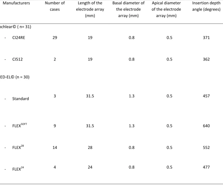
The implants from MED-EL© (Standard, FLEXSOFT, FLEX28, and FLEX24) were straight arrays inserted according to the standard procedure of insertion through the round window membrane. The characteristics of the electrode arrays are summarized in table 1.
The electrodes were fully inserted in all cases and electrophysiological recordings were performed in all cases to control the electrode integrity.
Imaging
All patients underwent CBCT using the same machine: 5G NewTom (NewTom, Verona, Italy). The system used a 200 x 25 mm flat panel detector at 650 mm from the radiation source. The 360° rotation of the X-ray tube took 18s. Tube voltage was 110kV, with a 19 mA charge at the terminals. Total filtrations were 2 mm and pitch 125 µm, with a field of view corresponding to a 12 x 7.5 cm diameter cylinder. The images were reconstructed in 125 µm isometric voxels and obtained in axial, coronal, and sagittal planes, using the NNT software provided by NewTom.
Image Analyses
Triple-blinded image analyses were performed by three senior neuroradiologists accustomed with CI imaging. They applied the same protocol for the reconstructions, measurements, and dislocation analyses.
- Reconstruction Technique: Three reconstructions were performed:
1) “The CBCT Cochlear view”: A coronal oblique section consisting of a view of the greatest axis of the cochlea. We determined an axis of the basal turn of the cochlea passing through the round window and reaching the pars ascendens on the lateral cochlear wall.
16
Then, we performed a double obliquity tilt to visualize the basal turn in one view. The thickness of the view was increased to 5mm to unroll the entire implant (Fig.1).
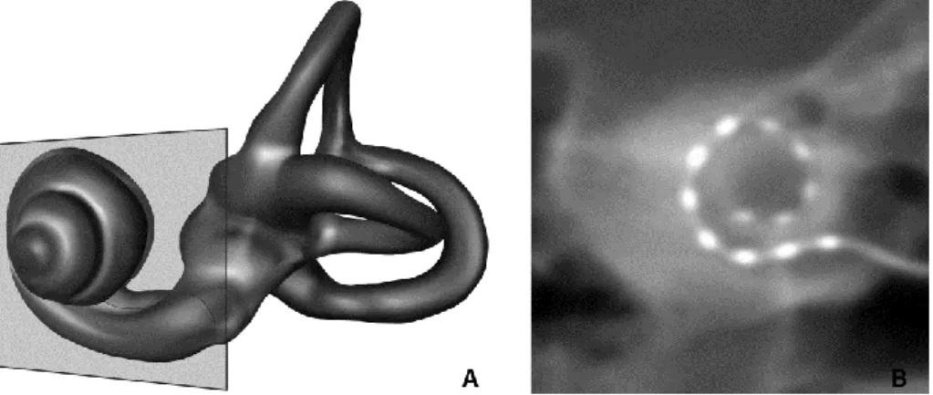
FIG. 1. Coronal oblique reconstruction: « CBCT Cochlear view ». A, Three-dimensional (3D) representation of a
left labyrinth demonstrating coronal oblique plane of section. B, two dimensional (2D) reconstruction from cone-beam computed tomography (CBCT).
2) An axial oblique section in the plane of the long axis of the modiolus, perpendicular to the “CBCT Cochlear view”. This section explored the cochlea from the round window to the lateral wall of the medial side of the basal turn (Fig.2).
FIG. 2.Sagittal oblique reconstruction.A, 3D representation of a left labyrinth demonstrating sagittal oblique
17
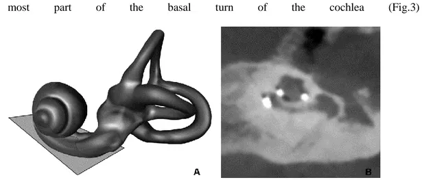
3) A sagittal oblique section perpendicular to the axial oblique and crossing the center of the modiolus. This section explored the cochlea from the inferior-most part to the superior-most part of the basal turn of the cochlea (Fig.3)
FIG. 3.Axial oblique reconstruction.A, 3D representation of a left labyrinth demonstrating axial oblique plane of
section.B, 2D reconstruction from CBCT.
- Measurements
We estimated the distances A and B according to the method developed by Escude on the “CBCT cochlear view” (12). Distance A was the largest distance measured from the center of the round window to the lateral wall crossing the modiolar axis. Distance B was the largest perpendicular cochlear measurement of A (Figure4).
FIG. 4.Measurements of distance A and B.A, Schematic 2D representation of a right cochlea and vestibule. The
A distance according to Escude (12) is the largest measurement from the center of the round window, through the modiolar axis, to the lateral wall. The B distance is the perpendicular distance of the cochlea crossing the modiolaraxis. B, Estimation of distance A and B on a 2D reconstruction of a right cochlea from CBCT.
18
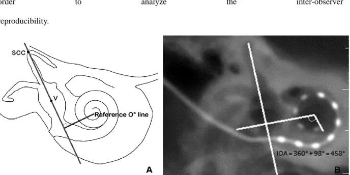
We estimated the insertion depth angle according to the method of Xu et al, on the “CBCT cochlear view” (13). We drew a line (SCC-V) from the superior semicircular canal (SCC) crossing the middle of the vestibule (V), as recommended by Xu et al. A perpendicular line to the SCC-V line was drawn crossing the spiral center of the cochlea establishing the zero-degree reference line. The insertion depth angle (IDA) was estimated in reference to the angular position of the most apical electrode relative to the zero–degree reference line (Figure5). These measurements were performed in
order to analyze the inter-observer
reproducibility.
FIG. 5. Measurements of the insertion depth angle (IDA). A, Schematic 2D representation of a right cochlea and
vestibule. Axis SCC-V connects the apex of the superior semicircular canal and the middle of the vestibule the reference line is perpendicular to the Axis SCC-V and crosses the cochlear spiral center. The IDA is estimated according to the reference line (13). B, Estimation of the IDA on 2D reconstruction of a right cochlea from CBCT.
- Dislocation Analysis
We defined a dislocation as a change in the location of the electrode array from the ST to the SV across the BM and/or osseous spiral lamina. The dislocation site was evaluated in sagittal and axial oblique reconstructions according to Lecerf et al (14). Scalar localization was determined by each neuroradiologist unaware of the surgical outcomes. The position of the electrode array was determined in reference to its position inside the cochlea; a posterior position was considered as an ST location
19
and an anterior position was considered as an SV location. A dislocation was defined as a change in the location of the electrode from the ST to the SV (Figure 6). A rereading was performed for all mismatches by the three radiologists. For each dislocation the angle of its occurrence in the cochlea was estimated, by locating on the sagittal and the axial oblique reconstruction the area where the electrode array changed from the posterior to the anterior part of the cochlea. This location identification was transferred to the cochlear view to estimate the depth of the angle of dislocation.
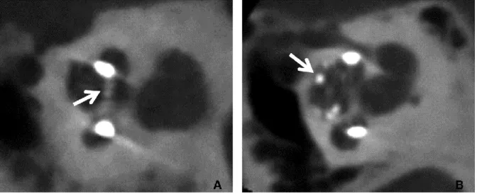
FIG. 6.Dislocation of the electrode array on a sagittal oblique reconstruction. A, Using a precurved perimodiolar
electrode from Cochlear©. The dislocation occurs at the end of the basal turn of the cochlea (arrow). B, Using a straight electrode from MED-EL©. The dislocation occurs in the apical part of the cochlea (arrow).
Statistical analysis
Data are presented as mean ± standard deviation. Measurements and dislocation occurrence mismatches were reread by the three neuroradiologists. Inter-rater agreement was calculated using the Fleiss kappa-like agreement coefficient for categorical data and an Intra-class Correlation Coefficient (ICC) for continuous variables. Distances A and B, and the IDA were averaged from the measurements given by the three neuroradiologists. Comparisons were made using Student’s t-test. A P-value of ≤ 0.05 was considered statistically significant. Data analyses were conducted using the Statistical Package for the Social Sciences (SPSS; version 17.0, SPSS Inc., Chicago, Illinois, USA). Statistical analyses were performed by our Clinical Center of Investigation.
20
Results
Fifty four patients and 61 implants were included (seven patients had a bilateral implantation), 25 women and 29 men, with a mean age of 50.2 years (Range 8 – 89).
Thirty one patients were implanted using the Nucleus implant system with the Contour Advance Electrode fitted with the AOS electrode insertion technique. Among the 31 implants from Cochlear©, 29 were CI24RE models and 2 were CI512.
Thirty patients were implanted using the implant from MED-EL© with the standard insertion technique, 9 FLEXSOFT, 14 FLEX28, 4 FLEX24, and 3 Standard electrode arrays were used.
The mean distance A was 8.81 ± 3.27 mm (ICC = 0.756, Confidence interval 95% (CfI95) = 0.548 - 0.877), the mean distance B was 6.71 ± 2.36 mm (ICC = 0.820, CfI95 = 0.666 - 0.909).
The mean insertion depth angle for the implants from Cochlear© was 371 ± 39.72 degrees for the CI24RE and 362 ± 24.6 degrees for the CI512. The mean insertion depth angle for the MED-EL© Standard implant was 457 ± 74 degrees, 640 ± 45 degrees for the FLEXSOFT, 552 ± 60.3 degrees for the FLEX28 and 477 ± 55.6 degrees for the FLEX24 (ICC = 0.589, CfI95 = 0.238 - 0.793). The mean values of the IDAs are summarized in Table 1.
21
Manufacturers Number of cases Length of the electrode array (mm) Basal diameter of the electrode array (mm) Apical diameter of the electrode array (mm) Insertion depth angle (degrees) Cochlear© ( n= 31) - CI24RE 29 19 0.8 0.5 371 - CI512 2 19 0.8 0.5 362 MED-EL© (n = 30) - Standard 3 31.5 1.3 0.5 457 - FLEXSOFT 9 31.5 1.3 0.5 640 - FLEX28 14 28 0.8 0.5 552 - FLEX24 4 24 0.8 0.5 477TABLE 1. Electrodes array specifications: length, basal, apical diameter and mean values of insertion depth
angle
.
We recorded 5 mismatches concerning the positioning of the electrode array. All three neuroradiologists agreed after rereading the images.
Scalar dislocation occurred in 9/61 cases (15%); in 8/31cases (26%) for the perimodiolar electrode arrays and in 1/30 case (3%) for the straight electrode arrays (Figure 6). The difference in scalar dislocation between the two types of arrays was statistically significant (Odd Ratio = 10, Fischer’s test, p = 0.026).
22
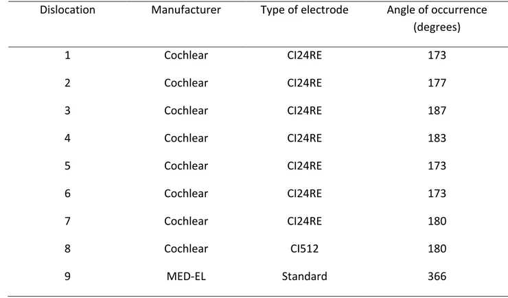
All dislocations of the implant from Cochlear© were located at an angle between 170° and 190°. The dislocation of the implant from MED-EL© was located at the angle of 380° (Table 2).
Dislocation
Manufacturer
Type of electrode
Angle of occurrence
(degrees)
1
Cochlear
CI24RE
173
2
Cochlear
CI24RE
177
3
Cochlear
CI24RE
187
4
Cochlear
CI24RE
183
5
Cochlear
CI24RE
173
6
Cochlear
CI24RE
173
7
Cochlear
CI24RE
180
8
Cochlear
CI512
180
9
MED-EL
Standard
366
TABLE 2. Cases of dislocation and angles of their occurrence
Statistical analysis of the site of dislocation between Cochlear and MED-EL devices was not performed because of the low statistical power.
23
Discussion
Atraumaticity of electrode array insertion is an essential objective, with the increasing rate of cochlear implantation, in patients with residual hearing, and the emergence of electro-acoustic stimulation. Although, patients with residual hearing represent a small part of the implanted population, the hearing preservation technique should be the gold standard for all cochlear implantation procedures. Moreover, in the pediatric population, repeated cochlear implantation surgery in the lifetime of the individual requires a non-traumatic insertion in the ST.
We compared the possible occurrence of a dislocation between 2 types of electrodes arrays: the perimodiolar Nucleus Contour Advance (CI24RE and CI512), and the straight MED-EL© FLEX and Standard electrodes. Since perimodiolar arrays are shorter than the straight arrays that we used, the insertion depth angle of perimodiolar arrays was smaller (15,16).We found a high degree of variation in the IDA for the same type of electrode array. This is attributed to the variations in the dimensions of the cochlea, and also to the position of the electrode array within the ST (medial position, midscala position or lateral position) (16).
A traumatic insertion of the electrode array is responsible for the elevation of the BM until it ruptures. The misplacement of the array in the SV involves a fracture of the osseous spiral lamina, damage to the vasculature and spiral ganglion cells (7,17,18). This sensorineural injury affects the residual hearing and increases electrical thresholds; resulting in poorer audiologic and speech results (1,19,20).
The total rate of dislocation in our study (15%) was in agreement with temporal bone and clinical studies (11-29.3%) (14,21,22). Our data demonstrated a significantly higher rate of dislocation for electrodes with a perimodiolar design, than electrodes with a lateral wall design. This difference has already been underlined in a recent publication (22). Despite the theoretical advantages of modiolar proximity, and the decrease of the threshold and comfort levels, the flexibility of the silicon and the nearness of the metal wires along the silicon body leads to a stiffness in the perimodiolar electrode array compared to the straight flexible array (23,24). In addition, data are largely lacking in the literature as to whether of not the AOS insertion technique has been used properly, with good
24
reproducibility and robustness. In our study, this problem was avoided because all of the electrode insertions followed a strict AOS insertion technique, performed by the same surgeon.
In our study, the straight electrode arrays resulted in only one dislocation. The basal and apical diameter of the electrode array did not influence the dislocation rate (the basal diameter of the FLEXSOFT and Standard is 1.3 mm versus 0.8 mm for the Contour Advance).
In the precurved perimodiolar array group, the electrode crossed from the ST to the SV at an approximate IDA of 180°, which corresponds to the ascending part of the basal turn of the cochlea. This “sensitive zone” has been described in several other studies (7,18,25-27). It corresponds to the point of lowest resistance of the basilar membrane, or the point where the force of electrode array insertion is the highest. Anatomically, this area corresponds to a narrowing of the ST (approximately 300µm), and an enlargement of the SV (28). In the straight array group, the electrode array crossed the basilar membrane at an IDA of around 370°. This area corresponds to the end of the second turn of the cochlea and the beginning of the apical turn, where a pressure point is described (29).
In our study, in all cases, we favored the use of a pure round window approach to surgery with a hearing preservation technique; even though the patients had no residual hearing. This approach reduces the acoustic trauma induced by the noise of drilling; decreases the risk of a perilymphatic fistula; and avoids an SV insertion (21,24,30). Moreover, a round window insertion potentiates the retention of the electrode array inside the ST (22,31). Our data show that a pure round window insertion can be performed in almost all cases (except two). We focused our investigation on this approach to compare the design of the electrodes without any bias from the insertion technique (round window versus cochleostomy insertion).
Nowadays, life expectancy of a newborn can reach a century; therefore, it is of utmost importance that neural structures within the cochlea are preserved during surgery to provide the best outcomes for re-implantation in the future. Similar objectives should also be applied in the adult patient, whether residual hearing is present preoperatively or not, since soft surgery techniques have become a gold standard in all recipients.
Our aim was not to compare audiological results in the subgroups that experienced a dislocation in the SV versus those with an exclusive ST positioning of the electrode array. Obviously,
25
we would expect that patients with a good positioning of the electrode in the ST would have better hearing performance than patients with a dislocation of the electrode array, but such was not the case in the present study. On the other hand, although patients from both subgroups exhibited the same audiological outcomes postoperatively, a high dislocation rate is to discouraged in cochlear implantation. In addition, comparison of the speech performance of patients implanted with different types of electrodes (with or without a dislocation) is difficult in such small cohorts. Such a comparison would require matching of the patients’ age, residual hearing, and type of CI. The aim of this investigation was not to compare audiological outcomes provided by both manufacturers. We rather focused our analyses on radiological examination, because post-implantation hearing performance involves many parameters and would require a larger population.
It has been suggested in a temporal bone study that the round window approach is not recommended with a perimodiolar electrode array (32). The perceived angle of insertion through the round window membrane via a posterior tympanotomy might have an effect on the location of the electrode array in the cochlea, and on the point of first contact of the electrode array with the pars ascendens(33). It remains to be determined whether or not the higher rate of dislocation observed in the present study, using a perimodiolar electrode array, was a consequence of the pure round window approach used. To answer this question, a comparative study in patients would be required. However, it has already been demonstrated in a clinical study that using an extended round window approach, a perimodiolar electrode array has a higher risk of dislocation, than a straight electrode array (34). To our knowledge, there is no evidence in the literature of a clinical study comparing a pure round window approach with a cochleostomy approach, using a perimodiolar electrode array.
CBCT is an innovative and useful tool in post-implantation imaging. It demonstrated in several temporal bone studies an accurate ability to detect the intracochlear position of the electrode array (35-37). It also improves visualization of the basilar membrane and the osseous spiral lamina (36). Furthermore, in patients implanted with a CI, CBCT has the ability to determine the scalar localization and the insertion depth; while reducing the amount of metal artifacts (19). Moreover, the volumetric acquisition using a cone-shaped beam with a flat panel detector reduces dramatically the radiation dose, the time of image acquisition, and the cost of the examination compared with HRCT
26
(10,11,38,39). In our study, the agreement among the 3 investigators in the measurements of A and B, and the IDA was statistically significant, thus demonstrating the reproducibility of inter-observer analyses. The evaluation of the dislocation carried only 5 mismatches, out of 61 CBCT scans (8%) analyzed, which necessitated a rereading for the agreement of the neuroradiologists. Visualization of a crossing from the ST to the SV was rather obvious in the basal turn of the cochlea. The neuroradiologists experienced greater difficulty in analyzing the second turn of the cochlea and the apex; however, they were still were able to identify a straight array dislocation in the second turn. This was because of the narrowing of the scalae and their close proximity to the apical part of the cochlea, which has already been described in the literature (33). Despite the fact that CBCT is known to decrease metal artifact, several examinations were still difficult to be interpreted because the patients’ moved during the procedure. The next generations of CBCT (with a faster acquisition or artifact attenuation) could be of great help in limiting this problem.
27
Conclusion
CBCT is a reliable tool to investigate the scalar position of the electrode array after cochlear implantation. It allows accurate evaluation of crossing from the ST to the SV. In our study, we investigated the occurrence of a dislocation with the use of a precurved perimodiolar array and a straight array. Favoring a pure round window technique, the straight flexible array had a higher chance of a strict scala tympani position than a perimodiolar precurved array.
29
Bibliographie
1. Finley CC, Holden TA, Holden LK, et al. Role of electrode placement as a contributor to variability in cochlear implant outcomes. OtolNeurotol 2008;29:920-8.
2. Lehnhardt E. Intracochlear placement of cochlear implant electrodes in soft surgery technique.
HNO 1993;41:356-9.
3. Briggs RJ, Tykocinski M, Xu J, et al. Comparison of round window and cochleostomy approaches with a prototype hearing preservation electrode. AudiolNeurootol 2006;11Suppl 1:42-8.
4. Hughes ML, Abbas PJ. The relation between electrophysiologic channel interaction and electrode pitch ranking in cochlear implant recipients. J AcoustSoc Am 2006;119:1527-37. 5. Hochmair I, Arnold W, Nopp P, et al. Deep electrode insertion in cochlear implants: apical
morphology, electrodes and speech perception results. ActaOtolaryngol 2003;123:612-7. 6. Gstoettner W, Plenk H, Jr., Franz P, et al. Cochlear implant deep electrode insertion: extent of
insertional trauma. ActaOtolaryngol 1997;117:274-7.
7. Eshraghi AA, Yang NW, Balkany TJ. Comparative study of cochlear damage with three perimodiolar electrode designs.Laryngoscope 2003;113:415-9.
8. Cohen LT, Xu J, Xu SA, et al. Improved and simplified methods for specifying positions of the electrode bands of a cochlear implant array. Am J Otol 1996;17:859-65.
9. Helbig S, Mack M, Schell B, et al. Scalar localization by computed tomography of cochlear implant electrode carriers designed for deep insertion. OtolNeurotol 2012;33:745-50.
10. Arweiler-Harbeck D, Monninghoff C, Greve J, et al. Imaging of Electrode Position after Cochlear Implantation with Flat Panel CT. ISRN Otolaryngol 2012;2012:728205.
11. Marx M, Risi F, Escude B, et al. Reliability of cone beam computed tomography in scalar localization of the electrode array: a radio histological study. Eur Arch Otorhinolaryngol 2014;271:673-9.
12. Escude B, James C, Deguine O, et al. The size of the cochlea and predictions of insertion depth angles for cochlear implant electrodes. AudiolNeurootol 2006;11 Suppl 1:27-33.
30
13. Xu J, Xu SA, Cohen LT, et al. Cochlear view: postoperative radiography for cochlear implantation. Am J Otol 2000;21:49-56.
14. Lecerf P, Bakhos D, Cottier JP, et al. Midmodiolar reconstruction as a valuable tool to determine the exact position of the cochlear implant electrode array. OtolNeurotol 2011;32:1075-81.
15. Franke-Trieger A, Jolly C, Darbinjan A, et al. Insertion depth angles of cochlear implant arrays with varying length: a temporal bone study. OtolNeurotol 2013;35:58-63.
16. Radeloff A, Mack M, Baghi M, et al. Variance of angular insertion depths in free-fitting and perimodiolar cochlear implant electrodes. OtolNeurotol 2008;29:131-6.
17. Lane JI, Driscoll CL, Witte RJ, et al. Scalar localization of the electrode array after cochlear implantation: a cadaveric validation study comparing 64-slice multidetector computed tomography with microcomputed tomography. OtolNeurotol 2007;28:191-4.
18. Wardrop P, Whinney D, Rebscher SJ, et al. A temporal bone study of insertion trauma and intracochlear position of cochlear implant electrodes. II: Comparison of Spiral Clarion and HiFocus II electrodes. Hear Res 2005;203:68-79.
19. Aschendorff A, Kromeier J, Klenzner T, et al. Quality control after insertion of the nucleus contour and contour advance electrode in adults.Ear Hear 2007;28:75S-9S.
20. Wanna GB, Noble JH, McRackan TR, et al. Assessment of electrode placement and audiological outcomes in bilateral cochlear implantation. OtolNeurotol 2011;32:428-32. 21. Coordes A, Ernst A, Brademann G, et al. Round window membrane insertion with
perimodiolar cochlear implant electrodes. OtolNeurotol 2013;34:1027-32.
22. Wanna GB, Noble JH, Carlson ML, et al. Impact of electrode design and surgical approach on scalar location and cochlear implant outcomes. Laryngoscope 2014: (Epub ahead of print). 23. Saunders E, Cohen L, Aschendorff A, et al. Threshold, comfortable level and impedance
changes as a function of electrode-modiolar distance. Ear Hear 2002;23:28S-40S.
24. Gstoettner W, Helbig S, Settevendemie C, et al. A new electrode for residual hearing preservation in cochlear implantation: first clinical results. ActaOtolaryngol 2009;129:372-9.
31
25. Roland JT, Jr. A model for cochlear implant electrode insertion and force evaluation: results with a new electrode design and insertion technique. Laryngoscope 2005;115:1325-39.
26. Stover T, Issing P, Graurock G, et al. Evaluation of the advance off-stylet insertion technique and the cochlear insertion tool in temporal bones. OtolNeurotol 2005;26:1161-70.
27. Tykocinski M, Saunders E, Cohen LT, et al. The contour electrode array: safety study and initial patient trials of a new perimodiolar design. OtolNeurotol 2001;22:33-41.
28. Biedron S, Prescher A, Ilgner J, et al. The internal dimensions of the cochlear scalae with special reference to cochlear electrode insertion trauma.OtolNeurotol 2010;31:731-7.
29. Verbist BM, Ferrarini L, Briaire JJ, et al. Anatomic considerations of cochlear morphology and its implications for insertion trauma in cochlear implant surgery.OtolNeurotol 2009;30:471-7.
30. Adunka O, Unkelbach MH, Mack M, et al. Cochlear implantation via the round window membrane minimizes trauma to cochlear structures: a histologically controlled insertion study.
ActaOtolaryngol 2004;124:807-12.
31. Connor SE, Holland NJ, Agger A, et al. Round window electrode insertion potentiates retention in the scala tympani. ActaOtolaryngol 2012;132:932-7.
32. Souter MA, Briggs RJ, Wright CG, et al. Round window insertion of precurvedperimodiolar electrode arrays: how successful is it? OtolNeurotol 2011;32:58-63.
33. Shapira Y, Eshraghi AA, Balkany TJ. The perceived angle of the round window affects electrode insertion trauma in round window insertion - an anatomical study. ActaOtolaryngol 2010;131:284-9.
34. van der Marel KS, Verbist BM, Briaire JJ, et al. Electrode migration in cochlear implant patients: not an exception. AudiolNeurootol 2012;17:275-81.
35. Cushing SL, Daly MJ, Treaba CG, et al. High-resolution cone-beam computed tomography: a potential tool to improve atraumatic electrode design and position. ActaOtolaryngol 2012;132:361-8.
32
36. Bartling SH, Gupta R, Torkos A, et al. Flat-panel volume computed tomography for cochlear implant electrode array examination in isolated temporal bone specimens. OtolNeurotol 2006;27:491-8.
37. Saeed SR, Selvadurai D, Beale T, et al. The Use of Cone Beam Computed Tomography to Determine Cochlear Implant Electrode Position in Human Temporal Bones. OtolNeurotol 2014: (Epub ahead of print).
38. Zeitler DM, Wang KH, Prasad RS, et al. Flat-panel computed tomography versus multislice computed tomography to evaluate cochlear implant positioning. Cochlear Implants Int 2012;12:216-22.
39. Dahmani-Causse M, Marx M, Deguine O, et al. Morphologic examination of the temporal bone by cone beam computed tomography: comparison with multislice helical computed tomography. Eur Ann Otorhinolaryngol Head Neck Dis 2011;128:230-5.
33
Remerciements officieux
A ma maman Sylvie, pour tout l’amour que tu me portes depuis ma naissance. Pour avoir été là à chaque instant de ma vie. Pour m’avoir soutenu dans les moments difficiles. Pour nous avoir si bien élevé toute seule, et nous avoir donné envie de croquer la vie à pleine dents. Je t’aime mamoune A ma sœur Delphine, pour ta volonté de fer, ta motivation, ta capacité à surmonter toutes les épreuves. Tu as toujours été un exemple pour moi
A Michel, pour ta joie de vivre, ton soutien toutes ces années et d’avoir rendu ma mère si heureuse. Le Mont-Blanc nous attend !!!!!
A Manou, de m’avoir donné goût au travail. Je suis si content que tu sois fière de ton petit fils. A ma tante Patricia ma deuxième maman, pour ton amour, ta passion de ton travail. A Paul et Cyril, vous êtes comme mes petits frères
A mon père Jean-Pierre, pour m’avoir transmis ta soif de savoir et ta curiosité. A loulou et Christine mes parrains marraines que je ne vois pas assez.
A mes beaux parents, de m’avoir accepté dans votre famille et d’avoir conçu cette perle d’Alexandra. A papi-jean qui doit nous regarder en ce moment
A Alexandre, pour ton enseignement de l’ORL, pour ton aide dans la rédaction de ma thèse. Pour la culture chirurgicale que tu m’as transmis en étant toujours disponible. Tu es un exemple pour nous tous internes et ton départ pour Saint-etienne est une grande perte pour moi.
A Junééééé, mon collègue, mon pote, mon frère. Ces années d’internat ont été merveilleuses et tu n’y es pas pour rien. J’espère que l’on continuera à travailler ensemble pendant longtemps. Brou
A tous mes co-internes, Etienne (sniff), Eléa, Jenny, l’ami des tous petits, Akil (mon petit tandoori), Rachel 4, Annie siiii gentille et Cindy.
A Alice pour ta bonne humeur et ta passion pour les gnomes.
A Ihab pour ton enseignement chirurgical et ta mélomanie du sifflement. A A nne Rivron
A tous mes co-internes d’ophtalmologie, marco (presque un ORL), papa mottet (et tous ses conseils). A toutes les infirmières du service, de votre patience et votre professionalisme malgré la difficulté de votre travail. Je vous adore !!!!!!
Aux infirmières de consultation (et à Jacqueline) pour leur expérience, tout ce que vous m’avez appris toutes ces années.
Aux infirmières du bloc : Jacquou le croquant, la bergère, la comtesse, katy, julie et tous les gens du bloc.
Aux secrétaires d’ORL pour votre bonne humeur et votre patience avec nos dictées moisies A tout le personnel du service d’ORL.
AuxPrs Gay, Brichon, Chaffanjon, au Dr Fonlupt qui m’ont si bien accueilli dans leurs services. A ali, Adrien, matthieu, diane, celine, François Berger et toutes les personnes de CLINATEC pour ce super Master II.
34
A mes amis d’enfance (Notre amitié vaut tous les trésors)
Souchy (mon Hodor…), mon plus vieux pote. Tu m’as accueilli comme un frère dans ta famille. Ton amitié est si précieuse. A Sbouby le cordon bleu et au ptitGab.
A puppet, dire qu’au début on ne s’entendait pas !!!!!! A tes faux pas en sortant de la tente. Au fait t’es né où déjà ?
A Niggers, loin des yeux près du cœur, tu nous manques avec ton style inimitable!!!! A copain, ma tige préférée, stabilise la Centrafrique et rentres vite !!!!
A Manchac, marie arthur et paul que vous êtes beaux tous les quatres.
A la noune, parce que tu m’as prêté ta serviette et que grâce à toi, je sais ce que c’est qu’une bague de pied. J’aimerai te voir plus souvent.
A Peddy, toujours enjoué, a notre passion commune de la montagne. A ta caravane. A soline medvedev pour ta crinière blonde, ton rire, ta bonne humeur.
A Floflo, la besogneuse. Merde pour demain. Tu vas les éclater !!!!!! A Regis, pour ton talent du fastfood corporel !!!!!
A toute la famille souchal : Jean et Marielle pour tous ces week-end où vous vous êtes occupé de moi. A Gaëlle et Pipo, A jobé (un vrai attaquant), A souch (un jour tu vas le gagner ce match de squash), a pierrot pour tintin, à Aurore.
A mes amis médecins :
A Micky, depuis le début tu es là. Tu as un cœur gros comme ça. Merci de ton amitié sans faille. A Yannou l’évier, mon ami, un confident. D’être si extraordinaire. A Lauren
A Minor, pour toutes ces virées au ski, toutes ces soirées, tes playlists, ta passion du Portugal. A Remy et Marion parce que vous êtes des parents admirables et que vous me manquez.
A Pafou., Mr Blague, l’attaquant Pivot. Qu’es ce que tu prends au petit déj ? Au B.R.A qui nous attend. A Carlota la plus patiente et la plus souriante de toutes.
A Paco pour ton caleçon du PSG. A Maë pour tes cupcakes !!
A Gabio, polo bayard, oliH et tous les abrutis. A Bibenbat, à Néné, à Cuge, à Juju a six fois six. A Marie Chaix, ma grande copine partie si loin.
A tous ceux de l’équipe de foot : Spirou, champi, papa casez, Nico, Guigui et l’audi. A guigui michel mon confrère suédois.
A tous ceux que j’ai oublié.
A mon amour Alexandra, depuis que tu es entrée dans ma vie elle prend tout son sens, j’ai l’impression de vivre dans une chanson. Tu me rends heureux depuis notre premier « Still loving you ». Le chemin est encore long pour nous deux et comme le dis Elton « How wonderful life is, while you’re in the world »