HAL Id: inserm-02134341
https://www.hal.inserm.fr/inserm-02134341
Submitted on 20 May 2019
HAL is a multi-disciplinary open access
archive for the deposit and dissemination of sci-entific research documents, whether they are pub-lished or not. The documents may come from teaching and research institutions in France or abroad, or from public or private research centers.
L’archive ouverte pluridisciplinaire HAL, est destinée au dépôt et à la diffusion de documents scientifiques de niveau recherche, publiés ou non, émanant des établissements d’enseignement et de recherche français ou étrangers, des laboratoires publics ou privés.
Cognitive Impairment Induced by
Delta9-tetrahydrocannabinol Occurs through
Heteromers between Cannabinoid CB1 and Serotonin
5-HT2A Receptors
Xavier Viñals, Estefanía Moreno, Laurence Lanfumey, Arnau Cordomí,
Antoni Pastor, Rafaël de la Torre, Paola Gasperini, Gemma Navarro, Lesley
Howell, Leonardo Pardo, et al.
To cite this version:
Xavier Viñals, Estefanía Moreno, Laurence Lanfumey, Arnau Cordomí, Antoni Pastor, et al.. Cog-nitive Impairment Induced by Delta9-tetrahydrocannabinol Occurs through Heteromers between Cannabinoid CB1 and Serotonin 5-HT2A Receptors. PLoS Biology, Public Library of Science, 2015, 13 (7), pp.e1002194. �10.1371/journal.pbio.1002194�. �inserm-02134341�
Cognitive Impairment Induced by
Delta9-tetrahydrocannabinol Occurs through
Heteromers between Cannabinoid CB
1
and
Serotonin 5-HT
2A
Receptors
Xavier Viñals1☯, Estefanía Moreno2,3☯, Laurence Lanfumey4, Arnau Cordomí5, Antoni Pastor6, Rafael de La Torre6, Paola Gasperini2,3,7, Gemma Navarro2,3,
Lesley A. Howell7, Leonardo Pardo5, Carmen Lluís2,3, Enric I. Canela2,3, Peter J. McCormick2,3,7‡*, Rafael Maldonado1‡*, Patricia Robledo1,6‡*
1 Neuropharmacology Laboratory, University Pompeu Fabra, Barcelona, Spain, 2 Centro de Investigación Biomédica en Red sobre Enfermedades Neurodegenerativas, Barcelona, Spain, 3 Department of Biochemistry and Molecular Biology, Faculty of Biology, University of Barcelona, Barcelona, Spain, 4 CPN, INSERM UMR S894, Université Paris Descartes, UMR S894, Paris, France, 5 Laboratori de Medicina Computacional, Unitat de Bioestadística, Facultat de Medicina, Universitat Autònoma de Barcelona, Bellaterra, Spain, 6 Integrative Pharmacology and Systems Neuroscience, IMIM-Hospital del Mar Medical Research Institute, Barcelona, Spain, 7 School of Pharmacy, University of East Anglia, Norwich Research Park, Norwich, United Kingdom
☯ These authors contributed equally to this work. ‡ These authors equally supervised this work.
*p.mccormick@uea.ac.uk(PJM);rafael.maldonado@upf.edu(RM); probledo@imim.es(PR)
Abstract
Activation of cannabinoid CB1 receptors (CB1R) by delta9-tetrahydrocannabinol (THC)
pro-duces a variety of negative effects with major consequences in cannabis users that consti-tute important drawbacks for the use of cannabinoids as therapeutic agents. For this reason, there is a tremendous medical interest in harnessing the beneficial effects of THC. Behavioral studies carried out in mice lacking 5-HT2Areceptors (5-HT2AR) revealed a
remarkable 5-HT2AR-dependent dissociation in the beneficial antinociceptive effects of
THC and its detrimental amnesic properties. We found that specific effects of THC such as memory deficits, anxiolytic-like effects, and social interaction are under the control of 5-HT2AR, but its acute hypolocomotor, hypothermic, anxiogenic, and antinociceptive effects
are not. In biochemical studies, we show that CB1R and 5-HT2AR form heteromers that are
expressed and functionally active in specific brain regions involved in memory impairment. Remarkably, our functional data shows that costimulation of both receptors by agonists reduces cell signaling, antagonist binding to one receptor blocks signaling of the interacting receptor, and heteromer formation leads to a switch in G-protein coupling for 5-HT2AR from
Gq to Gi proteins. Synthetic peptides with the sequence of transmembrane helices 5 and 6 of CB1R, fused to a cell-penetrating peptide, were able to disrupt receptor heteromerization
in vivo, leading to a selective abrogation of memory impairments caused by exposure to THC. These data reveal a novel molecular mechanism for the functional interaction between CB1R and 5-HT2AR mediating cognitive impairment. CB1R-5-HT2AR heteromers
a11111
OPEN ACCESS
Citation: Viñals X, Moreno E, Lanfumey L, Cordomí A, Pastor A, de La Torre R, et al. (2015) Cognitive Impairment Induced by Delta9-tetrahydrocannabinol Occurs through Heteromers between Cannabinoid CB1and Serotonin 5-HT2AReceptors. PLoS Biol
13(7): e1002194. doi:10.1371/journal.pbio.1002194 Academic Editor: Eric J. Nestler, Mount Sinai School of Medicine, UNITED STATES Received: November 12, 2014 Accepted: June 3, 2015 Published: July 9, 2015
Copyright: © 2015 Viñals et al. This is an open access article distributed under the terms of the
Creative Commons Attribution License, which permits unrestricted use, distribution, and reproduction in any medium, provided the original author and source are credited.
Data Availability Statement: All relevant data are within the paper and its Supporting Information files. Funding: This study was supported by grants from the Spanish‘Ministerio de Ciencia e Innovación’ (SAF2011-29864) to RM, (SAF2010-18472) to PJM and (SAF2011-23813) to EC.‘Ministerio de Economía y Competitividad’ (SAF2014-59648-P) to RM. The Spanish‘Instituto de Salud Carlos III’ (P1070709 and PI14/00210) to PR and (RD06/001/ 001) to RM, the Catalan government (SGR2009-00131) to RM. PJM was supported by a Ramon y Cajal Fellow and internal funds from UEA, and PJM
are thus good targets to dissociate the cognitive deficits induced by THC from its beneficial antinociceptive properties.
Author Summary
Delta-9-tetrahydrocannabinol (THC), the main psychoactive compound of marijuana, induces numerous undesirable effects, including memory impairments, anxiety, and dependence. Conversely, THC also has potentially therapeutic effects, including analgesia, muscle relaxation, and neuroprotection. However, the mechanisms that dissociate these responses are still not known. Using mice lacking the serotonin receptor 5-HT2A, we revealed that the analgesic and amnesic effects of THC are independent of each other: while amnesia induced by THC disappears in the mutant mice, THC can still promote analgesia in these animals. In subsequent molecular studies, we showed that in specific brain regions involved in memory formation, the receptors for THC and the 5-HT2A receptors work together by physically interacting with each other. Experimentally interfer-ing with this interaction prevented the memory deficits induced by THC, but not its anal-gesic properties. Our results highlight a novel mechanism by which the beneficial
analgesic properties of THC can be dissociated from its cognitive side effects.
Introduction
The administration of delta-9-tetrahydrocannabinol (THC), the main psychoactive compound inCannabis sativa, induces numerous behavioral responses related to undesirable effects, including memory impairments [1,2], anxiogenic effects [3], and dependence [4,5]. However, other effects of THC are associated with potential therapeutic applications, including analgesia [6] and anxiolytic-like and neuroprotective effects [3,5,7]. One major challenge in the field of cannabinoids is to identify new mechanisms that could be used to dissociate these responses in order to improve the benefit-risk ratio of cannabinoid agonists. Cannabinoid behavioral effects are mainly due to the activation of central CB1cannabinoid receptors (CB1R) [5]. CB1R activa-tion has been shown to modulate a wide range of neurotransmitters in the brain, including glu-tamate,γ-aminobutyric acid (GABA), opioids, dopamine, and serotonin, which could
participate in THC pharmacological responses [4,5]. Recent evidence shows that THC and other cannabinoids modulate behaviors typically mediated by serotonin 2A receptors’
(5-HT2AR) activation [8–10]. In addition, mice lacking CB1R exhibit a dysregulation of seroto-nergic activity in the prefrontal cortex [11] and reduced head twitches induced by the 5-HT2AR agonist, (±)-1-(2,5-dimethoixy-4-odophenyl)-2-aminopropane (DOI) [12]. Reciprocally, the activation of 5-HT2AR expressed in cells stimulates the formation and release of the endocan-nabinoid, 2-arachidonoylglycerol (2-AG) [13,14]. Both 5-HT2AR and CB1R are expressed in brain structures involved in regulating emotions, learning, and memory, including the amyg-dala, cerebral cortex, and hippocampus [15–17]. CB1Rs are highly expressed presynaptically in the prefrontal cortex [18–20] and in the hippocampus [21], where moderate expression of 5-HT2AR has also been observed [16,22,23]. In the rat striatum, CB1R expression has been detected in dendrites of spiny- and aspiny-type somata [24], coinciding with the observed den-dritic expression of 5-HT2AR in this area [25–27]. Moreover, 5-HT2AR is involved in different psychotic manifestations [28,29], while adolescent consumption of cannabis enhances the inci-dence of psychotic symptoms [30,31]. Collectively, these data suggest possible bidirectional
CB1R-5-HT2AR Heteromers Mediate THC-Induced Cognitive Deficits
and LP participate in the European COST Action CM 1207 (GLISTEN). FEDER funds partial support is also acknowledged. The funders had no role in study design, data collection and analysis, decision to publish, or preparation of the manuscript. Competing Interests: The authors have declared that no competing interests exist.
Abbreviations: 2-AG, 2-arachidonoylglycerol; 5-HT, serotonin; 5-HT2AR, serotonin 2A receptors; A1,
adenosine receptor type 1; aCSF, artificial cerebrospinal fluid; AEA, anandamide; Akt, protein kinase B; BiFC, bimolecular fluorescence complementation; BRET, bioluminescent resonance energy transfer; cAMP, cyclic adenosine
monophosphate; CB1R, cannabinoid receptor type 1;
CLAHE, contrast limited adaptive histogram equalization; CTX, cholera toxin; D1, dopamine
receptor type 1; DAPI, 4',6-diamidino-2-phenylindole; DI, discrimination index; DMEM, Dulbecco’s modified Eagle’s medium; DMR, dynamic mass redistribution; DMSO, dimethyl sulfoxide; DOI, (±)-1-(2,5-dimethoixy-4-odophenyl)-2-aminopropane; DR, dorsal raphe; EPM, elevated plus maze; ERK, extracellular signal-regulated kinase; FBS, fetal bovine serum; FR1, fixed ratio 1; GABA, γ-aminobutyric acid; GPCR, G protein-coupled receptor; GWS, global withdrawal score; HEK, human embryonic kidney; HIV, human immunodeficiency virus; HTRF, homogeneous time-resolved fluorescence; ICV, intracerebroventricular; IP, intraperitoneally; KO, knockout; LC-MS-MS, liquid chromatography—mass spectrometry; mBU, milli BRET unit; mGlu2R, metabotropic glutamate receptor type 2; PBS, phosphate-buffered saline; PLA, proximity ligation assay; PTX, pertussis toxin; RIM, rimonabant; Rluc, renilla luciferase; ROI, region of interest; SD, standard deviation; SEM, standard error of the mean; TBS, Tris-buffered saline; THC, delta9-tetrahydrocannabinol; TM, transmembrane helix; TR-FRET, time-resolved fluorescent resonance energy transfer; VEH, vehicle; WT, wild type; YFP, yellow fluorescent protein.
interactions between CB1R- and 5-HT2AR-mediated pharmacological responses, although a cellular mechanism for this cross talk has yet to be discovered. Here we sought to understand at what level the interactions of these two systems occur. Using a variety of in vivo and in vitro assays, we reveal a new molecular mechanism by which the cognitive deficits of THC can be dissociated from its beneficial antinociceptive properties.
Using transgenic mice lacking 5-HT2AR, we found that these two receptors do indeed inter-act in heteromeric complexes. Importantly, these heteromers are specifically required for the amnesic, anxiolytic, and social interaction effects caused by THC, but not for other pharmaco-logical responses, such as antinociception and hypolocomotion. Interestingly, the activation of this complex in cells expressing both receptors resulted in a reduction of intracellular signaling through adenylate cyclase, arrestin recruitment, and the extracellular signal-regulated kinase (ERK) 1/2 and protein kinase B (Akt) pathways, confirming altered downstream signaling. The formation of this receptor complex in the brain and its selective involvement in THC-induced memory and anxiety-like responses was further evidenced by the use of transmembrane helix (TM) 5 and TM 6 interference peptides in vivo. Hence, the disruption of the CB1R-5-HT2AR heteromer by intracerebroventricular (ICV) infusion of these peptides abolished the memory deficits induced by THC and its anxiolytic-like effects, but not its antinociceptive properties. Our findings reveal a new molecular mechanism by which the cognitive deficits of THC can be dissociated from its beneficial antinociceptive properties.
Results
5-HT
2AR Modulates THC’s Effects on Acute Amnesia, Anxiety, and
Social Interaction and Its Effect on Dorsal Raphe Neuronal Activity
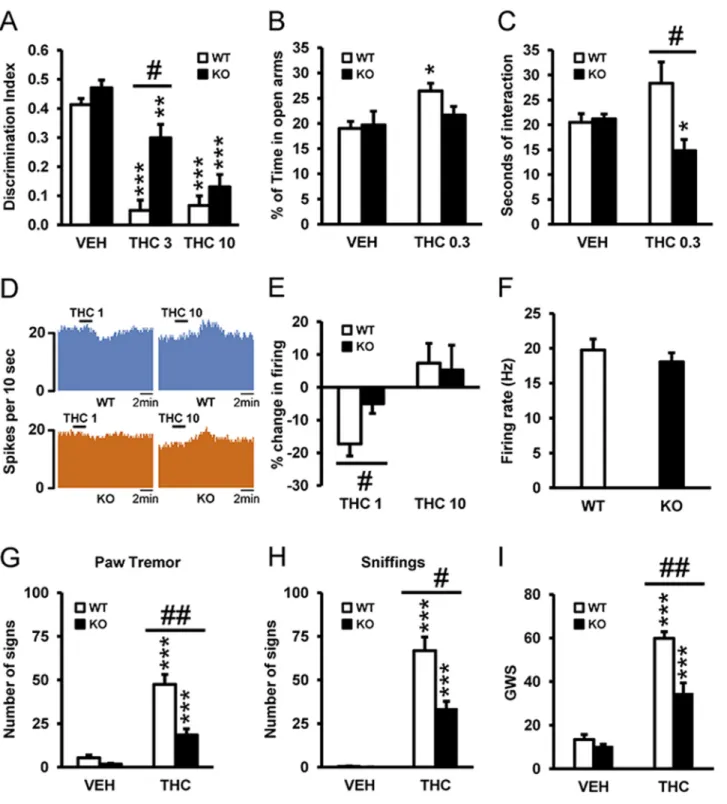
Due to the compelling data that 5-HT2AR may interact with CB1R, we sought to broadly iden-tify which behavioral effects of THC might be dependent on 5-HT2AR. Using wild-type (WT) and 5-HT2AR knockout (KO) animals, we evaluated THC’s amnesic-like effects at two doses (3 and 10 mg/kg) known to induce memory deficits in the novel object recognition paradigm [1]. Acute THC administration of both doses of THC induced amnesic-like effects in WT mice, and this effect was significantly reduced in 5-HT2AR KO animals only at the dose of 3 mg/kg (Fig 1A), suggesting that 5-HT2AR increases the potency of THC to induce memory impair-ments. Previous studies have shown that cannabinoid agonists can induce anxiogenic and anxi-olytic-like responses in rodents, depending on the dose and the environmental conditions [3,7]. Low doses of cannabinoids usually induce an anxiolytic-like effect, whereas higher doses cause the opposite response. Using the previous reported dose of 0.3 mg/kg, we evaluated the involvement of 5-HT2AR in the anxiolytic-like effects of THC [32]. Acute administration of THC induced an anxiolytic-like effect in WT animals, while in KO mice this response was reduced (Fig 1B). In agreement, a similar acute low dose of THC increased social interaction in WT mice, whereas it decreased this response in KO animals (Fig 1C). Since anxiolytic-like behavior has been associated with changes in the activity of dorsal raphe (DR) neurons [33], we determined whether variations in DR neuronal activity induced by THC were modified in 5-HT2AR KO mice using extracellular electrophysiological recordings in slice preparations of DR nucleus (Fig 1D). Basal activity of DR neurons was not modified in KO with respect to WT mice (Fig 1F), indicating that 5-HT2AR do not exert a tonic modulation of serotonergic activity in the DR nucleus, in line with previous microdialysis data showing similar basal extracellular levels of 5-HT in the cortex of both genotypes [34]. However, KO animals were less sensitive than WT mice to the decrease in neuronal firing evoked by THC (1 nM), an effect lost at a higher THC concentration (10 nM), probably because of a ceiling effect (Fig 1E). These data
Fig 1. 5-HT2AR mediates THC-induced amnesic- and anxiolytic-like effects. (A) The administration of THC (3 and 10 mg/kg) induced memory
impairments in the novel object recognition test in WT mice as compared to vehicle (VEH) treatment (n = 8–11), and this effect was significantly abrogated in 5-HT2AR KO mice at the dose of 3 mg/kg, but not at the dose of 10 mg/kg. (B) The anxiolytic effects of THC (0.3 mg/kg) observed in WT mice tested in the
elevated plus maze were blocked in 5-HT2AR KO mice (n = 9–12). (C) The increase in social interaction induced by THC (0.3 mg/kg) in WT mice was
abolished in 5-HT2AR KO mice (n = 5–7). (D) Neuronal firing of representative dorsal raphe (DR) neurons before and after THC administration (1 and 10 nM)
in WT (upper panel) and 5-HT2AR KO (lower panel) animals. (E) A challenge with THC (1 nM) reduced the percent change in firing rate of DR neurons from
WT mice, and this effect was blunted in DR neurons from 5-HT2AR KO mice. No significant differences between genotypes were observed following a
challenge with THC at 10 nM (n = 7–14). (F) The basal firing rate of DR neurons was similar in WT and 5-HT2AR KO animals (n = 20–24). (G–I) Withdrawal
symptoms, including paw tremor (G), sniffing (H), and global withdrawal score (GWS) (I), were reduced in 5-HT2AR KO mice when compared to WT mice.
Data are mean + standard error of the mean (SEM).* p < 0.05, ** p < 0.01, and *** p < 0.001 versus VEH; # p < 0.05 versus WT mice. The statistical analyses used and their corresponding F andp-values are shown inS1 Table.
suggest that specific effects of THC such as memory deficits, anxiolytic-like effects, social inter-action, and DR neuronal activity are under the control of 5-HT2AR.
We also evaluated the role of 5-HT2AR in responses to chronic THC exposure. Rimona-bant-precipitated THC withdrawal syndrome was evaluated in 5-HT2AR KO mice after chronic THC treatment. Several somatic signs of abstinence including paw tremor (Fig 1G) and sniffing (Fig 1H) were significantly attenuated in 5-HT2AR KO mice compared to WT animals, as well as the global withdrawal score (GWS) calculated by giving each sign a proportional weight (Fig 1I), indicating that 5-HT2AR is necessary for the full expression of THC withdrawal. Next, we assessed by western blot analysis whether CB1R levels were modified in 5-HT2AR KO mice following chronic treatment with THC. As a first step, we verified the specificity of the CB1R antibody (S1A Fig) in naïve WT and KO mice. These results show that CB1R are expressed at high levels in the cortex, striatum, nucleus accumbens, and hippocampus of WT mice but are virtually absent in KO mice. In animals chronically treated with THC, CB1R levels decreased in the hippocampus (S1B Fig) and cerebellum (S1C Fig) of both WT and KO, in agreement with previous data [35]. In the hippocampus, but not in the cerebellum, this receptor down-regulation was greater in KO mice than in WT mice treated with THC. In addition, we deter-mined the state of the endocannabinoid system in KO mice as a control. Anandamide levels were slightly but significantly reduced (S1D Fig), while 2-arachidonoylglycerol levels were not modified in KO mice versus WT mice (S1E Fig). These findings highlight the key role played by hippocampal 5-HT2AR in the adaptive responses induced by chronic THC exposure.
5-HT
2AR Does Not Affect the Hypolocomotor, Hypothermic, Anxiogenic,
and Analgesic Effects of THC or the Reinforcing Effects of Cannabinoid
Agonists
Following the above-described differential effects in WT and 5-HT2AR KO animals in memory and social phenotypes, we next sought to explore other behaviors to appreciate how general the influence of 5-HT2AR is on THC's effects. Surprisingly, THC decreased locomotor activity in a similar dose-dependent manner in WT and KO mice (Fig 2A). Likewise, THC reduced body temperature dose-dependently and induced a profound hypothermia at 10 mg/kg in both genotypes (Fig 2B). Next, we determined the antinociceptive effects of THC in WT and KO mice using two different behavioral tests, namely, the tail-immersion and hot-plate tests. In the tail-immersion test, THC induced a comparable dose-dependent analgesic effect in WT and KO mice (Fig 2C). Similarly, the antinociception observed in the hot-plate test in terms of fore-paw licking (Fig 2D) and jumping (Fig 2E) responses was comparable in WT and KO mice. To evaluate THC-induced anxiogenic-like behavior, we used the dose of 3 mg/kg, in order to avoid the locomotor suppressant effects of THC at higher doses. The acute administration of this dose induced an anxiogenic-like response in both WT and KO mice as revealed by a decrease in the percentage of time spent in the open arms of the elevated plus maze, with simi-lar extent in both WT and KO mice (Fig 2F). Finally, the reinforcing properties of the CB1R agonist, WIN 55,212–2, were investigated using the intravenous operant self-administration model [36], which is the most reliable paradigm in rodents to evaluate the addictive potential of drugs of abuse [37]. Both WT and KO mice learned to discriminate between the active and inactive nose pokes and self-administered WIN 55,212–2 in a similar manner, indicating that 5-HT2AR does not play a role in the reinforcing properties of this cannabinoid agonist (Fig
2G), in contrast to the role played by this receptor on the reinforcing properties of psychosti-mulants [34]. Together, our behavioral data comparing THC's effects in WT and 5-HT2AR KO animals strongly point to a cross talk between CB1R and 5-HT2AR, particularly at the level of memory, anxiolytic-like behavior, social interaction, and withdrawal syndrome.
Fig 2. 5-HT2AR does not mediate THC-induced hypolocomotion, hypothermia, analgesia, anxiogenic-like behavior, or the reinforcing properties of
WIN 55,212–2. (A) Locomotor activity (% of baseline) was dose-dependently reduced in both WT and 5-HT2AR KO mice treated with THC (0.3, 1, 3, and 10
mg/kg) as compared to vehicle (VEH) administration (n = 5–15). (B) THC induced hypothermia at the dose of 10 mg/kg to a similar extent in WT and 5-HT2AR
KO mice (n = 13–24). (C) In the tail-immersion test, percent analgesia was dose-dependently increased by THC (1, 3, and 10 mg/kg) in both WT and 5-HT2AR KO mice (n = 10–16). In the hot-plate test, the percent of analgesia as calculated from the latency to paw-licking (D) and to jumping behavior (E)
was similar in WT and KO animals treated with THC (1, 3, and 10 mg/kg) as compared to VEH administration (n = 7–16). (F) No significant differences CB1R-5-HT2AR Heteromers Mediate THC-Induced Cognitive Deficits
Coexpression of 5-HT
2AR and CB
1R Causes Cell Signaling via Gi
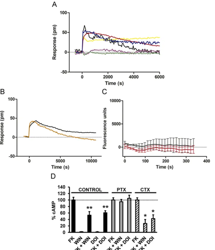
Next, we sought to explore at the cellular level how 5-HT2AR and CB1R might achieve the above-described cross talk between receptors. We first measured the global cellular response using dynamic mass redistribution (DMR) label-free assays, which detect changes in light dif-fraction in the bottom 150 nm of a cell monolayer [38]. Both CB1R agonists, THC and WIN 55,212–2, induced dose- and time-dependent signaling in cells only expressing CB1R (S2A
Fig). The 5-HT2AR receptor agonist DOI was unable to signal, and the antagonist MDL100, 907 was unable to revert the WIN 55,212-2-induced signal in cells expressing CB1R (S2B Fig). The 5-HT2AR agonists, DOI and serotonin, induced dose- and time-dependent signaling in cells only expressing 5-HT2AR (S2C Fig). Furthermore, WIN 55,212–2 was unable to signal, and the antagonist rimonabant was unable to revert the DOI-induced signaling in cells express-ing 5-HT2AR (S2D Fig), demonstrating the selectivity of the ligands. In cells stably expressing CB1R, WIN 55,212–2 induced a time-dependent cell signal that was inhibited by pertussis toxin (PTX), but not by cholera toxin (CTX) (S3A Fig), confirming that CB1R are coupled to Gi in these cells [39]. Accordingly, WIN 55,212–2 reduced the forskolin-induced cyclic
adeno-sine monophosphate (cAMP), an effect blocked by PTX, but not by CTX or the Gq inhibitor YM-254890 (S3B Fig). In cells only expressing 5-HT2AR, the cell signal induced by DOI was not blocked by CTX or PTX, suggesting something other than a Gi or Gs coupling (S3C Fig). Moreover, DOI was not able to increase cAMP or decrease forskolin-induced cAMP (S3D Fig), bolstering previous studies showing that 5-HT2AR is coupled to Gq. In agreement, the Gq inhibitor YM-254890 completely blocked the cell response to DOI (S3E Fig). DOI was able to induce intracellular calcium release in these cells, an effect that was blocked by YM-254890 (S3F Fig), confirming that when expressed alone, 5-HT2AR are coupled to a Gq protein. Impor-tantly, in cells coexpressing CB1R and 5-HT2AR, the DMR signal induced by both WIN 55,212–2 and DOI was inhibited by PTX, but not by CTX (Fig 3A). The Gq inhibitor, YM-254890, had no effect on DOI signaling (Fig 3B), and neither DOI nor WIN 55,212–2 induced
intracellular calcium release in these cells (Fig 3C). These results suggest that coexpression of CB1R and 5-HT2AR causes Gi coupling. Thus, blocking of Gi is sufficient to block receptor sig-naling. To further support this finding, we measured changes in cAMP production upon recep-tor activation. In cells expressing CB1R and 5-HT2AR, both WIN 55,212–2 and DOI
treatments led to a reduction in forskolin-activated cAMP production, and the effect of both ligands was sensitive to PTX, but not to CTX (Fig 3D). In addition, WIN 55,212–2 or DOI
alone, in the absence of forskolin, did not modify cAMP in these cells (Fig 3D). This change of G-protein coupling of 5-HT2AR by coexpression of CB1R suggests the formation of CB1 R-5-HT2AR heteromers.
CB
1R and 5-HT
2AR Form Heteromers
CB1R and 5-HT2AR have been traditionally considered as monomeric structural units that are coupled to intracellular heterotrimeric G-proteins. More recent evidence suggests that they can also assemble into homomers or heteromers with other G protein-coupled receptors (GPCRs) [40,41]. We hypothesized, based on the above results, that CB1R and 5-HT2AR form heteromers and their different expression in brain regions might account for the dissociation of THC behav-ioral responses. To test this hypothesis, we first used a bioluminescent resonance energy transfer
between genotypes were observed in anxiogenic-like behavior induced by THC (3 mg/kg) (n = 6–10). (G) Both WT and 5-HT2AR KO mice acquired WIN
55,212–2 self-administration behavior and responded equally for this drug during the 12 d of training (n = 12–15). Data are mean + SEM. * p < 0.05, ** p < 0.01, and *** p < 0.001 versus VEH-treated animals. The statistical analyses used and their corresponding F and p-values are shown inS1 Table. doi:10.1371/journal.pbio.1002194.g002
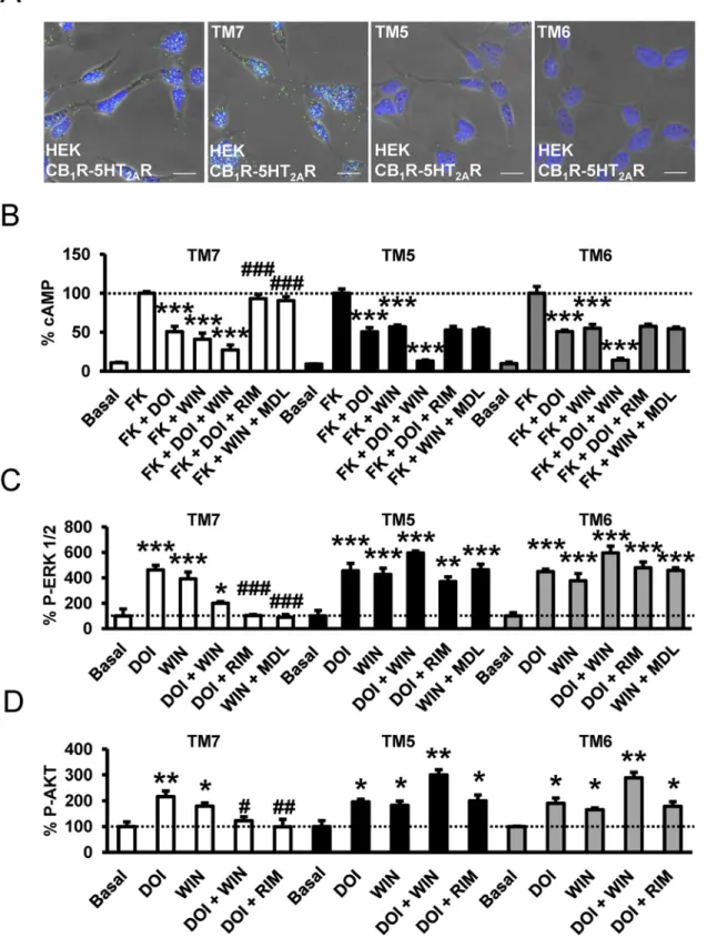
Fig 3. CB1R and 5-HT2AR are associated to Gi signaling when coexpressed. HEK-293Tcells expressing CB1R and 5-HT2AR receptors were used. In
(A and B), the DMR analysis was performed. In (A), cells were pretreated overnight with medium (black line), with 10 ng/ml pertussis toxin (purple line), or with 100 ng/ml cholera toxin (yellow line) and were stimulated with 100 nM DOI. Alternatively, cells pretreated overnight with medium (red line), with 10 ng/ml pertussis toxin (green line), or with 100 ng/ml cholera toxin (blue line) were stimulated with 50 nM WIN 55,212–2. In (B), cells were pretreated 30 min with medium (black line) or with 1μM of the Gq protein inhibitor YM-254890 (orange line) and were stimulated with 100 nM DOI. In all cases, the resulting
(BRET) assay. This assay has been well established for studying GPCR interactions and has the advantage over classical immunoprecipitation approaches that it is performed in live cells over a range of protein expression levels [42,43]. A saturable BRET curve (BRET-max of 64 ± 8 milli BRET unit [mBU] and BRET50of 4 ± 2) in cells expressing a constant amount of 5-HT2AR— renilla luciferase (Rluc) and increasing amounts of CB1R yellow fluorescent protein (YFP) was obtained (Fig 4A), indicating a specific interaction. Low and linear plots were observed using either dopamine D1R-Rluc as the donor or adenosine A1R-YFP as the acceptor as negative con-trols (Fig 4A), results consistent with nonspecific interactions [43]. These results indicate that 5-HT2AR and CB1R can form heteromers when coexpressed in cells. Further support for hetero-mer formation was obtained by bimolecular fluorescence complementation (BiFC) assays out-lined inFig 4B. In this assay, fluorescence only appears after correct folding of two YFP Venus hemiproteins. This occurs when two receptors fused to hemi-YFP Venus proteins (cYFP or nYFP) come within proximity. Fluorescence was detected in HEK-293T cells transfected with different amounts of cDNA corresponding to both 5-HT2AR-cYFP and CB1R-nYFP, but not in negative controls in which cells were transfected with cDNA corresponding to 5-HT2AR-cYFP and the noninteracting A1R-nYFP or CB1R-nYFP and the noninteracting D1R-cYFP (Fig 4B). Finally, in a third technique, we provided additional evidence of heteromer formation via prox-imity ligation assays (PLAs). This technique permits the direct detection of molecular interac-tions between two endogenous proteins or transfected proteins, without the need of fusion proteins. This technique is similar to immunoprecipitation but has an additional advantage of not requiring membrane solubilization. Labeling heterodimers by PLA requires both receptors to be sufficiently close to allow the two antibody-DNA probes to form double stranded segments (<17 nm), a signal that is further amplified in the presence of fluorescent nucleotides [44]. CB1R-5-HT2AR heteromers were observed as green punctate staining in HEK-293T cells coex-pressing CB1R and 5-HT2AR (Fig 4C), but not in negative controls in HEK-293T cells express-ing CB1and D1receptors or for the noninteracting CB1and transferrin receptors, in spite of the expression and high colocalization of these pairs at the membrane level (S4A and S4B Fig). Also as a negative control, no PLA staining was detected in samples in which cells only expressing CB1R or 5-HT2AR alone were mixed at a 1:1 ratio (S4C Fig). Previous studies have shown PLA to be semiquantitative and particularly useful at lower expression levels [45]. To estimate the rel-ative sensitivity of the PLA for GPCRs, experiments were performed in cells transfected with increasing cDNA amounts of 5-HT2AR and CB1R. In each case, PLA was quantified as the ratio between the number of green spots and the number of cells expressing spots (ratio r). This ratio was then represented as a function of the receptor’s cDNA transfected (Fig 4D). We observed an increase in PLA signal with increasing cDNA (Fig 4D.) These three independent approaches provide strong support for the formation of CB1R-5-HT2AR heteromers.
Functional Characteristics of CB
1R-5-HT
2AR Heteromers
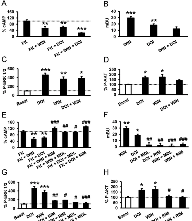
A common consequence of heteromer formation is altered downstream signaling upon dual stimulation of the receptors in the heteromer [46–48]. To examine whether this may be the
picometer shifts of reflected light wavelength (pm) were monitored over time. Each curve is the mean of a representative optical trace experiment carried out in triplicates. In (C), intracellular calcium release was monitored in cells expressing CB1R and 5-HT2AR receptors treated with 100 nM DOI (black curve) or
with 50 nM WIN 55,212–2 (red curve). Values are mean ± SEM of n = 3. In (D), cAMP production was determined in cells treated with medium (control), treated overnight with 10 ng/ml pertussis toxin (PTX), or treated 1 h with 100 ng/ml cholera toxin (CTX). Cells were stimulated with 100 nM DOI or 100 nM WIN 55,212–2 (WIN) in the absence or in the presence of 0.5 μM forskolin (FK). Values (cAMP produced in each condition minus basal stimulation in the absence of forskolin or agonists) represent mean± SEM of n = 3–4 and are expressed as the percentage of the forskolin-treated cells in each conditions (120–150 pmols cAMP/106cells). One-way ANOVA followed by a Dunnett’s multiple comparison test showed a significant effect over the forskolin-alone effect in each condition (* p < 0.05, ** p < 0.01).
Fig 4. 5-HT2AR and CB1R form heteromers in transfected cells. In (A), BRET saturation experiments were performed in HEK-293T cells transfected with
0.025μg of 5-HT2AR-Rluc cDNA and increasing amounts of CB1R-YFP cDNA (0.05μg to 1.5 μg, black curve), with 0.5 μg of dopamine D1R-Rluc cDNA and
increasing amounts of CB1R-YFP cDNA (0.5μg to 6 μg, yellow line), or with 0.025 μg of 5-HT2AR-Rluc cDNA and increasing amounts of adenosine A1R-YFP
cDNA (0.05μg to 1.5 μg, red line). The relative amount of BRET is given as a function of 100 x the ratio between the fluorescence of the acceptor (YFP) and the luciferase activity of the donor (Rluc). BRET is expressed as milli BRET units (mBU) and is given as the mean± standard deviation (SD) of 3–6
case for CB1R-5-HT2AR- heteromers, we determined signaling through adenylate cyclase, arrestin recruitment, the ERK 1/2 pathway, and the Akt pathway in cells expressing both recep-tors. In cells stimulated with forskolin and treated with WIN 55,212–2, DOI, or both, we found that costimulation led to reduced cAMP production (Fig 5A). To examine whether costimula-tion led to changes inβ-arrestin II recruitment compared to single stimulation, the agonist-induced interaction of arrestin with the receptors was measured by BRET in cells expressing β-arrestin II-Rluc, 5-HT2AR-YFP, and CB1R. Both agonists, DOI and WIN 55,212–2, recruited β-arrestin II, but costimulation led to a significant decrease in arrestin recruitment (Fig 5B), indicating that costimulation reduces cell signaling. Both CB1R and 5-HT2AR agonists induced the activation of ERK 1/2 and Akt pathways in a time- and dose-dependent manner (S5 Fig). Measuring ERK 1/2 phosphorylation (Fig 5C) or Akt phosphorylation (Fig 5D), the costimula-tion with WIN 55,212–2 and DOI, surprisingly, did not increase the phosphorylation levels reached by each agonist separately. This response was not due to a change in the optimum time response for ERK 1/2 (S6A Fig) or Akt phosphorylation (S6B Fig). Taken together, these data suggest that costimulation of CB1R-5-HT2AR heteromers leads to reduced cell signaling. Some GPCR heteromers have been found to display cross antagonism, the ability of an antago-nist of one receptor to antagonize the signaling of the partner receptor [49,50]. Cross antago-nism requires direct protein—protein interaction since antagonists do not signal on their own. When cells coexpressing both receptors were pretreated with the CB1R antagonist rimonabant and then stimulated with the CB1R agonist WIN 55,212–2 or the 5-HT2AR agonist DOI, sur-prisingly no decreases in cAMP (Fig 5E), noβ-arrestin II recruitment (Fig 5F), and no phos-pho-ERK 1/2 (Fig 5G) or phospho-Akt (Fig 5H) were observed. These results indicate that rimonabant blocks both CB1R and 5-HT2AR signaling. Analogously, the signaling in cAMP (Fig 5E),β-arrestin II recruitment (Fig 5F), and phospho-ERK 1/2 (Fig 5G) or phospho-Akt (Fig 5H) induced by both the CB1R agonist WIN 55,212–2 and the 5-HT2AR agonist DOI were completely blocked when cells were pretreated with the 5-HT2AR antagonist MDL 100,907. This cross antagonism is not due to the lack of specificity of the ligands since the 5-HT2AR ago-nist DOI and the antagoago-nist MDL100,907 were unable to signal or modify the CB1R signaling in cells only expressing CB1R (S6C Fig). Furthermore, the CB1R agonist WIN 55,212–2 and the antagonist rimonabant did not modify the 5-HT2AR signaling in cells only expressing
5-HT2AR (S6D Fig). In total, these results demonstrate that CB1R-5-HT2AR heteromers display bidirectional cross antagonism.
Molecular Basis of Cross Antagonism in CB
1R-5-HT
2AR Heteromers
To understand how receptor—receptor interactions might facilitate the above-mentioned cross antagonism and to potentially design a biochemical tool to disrupt these interactions, we took advantage of the exponential growth in the number of solved GPCR structures, in the form of
experiments grouped as a function of the amount of BRET acceptor. In (B), a schematic representation of fluorescence complementation experiments is depicted in the left panel showing that fluorescence only appears after the YFP Venus hemiprotein complementation due to the proximity of two receptors fused to hemi-YFP Venus proteins (cYFP or nYFP). In the right panel, fluorescence at 530 nm was detected in HEK-293T cells transfected with different amounts of cDNA corresponding to both 5-HT2AR-cYFP and CB1R-nYFP (equal amount for each construct), but not in negative controls in which cells were
transfected with cDNA corresponding to 5-HT2AR-cYFP and the noninteracting adenosine A1receptor-nYFP or CB1R-nYFP and the noninteracting
dopamine D1receptor-cYFP. One-way ANOVA followed by a Dunnett’s multiple comparison test showed a significant fluorescence over basal values in
HEK-293T cells (** p < 0.01, *** p < 0.001). In (C), PLAs were performed in HEK-293T cells expressing CB1R and 5-HT2AR. Confocal microscopy images
(superimposed sections) are shown in which heteromers appear as green spots. In all cases, cell nuclei were stained with DAPI (blue). Scale bars = 20μm. In (D), PLAs were performed in nontransfected HEK-293T cells, cells transiently transfected with 0.5μg of CB1R or 5-HT2AR cDNA (negative controls, white
columns), or with increasing amounts of CB1R and 5-HT2AR cDNA (black columns). In each case, the ratio between the number of green spots and the
number of cells showing spots (ratio r) was calculated. One-way ANOVA followed by a Dunnett’s multiple comparison test showed a significant PLA staining over nontranfected cells (*** p < 0.001).
Fig 5. A profile of CB1R-5-HT2AR heteromer signaling. In (A and E), cAMP production was determined in HEK-293Tcells expressing CB1R and 5-HT2AR
after stimulation with 100 nM DOI, 100 nM WIN 55,212–2 (WIN), or both in the absence or in the presence of 0.5 μM forskolin. In (E), cells were first preincubated either with the CB1R antagonist rimonabant (1μM, RIM) or the 5-HT2AR antagonist MDL 100,907 (300 nM) for 20 min prior to being stimulated.
Values (cAMP produced in each condition minus basal stimulation in the absence of forskolin or agonists) represent mean± SEM of n = 3–4 and are expressed as the percentage of the forskolin-treated cells in each condition (120–150 pmols cAMP/106cells). One-way ANOVA followed by a Bonferroni
monomers or homo-oligomers, bound to either agonists, antagonists, inverse agonists, or in complex with the G-protein to model heteromer activation (S7 Fig) [51]. Agonist binding at the extracellular side triggers small local structural changes near the binding site [52] that are translated into larger-scale helix movements at the intracellular site [53]. Specifically, agonists increase signaling by opening an intracellular cavity, required for the binding of the C-terminal α5 helix of the G-protein, through the movement of TM 5 and TM 6. Conversely, inverse ago-nists decrease the basal, agonist-independent level of signaling by closing this cavity. Our find-ings that CB1R-5-HT2AR heteromers display bidirectional cross antagonism led us to suggest that the antagonist-bound conformation of protomer A allosterically prevents the opening of the intracellular cavity of protomer B. To our knowledge, the molecular basis of this bidirec-tional cross antagonism has not been described. Recently, the crystal structure of theμ-opioid receptor has shown a novel mode of receptor dimerization via TMs 5 and 6 [54]. In this assem-bly, TMs 5 and 6 of protomer A form a very stable four-helix bundle with TMs 5 and 6 of pro-tomer B (S7A Fig). This high surface complementarity in the heteromer, within the four-helix bundle interface, prevents the opening of the intracellular cavity (S7A Fig). Thus, we hypothe-sized that bidirectional cross antagonism in the CB1R-5-HT2AR heteromer is due to antagonist binding to either protomer that stabilizes the inactive conformation of TM 5 and TM 6 and to the subsequent formation of this very stable four-helix association. As a consequence, the action of agonists is blocked at both receptors (seeFig 6A–6Dfor details). To test this hypothe-sis, we investigated if synthetic peptides with the sequence of TMs 5, 6, and 7 (as negative con-trol) of CB1R, fused to HIV TAT, were able to disrupt receptor heterodimerization and the observed bidirectional cross antagonism. This approach has been used by us and others previ-ously [55,56]. We first checked by immunocytochemistry that TM 5, TM 6, and TM 7 interfer-ence peptides do not appreciably change the expression and colocalization of CB1R and 5-HT2AR at the membrane level (S8 Fig). We found that pretreatment with TM 5 and TM 6 (but not TM 7) interference peptides of cells expressing CB1R-nYFP and 5-HT2AR-cYFP dis-rupt the heteromer structure as revealed by a loss of fluorescence in BiFC assays (S9 Fig). These results were further confirmed by PLA assays. CB1R-5-HT2AR heteromers were observed as green punctate staining in HEK-293T cells not treated or treated with the TM 7 interference peptide, but they were absent in cells treated with TM 5 or TM 6 interference peptides (Fig 7A). Notably, the cross antagonism was not observed at the level of cAMP (Fig 7B), ERK 1/2 phosphorylation (Fig 7C), and Akt phosphorylation (Fig 7D) in HEK-293T cells expressing CB1R and 5-HT2AR and treated with TM 5 or 6 peptides. The effect of the peptides was specific to the heteromer, as single activation of the individual receptors still led to signaling in the presence of the peptides. Importantly, these results indicate that negative cross talk and cross antagonism require receptor—receptor interaction and are specific biochemical characteristics of the heteromer that can be used as a fingerprint to detect the heteromer [47,49,57]. These results suggest that receptor heterodimerization occurs via TM 5 and TM 6, which facilitates
post hoc tests showed a significant effect over the forskolin-alone effect in each condition (** p < 0.01, *** p <0.001) or of the antagonist plus agonist treatment over the agonist treatment (#p < 0.05, ## p < 0.01, ### p < 0.001). In (B and F), β arrestin II recruitment was measured by BRET experiments in cells transfected with 2μg of CB1R cDNA, 0.2μg of β-arrestin II-Rluc cDNA, and 1 μg of 5-H2AR-YFP cDNA. Cells were not preincubated with antagonist (B)
or were preincubated (F) for 20 min with vehicle, rimonabant (1μM, RIM), or MDL 100,907 (300 nM, MDL). Cells were not treated (BRET < 10) or were treated for 7 min with WIN 55,212–2 (100 nM, WIN) or DOI (100 nM) before BRET determination. Values represent mean ± SEM of n = 4–6. One-way ANOVA followed by a Bonferroni post hoc tests showed a significant effect over not-treated cells (* p < 0.05, ** p < 0.01, *** p <0.001) or of the antagonist plus agonist treatment over the agonist treatment (##p < 0.01, ### p < 0.001). In (C, D, G, and H), cells expressing CB1R and 5-HT2AR were not
preincubated with antagonist (C and D) or were preincubated for 15 min with rimonabant (1μM, RIM) or MDL 100,907 (300 nM, MDL) (G and H) and stimulated for 5 min with WIN 55,212–2 (100 nM,WIN) or DOI (100 nM). Quantification of phosphorylated ERK 1/2 (C and G) or Akt (D and H) was determined by western blot. Values, expressed as percentage of basal (nontreated cells), were mean± SEM of n = 3–10. One-way ANOVA followed by a Bonferroni post hoc tests showed a significant effect over basal (* p < 0.05, ** p < 0.01, ** p < 0.001) or of the antagonist plus agonist treatment over the agonist treatment (#p < 0.05, ## p < 0.01, ### p < 0.001).
the observed cross antagonism. GPCRs are dynamic proteins that permit rapid small-scale structural fluctuations and pass through an energy landscape to adopt a number of conforma-tions ranging from inactive to active. Our results have shown that in the case of receptor het-erodimerization via TM 5 and TM 6, one of the protomers allosterically modulates the functional properties (energy landscape) of the interacting receptor (S7B Fig).
Differential Expression of Functional CB
1R-5-HT
2AR Heteromers in the
Brain
CB1R-5-HT2AR heteromer expression in tissue was analyzed by in situ PLA using specific pri-mary antibodies directed against CB1R and 5-HT2AR that have been validated in WT, CB1R KO, and 5-HT2AR KO mice (Fig 8) [58]. CB1R-5-HT2AR heteromers were observed as punc-tate green spots in cells with DAPI-stained nuclei in slices from hippocampus (CA3 region), dorsal striatum (caudate-putamen), and cortex (somatomotor layers 1, 2, and 3), all areas where both receptors are expressed [13], of WT animals, but not in 5-HT2AR or CB1R KO ani-mals (Fig 8A) or in negative controls (S10 Fig). In all cases, staining was observed in a relatively high percentage of cells (60%–70%) (Fig 8B). Interestingly, no green spots were detected in slices from nucleus accumbens in either WT or KO animals, indicating a differential expression of heteromers in the brain (Fig 8). To further support the existence of heteromer expression in these brain regions, we used the heteromer specific biochemical characteristics identified above (negative cross talk and cross antagonism) as a heteromer fingerprint to detect heteromers in situ. We measured ERK 1/2 phosphorylation in isolated brain slices from the hippocampus
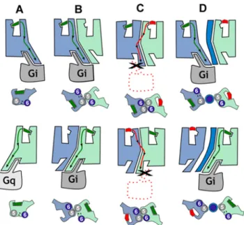
Fig 6. Proposed functional properties of CB1R-5-HT2AR heteromers. In (A), agonist binding to CB1R
(blue) or 5-HT2AR (light green) triggers the conformational changes of TMs 5 and 6, opening the intracellular
cavity for Gi and Gq binding, respectively. In (B), the formation of the CB1R-5-HT2AR heteromer makes both
receptors signal via Gi. In (C), rimonabant binding to CB1R or MDL 100,907 to 5-HT2AR stabilizes the closed
conformation of the receptor, facilitating heterodimerization via TMs 5 and 6 as in the crystal structure of theμ-opioid receptor. In this assembly, both protomers are locked in the closed conformation since the opening of TMs 5 and 6 for G-protein binding is not feasible (seeS7 Fig). Bidirectional cross antagonism is due to the fact that antagonist binding to any protomer must, in addition to its common role in a monomeric signaling unit, disrupt this very stable four-helix association. (D) In agreement, bidirectional cross antagonism is abrogated following treatment with TM 5 or TM 6 interference peptides (dark blue), which disrupt the heteromer structure.
doi:10.1371/journal.pbio.1002194.g006
Fig 7. Interacting protomer domains in CB1R-5-HT2AR heteromers and heteromer disruption by TM interference peptides. In (A), HEK-293T cells
expressing CB1R and 5-HT2AR were treated for 4 h with vehicle (left panel) or 4μM of CB1R TM 7, TM 5, or TM 6 interference peptides before performing
proximity ligation assays. Confocal microscopy images (superimposed sections) are shown in which heteromers appear as green spots in cells treated with vehicle and with TM 7 interference peptide, but not in cells treated with TM 5 or TM 6 interference peptides. In all cases, cell nuclei were stained with DAPI (blue). Scale bars = 20μm. In (B–D), HEK-293T cells expressing CB1R and 5-HT2AR were preincubated for 20 min with rimonabant (1μM, RIM) or MDL
(Fig 9A), dorsal striatum (Fig 9B), cortex (Fig 9C), or nucleus accumbens (Fig 9D) of WT mice. As expected when slices were treated with WIN 55,212–2 or DOI, ERK 1/2 phosphorylation was induced. As in cells, hippocampal, dorsal striatal, and cortical cell signaling was not increased when slices were coactivated with both agonists, again suggesting reduced signaling
100,907 (300 nM, MDL) before stimulation for 10 min (B) or 5 min (C, D) with the CB1R agonist WIN 55,212–2 (100 nM), the 5-HT2AR agonist DOI (100 nM),
or both in the presence (B) or absence (C, D) of 0.5μM forskolin. In (B), cAMP production was determined. Values represent mean ± SEM of n = 3–9 and are expressed as the percentage of the cAMP produced in forskolin-treated cells. Quantification of phosphorylated ERK 1/2 (C) or Akt (D) was determined by western blot. Values, expressed as a percentage of basal (nontreated cells), were mean± SEM of n = 3–6. One-way ANOVA followed by a Bonferroni post hoc tests showed a significant effect over forskolin’s effects alone in each condition (B) or over basal (C, D) (* p < 0.05, ** p < 0.01, *** p <0.001) or of the antagonist plus agonist treatment over the agonist treatment (#p < 0.05, ## p < 0.01, ### p < 0.001).
doi:10.1371/journal.pbio.1002194.g007
Fig 8. Differential expression of CB1R-5-HT2AR heteromers in the brain detected by in situ PLAs. In (A), PLAs were performed using slices of mouse
hippocampus CA3, caudate-putamen (striatum), cortex (somatomotor layers 1, 2, and 3) or nucleus accumbens (NaC). Confocal microscopy images (superimposed sections) are shown in which heteromers appear as green spots in WT mice, but not in 5-HT2AR KO or CB1R KO mice in the hippocampus,
caudate-putamen, and cortex. Any staining was observed in the nucleus accumbens of either WT or KO animals. In all cases, cell nuclei were stained with DAPI (blue). Scale bars = 20μm. In (B), the number of cells containing one or more green spots is expressed as the percentage of the total number of cells (blue nucleus) in the hippocampus, striatum, cortex, and nucleus accumbens of WT (white bars), 5-HT2AR KO (black bars), or CB1R KO (grey bars) mice.
Data (percentage of positive cells) are the mean± SEM of counts in 4–9 different fields (see experimental procedures). Student’s t test showed a significant effect over 5-HT2AR KO or over CB1R KO mice in each condition (*** p < 0.001).
doi:10.1371/journal.pbio.1002194.g008
Fig 9. Differential expression of CB1R-5-HT2AR heteromers in the brain detected by heteromer
signaling. Slices from the hippocampus (A), caudate-putamen (B), cortex (C), and nucleus accumbens (D) of WT mice (white bars) and 5-HT2AR KO mice (black bars) were preincubated or not with CB1R antagonist
rimonabant (1μM, RIM) or the 5-HT2AR antagonist MDL 100,907 (300 nM, MDL) for 20 min before the
(Fig 9A–9C, white bars). In addition, p-ERK 1/2 levels induced after treatment with DOI were lowered when the slices were pretreated with the CB1R antagonist rimonabant, while the 5-HT2AR antagonist MDL 100,907 blocked the activation induced by WIN 55,212–2 (Fig 9A–
9C, white bars). This cross antagonism mirrors what was observed in transfected cells and serves as biochemical evidence that heteromers are both expressed and functional. Importantly, nucleus accumbens from WT mice showed increased signaling upon dual stimulation and no cross antagonism (Fig 9D, white bars), supporting the lack of expression of the heteromer as previously observed by the lack of PLA staining. To confirm that the results were indeed due to the expression of heteromers, we repeated the experiments in 5-HT2AR KO mice. No cross talk or cross antagonism was observed in slices from the cortex, striatum, and hippocampus of KO mice (Fig 9, black bars). The lack of heteromerization in the nucleus accumbens is not due to a lack of receptor expression in this tissue since both agonists DOI and WIN 55,212–2 induced a signal very similar to the one induced in the other brain regions, where the heteromer finger-print or PLA staining was observed (Fig 9A–9D). The above results demonstrate the differen-tial expression of functional CB1R-5-HT2AR heteromers in brain tissue.
CB
1R-5-HT
2AR Heteromers Are Involved in the Amnesic and
Anxiolytic-like Behavior Induced by THC
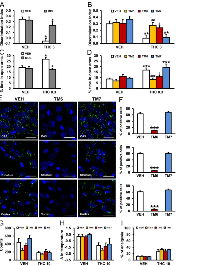
In order to implicate the involvement of the heteromer in the behavioral effects of THC in vivo, we evaluated cross antagonism in WT mice. Thus, the effects of the 5-HT2AR antagonist MDL 100,907 on THC-induced memory impairments using the object recognition test and on its anxiolytic-like properties using the elevated plus maze were evaluated. THC (3 mg/kg) induced significant memory impairments in vehicle-treated animals, but not in mice pretreated with MDL 100,907 (0.01 mg/kg) (Fig 10A). We further confirmed that this effect was mediated by the heteromer since THC-induced memory impairments were not observed in WT animals previously treated with TM5 or TM6 peptides (0.2μg/2 μl ICV) but were present in animals receiving the TM7 peptide (Fig 10B). Similarly, THC-induced anxiolytic-like effects were pre-vented by MDL 100,907 administration (Fig 10C) and by ICV infusion of TM5 and TM6 pep-tides, but not by TM7 (Fig 10D). Importantly, using PLA we were able to demonstrate that administration of TM5 and TM6, but not TM7, peptides was able to disrupt the heteromer in vivo. In hippocampal CA3, striatal (caudate-putamen) or cortical (somatomotor layers 1, 2, and 3) slices from mice treated with vehicle or TM7 peptide (0.2μg/2μl ICV) heteromers appear as green spots, a staining not seen in mice treated with equivalent amounts of TM6 pep-tide (Fig 10E and 10F). These results demonstrate that CB1R-5-HT2AR heteromers are involved in the amnesic and anxiolytic-like behavior induced by THC.
In support of the involvement of 5-HT2AR in only certain effects of THC, preadministration (0.2μg/2 μl ICV) of TM 5, TM 6, or TM 7 interference peptides did not change THC-induced hypolocomotion (Fig 10G), hypothermia (Fig 10H), or analgesia (Fig 10I). This differential effect was also observed when 5-HT2AR KO mice and WT littermates were compared (S11
Fig). Preadministration of TM 6, but not TM 7, peptides blocked the THC-induced changes in the discrimination index and percent of time in open arms in WT mice (S11A and S11B Fig),
additional incubation period of 10 min. ERK 1/2 phosphorylation was determined by western blot. Immunoreactive bands from three to seven slices obtained from ten WT or KO animals were quantified for each condition. Values represent mean± SEM of the percentage of phosphorylation relative to basal levels found in untreated slices. No significant differences were obtained between the basal levels of the WT and the KO mice. One-way ANOVA followed by Bonferroni post hoc tests showed a significant (* p < 0.05, ** p < 0.01, *** p < 0.001) effect over basal or of the antagonist plus agonist treatment over the agonist treatment (#p < 0.05, ## p < 0.01).
doi:10.1371/journal.pbio.1002194.g009
Fig 10. Prevention of THC-induced amnesic and anxiolytic-like effects by pharmacological blockade of 5-HT2AR or by CB1R-5-HT2AR heteromer
disruption with TM interference peptides. The amnesic effects of THC (3 mg/kg) observed in C57BL/6J mice in the novel object recognition test were abrogated by pretreatment with the 5-HT2AR antagonist, MDL 100,907 (0.01 mg/kg) (A) and by pretreatment with TM 5 and TM 6, but not TM 7, interference
peptides (0.2μg/ 2μl ICV) (B) (n = 5–9). The anxiolytic effects of THC (0.3 mg/kg) observed in the elevated plus maze in C57BL/6J mice were blocked by pretreatment with the 5-HT2AR antagonist, MDL 100,907 (0.01 mg/kg) (C) and by pretreatment with TM 5 and TM 6, but not TM 7, interference peptides
but neither TM 6 nor TM 7 altered the lack of effect of THC in KO mice (S11F and S11G Fig). In both WT (S11C–S11E Fig) and 5-HT2AR KO (S11H–S11J Fig) mice, the effect of THC on locomotion, body temperature, and analgesia was not altered by preadministration of TM 6 or TM 7 peptides. These findings provide evidence for the in vivo requirement of the CB1 R-5-HT2AR heteromer to be intact in order to observe the amnesic and anxiolytic-like behavior induced by THC but not in other THC-mediated effects.
Discussion
While exploring the neurobiological mechanisms underlying THC-induced cognitive impairment, we discovered an unexpected role of the 5-HT2AR. Our findings lead to three major conclusions. First, behavioral studies carried out in mice lacking 5-HT2AR revealed a remarkable 5-HT2AR-dependent dissociation in the beneficial antinociceptive effects of THC and its detrimental amnesic properties. Second, CB1R and 5-HT2AR form heteromers that are expressed and functionally active in specific brain regions involved in memory impairment. Third, to observe the negative cognitive effects of THC, these receptors must be functionally interacting, as administration of a 5-HT2AR antagonist or selective disruption of the CB1 R-5-HT2AR heteromers by ICV infusion of synthetic interference peptides in WT mice abrogated the memory deficits induced by THC and its anxiolytic-like effects, but not its antinociceptive properties.
Previous studies have suggested interactions between endocannabinoids and 5-HT, although the extent of this alleged reciprocal interaction and the molecular mechanisms involved have been difficult to ascertain. Here we found that the amnesic, anxiolytic, and pro-social-like effects induced by THC, as well as the manifestations of THC withdrawal syndrome, were reduced in mice with constitutive deletions of 5-HT2AR. In contrast, 5-HT2AR deletion did not modulate the acute hypolocomotor, hypothermic, anxiogenic, and antinociceptive effects of THC or the reinforcing effects of the cannabinoid agonist, WIN 55,212–2. These data demonstrate for the first time, to our knowledge, that 5-HT2AR modulates specific behavioral responses related to CB1R activation by THC. There seemed to be three plausible explanations for the differential effects of THC and its dependence on 5-HT2AR: (1) interactions only at the level of circuitry, (2) circumstantial cross talk at the level of intracellular signaling, or (3) direct protein—protein interaction that can modify receptor function. Although our study cannot completely count out interactions at the level of circuitry, we have clearly observed cross talk in transfected cells that would circumvent the need for circuitry connections. Indeed, both recep-tors are coexpressed in the hippocampus, where they participate in memory processing [16,17,59]. They are also colocalized in the cerebral cortex, hypothalamus, striatum, and nucleus accumbens, brain areas implicated in reward processing and affective disorders, including anxiety [60–63]. To delineate between circumstantial intracellular cross talk and direct protein—protein interaction, we tested whether the receptors could form complexes if coexpressed, which we found to be the case. We then were able to show that the signaling cross
(0.2μg/ 2μl ICV) (D) (n = 4–11). The data represent mean + SEM. * p < 0.05, ** p < 0.01, *** p < 0.001 versus vehicle, # p < 0.05 versus THC-treated mice. In (E), PLA performed in hippocampal CA3, striatal (caudate-putamen), and cortical (somatomotor layers 1, 2, and 3) slices from mice treated with VEH, TM 6, and TM 7 interference peptides (0.2μg/ 2μl ICV). Confocal microscopy images (superimposed sections) are shown in which heteromers appear as green spots in VEH and TM 7-treated mice, but not in mice treated with TM 6 interference peptides. In all cases, cell nuclei were stained with DAPI (blue). Scale bars = 20μm. In (F), the number of cells containing one or more green spots is expressed as the percentage of the total number of cells (blue nucleus) in the hippocampus, striatum, and cortex (top to bottom). Data (percentage of positive cells) are the mean± SEM of counts in 8–12 different fields. *** p < 0.001 versus vehicle-treated mice. Pretreatment with TM 5, TM 6, or TM 7 peptides (0.2μg/ 2μl ICV) had no significant effects on hypolocomotion (G), hypothermia (H), or analgesia (I) induced by THC (10 mg/kg) in C57BL/6J mice. The statistical analyses used and their corresponding F andp-values are shown inS1 Table.
doi:10.1371/journal.pbio.1002194.g010
talk observed required this receptor—receptor interaction both in vitro and in vivo. The disso-ciation observed regarding the involvement of this heteromeric complex in the memory impairments and the antinociception observed following THC administration were corrobo-rated in our in vivo studies. When the heteromer was disrupted by ICV infusion of interference peptides TM5 and TM6, we observed blunted amnesic and anxiolytic, but not antinociceptive, effects of THC selectively in WT mice. Interference peptides have been successfully used in ear-lier studies to ascertain the role of heterodimer formation in physiological functions [56] and in behavioral models of mood disorders [64]. It appears then that serendipitous signaling cross talk is not a sufficient explanation for the dependence on 5-HT2AR for THC’s effects. A more plausible explanation might be that these receptors present different degrees of interaction depending on the cell type or cell location. Such a scenario would predict the existence of dif-ferent populations of CB1R. One population when stimulated with THC provides a certain level of cellular signaling that impacts on neurons influencing locomotion or antinociceptive effects, while a separate population of CB1R coupled to 5-HT2AR would provide altered cell sig-naling upon exposure to THC that directly influences memory or anxiolytic-like effects. In agreement, CB1R in glutamatergic cells have been recently reported to have a much higher cou-pling to G-proteins than CB1R in GABAergic cells, sustaining the possibility of different func-tional populations of the receptor [65]. Support for this idea is provided by our results showing that 5-HT2AR and CB1R form heteromers in specific brain structures, such as the cortex, hip-pocampus, and striatum, but not in the nucleus accumbens, a key structure of the reward cir-cuit. Several drugs of abuse, including cannabis, increase dopamine release in the nucleus accumbens [66], and an interaction between CB1R and dopamine D2receptor signaling has been suggested in this area [67]. Our results showing no heteromer formation in the nucleus accumbens, together with the finding that 5-HT2AR does not modulate cannabinoid (WIN 55,212–2) reinforcing properties, suggest that these receptors are not involved in the modula-tion of dopamine responses in this structure. Moreover, our data showing a reducmodula-tion in p-ERK 1/2 via the heteromer are particularly interesting since p-ERK signaling is important for long-term synaptic plasticity [68], which plays a crucial role in learning and memory.
Although we are unable to speculate on the amount of heteromers in the different regions, it is clear that the lack of heteromers in the nucleus accumbens is not due to lack of expression, as both receptors were still able to signal upon receptor stimulation at levels equal to the other brain regions. These data suggest that heteromer formation is not simply due to overexpression of the receptors in these regions and that there exists a mechanism to regulate heteromer for-mation within the brain. Differential expression levels of CB1R do not always correlate with the ability to couple to G-proteins [65], which reinforces the idea that more subtle mechanisms are at play than simple expression. The precise subcellular localization of this heteromeric popula-tion of receptors is still not known. However, PLA positive elements could be revealing both pre- and postsynaptic heterodimers, consistent with data showing that CB1R and 5-HT2AR are colocalized at pre-and postsynaptic levels in different areas of the brain [16,18–27,69,70].
Furthermore, the ICV infusion technique ensured that CB1R-5-HT2AR heteromers were disrupted by TM interference peptides in key brain areas mediating the observed effects since most of these structures, including the hippocampus, dorsal raphe nucleus, and periaqueductal grey, are in close proximity to the ventricles. Pharmacological targeting of heteromers is of great interest, in part because the GPCR heteromers are unique signaling units with functional properties different from homomers [40,71]. Indeed, our functional data show that costimula-tion of CB1R-5-HT2AR heteromers by agonists reduces cell signaling, whereas antagonist bind-ing to one of the receptors blocks the signalbind-ing of the interactbind-ing receptor (bidirectional cross antagonism). Importantly, we also found that formation of the CB1R-5-HT2AR heteromers presents a different G-protein coupling, with 5-HT2AR coupling to Gi instead of Gq, and a
signaling profile different from the single receptors (similar results were previously seen with the mGlu2R-5-HT2AR heteromer [72]). In pioneering work, the group of Kobilka has shown that GPCRs are dynamic proteins, adopting a number of conformations through an energy landscape [73]. Ligand or G-protein binding changes the shape of the energy landscape, favor-ing or disfavorfavor-ing the intracellular signal. Based on our findfavor-ings, we propose that in the case of GPCR heteromers one of the protomers allosterically modulates the functional properties of the interacting receptor, and this can be conceptualized using energy landscapes (S7 Fig).
CB1R activation by cannabinoids such as THC produces a variety of negative effects, includ-ing cognitive impairments [1,2] and anxiogenic- and addictive-like responses [5], which have major consequences in cannabis users and constitute important drawbacks for the use of can-nabinoids as therapeutic agents [74]. The genetic, molecular, and pharmacological data pre-sented here demonstrate the requirement for CB1R-5-HT2AR heteromers for the negative cognitive effects of THC. These heterocomplexes could be potentially modulated in the form of disruption or by their selective pharmacological blockade in order to dissociate the cognitive impairment induced by THC from its beneficial antinociceptive properties.
Materials and Methods
All the procedures involving animals were performed by observers blind to experimental con-ditions following standard ethical guidelines (European Communities Directive 86/60-EEC) and were approved by the local ethical committee (Comitè Ètic d'Experimentació Animal-Parc de Recerca Biomèdica de Barcelona, CEEA-PRBB). The PRBB also has Animal Welfare Assur-ance (#A5388-01, Institutional Animal Care and Use Committee approval date 06/08/2009) granted by the Office of Laboratory Animal Welfare (OLAW) of the United States National Institutes of Health. As of June 2010, the programme of care and use of laboratory animals at the PRBB has the full accreditation from the Association for Assessment and Accreditation of Laboratory Animal Care (AAALAC). Mice were anesthetized with a mixture of ketamine/xyla-zine and euthanized with carbon dioxide (concentrations between>70% and <100%).
Animals and Drug Treatment
The 5-HT2AR KO and WT littermates were originally generated at Columbia University (US) on a 129S6/SvEv background [29,75]. Animals were backcrossed over at least ten generations onto the inbred C57BL/6J line. Male and female 5-HT2AR KO and WT mice were genotyped as previously described [76]. C57BL/6J male, 9-wk-old mice (Charles River L’Arbresle, France) were used for the pharmacological and behavioral experiments. Constitutive CB1R KO mice were bred by backcrossing chimeric animals to the C57BL/6J background and crossing hetero-zygotes [77]. Mice weighing 20–25 g at the beginning of the experiments were initially housed
four per cage in a temperature-controlled (21 ± 1°C) and humidity-controlled (55 ± 10%) envi-ronment, where food and water were available ad libitum. All the experiments were performed during the light phase of a 12 h light/dark cycle (lights on at 8 a.m. and off at 8 p.m.), except for the WIN 55,212–2 self-administration experiment that was conducted in the dark phase of the cycle. The CB1R ligands THC (Pharm GmbH, Frankfurt, Germany) and rimonabant (Sanofi-Aventis Recherche, Montpellier, France) were diluted in 5% ethanol, 5% Cremophor-EL (Sigma-Aldrich), and 90% saline. WIN 55,212–2 (Sigma-Aldrich) was dissolved in one drop of Tween 80 (Sigma-Aldrich) and diluted in physiological saline. For the self-administration experiment, WIN 55,212–2 was administered by intravenous route at 12.5 μg/kg/infusion. The 5-HT2AR antagonist, MDL 100,907 (Sigma-Aldrich), was dissolved in saline solution using a drop of Tween 80. Except for WIN 55,212–2, all compounds were administered intraperitone-ally (IP) at a volume of 10 ml/kg.
Behavioral Experiments
In mice, cannabinoids produce the so-called‘‘tetrad model” of cannabimimetic activity in the same dose range and within the same time frame, consisting of hypolocomotion, hypothermia, antinociception, and catalepsy. Accordingly, we tested the hypolocomotor, hypothermic, and analgesic effects of THC using a complete dose response, as published in previous studies [78]. For memory deficits induced by THC in mice, previous data from our laboratory [79] have revealed significant effects in the object discrimination test only with 3 and 10 mg/kg of THC, which were both tested in the present study. For anxiety-like behavior, it has been shown that THC produces biphasic effects, with lower and higher doses inducing anxiolytic- and anxio-genic-like responses, respectively [3,7]. Accordingly, we measured the anxiolytic-like effect of THC, as well as the consequent increase in social interaction observed, using a low dose (0.3 mg/kg), and the anxiogenic-like response using a high dose (3 mg/kg). The different behavioral effects produced by specific doses of THC suggest that a different level of receptor occupancy is required for each behavioral response [80].
Determination of Locomotor Activity, Social Interaction, Body
Temperature, and Analgesia in Mice
Locomotor responses to acute administration of THC were evaluated by using individual loco-motor activity boxes (9 x 20 x 11 cm; Pessac, France) provided with two lines of photocells in a low-luminosity environment (20–25 lux). Mice were placed in the boxes for 45 min (15 min after THC [0.3, 1, 3, and 10 mg/kg] or vehicle administration), and total horizontal activity was analyzed. The social interaction test was performed in an open field (40 x 40 cm) in which mice were habituated for 2 consecutive d in order to maximize the duration of interactions. Next, animals were treated with THC (0.3 mg/kg) or vehicle, and 30 min after the injection, a previously established pair of mice (same strain and size and unfamiliar with each other) was placed into the arena for 10 min. The social interaction test was performed as previously described [81]. Total time spent in active social interactions (defined as sniffing, fighting, chas-ing, groomchas-ing, or crawling under and over each other) was scored from a recorded video. Data were expressed as the amount of interaction time per couple. Body temperature was measured before (basal) and 60 min after THC (1, 3, and 10 mg/kg) or vehicle administration using a lubricated thermo-coupled flexible probe (Panlab, Madrid, Spain) placed into the rectum for 10 s. Data were expressed as change in temperature from basal recording. Antinociceptive effects were evaluated using the tail-immersion and hot-plate tests. The tail-immersion test measures spinal pain responses, which are modulated by descending influences from the brain stem, cerebellum, basal ganglia, and cerebral cortices. All of these structures comprise CB1R that play an important role in pain responses [82,83]. The hot-plate test measures supraspinal antinociception, and direct evidence for supraspinal sites of cannabinoid analgesic action has been provided (see Palazzo et al., 2010, for review [83]). Sixty minutes after the injection of THC (1, 3, and 10 mg/kg) or vehicle, the tail-immersion test was carried out, as previously described [84]. The water temperature was maintained at 50 ± 0.5°C using a thermo-regulated water-circulating pump (Clifton, North Somerset, United Kingdom). The latency to a rapid tail flick was registered, and in its absence a 10-s cutoff was used to prevent tissue damage. Subse-quently, the hot-plate test was performed, as previously reported [85], 90 min after THC (1, 3, and 10 mg/kg) or vehicle injection. The surface of the plate was kept at 50 ± 0.1°C (Columbus Instruments, Columbus, Ohio, US), and the nociceptive threshold was evaluated by measuring licking and jumping responses. A 5-min cutoff was determined to avoid tissue damage. For both nociceptive models, data were expressed as a percentage of the cutoff latency.