Structure and dynamics of dynorphin peptide and its receptor
Guillaume Ferré*, Georges Czaplicki, Pascal Demange, Alain Milon*
,
31077 Toulouse, France
* Corresponding authors: Email addresses: [email protected] (G. Ferré);
[email protected] (A. Milon)
Vitamins and Hormones, 2019, in press. A ’ version.
Abstract
Dynorphin is a neuropeptide involved in pain, addiction and mood regulation. It exerts its activity
by binding to the kappa opioid receptor (KOP) which belongs to the large family of G-protein
coupled receptors. The dynorphin peptide was discovered in 1975, while its receptor was cloned
in 1993. This review will describe: a) the activities and physiological functions of dynorphin and
its receptor, b) early structure-activity relationship studies performed before cloning of the
receptor (mostly pharmacological and biophysical studies of peptide analogues), c)
structure-activity relationship studies performed after cloning of the receptor via receptor mutagenesis and
the development of recombinant receptor expression systems, d) structural biology of the opiate
receptors culminating in X-ray structures of the four opioid receptors in their inactive state and
structures of MOP and KOP receptors in their active state. X-ray and EM structures are combined
with NMR data, which gives complementary insight into receptor and peptide dynamics.
Molecular modelling greatly benefited from the availability of atomic resolution 3D structures of
receptor-ligand complexes and an example of the strategy used to model a dynorphin-KOP
receptor complex using NMR data will be described. These achievements have led to a better
understanding of the complex dynamics of KOP receptor activation and to the development of
new ligands and drugs.
Keywords: membrane protein, GPCR, site-directed mutagenesis, heterologous expression
systems, NMR, X-ray crystallography, electron microscopy, molecular dynamics, docking, drug
I.
Dynorphin: a neuropeptide involved in pain, addiction and mood regulation
Dynorphin is an endogenous neuropeptide first isolated from porcine pituitary (Cox, Opheim,
Teachemacher, & Goldstein, 1975) with a particularly potent opioid activity (Goldstein,
Tachibana, Lowney, Hunkapiller, & Hood, 1979). The dynorphin A 1-17 isoform was the first to
be fully sequenced (Goldstein, Fischli, Lowney, Hunkapiller, & Hood, 1981) and revealed an
amino-terminal sequence identical to leu-enkephalin opioid peptide (YGGFL) with a basic
carboxy-terminal extension. Several dynorphin isoforms were further identified: dynorphin A 1-8,
dynorphin B 1-13, big dynorphin and leumorphin (Charles Chavkin, 2013) (Fig. 1). All
dynorphin isoforms and α- β-neo-endorphin, which are also leu-enkephalin-based opioid
peptides, derive from a common precursor named prodynorphin (C. Chavkin, Bakhit, Weber, &
Bloom, 1983; Kakidani et al., 1982; Weber, Evans, & Barchas, 1982; Zamir, Palkovits, Weber,
MEzey, & Brownstein, 1984) while other opioids are derived from the precursors proenkephalin
and proopiomelanocortin (Charles Chavkin, 2013). Prodynorphin and its processing enzyme,
prohormone convertase 2 (PC2), are largely expressed in the central nervous system (Berman et
al., 2000; Civelli, Douglass, Goldstein, & Herbert, 1985) and dynorphin peptides are present in
presynaptic neurosecretory vesicles (Molineaux & Cox, 1982; Pickel, Chan, & Sesack, 1993;
Pickel, Chan, Veznedaroglu, & Milner, 1995; Whitnall, Gainer, Cox, & Molineaux, 1983). They
can be released by membrane depolarization (C. Chavkin, et al., 1983) and subsequently activate
the kappa opioid receptor (KOP) (Wagner, Evans, & Chavkin, 1991) which modulates
neurotransmitter release and postsynaptic neural activity (Wagner, Terman, & Chavkin, 1993).
KOP belongs to the opioid receptor family which is composed of several subtypes, originally
defined by the pharmacological profiles of the first receptors to be characterized (Dhawan et al.,
1996): µ δ p p MOP and DOP). KOP was initially named from
ketocyclazocine opioid activity (Martin, Eades, Thompson, Huppler, & Gilbert, 1976) and highly
KOP-specific agonists were synthesized (Dhawan, et al., 1996) such as U50488 (Lahti,
VonVoigtlander, & Barsuhn, 1982) or U69593 (Lahti, Mickelson, McCall, & Von Voigtlander,
1985). Opioid receptor cloning led to the identification of a fourth member of the family, the
nociceptin opioid receptor (NOP), and of its endogenous ligand, nociceptin (J-C. Meunier et al.,
1995; Mollereau et al., 1994). Several studies reported the cloning of KOP from rodents (Li et al.,
1993; Meng et al., 1993; Yasuda et al., 1993) and then from human
Figure 1: Dynorphin-related opioid peptides. Met-enkephalin, leu-enkephalin, dynorphin A 1-13 and
prodynorphin-derived peptides amino acid sequence and affinity for opioid receptors. The common N-terminal leu-enkephalin "message" sequence is colored in green and C-N-terminal "address" residues conserved with dynorphin A 1-17 in blue. In order to compare affinities in homogenous systems, pKi values from (Mansour, Hoversten, Taylor, Watson, & Akil, 1995) are reported where competition binding experiments were conducted against rat opioid receptors transiently expressed in COS-1 cells. Binding properties have been additionally reviewed from the indicated references from IUPHAR/BPS guide to pharmacology (Alexander et al., 2017): 1 (Meng, et al., 1993), 2 (Raynor et al., 1994), 3 (Yasuda, et al., 1993), 4 (Toll et al., 1998), 5 (Simonin, et al., 1995), 6 (Merg et al., 2006), 7 (J. Zhu, et al., 1995), 8 (J. Zhu, Luo, Li, Chen, & Liu-Chen, 1997). Similar trends are observed despite discrepancies arising from differences in experimental models and conditions.
(Simonin et al., 1995; J. Zhu et al., 1995), showing that it, together with the other opioid
receptors, belongs to the G-protein-coupled receptors (GPCR) superfamily. Opioid receptors are
mainly Gi/Go-coupled (Al-Hasani & Bruchas, 2011; Prather et al., 1995). They display a small
basal intracellular signaling activity, at about 10% of the maximal response for KOP, in the
absence of any ligand, and this is modulated by extracellular ligand binding (D. Wang, Sun, &
Sadee, 2007).
Opioid receptor signaling controls a multitude of intracellular effectors by both
G-protein-dependent and G-protein-inG-protein-dependent pathways (Al-Hasani & Bruchas, 2011; Bruchas &
Chavkin, 2010; Law, Wong, & Loh, 2000). G-protein-dependent opioid receptor signaling
predominantly results in neuronal excitability and synaptic transmission inhibition. G
i/G
o-protein
activation promotes G-protein-coupled inwardly rectifying potassium channel (GIRK) activity
(Barchfeld & Medzihradsky, 1984; Childers & Snyder, 1978; Minneman & Iversen, 1976). It
causes neuronal membrane hyperpolarization and thus attenuates the neuron’ ability to generate
and propagate action potentials (Henry, Grandy, Lester, Davidson, & Chavkin, 1995; Sadja,
Alagem, & Reuveny, 2003; Schneider, Eckert, & Light, 1998). Activation of Gi/Go-proteins also
inhibits voltage-dependent calcium channels, diminishing calcium influx in response to action
potentials, and thus prevents neurotransmitter synaptic release (Bourinet, Soong, Stea, & Snutch,
1996; Rhim & Miller, 1994; Rusin, Giovannucci, Stuenkel, & Moises, 1997; Zamponi & Snutch,
1998). Activated Gi/Go-proteins also inhibit adenylate cyclase thus decreasing intracellular cyclic
adenosine monophosphate (cAMP) concentration (Taussig, Iniguez-Lluhi, & Gilman, 1993),
which in turn regulates numerous targets. Opioid receptors also signal through
G-protein-independent pathways. Following agonist stimulation, they can be intracellularly phosphorylated
by G protein-coupled receptor kinases (GRKs) leading to receptor w β-arrestins
(Al-Hasani & Bruchas, 2011; Law, et al., 2000). This interaction is involved in receptor
internalization, one of the consequences of which is to prevent exposition to extracellular ligands.
It is also responsible for intracellular signaling per se, notably through mitogen activated kinase
(MAPK) pathways that regulate predominant cellular processes such as gene transcription,
proliferation and differentiation (Bruchas & Chavkin, 2010; Raman, Chen, & Cobb, 2007).
Because of their involvement in cellular communication, opioid peptides and their receptors
participate in numerous physiological processes (Y. Feng et al., 2012). First, opioid receptor
activation results in spinal and supra-spinal pain modulation (Ahlbeck, 2011; Waldhoer, Bartlett,
& Whistler, 2004). The afferent nociceptive influx from peripheral tissues is regulated by
descending neurons and opioid receptors participate in this system at multiple levels (Vanderah,
2010). As an example, KOP has been found to be present in spinal cord dorsal root ganglia
(Attali & Vogel, 1989; Corder, Castro, Bruchas, & Scherrer, 2018; Ji et al., 1995) where it
inhibits synaptic transmission between primary and secondary afferent neurons (Vanderah, 2010)
in response to agonists released by descending neurons. Opioid peptides and their receptors also
regulate monoaminergic systems and notably mesolimbic dopaminergic functions (Lutz &
Kieffer, 2013). KOP agonists inhibit dopamine neuron release activity by their receptor-specific
action both in the nucleus accumbens and the ventral tegmental area (Lutz & Kieffer, 2013;
Margolis, Hjelmstad, Bonci, & Fields, 2003; R. Spanagel, A. Herz, & Shippenberg, 1992),
resulting in an aversive effect (R. Bals-Kubik, A. Ableitner, A. Herz, & Shippenberg, 1993). In
contrast, MOP agonists cause a rewarding effect (R. Bals-Kubik, et al., 1993) because they
indirectly stimulate dopamine release by diminishing inhibitory gamma-aminobutyric acid
(GABA) neuron activity in the ventral tegmental area (Lutz & Kieffer, 2013; R. Spanagel, et al.,
1992). Opioid receptors thus mediate abusive opioid seeking behavior but are also more generally
involved in modulating drug addictions with pronounced subtype discrepancies (Kreek et al.,
2012). In addition, the mesolimbic dopamine reward system is linked to mood disorders (Nestler
& Carlezon, 2006) and opioid receptors further regulate serotonin and noradrenaline neurons
(Lutz & Kieffer, 2013), thought to participate in depression (Krishnan & Nestler, 2010).
Consistent with this, opioid receptors and their ligands are involved in depression-like behaviours
and KOP antagonists such as JDTic induce antidepressant-like effects (Lutz & Kieffer, 2013).
Furthermore, opioid peptides and their receptors, especially dynorphin and KOP, control the
hypothalamic–pituitary–adrenal axis (HPA) and are thus involved in stress-related phenomenon
with implications in addiction, depression and anxiety disorders (Bruchas, Land, & Chavkin,
2010; Knoll & Carlezon, 2010; Kreek, et al., 2012; Ribeiro, Kennedy, Smith, Stohler, & Zubieta,
2005). Beside their predominant role in pain, addiction and mood control, opioid receptors have
been linked to a multitude of functions such as immunity, neuroprotection, cell proliferation,
neural differentiation, cardiovascular system regulation and feeding (Y. Feng, et al., 2012). Of
particular importance, opioid receptors signaling stimulation can influence the respiratory system
function (Pattinson, 2008) with strong respiratory depression observed upon activation of MOP
(but not KOP). Together with its impact on breathing, MOP agonism induces an important
inhibition of gastrointestinal transit (A. Tavani, P. Petrillo, A. La Regina, & Sbacchi, 1990),
which represents a significant limitation of opioid use in the clinic.
The biological functions of opioid peptides and their receptors renders them key targets to
interfere in pain, addiction and mood disorders. There is considerable interest in discovering new
opioids with reduced side effects and compounds that target the dynorphin / KOP system are
being developed in order to produce analgesic, antidepressant, anxiolytic or anti-addiction drugs
(Dogra & Yadav, 2015; Zheng et al., 2017). Because of the biological significance of this system,
/ KOP interaction and the resulting modulation of receptor signaling activity. These studies,
which form the subject of this review, could also help in the design of novel KOP ligands with
pharmacologically relevant properties such as biased agonism or allosteric modulation.
II.
The molecular mechanism of action of dynorphin: research and hypotheses prior
to KOP cloning
The ability to produce dynorphin and analogues by solid-phase peptide synthesis (Goldstein, et
al., 1979) opened the way to a wide range of structure-activity relationship studies on various
peptide structures. These have been reviewed thoroughly elsewhere (Aldrich & McLaughlin,
2009; Lapalu et al., 1997; Naqvi, Haq, & Mathur, 1998; Ramos-Colon et al., 2016). In brief, the
17 amino-acid long dynorphin 1-17 may be shortened at its C-terminus to dynorphin 1-13
without affecting its activity (activation of KOP is typically assessed on the guinea pig ileum
electrical contraction assay), and further to dynorphin 1-8 with a 50-fold reduction in activity and
no loss in affinity (Fig. 1 and (Mansour, et al., 1995)). Further shortening is deleterious for both
affinity and activity. Substitution of Gly2 by L-amino-acids resulted in a reduction in activity
while substitution by D-amino-acids resulted in reduced selectivity for KOP versus MOP, since
D amino-acids tend to increase activity on MOP and to decrease it on KOP. Substitution of Pro10
by a D-proline led to a marked increase in selectivity for KOP over the other opiate receptors.
The further alkylation of the amino group of Tyr1 gave rise to highly KOP-selective peptide
ligands such as N–Benzyl[D–Pro10]–Dyn A(1–11) (Choi, Murray, DeLander, Caldwell, &
Aldrich, 1992).
Peptide synthesis also enabled biophysical studies of dynorphin conformation and dynamics in
solution and in membrane biomimetic media (Lancaster et al., 1991; Lind, Graslund, & Maler,
2006; Naito & Nishimura, 2004; Spadaccini, Crescenzi, Picone, Tancredi, & Temussi, 1999).
These included circular dichroism, Raman, FT-IR and NMR spectroscopies. In aqueous solution,
dynorphin is largely disordered. It may adopt secondary structures in specific environments, such
yp β-turn involving the first five residues in DMSO (Renugopalakrishnan, Rapaka, Huang,
Moore, & Hutson, 1988) or a
-helical conformation from residue Gly3 to Arg9 when bound to
dodecylphosphocholine detergent micelles (Kallick, 1993). Limiting the conformational space
available to constrained peptide analogues increases receptor binding affinity and specificity
(Naqvi, et al., 1998). However, without knowing the structure adopted by dynorphin upon KOP
binding, it was difficult to derive a clear understanding of the binding mechanism from these
results.
A detailed study of dynorphin and dynorphin analogues bound to lipid bilayers was performed in
the early eighties. Using a combination of infrared attenuated total reflection spectroscopy and
capacitance minimization, dynorphin was found to b O p b y by f
α-helix from Tyr1 to Pro10, which inserts into the bilayer perpendicular to the bilayer plane (Erne,
Sargent, & Schwyzer, 1985). In the same study, the affinity for a neutral bilayer was determined
to have a Kd of 11 µM. In a subsequent theoretical estimation of the preferred orientation and
binding energy of a series of dynorphin analogues of various lengths (from 1-13 to 1-5), a good
correlation between the peptide amphiphilic moment, the affinity for lipid bilayers and the KOP
receptor subtype specificity was found. Loss of the C-terminal positively-charged residues
converts a KOP-selective peptide dynorphin, into a MOP/DOP-selective peptide Leu-enkephalin
(Fig. 1). This, and similar observations on other neuropeptide families, gave rise to two major
hypotheses: a) the message-address concept, first introduced in 1977 (Schwyzer, 1977)), in
which the N-terminal 5 residues (message) are responsible for specific binding and receptor
activation, while the positively charged C-terminus (address) is responsible for KOP receptor
selectivity by concentrating the peptide in the vicinity of the receptor; b) the membrane
compartment concept, in which the lipid bilayer plays an active role in catalyzing the
peptide-receptor interaction (Auge, Bersch, Tropis, & Milon, 2000; Axelrod & Wang, 1994; Bersch,
Koehl, Nakatani, Ourisson, & Milon, 1993; Czaplicki & Milon, 1998, 2005; Milon, Miyazawa, &
Higashijima, 1990; Sargent & Schwyzer, 1986). One reason for the correlation between KOP
receptor subtype specificity and the positively charged C-terminus (besides the potential role of
the bilayer itself) became clear when the opiate receptors were cloned in 1992-1994 (Chen,
Mestek, Liu, Hurley, & Yu, 1993; Evans, Keith, Morrison, Magendzo, & Edwards, 1992; Kieffer,
Befort, Gaveriaux-Ruff, & Hirth, 1992; Mollereau, et al., 1994; Yasuda, et al., 1993). Indeed, it
appeared that a specific feature of KOP and NOP receptors as compared to MOP and DOP
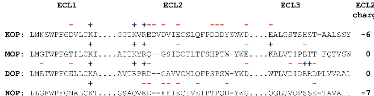
receptors is the high negative potential surrounding the extracellular loops 2 (Fig. 2), which is
expected to increase the local concentration of highly positively-charged neuropeptides (such as
extracellular loop 2 was further confirmed experimentally (Bjorneras et al., 2014). The sole effect
of an attractive electrostatic potential may account for increased on-rate binding kinetics and
receptor binding affinity by at least two orders of magnitude (Fersht, 1999).
Figure 2: Primary sequences of the extracellular loops of the opioid receptor subtypes, KOP, MOP, DOP
and NOP showing that the extracellular loop 2 (ECL2) is particularly rich in negative charges in KOP and NOP, while it is neutral for MOP and DOP. A similar trend is observed for the entire extracellular surface. This characteristic contributes to KOP specificity of positively charged dynorphin analogues as shown with MOP/KOP chimeric receptors (J. B. Wang, Johnson, Wu, Wang, & Uhl, 1994).
III.
KOP cloning, site-directed mutagenesis and heterologous expression systems
KOP (Li, et al., 1993; Meng, et al., 1993; Simonin, et al., 1995; Yasuda, et al., 1993; J. Zhu, et
al., 1995) and the other three opioid receptors DOP, MOP and NOP (Chen, et al., 1993; Evans, et
al., 1992; Kieffer, et al., 1992; Mollereau, et al., 1994; Yasuda, et al., 1993) were cloned in the
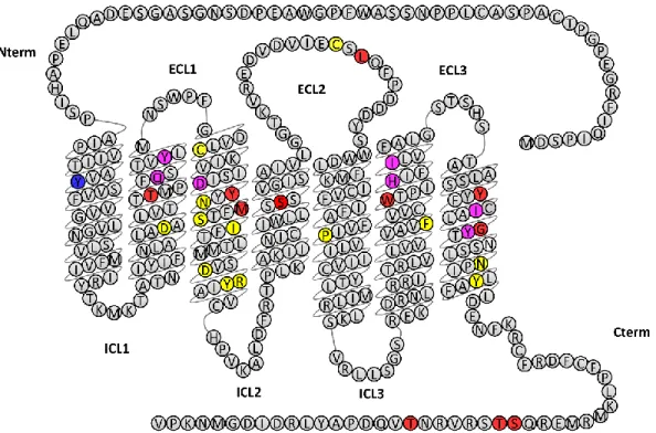
early nineties. KOP shares 60% sequence similarity with the other receptors. The highest
sequence diversity is located in the N-terminus, C-terminus and extracellular loops (Waldhoer, et
al., 2004). Moreover, all four opioid receptors possess a remarkable signature, specific to class A
GPCRs, which includes a sodium binding pocket (residues numbering corresponds to human
KOP; superscripts are given according to Ballesteros-Weinstein numbering (Ballesteros &
Weinstein, 1995)): D105
2.50, N141
3.35, S145
3.39; a PIF micro-switch P238
5.50, I146
3.40, F283
6.44; a
NPxxY motif N326
7.49, Y330
7.53; a DRY motif D155
3.49, R156
3.50, Y157
3.51and a cysteine
disulfide bridge C131
3.25, C210ECL2 (Fig. 3). Heterologous expression and site-directed
mutagenesis experiments were then used intensively to better understand the receptor architecture,
ligand recognition specificity and activation mechanisms. It was shown that the extracellular
loops play a major role in receptor subtype specificity (Metzger & Ferguson, 1995; Seki et al.,
1998). Several key transmembrane residues were identified, such as S187
4.54(Claude et al., 1996),
E297
6.58(Larson, Jones, Hjorth, Schwartz, & Portoghese, 2000; Sharma, Jones, Metzger,
Ferguson, & Portoghese, 2001), Y312
7.35(Metzger, Paterlini, Ferguson, & Portoghese, 2001) and
I316
7.39(Owens & Akil, 2002). Using KOP/DOP chimeric receptor, Kong et al. obtained indirect
“ b ff f k pp p
p ” (Kong et al., 1994). However, recent structures of KOP in the presence of antagonist (H.
Wu et al., 2012) and agonist (Che et al., 2018) do not support this statement, illustrating the
difficulties of drawing firm conclusions from these approaches in the absence of precise 3D
structures. Using the 3D structures of KOP, extensive mutagenesis experiments were performed
to characterize the interaction of KOP with dynorphin, as well as with other non-peptide ligands
such as salvinorin, which are summarized, together with previous studies, in Figure 3 and Table 1.
KOP
mutant
Binding Potency
References
T111
2.56↔
↓
3
Q115
2.60↓
↓
2
Y119
2.64↓
↓
1, 2
D138
3.32↓
↓
2, 3
Y139
3.33↔
↓
1, 2, 3
M142
3.36↔
↓
2
W287
6.48↔
↓
3
H291
6.52↓
↓
2, 3
I294
6.55↓
↓
2
Y312
7.34↔
↓
1, 2, 3
Y313
7.35↓
↓
1, 3
I316
7.39↓
↓
2
G319
7.42↔
↓
3
Y320
7.43↓
↓
1, 2, 3
Table 1: Main point mutations of KOP and their consequences on dynorphin binding and functional
activity (cAMP inhibition assay). References: 1 (Yan et al., 2005); 2 (Vardy, et al., 2013); 3 (Che, et al., 2018).
Figure 3: Primary sequence of human KOP highlighting mutations shown to affect KOP-dynorphin
. f α-helices were defined according to the activated KOP 3D structure (Che, et al., 2018). In the three-dimensional structure of inactive KOP (H. Wu, et al., 2012) these limits ff y f α-helix 5 (D2185.30
to S2605.72 instead of W2215.33 to S2555.67 α-helix 6 (R2676.28 to L2996.60 instead of R2636.24 to G3006.61 α-helix 7 (L3097.32 to L3337.56 instead of T3067.29 to D3347.57). Mutations affecting dynorphin binding (more than 10-fold increase in Kd) are colored in blue (Y66
1.39 ), those affecting signaling (more than 10-fold increase in EC50 in cAMP inhibition assay) are colored in red (T1112.56, Y1393.33, M1423.36, S1874.54, C210ECL2, L212ECL2, W2876.48, Y3127.35, G3197.43, S356Ct, T357Ct, T363Ct) and those affecting both are colored in magenta (Q1152.60, Y1192.64, D1383.32, H2916.52, I2946.55, I3167.39, Y3207.43). Other residues generally considered to play a role in opioid receptor activation (Che, et al., 2018; Koehl et al., 2018) are colored in yellow (cysteine disulfide bridge C1313.25, C210ECL2; sodium binding pocket: D1052.50, N1413.35, S1453.39; PIF microswitch: P2385.50, I1463.40, F2836.44; NPxxY motif: N3267.49, Y3307.53; DRY motif: D1553.49, R1563.50, Y1573.51). Residues D3.32 and Y7.43 (conserved in all four opioid receptors) were shown to form direct contacts with DAMGO N-terminus in the DAMGO-MOP-Gi structure (Koehl, et al., 2018) and thus presumably with the N-terminus of dynorphin in the case of KOP. Residue H6.52 is conserved in MOP, KOP and DOP and forms a water-mediated contact with the phenol group of Tyr1 in the same MOP structure. Interestingly, this residue H6.52 is mutated to a glutamine in NOP, whose ligand nociceptin possesses a phenylalanine at position 1 and is thus devoid of the phenol hydroxyl group. It should be noted that for clarity, this figure presents data obtained with dynorphin
(mostly from (Claude, et al., 1996, Che, 2018 #199; Vardy et al., 2013) and not with other non-peptide ligands for which other deleterious mutations have been described. Superscripts are given according to Ballesteros-Weinstein numbering (Ballesteros & Weinstein, 1995); residues numbering corresponds to human KOP.
IV.
Structure and dynamics of dynorphin and its receptor based on experimental 3D
structures
Three-dimensional structures of GPCRs at atomic resolution began to appear with the structure of
rhodopsin A (Palczewski et al., 2000). However, rhodopsin was a specific case due to its unusual
stability and availability from natural sources, and further efforts were necessary to solve the
three-dimensional structures of recombinant GPCRs. Several international consortia developed
crucial methodologies in protein expression systems, receptor stabilization by mutagenesis,
fusion proteins, the selection of stabilizing ligands, binding to antibodies (particularly
nanobodies), the development of better solubilizing and crystallizing media (Caffrey & Cherezov,
2009; Cherezov, 2011; Granier & Kobilka, 2012; Kobilka & Schertler, 2008; Rosenbaum,
Rasmussen, & Kobilka, 2009; Stevens et al., 2013; Tate, 2012).
Most GPCR structures were obtained from membrane proteins expressed in eukaryotic insect
cells where the flexible N- and C-termini, as well as the intracellular loops (mostly ICL3), were
deleted or replaced by exogenous protein domains promoting thermostability and crystallization,
such as T4 lysozyme or the thermostabilized apocytochrome b562 RIL (BRIL) (Chun et al.,
2012; Lv et al., 2016). Other expression systems have been used such as the methylotrophic yeast
P. pastoris (Talmont, Sidobre, Demange, Milon, & Emorine, 1996), which allows stable isotope
labelling, including perdeuteration (Massou et al., 1999), mostly for NMR experiments (Eddy et
al., 2018). E. coli is also an interesting host for isotope-labelled GPCR biosynthesis which can be
achieved by receptor expression as inclusion bodies followed by in vitro refolding during the
protein purification (Baneres et al., 2003; Baneres, Popot, & Mouillac, 2011; Casiraghi et al.,
2016). We have shown this strategy to be efficient for KOP: the dynorphin-KOP interaction was
measured in our laboratory using KOP expressed in E. coli and refolded (unpublished results),
and the same results were obtained as with KOP produced and purified from sf9 cell membranes
(O'Connor et al., 2015).
These methodological developments allowed the first three-dimensional structure of a
rec b G β2-adrenergic receptor, to be solved in 2007 (Rosenbaum et al., 2007).
This marked the beginning of a new era of GPCR structural biology: according to the database
GPCRdb, 270 structures of receptor-ligand complexes had been solved by September 2018,
including 52 unique receptor complexes (
http://gpcrdb.org/structure/statistics
). In 2011 the first
f β2-adrenergic receptor in its active form (that is in the presence of an agonist and
a G-protein or a nanobody mimicking the G-protein) was solved, thus revealing for the first time
the atomic details of an activation mechanism of a GPCR (Rasmussen, Choi, et al., 2011;
Rasmussen, DeVree, et al., 2011). The field of opioid receptors followed closely this revolution,
with structures of the four opioid receptors solved in their inactive state in 2012 (Granier et al.,
2012; Manglik et al., 2012; Thompson et al., 2012; H. X. Wu et al., 2012), and later in their
active states for MOP (Huang et al., 2015; Koehl, et al., 2018) and KOP (Che, et al., 2018). An
overlay of KOP in its inactive state, in complex with the antagonist JDTic (PDB 4DJH), and in its
activated state, in complex with the agonist MP1104 and a nanobody mimicking G-protein
(Nb39) (PDB 6B7S) illustrates the general mechanism of activation (Fig. 4): it is characterized
by outward movements of transmembrane helix 6 (TM6) (by 10 Å) and ICL2 and inward
movements of TM5 and TM7, leading to the creation of an intracellular pocket into which
G-proteins can penetrate. These movements are associated with a contraction (10% reduction in
volume of the ligand binding pocket) of the extracellular portion in the active-state KOP, with
extracellular loop 2 (ECL2) and TM4 and TM6 moving closer to the receptor core (Che, et al.,
2018). Both ligands, the antagonist JDTic and the agonist MP1104 bind at a similar location, with
conserved contacts, in particular a salt bridge to D138
3.32in TM3 and a water-mediated hydrogen
bond with the backbone carbonyl oxygen of K227
5.39. Comparison of the active and inactive
states of KOP indicates structural changes involving several residues of TM3, which are thus
believed to be critical for coupling ligand-mediated changes in the orthosteric site and the
transducer interface. This coupling is in part mediated by changes in a sodium binding pocket
(formed by residues D105
2.50, N141
3.35and S145
3.39) which acts as a negative allosteric modulator
at opioid receptors (Fenalti et al., 2014; V. Katritch et al., 2014; Pasternak, Snowman, & Snyder,
1975).
With these data in hand, it may appear that the structural biology of opioid receptors and the
molecular details of their activation mechanism are now well understood. This is not entirely true
for several reasons: firstly, GPCRs in general and opioid receptors in particular must be
understood in terms of interactions with other intracellular protein partners such as arrestins
(Kang et al., 2015; Zhou et al., 2017), and with phospholipids and cholesterol within membrane
domains (Dawaliby et al., 2016; Lagane et al., 2000; Meral et al., 2018; Pucadyil &
Chattopadhyay, 2006; Xu et al., 2006), where they can form homo- and hetero-oligomers (Ferre
et al., 2014; Jordan & Devi, 1999). Secondly, another important emerging characteristic of
GPCRs is their extremely complex
Figure 4: Superposition of the X-ray structures of KOP in its inactive state (in blue, PDB 4DJH) and in its
active state (in green, PDB 6B7S). The nanobody present in the active state is shown in grey, penetrating a pocket created by the displacement of TM6. The antagonist and agonist in the inactive and active states respectively are not displayed.
conformational landscape, within which the X-ray structures determined to date should be viewed
as specific snapshots (Casiraghi et al., 2016; Deupi & Kobilka, 2010; Hilger, Masureel, &
Kobilka, 2018). Thirdly, it is extremely difficult to solve three-dimensional structures of
complexes of a GPCR with its natural peptide agonist. So far, the structure of DOP in complex
with a non-natural peptide antagonist has been solved (Fenalti et al., 2015), and the structure of
MOP in complex with DAMGO, a highly specific synthetic peptide agonist analogue, and Gi
heterotrimeric protein has been solved at 3.5 Å resolution by cryo-electron microscopy (Koehl, et
al., 2018).
NMR has proven highly efficient in demonstrating the conformational heterogeneity of GPCRs
and their ligands (Bokoch et al., 2010; Casiraghi, et al., 2016; Didenko, Liu, Horst, Stevens, &
Wuthrich, 2013; Nygaard et al., 2013), including opioid receptors (Sounier et al., 2015). In
collaboration with R.C. Stevens and K. Wüthrich, we have solved the structure of dynorphin 1-13
bound to KOP in the absence of G-proteins by NMR, thus in its low affinity state for agonists
(O'Connor et al., 2015). A well-defined α-helical conformation forms from Leu5 to Arg9 upon
receptor binding (Fig. 5A). Most interestingly,
15N relaxation measurements indicate that the
peptide remains flexible on a nanosecond time scale in its receptor-bound state (Fig. 5B). This
was expected for the C-terminus in which non-specific electrostatic interaction contribute to
receptor binding . . f “ ” p f y p . It was however unexpected for the first
four amino acids Tyr1-Gly2-Gly3-Phe4 which form the signature of opioid peptides (the
“ ” and which cannot be modified without affecting receptor binding and activation
(Naqvi, et al., 1998). This mobility may be characteristic of an intermediate binding state
observed for the G-protein uncoupled receptor, and work is in progress to determine whether
N-terminal immobilization occurs in the high-affinity ternary complex of peptide, receptor and
G-protein or the Nb39 nanobody used to stabilize the active conformation (Che, et al., 2018).
Figure 5: A) receptor-bound conformation of dynorphin 1-13: α-helical conformation is formed
between Phe4 and Arg9; B) Order parameter profile of dynorphin N-H bonds in the receptor-bound state.
Grey: experimental data; White: calculated S2 profiles from molecular dynamics simulations of dynorphin-receptor complexes. Note that both the N- and C-termini remain flexible in the receptor-bound state.
V.
Building 3D models of dynorphin-KOP complexes
Modelling structures of complexes formed by KOP and its agonists or antagonists has been
attempted for more than 20 years (Alonso, Bliznyuk, & Gready, 2006; Bailey & Husbands, 2018;
Benyhe, Zador, & Otvos, 2015; Bera, Marathe, Payghan, & Ghoshal, 2018; Gentilucci, Tolomelli,
De Marco, & Artali, 2012; Johnson, 2017; Kane, Svensson, & Ferguson, 2006; Kaserer, Lantero,
Schmidhammer, Spetea, & Schuster, 2016; Kolinski & Filipek, 2010; Lavecchia, Greco,
Novellino, Vittorio, & Ronsisvalle, 2000; Martinez-Mayorga et al., 2013; Patra, Kumar, Pasha, &
Chopra, 2012; Tessmer, Meyer, Hruby, & Kallick, 1997; Wu, Song, Graaf, & Stevens, 2017;
Yongye & Martínez-Mayorga, 2012). Some of these models specifically focused on dynorphin
(Bjorneras, et al., 2014; Charles Chavkin, 2013; Iadanza, Höltje, Ronsisvalle, & Höltje, 2002;
Kang, et al., 2015; O'Connor, et al., 2015; Paterlini, Portoghese, & Ferguson, 1997;
Sankararamakrishnan & Weinstein, 2000; Smeets et al., 2016; Vardy, et al., 2013). Early studies,
performed before any experimentally determined receptor structures were available, were based
entirely on modelling (Iadanza, et al., 2002; Paterlini, et al., 1997; Wan et al., 2000). Dynorphin
was positioned within the receptor such that it was in agreement with mutagenesis data.
Specifically, the spatial proximity between the N-terminus of dynorphin and residue D138
3.32was
preserved. Recent progress in obtaining X-ray and electron microscopy structures of the opioid
receptors has enabled significant advances, due to the wealth of details for both the receptor
structure and the binding modes of associated ligands. A binding mode was proposed for
dynorphin 1-8, in which the peptide's N terminal mimics the orientation of the phenol-like ring of
the JDTic antagonist (Vardy, et al., 2013). The structure of dynorphin 1-13 in the bound state was
determined using transferred NOE experiments and that of the dynorphin-KOP complex was
modelled using restraints from NMR, in particular N-H bond order parameters derived from
15N
R
2relaxation rates, as discussed below (O'Connor, et al., 2015),.
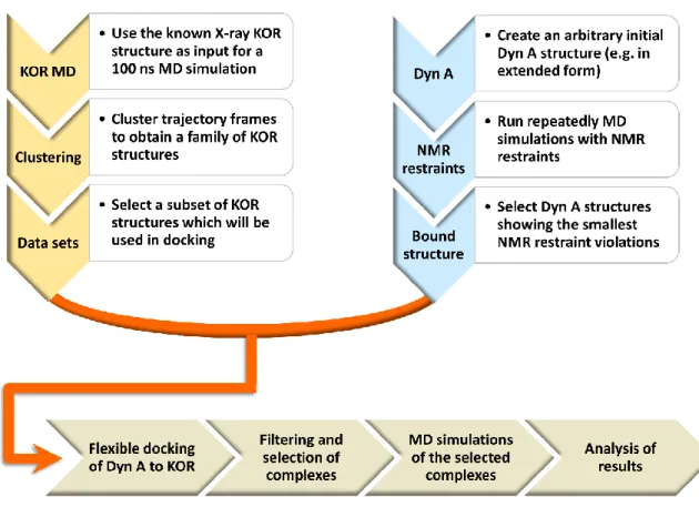
A protocol has been developed for creating three-dimensional structures of dynorphin-KOP
complexes (O'Connor, et al., 2015). The molecular modelling approach (Fig. 6) starts with the
separate preparation of the two molecules. Although a KOP structure in complex with an
antagonist was known from X-ray studies (H. Wu et al., 2012) a MOP structure mutated into
KOP was used as starting point to avoid possible structural distortions caused by JDTic binding.
Following the addition of missing residues and the prediction of rotamers of their side-chains
(Krivov, Shapovalov, & Dunbrack, 2009; Nagata, Randall, & Baldi, 2012), the structure was
relaxed and equilibrated in a 100 ns molecular dynamics (MD) run (R Salomon-Ferrer, Case, &
Walker, 2013; Romelia Salomon-Ferrer, Götz, Poole, Le Grand, & Walker, 2013), followed by a
clustering procedure which allowed major conformers of the receptor to be identified. The use of
a cluster radius of 2 Å resulted in eight families of structures, whose representative members
were selected as those being closest to the cluster centroids. Six major KOP structures were
retained for docking. The modelling of dynorphin involved a typical MD simulation coupled with
a simulated annealing protocol (Nilges, Clore, & Gronenborn, 1988), run in the presence of NMR
constraints to preserve the peptide structure previously determined by NMR. The results
indicated the existence of an α-helical turn involving residues Leu5-Ile8, and a disordered peptide
elsewhere. In the subsequent docking procedure, the backbone of residues Leu5-Ile8 was
therefore held fixed, while the rest of the molecule (backbone and side-chains) remained flexible.
The KOP molecule was mostly held rigid, except for 16 residues whose side-chains were allowed
to be flexible as they were considered likely to interact with the peptide in the binding pocket.
Flexible docking was performed with the AutoDock Vina program (Trott & Olson, 2009), by
launching multiple runs on each of the six retained KOP structures. The results were filtered to
keep the 10 best poses per each KOP structure. This produced a set of 60 structures of the
dynorphin-KOP complex, which was reduced to a set of 22 structures after selecting the best
representatives from each family, characterized by lowest energies. The stability of the
complexes was verified by running further MD simulations for times ranging from 50 to 100 ns.
The crucial final step consisted of comparing the values of the order parameters calculated from
the generated structures with those obtained from NMR experiments. The calculations were
based on the hypothesis that the flexible peptide adopts varying conformations and that its order
parameter should be averaged over different conformers. The exchange is fast on the NMR time
scale, but beyond the reach of MD simulations (1µs – 1ms). To find the minimum number of
conformations required to reproduce the experimental order parameters, all combinations of
modelled structures were taken into account. As a result, five major dynorphin conformations
were identified, revealing significant structural diversity in both N- and C- termini.
Analysis of the resulting complexes allowed us to conclude that the position of a phenol-like
functional group in the orthosteric site was largely conserved, for antagonist-bound structures of
KOP-JDTic, as well as DOP-DIPP and MOP-funaltrexamine. One of the structural models
(available in (O'Connor, et al., 2015)) revealed Tyr1 in a position near the phenol-piperidine
fused-ring system of JDTic, resembling a previously proposed binding mode (Vardy, et al., 2013).
Another model suggested that the side-chain of Tyr1 is close to the allosteric sodium binding site.
D138
3.32makes polar contacts with Tyr1, Gly2 and Gly3 in both of these models. However, only
the latter features polar contacts of both Tyr1 and Arg7 with W287
6.48and N322
7.45.
Figure 6: Molecular modelling protocol leading to three-dimensional structures of dynorphin-KOP
complexes. Briefly, plausible KOP structures were obtained from MD simulations using an X-ray structure as input, while dynorphin models were obtained from simulations with NMR constraints. Subsequent docking, filtering and verification of stability of complex structures thus obtained permitted a selection of the optimal result. See the text for a more detailed description of the procedure.
VI.
Development of novel KOP ligands based on structural knowledge
Opioids possess powerful properties and are currently the most effective analgesics available, but
development is still needed to reduce their undesired side-effects (Dogra & Yadav, 2015). KOP
agonists produce analgesia but, for most, their use is limited by centrally-mediated adverse
effects such as dysphoria. Nalfurafine (TRX-820)
,
that does not induce dysphoria
,
has been
registered in Japan since 2009 for the treatment of uremic pruritus (Kozono, Yoshitani, &
Nakano, 2018).
Several peripherally-restricted (to avoid undesired effects) KOP agonists are
currently in clinical trials: asimadoline (EMD-61753) (Delvaux et al., 2004; Szarka et al., 2007)
and difelikefalin (CR845) (Hesselink, 2017) for irritable bowel syndrome and chronic or
post-operative pain (
https://clinicaltrials.gov/
). Another promising strategy is the development of
KOP-biased ligands: by acting as agonists for the protein-dependent signaling and not for
G-protein-independent pathways, they could induce analgesia without dysphoria (Dogra & Yadav,
2015). In addition, there is a strong potential for KOP antagonists as antidepressant, anxiolytic or
anti-addiction drugs (Zheng, et al., 2017). The knowledge obtained from the structures of inactive
KOP (H. X. Wu, et al., 2012), KOP-bound dynorphin (O'Connor, et al., 2015) and G-protein
bound KOP (Che, et al., 2018) offers new strategies for finding novel KOP ligands (Shang &
Filizola, 2015).
Among the various ways of using structural knowledge in drug discovery, structure-based virtual
screening has proven successful in designing GPCR ligands, such as for D3 dopamine (Carlsson
et al., 2011; Lane et al., 2013) β2 adrenergic (Kolb et al., 2009; Weiss et al., 2013) and A2A
adenosine (Carlsson et al., 2010; Vsevolod Katritch et al., 2010) receptors. It has allowed the
discovery of a G-protein-biased MOP agonist that produces analgesia with diminished
side-effects (Manglik et al., 2016). Studies using virtual screening from the inactive JDTic-bound
KOP structure have discovered new KOP agonists (Negri et al., 2013) and new G-protein–biased
agonists scaffolds (White et al., 2013). More recently, ligand-guided receptor optimization was
applied to the inactive JDTic-bound KOP structure to generate alternative orthosteric pocket
models that served to increase the prediction power of the methods (Zheng, et al., 2017). Virtual
screening on the initial and alternative binding site models, followed by Tanimoto
distances-based selection of novel chemotypes, revealed ligands in the micromolar range with a 32% hit
rate. An initial optimization round generated eleven compounds with sub-micromolar affinities
and functional assays defined two potent antagonists and one G-protein-biased agonist. The
accumulation of precise structural knowledge on the modulation of KOP signaling activity by
orthosteric ligands and allosteric modulators will certainly play a major role in future drug
discovery programs.
Future research into the dynorphin-KOP structure and dynamics must address one major
question: what defines a ligand as a full, partial, unbiased or biased agonist, an antagonist or an
inverse agonist (Wacker, Stevens, & Roth, 2017). As GPCR signaling involves multiple receptor
conformations in dynamic exchange, extensive research aims to characterize their conformational
landscape and their modulation by ligands and signaling partners (Weis & Kobilka, 2018).
Crystallography and electron microscopy provide structures of lowest-energy populated states
(Wacker, et al., 2017). Spectroscopic methods such as NMR allow the characterization of
dynamic properties (Weis & Kobilka, 2018), such as the weak allosteric coupling between the
orthosteric site and the signaling interface, as described for MOP (Sounier, et al., 2015). One may
take advantage of yeast or bacterial expression systems to produce specifically labelled GPCRs
and perform advanced relaxation-based analyses to assess receptor dynamics, as was done
recently for BLT2 (Casiraghi, et al., 2016) and A2A adenosine receptors (Clark et al., 2017; Eddy,
et al., 2018). We are currently applying the methodologies we developed to determine the
conformation and dynamics of KOP-bound dynorphin (O'Connor, et al., 2015) to the ternary
dynorphin-KOP-Nb39 complex (where Nb39 is a nanobody which mimics G-proteins and
confers high affinity binding to agonists). We thus hope to explain the 10-fold gain in dynorphin
binding affinity in the presence of G-proteins. The conformational dynamics of G proteins and
arrestins themselves can be modulated by the ligands (Hilger, et al., 2018). The question of
conformational landscape and allosteric coupling must therefore be extended to entire GPCR
signaling complexes and posed in the context of real cellular environment where modulation by
lipids, membrane domains and other receptors do occur. While using information derived from
structural biology, one should always bear in mind that recombinant G protein-coupled receptors
in vitro may not recapitulate all the properties of native receptors naturally expressed in tissues as
was shown for instance in (Broad et al., 2016).
References
A. Tavani, P. Petrillo, A. La Regina, & Sbacchi, M. (1990). Role of peripheral mu, delta, and kappa opioid receptors in opioid-induced inhibition of gastrointestinal transit in rats. The Journal
Ahlbeck, K. (2011). Opioids: a two-faced Janus. Current Medical Research and Opinion, 27(2), 439-448. doi:10.1185/03007995.2010.545379
Al-Hasani, R., & Bruchas, M. R. (2011). Molecular Mechanisms of Opioid Receptor-Dependent Signaling and Behavior. Anesthesiology, 115, 1363–1381.
Aldrich, J. V., & McLaughlin, J. P. (2009). Peptide kappa opioid receptor ligands: potential for drug development. The AAPS journal, 11(2), 312-322. doi:10.1208/s12248-009-9105-4
Alexander, S. P. H., Christopoulos, A., Davenport, A. P., Kelly, E., Marrion, N. V., Peters, J. A., . . . Collaborators, C. (2017). The concise guide to pharmacology 2017/18: G protein-coupled receptors. British Journal of Pharmacology, 174, S17-S129.
Alonso, H., Bliznyuk, A. A., & Gready, J. E. (2006). Combining Docking and Molecular Dynamic Simulations in Drug Design. Medicinal Research Reviews, 26, 531-568.
Attali, B., & Vogel, Z. (1989). Characterization of kappa opiate receptors in rat spinal cord-dorsal root ganglion cocultures and their regulation by chronic opiate treatment. Brain research,
517, 182-188.
Auge, S., Bersch, B., Tropis, M., & Milon, A. (2000). Characterization of substance P-membrane interaction by transferred nuclear Overhauser effect. Biopolymers, 54(5), 297-306.
Axelrod, D., & Wang, M. D. (1994). Reduction-of-Dimensionality Kinetics at Reaction-Limited Cell-Surface Receptors. Biophysical Journal, 66(3), 588-600. doi:Doi 10.1016/S0006-3495(94)80834-3
Bailey, S. J., & Husbands, S. M. (2018). Targeting opioid receptor signaling in depression: do we need selective κ opioid receptor antagonists? Neuronal Signaling, 2(2).
Ballesteros, J. A., & Weinstein, H. (1995). Integrated methods for the construction of three-dimensional models and computational probing of structure-function relations in G protein-coupled receptors. Methods Neurosci, 25, 366-428.
Barchfeld, C. C., & Medzihradsky, F. (1984). Receptor-mediated stimulation of brain GTPase by opiates in normal and dependent rats. Biomedical and biophysical reasearch communications,
121, 641-648.
Benyhe, S., Zador, F., & Otvos, F. (2015). Biochemistry of opioid (morphine) receptors: binding, structure and molecular modelling. Acta Biologica Szegediensis, 59, 17-37.
Bera, I., Marathe, M. V., Payghan, P. V., & Ghoshal, N. (2018). Identification of novel hits as highly prospective dual agonists for mu and kappa opioid receptors: an integrated in silico approach. Journal of Biomolecular Structure and Dynamics, 36(2), 279-301.
Berman, Y., Mzhavia, N., Polonskaia, A., Furuta, M., Steiner, D. F., Pintar, J. E., & Devi, L. A. (2000). Defective Prodynorphin Processing in Mice Lacking Prohormone Convertase PC2. Journal of
neurochemistry, 75, 1763–1770.
Bersch, B., Koehl, P., Nakatani, Y., Ourisson, G., & Milon, A. (1993). 1H nuclear magnetic resonance determination of the membrane-bound conformation of senktide, a highly selective neurokinin B agonist. Journal of biomolecular NMR, 3(4), 443-461.
Bjorneras, J., Kurnik, M., Oliveberg, M., Graslund, A., Maler, L., & Danielsson, J. (2014). Direct detection of neuropeptide dynorphin A binding to the second extracellular loop of the kappa opioid receptor using a soluble protein scaffold. Febs Journal, 281(3), 814-824. doi:10.1111/febs.12626
Bokoch, M. P., Nygaard, R., Zou, Y. Z., Rasmussen, S. G. F., Pardo, L., Prosser, R. S., . . . Kobilka, B. K. (2010). Conformational Changes in GPCR Surface and Core Probed by [C-13]-Methyl NMR Spectroscopy. Biophysical Journal, 98(3), 418a-418a. doi:DOI 10.1016/j.bpj.2009.12.2257 Bourinet, E., Soong, T. W., Stea, A., & Snutch, T. P. (1996). Determinants of the G protein-dependent
opioid modulation of neuronal calcium channels. Proc. Nati. Acad. Sci. USA, 93, 1486-1491. Broad, J., Maurel, D., Kung, V. W. S., Hicks, G. A., Schemann, M., Barnes, M. R., . . . Sanger, G. J. (2016).
10.1038/srep30797
Bruchas, M. R., & Chavkin, C. (2010). Kinase cascades and ligand-directed signaling at the kappa opioid receptor. Psychopharmacology, 210, 137-147.
Bruchas, M. R., Land, B. B., & Chavkin, C. (2010). The dynorphin/kappa opioid system as a modulator of stress-induced and pro-addictive behaviors. Brain research, 1314, 44-55. doi:10.1016/j.brainres.2009.08.062
Caffrey, M., & Cherezov, V. (2009). Crystallizing membrane proteins using lipidic mesophases.
Nature Protocols, 4(5), 706-731. doi:10.1038/nprot.2009.31
Carlsson, J., Coleman, R. G., Setola, V., Irwin, J. J., Fan, H., Schlessinger, A., . . . Shoichet, B. K. (2011). Ligand discovery from a dopamine D3 receptor homology model and crystal structure.
Nature Chemical Biology, 7(11), 769-778. doi:10.1038/nchembio.662
Carlsson, J., Yoo, L., Gao, Z.-G., Irwin, J. J., Shoichet, B. K., & Jacobson, K. A. (2010). Structure-Based Discovery of A2AAdenosine Receptor Ligands. Journal of medicinal chemistry, 53(9), 3748-3755. doi:10.1021/jm100240h
Casiraghi, M., Damian, M., Lescop, E., Point, E., Moncoq, K., Morellet, N., . . . Catoire, L. J. (2016). Functional Modulation of a G Protein-Coupled Receptor Conformational Landscape in a Lipid Bilayer. Journal of the American Chemical Society, 138(35), 11170-11175. doi:10.1021/jacs.6b04432
Chavkin, C. (2013). Dynorphin–Still an Extraordinarily Potent Opioid Peptide. Molecular
Pharmacology, 83(4), 729-736.
Chavkin, C., Bakhit, C., Weber, E., & Bloom, F. E. (1983). Relative Contents and Concomitant Release of Prodynorphin Neoendorphin-Derived Peptides in Rat Hippocampus. Proceedings of the
National Academy of Sciences of the United States of America-Biological Sciences, 80(24),
7669-7673. doi:DOI 10.1073/pnas.80.24.7669
Che, T., Majumdar, S., Zaidi, S. A., Ondachi, P., McCorvy, J. D., Wang, S., . . . Roth, B. L. (2018). Structure of the Nanobody-Stabilized Active State of the Kappa Opioid Receptor. Cell, 172(1-2), 55-+. doi:10.1016/j.cell.2017.12.011
Chen, Y., Mestek, A., Liu, J., Hurley, J. A., & Yu, L. (1993). Molecular-Cloning and Functional Expression of a Mu-Opioid Receptor from Rat-Brain. Molecular Pharmacology, 44(1), 8-12. Cherezov, V. (2011). Lipidic cubic phase technologies for membrane protein structural studies.
Current Opinion in Structural Biology, 21(4), 559-566.
doi:https://doi.org/10.1016/j.sbi.2011.06.007
Childers, S. R., & Snyder, S. H. (1978). Guanine Nucleotides Differentiate Agonist and Antagonist Interactions with opiate receptors. Life sciences, 23, 759-762.
Choi, H., Murray, T. F., DeLander, G. E., Caldwell, V., & Aldrich, J. V. (1992). N-terminal alkylated derivatives of [D-Pro10]dynorphin A-(1-11) are highly selective for kappa-opioid receptors.
Journal of medicinal chemistry, 35(24), 4638-4639.
Chun, E., Thompson, A. A., Liu, W., Roth, C. B., Griffith, M. T., Katritch, V., . . . Stevens, R. C. (2012). Fusion partner toolchest for the stabilization and crystallization of G protein-coupled receptors. Structure, 20(6), 967-976. doi:10.1016/j.str.2012.04.010
Civelli, O., Douglass, J., Goldstein, A., & Herbert, E. (1985). Sequence and expression of the rat prodynorphin gene. Proceedings of the National Academy of Sciences of the United States of
America, 82(12), 4291-4295.
Clark, L. D., Dikiy, I., Chapman, K., Rodstrom, K. E., Aramini, J., LeVine, M. V., . . . Rosenbaum, D. M. (2017). Ligand modulation of sidechain dynamics in a wild-type human GPCR. Elife, 6. doi:10.7554/eLife.28505
Claude, P. A., Wotta, D. R., Zhang, X. H., Prather, P. L., McGinn, T. M., Erickson, L. J., . . . Law, P. Y. (1996). Mutation of a conserved serine in TM4 of opioid receptors confers full agonistic properties to classical antagonists. Proceedings of the National Academy of Sciences of the
Corder, G., Castro, D. C., Bruchas, M. R., & Scherrer, G. (2018). Endogenous and Exogenous Opioids in Pain. Annu Rev Neurosci, 41, 453-473. doi:10.1146/annurev-neuro-080317-061522
Cox, B. M., Opheim, K. E., Teachemacher, H., & Goldstein, A. (1975). A peptide-like substance from pituitary that acts like morphine. 2. Purification and properties. Life sciences, 16, 1777-1782. Czaplicki, J., & Milon, A. (1998). Simulations of transferred NOE in a ternary peptide-receptor-lipid
complex. Chem Phys Chem (ex J Chim Phys), 95, 196-207.
Czaplicki, j., & Milon, A. (2005). Quantitative analysis of transferred nuclear overhauser effects in complex spin systems by full relaxation matrix analysis. ACTA PHYSICA POLONICA A, 108(1), 25-32.
Dawaliby, R., Trubbia, C., Delporte, C., Masureel, M., Van Antwerpen, P., Kobilka, B. K., & Govaerts, C. (2016). Allosteric regulation of G protein-coupled receptor activity by phospholipids. Nature
Chemical Biology, 12(1), 35-+. doi:10.1038/Nchembio.1960
Delvaux, M., Beck, A., Jacob, J., Bouzamondo, H., Weber, F. T., & Frexinos, J. (2004). Effect of asimadoline, a kappa opioid agonist, on pain induced by colonic distension in patients with irritable bowel syndrome. Aliment Pharmacol Ther, 20(2), 237-246. doi:10.1111/j.1365-2036.2004.01922.x
Deupi, X., & Kobilka, B. K. (2010). Energy Landscapes as a Tool to Integrate GPCR Structure, Dynamics, and Function. Physiology, 25(5), 293-303. doi:10.1152/physiol.00002.2010 Dhawan, B. N., Cesselin, F., Raghubir, R., Reisine, T., Bradley, P. B., Portoghese, P. S., & Hamon, M.
(1996). International Union of Pharmacology. XII. Classification of Opioid Receptors.
Pharmacological Reviews, 48, 567-592.
Didenko, T., Liu, J. J., Horst, R., Stevens, R. C., & Wuthrich, K. (2013). Fluorine-19 NMR of integral membrane proteins illustrated with studies of GPCRs. Current Opinion in Structural Biology,
23(5), 740-747. doi:10.1016/j.sbi.2013.07.011
Dogra, S., & Yadav, P. N. (2015). Biased agonism at kappa opioid receptors: Implication in pain and
mood disorders. European journal of pharmacology, 763, 184-190.
doi:10.1016/j.ejphar.2015.07.018
Eddy, M. T., Lee, M. Y., Gao, Z. G., White, K. L., Didenko, T., Horst, R., . . . Wuthrich, K. (2018). Allosteric Coupling of Drug Binding and Intracellular Signaling in the A(2A) Adenosine Receptor. Cell,
172(1-2), 68-+. doi:10.1016/j.cell.2017.12.004
Erne, D., Sargent, D. F., & Schwyzer, R. (1985). Preferred conformation, orientation, and accumulation of dynorphin A-(1-13)-tridecapeptide on the surface of neutral lipid membranes. Biochemistry, 24(16), 4261-4263.
Evans, C. J., Keith, D. E., Jr., Morrison, H., Magendzo, K., & Edwards, R. H. (1992). Cloning of a delta opioid receptor by functional expression. Science, 258(5090), 1952-1955.
Fenalti, G., Giguere, P. M., Katritch, V., Huang, X. P., Thompson, A. A., Cherezov, V., . . . Stevens, R. C. (2014). Molecular control of delta-opioid receptor signalling. Nature, 506(7487), 191-196. doi:10.1038/nature12944
Fenalti, G., Zatsepin, N. A., Betti, C., Giguere, P., Han, G. W., Ishchenko, A., . . . Cherezov, V. (2015). Structural basis for bifunctional peptide recognition at human delta-opioid receptor. Nature
Structural & Molecular Biology, 22(3), 265-268. doi:10.1038/nsmb.2965
Ferre, S., Casado, V., Devi, L. A., Filizola, M., Jockers, R., Lohse, M. J., . . . Guitart, X. (2014). G Protein-Coupled Receptor Oligomerization Revisited: Functional and Pharmacological Perspectives.
Pharmacological Reviews, 66(2), 413-434. doi:10.1124/pr.113.008052
Fersht, A. R. (1999). Structure and Mechanism in Protein Science: Freeman, New York.
Gentilucci, L., Tolomelli, A., De Marco, R., & Artali, R. (2012). Molecular Docking of Opiates and Opioid Peptides, a Tool for the Design of Selective Agonists and Antagonists, and for the Investigation of Atypical Ligand-Receptor Interactions. Current Medicinal Chemistry, 19, 1587-1601.
Goldstein, A., Fischli, W., Lowney, L. I., Hunkapiller, M., & Hood, L. (1981). Porcine pituitary dynorphin: complete amino acid sequence of the biologically active heptadecapeptide.
Proceedings of the National Academy of Sciences of the United States of America, 78(11),
7219-7223.
Goldstein, A., Tachibana, S., Lowney, L. I., Hunkapiller, M., & Hood, L. (1979). Dynorphin-(1-13), an extraordinarily potent opioid peptide. Proceedings of the National Academy of Sciences of the
United States of America, 76(12), 6666-6670.
Granier, S., & Kobilka, B. (2012). A new era of GPCR structural and chemical biology. Nature
Chemical Biology, 8(8), 670-673. doi:10.1038/nchembio.1025
Granier, S., Manglik, A., Kruse, A. C., Kobilka, T. S., Thian, F. S., Weis, W. I., & Kobilka, B. K. (2012). Structure of the delta-opioid receptor bound to naltrindole. Nature, 485(7398), 400-U171. doi:10.1038/nature11111
Henry, D. J., Grandy, D. K., Lester, H. A., Davidson, N., & Chavkin, C. (1995). K-Opioid Receptors Couple to Inwardly Rectifying Potassium Channels when Coexpressed by Xenopus Oocytes.
Molecular Pharmacology, 47, 551-557.
Hesselink, J. M. K. (2017). CR845 (Difelikefalin), A Kappa Receptors Agonist In Phase III By CARA Therapeutics: A Case Of ‘Spin’ In Scientific Writing? J of Pharmacol & Clin Res, 2(3), 555588. doi:10.19080/JPCR.2017.02.555588
Hilger, D., Masureel, M., & Kobilka, B. K. (2018). Structure and dynamics of GPCR signaling complexes. Nature Structural & Molecular Biology, 25(1), 4-12. doi:10.1038/s41594-017-0011-7
Huang, W. J., Manglik, A., Venkatakrishnan, A. J., Laeremans, T., Feinberg, E. N., Sanborn, A. L., . . . Kobilka, B. K. (2015). Structural insights into mu-opioid receptor activation. Nature,
524(7565), 315-+. doi:10.1038/nature14886
Iadanza, M., Höltje, M., Ronsisvalle, G., & Höltje, H.-D. (2002). κ-Opioid Receptor Model in a Phospholipid Bilayer: Molecular Dynamics Simulation. Journal of medicinal chemistry, 45(22), 4838-4846.
J-C. Meunier, C. Mollereau, L. Toll, C. Suaudeau, C. Moisand, P. Alvinerie, . . . Costentin, J. (1995). Isolation and structure of the endogenous agonist of opioid receptor-like ORL1 receptor.
Nature, 377, 532-535.
Ji, R.-R., Zhang, Q., Law, P. Y., Low, H. H., Elde, R., & Hökfelt, T. (1995). Expression of mu-, delta-, and kappa- opioid receptor-like Immunoreactivities in rat dorsal roort ganglia after carrageenan-induced inflammation. The Journal of Neuroscience, 15, 8155-8166.
Johnson, S. (2017). Design and Synthesis of Functionally Selective Kappa Opioid Receptor Ligands.
University of Kansas, Lawrence. Retrieved from
https://kuscholarworks.ku.edu/handle/1808/26154
Jordan, B. A., & Devi, L. A. (1999). G protein coupled receptor heterodimerization modulates receptor function. Nature, 399(6737), 697-700.
Kakidani, H., Furutani, Y., Takahashi, H., Noda, M., Morimoto, Y., Hirose, T., . . . Numa, S. (1982). Cloning and sequence analysis of cDNA for porcine beta-neo-endorphin/dynorphin precursor. Nature, 298, 245-249.
Kallick, D. A. (1993). Conformation of Dynorphin a(1-17) Bound to Dodecylphosphocholine Micelles.
Journal of the American Chemical Society, 115(20), 9317-9318. doi:DOI
10.1021/ja00073a069
Kane, B. E., Svensson, B., & Ferguson, D. M. (2006). Molecular recognition of opioid receptor ligands.
The AAPS journal, 8(1), E126-E137.
Kang, Y. Y., Zhou, X. E., Gao, X., He, Y. Z., Liu, W., Ishchenko, A., . . . Xu, H. E. (2015). Crystal structure of rhodopsin bound to arrestin by femtosecond X-ray laser. Nature, 523(7562), 561-+. doi:10.1038/nature14656
Kaserer, T., Lantero, A., Schmidhammer, H., Spetea, M., & Schuster, D. (2016). μ Opioid receptor: novel antagonists and structural modeling. Scientific Reports, 6, 21548.
Katritch, V., Fenalti, G., Abola, E. E., Roth, B. L., Cherezov, V., & Stevens, R. C. (2014). Allosteric sodium in class A GPCR signaling. Trends in Biochemical Sciences, 39(5), 233-244. doi:10.1016/j.tibs.2014.03.002
Katritch, V., Jaakola, V.-P., Lane, J. R., Lin, J., Ijzerman, A. P., Yeager, M., . . . Abagyan, R. (2010). Structure-Based Discovery of Novel Chemotypes for Adenosine A2AReceptor Antagonists.
Journal of medicinal chemistry, 53(4), 1799-1809. doi:10.1021/jm901647p
Kieffer, B. L., Befort, K., Gaveriaux-Ruff, C., & Hirth, C. G. (1992). The delta-opioid receptor: isolation of a cDNA by expression cloning and pharmacological characterization. Proceedings of the
National Academy of Sciences of the United States of America, 89(24), 12048-12052.
Knoll, A. T., & Carlezon, W. A. (2010). Dynorphin, stress, and depression. Brain research, 1314, 56-73. doi:10.1016/j.brainres.2009.09.074
Kobilka, B., & Schertler, G. F. X. (2008). New G-protein-coupled receptor crystal structures: insights
and limitations. Trends in pharmacological sciences, 29(2), 79-83.
doi:10.1016/j.tips.2007.11.009
Koehl, A., Hu, H., Maeda, S., Zhang, Y., Qu, Q., Paggi, J. M., . . . Kobilka, B. K. (2018). Structure of the µ-opioid receptor–Gi protein complex. Nature. doi:10.1038/s41586-018-0219-7
Kolb, P., Rosenbaum, D. M., Irwin, J. J., Fung, J. J., Kobilka, B. K., & Shoichet, B. K. (2009). Structure-based discovery of beta2-adrenergic receptor ligands. Proceedings of the National Academy
of Sciences, 106(16), 6843-6848. doi:10.1073/pnas.0812657106
Kolinski, M., & Filipek, S. (2010). Study of a structurally similar kappa opioid receptor agonist and antagonist pair by molecular dynamics simulations. Journal of Molecular Modeling, 16(10), 1567–1576.
Kong, H., Raynor, K., Yano, H., Takeda, J., Bell, G. I., & Reisine, T. (1994). Agonists and antagonists bind to different domains of the cloned kappa opioid receptor. Proceedings of the National
Academy of Sciences of the United States of America, 91(17), 8042-8046.
Kozono, H., Yoshitani, H., & Nakano, R. (2018). Post-marketing surveillance study of the safety and efficacy of nalfurafine hydrochloride (Remitch((R)) capsules 2.5 mug) in 3,762 hemodialysis patients with intractable pruritus. Int J Nephrol Renovasc Dis, 11, 9-24. doi:10.2147/IJNRD.S145720
Kreek, M. J., Levran, O., Reed, B., Schlussman, S. D., Zhou, Y., & Butelman, E. R. (2012). Opiate addiction and cocaine addiction: underlying molecular neurobiology and genetics. Journal of
Clinical Investigation, 122(10), 3387-3393. doi:10.1172/jci60390
Krishnan, V., & Nestler, E. J. (2010). Linking Molecules to Mood: New Insight Into the Biology of
Depression. American Journal of Psychiatry, 167(11), 1305-1320.
doi:10.1176/appi.ajp.2009.10030434
Krivov, G. G., Shapovalov, M. V., & Dunbrack, R. L. (2009). Improved prediction of protein side-chain conformations with SCWRL4. Proteins, 77(4), 778-795.
Lagane, B., Gaibelet, G., Meilhoc, E., Masson, J. M., Cezanne, L., & Lopez, A. (2000). Role of sterols in modulating the human mu-opioid receptor function in Saccharomyces cerevisiae. Journal of
Biological Chemistry, 275(43), 33197-33200. doi:DOI 10.1074/jbc.C000576200
Lahti, R. A., Mickelson, M. M., McCall, J. M., & Von Voigtlander, P. F. (1985). U-69593 a highly selective ligand for the opioid kappa receptor. European journal of pharmacology, 109, 281-284. Lahti, R. A., VonVoigtlander, P. F., & Barsuhn, C. (1982). Properties of a selective kappa agonist,
U-50,488H. Life sciences, 31, 2257.
Lancaster, C. R. D., Mishra, P. K., Hughes, D. W., Stpierre, S. A., Bothnerby, A. A., & Epand, R. M. (1991). Mimicking the Membrane-Mediated Conformation of Dynorphin a-(1-13)-Peptide - Circular-Dichroism and Nuclear-Magnetic-Resonance Studies in Methanolic Solution. Biochemistry,