HAL Id: hal-02974826
https://hal.archives-ouvertes.fr/hal-02974826
Submitted on 22 Oct 2020HAL is a multi-disciplinary open access archive for the deposit and dissemination of sci-entific research documents, whether they are pub-lished or not. The documents may come from teaching and research institutions in France or abroad, or from public or private research centers.
L’archive ouverte pluridisciplinaire HAL, est destinée au dépôt et à la diffusion de documents scientifiques de niveau recherche, publiés ou non, émanant des établissements d’enseignement et de recherche français ou étrangers, des laboratoires publics ou privés.
and Tumor Segmentation
Théo Estienne, Marvin Lerousseau, Maria Vakalopoulou, Emilie Andres, Enzo
Battistella, Alexandre Carré, Siddhartha Chandra, Stergios Christodoulidis,
Mihir Sahasrabudhe, Roger Sun, et al.
To cite this version:
Théo Estienne, Marvin Lerousseau, Maria Vakalopoulou, Emilie Andres, Enzo Battistella, et al.. Deep Learning-Based Concurrent Brain Registration and Tumor Segmentation. Frontiers in Computa-tional Neuroscience, Frontiers, 2020, Multimodal Brain Tumor Segmentation and Beyond, �10.3389/fn-com.2020.00017�. �hal-02974826�
Deep Learning-Based Concurrent Brain
Registration and Tumor Segmentation
Th ´eo Estienne1,4,5,6∗,‡, Marvin Lerousseau1,3,4,5,‡, Maria Vakalopoulou1,6,
Emilie Alvarez Andres1,4,5, Enzo Battistella1,4,5,6, Alexandre Carr ´e1,4,5,
Siddhartha Chandra3, Stergios Christodoulidis2, Mihir Sahasrabudhe3, Roger
Sun1,3,4,5, Charlotte Robert1,4,5, Hugues Talbot3, Nikos Paragios1 and Eric
Deutsch1,4,5
1 Gustave Roussy-CentraleSup ´elec-TheraPanacea Center of Artificial Intelligence in Radiation Therapy and Oncology, Gustave Roussy Cancer Campus, Villejuif, France
2 ARTORG Center for Biomedical Engineering Research, Bern University, Bern, Switzerland
3 CVN, CentraleSup ´elec, University Paris-Saclay and INRIA Saclay, France 4 INSERM, U1030, Paris, France
5 University Paris Sud, UFR de m ´edecine, Paris, France
6 Laboratoire MICS, CentraleSup ´elec - University Paris-Saclay, France Correspondence*:
Th ´eo Estienne
ABSTRACT 2
Image registration and segmentation are the two most studied problems in medical image
3
analysis. Deep learning algorithms have recently gained a lot of attention due to their success
4
and state-of-the-art results in variety of problems and communities. In this paper, we propose
5
a novel, efficient, and multi-task algorithm that addresses the problems of image registration
6
and brain tumor segmentation jointly. Our method exploits the dependencies between these
7
tasks through a natural coupling of their interdependencies during inference. In particular, the
8
similarity constraints are relaxed within the tumor regions using an efficient and relatively simple
9
formulation. We evaluated the performance of our formulation both quantitatively and qualitatively
10
for registration and segmentation problems on two publicly available datasets (BraTS 2018 and
11
OASIS 3), reporting competitive results with other recent state-of-the-art methods. Moreover, our
12
proposed framework reports significant amelioration (p < 0.005) for the registration performance
13
inside the tumor locations, providing a generic method that does not need any predefined
14
conditions (e.g. absence of abnormalities) about the volumes to be registered.
15
16
Keywords: brain tumor segmentation, deformable registration, multi-task networks, deep learning, convolutional neural networks. 17
1
INTRODUCTION
Brain tumors and more specifically gliomas as one of the most frequent types, are across the most
18
dangerous and rapidly growing types of cancer (C. Holland, 2002). In clinical practice, multi-modal
19
magnetic resonance imaging (MRI) is the primary method of screening and diagnosis of gliomas. While
20
gliomas are commonly stratified into Low grade and High grade due to different histology and imaging
21
aspects, prognosis and treatment strategy, radiotherapy is one of the mainstays of treatment
(Sep´ulveda-22
S´anchez et al., 2018; Stupp et al., 2014). However, radiotherapy treatment planning relies on tumor manual
23
segmentation by physicians, making the process tedious, time-consuming, and sensitive to bias due to low
24
inter-observer agreement (Wee et al., 2015).
25
In order to overcome these limitations, numerous methods have been proposed recently that try to provide
26
tools and algorithms that will make the process of gliomas segmentation automatic and accurate (Parisot
27
et al., 2016; Zhao et al., 2018). Towards this direction, the multimodal brain tumor segmentation challenge
28
(BraTS) (Bakas et al., 2017b,c,a; Menze et al., 2015) is annually organized, in order to highlight
29
efficient approaches and indicate the way towards this challenging problem. In recent years, most of
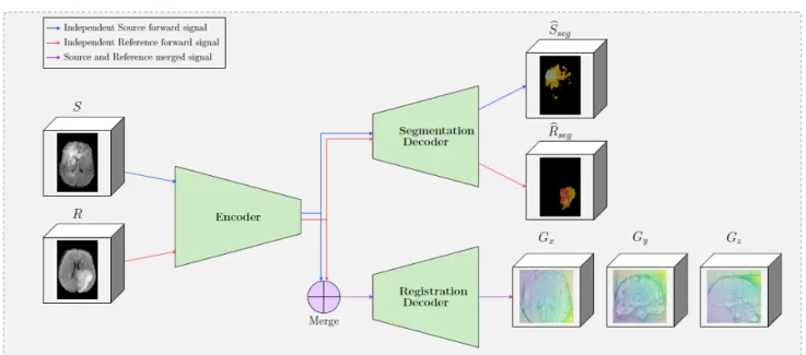
30
the approaches that exploit BraTS have been based on deep learning architectures using 3D convolutional
31
neural networks (CNNs) similar to VNet (Milletari et al., 2016). In particular, the best performing
32
approaches use ensembles of deep learning architectures (Kamnitsas et al., 2018; Zhou et al., 2018), with
33
autoencoder regularization (Myronenko, 2018) or they even combine deep learning architectures together
34
with algorithms such as conditional random fields (CRFs) (Chandra et al., 2019).Other top-performing
35
methods at the BraTS 2017 and 2018 used cascaded networks, multi-view and multi-scale approaches
36
(Wang et al., 2017), generic UNet architecture with data augmentation and post processing (Isensee et al.,
37
2018), dilated convolutions and label uncertainty loss (McKinley et al., 2018), and context aggregation and
38
localization pathways (Isensee et al., 2017).A more detailed comparison and presentation of the last years
39
challenges on BraTS is presented and summarized in (Bakas et al., 2018).
40
Image registration is a challenging task for medical image analysis in general and for rapidly evolving
41
brain tumors in particular, where longitudinal assessment is critical. Image registration seeks to determine
42
a transformation that will map two volumes (source and reference) to the same coordinate system. In
43
practice, we seek a volume mapping function that changes the coordinate system of the source volume
44
into the coordinate system of the reference volume. Among the different types of methods employed in
45
medical applications, deformable or elastic registration is the most commonly used (Sotiras et al., 2013).
46
Linear methods are an alternative but in that case a linear global transformation is sought for the entire
47
volume. Deformable registration has been addressed with a variety of methods, including for example
48
surface matching (Robinson et al., 2018; Postelnicu et al., 2009) or graph based approaches (Glocker et al.,
49
2009). These methods have been extended to address co-registration of multiple volumes (Ou et al., 2011).
50
Moreover, some of the most popular methods traditionally used for the accurate deformable registration
51
include (Klein et al., 2009; Avants et al., 2008; Shi et al., 2013). Recently a variety of deep learning
52
based methods have been proposed, reducing significantly the computational time but maintaining the
53
accuracy and robustness of the registration (Dalca et al., 2018; Christodoulidis et al., 2018). In particular,
54
the authors in (Dalca et al., 2018) presented a deep learning framework trained for atlas-based registration
55
of brain MR images, while in (Christodoulidis et al., 2018) the authors present a scheme for a concurrent
56
linear and deformable registration of lung MR images. However, when it comes to anatomies that contain
57
abnormalities such as tumoral areas, these methods fail to register the volumes at certain locations, due to
58
lack of similarity between the volumes. This most of the times ends to complete distortion of the tumor
59
area of the deformed image.
To overcome this problem, in this paper, we propose a dual deep learning based architecture that addresses
61
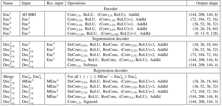
registration and tumor segmentation simultaneously, relaxing the registration constraints inside the predicted
62
tumor areas, providing displacements and segmentation maps at the same time. Our framework bears
63
concept similarities with the work presented in (Parisot et al., 2012) where a Markov Random Field (MRF)
64
framework has been proposed to address both of tumor segmentation and image registration jointly. Their
65
method required approximately 6 minutes for the registration of one pair and the segmentation of one
66
class tumor region was performed with handcrafted features and classical machine learning techniques
67
using only one MRI modality. Moreover, there are methods in the literature that try to address the problem
68
of registration of brain tumor MRI by registering on atlases or MRIs without tumoral regions (Gooya
69
et al., 2010, 2012). Here, we introduce a highly scalable, modular, generic and precise 3D-CNN for
70
both registration and segmentation tasks and provide a computationally efficient and accurate method for
71
registering any arbitrary subject involving possible abnormalities. To the best of our knowledge this is the
72
first time that a joint deep learning-based architecture is presented, showing very promising results in two
73
publicly available datasets for brain MRI. The proposed framework provides a very powerful formulation
74
introducing the means to elucidate clinical, or functional trends in the anatomy or physiology of the brain
75
due to the registration part. Moreover, it enables the modeling and the detection of brain tumor areas due to
76
the synergy with the segmentation part.
77
2
MATERIALS AND METHODS
Consider a pair of medical volumes from two different patients —a source S, and a reference R together
78
with their annotations for the tumor areas (Sseg and Rseg). The framework consists of a bi-cephalic structure
79
with shared parameters, depicted in Figure 1. During training the network uses as input a source S and
80
a reference R volumes and outputs their brain tumor segmentation masks bSseg and bRseg and the optimal
81
elastic transformation G which will project or map the source volume to the reference volume. The goal of
82
the registration part is to find the optimal transformation to transform the source (S) to the reference (R)
83
volume. In this section, we present the details for each of the blocks as well as our final formulation for the
84
optimization.
85
2.1 Shared encoder
86
One of the main differences of the proposed formulation with other registration approaches in the literature
87
is the way that the source and reference volumes are combined. In particular, instead of concatenating
88
the two initial volumes, these volumes are independently forwarded in a unique encoder, yielding two
89
sets of features maps (called latent codes) Csourceand Creferencefor the source and the reference volumes
90
respectively. These two codes are then independently forwarded into the segmentation decoder, providing
91
the predicted segmentation maps Sseg and Rseg. Simultaneously, the two codes are merged before being
92
forwarded in the registration decoder — this operation is depicted in the ”Merge” block in Figure 1.
93
The motivation behind adopting this strategy is based on forcing the encoder to extract meaningful
94
representations from individual volumes instead of a pair of volumes. This is equivalent to asking the
95
encoder discovering a template, ”deformation-free” space for all volumes, and encoding each volume
96
against this space (Shu et al., 2018), instead of decoding the deformation grid between every possible pair
97
of volumes. Besides, from the segmentation point of view, there are no relationship between the tumor
98
maps of the source volume and the reference volume, so the codes to be forwarded into the segmentation
99
decoder should not depend on each other.
We tested two merging operators, namely concatenation and subtraction. Both source and reference
101
images are 4D volumes whose first dimension corresponds to the 4 different MRI modalities that are used
102
per subject. After the forward to the encoder, the codes Csource and Creferenceare also 4D volumes with
103
the first dimension corresponding to nf, which is the number of convolutional filters of the last block
104
of the encoder. Before Csource and Creference are inserted into the registration decoder, they are merged,
105
outputting one 4D volume of size 2 × nf in the case of the concatenation, and of size nf for the elementwise
106
subtraction operator, both leaving the rest of the dimensions unchanged. In particular, the subtraction
107
presents the following natural properties for every coding image CI:
108
• ∀CI ∈ R4: M erge(CI, CI) = 0
109
• ∀CI, CJ ∈ R4× R4 : M erge(CI, CJ) = −M erge(CJ, CI)
110
2.2 Brain tumor segmentation decoder
111
Inspired by the latest advances reported on the BraTS 2018 dataset, we adopt a powerful autoencoder
112
architecture. The segmentation and registration decoders share the same encoder (Section 2.1) for feature
113
extraction and they provide brain tumor segmentation masks ( bSseg and bRseg) for the source and the
114
reference images. These masks refer to valuable information about the regions that cannot be registered
115
properly as there is no corresponding anatomical information on the pair. This information is integrated
116
into the optimisation of the registration component, relaxing the similarity constraints and preserving to a
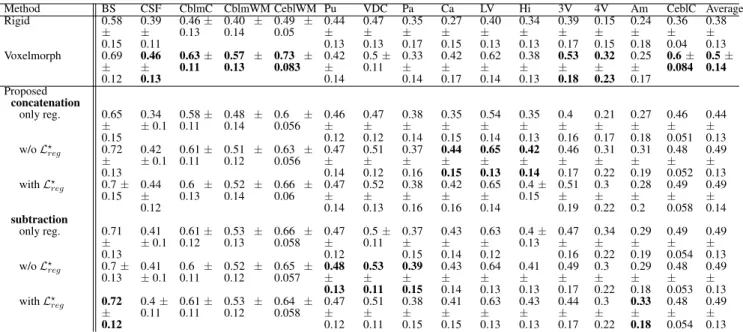
117
certain extent the geometric properties of the tumor.
118
Variety of loss functions have been proposed in the literature for the semantic segmentation of 3D medical
119
volumes. In this paper, we performed all our experiments using weighted categorical cross-entropy loss
120
and optimising 3 different segmentation classes for the tumor area as provided by the BraTS dataset. In
121
particular,
122
Lseg = CE(Sseg, bSseg) + CE(Rseg, bRseg) (1) where CE denotes the weighted cross entropy loss. The cross entropy is calculated for both the source
123
and reference images and the overall segmentation loss is the sum of the two. Here we should note that
124
different segmentation losses can be applicable as for example the dice coefficient (Sudre et al., 2017),
125
focal loss (Lin et al., 2017), e.t.c.
126
2.3 Elastic registration decoder
127
In this paper, the registration strategy is based on the one presented in (Christodoulidis et al., 2018), with
128
the main component being the 3D spatial transformer. A spatial transformer deforms (or warps) a given
129
image S with a deformation grid G. It can be represented by the operation,
130
D = W(S, G),
where W(·, G) indicates a sampling operation W under the deformation G and D the deformed image.
131
The deformation is hence fed to the transformer layer as sampling coordinates for a backward trilinear
132
interpolation sampling, adapting a strategy similar to (Shu et al., 2018). The sampling process is then
133
described by
D(~p) = W(S, G)(~p) =X ~ q S(~q)Y d max (0, 1 − |[G(~p)]d− ~qd|) ,
where ~p and ~q denote pixel locations, d ∈ {x, y, z} denotes an axis, and [G(~p)]ddenotes the d-component
135
of G(~p). Moreover, instead of regressing per-pixel displacements, we predict a matrix Ψ of spatial gradients
136
between consecutive pixels along each axis. The actual grid G can then be obtained by applying an
137
integration operation on Ψ along the x-, y- and z-axes, which is approximated by the cumulative sum in
138
the discrete case. Consequently, two pixels ~p and ~p + 1 will have moved closer, maintained distance, or
139
moved apart in the warped image, if Ψ~pis respectively less than 1, equal to 1, or greater than 1.
140
2.4 Network Architecture
141
Our network architecture is a modified version of the fully convolutional VNet (Milletari et al., 2016)
142
for the underlying encoder and decoders parts, maintaining the depth of the model and the rest of the
143
filter’s configuration unchanged. The model, whose computational graph is displayed in Table 1, comprises
144
several sequential residual convolutional blocks made of one to three convolutional layers, followed by
145
downsampling convolutions for the encoder part and upsampling convolutions for the decoder part. We
146
replaced the initial 5 × 5 × 5 convolutions filter-size by 3 × 3 × 3 in order to reduce the number of parameters
147
without changing the depth of the model, and also replace PReLu activations by ReLU ones. In order to
148
speed up its convergence, the model uses residual connections between each encoding and corresponding
149
decoding stage for both the segmentation and the registration decoder. This allows every layer of the
150
network, particularly the first ones, to be trained more efficiently since the gradient can flow easier from
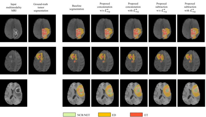
151
the last layers to the first ones with less vanishing or exploding gradient issues. The encoder part deals with
152
4-inputs per volume, representing the 4 different MRI modalities that are available on the BraTS dataset,
153
an extra 1 × 1 × 1 convolution is added to fuse the initial modalities. Moreover, the architecture contains 2
154
decoders of identical blocks, 1 dedicated to the segmentation of tumors for the source and reference image
155
and 1 dedicated to the optimal displacement that will map the source to the reference image.
156
2.5 Optimization
157
The network is trained to minimize the segmentation and registration loss functions jointly. For the
158
segmentation task the loss function is summarized in Eq. 1. For registration, the classical optimization
159
scheme is to minimize the Frobenius norm between the R and D image intensities:
160
Lreg= ||(R − D)||2+ α kΨ − ΨIk1 (2)
Here, in order to better achieve overall registration, the Frobenius norm within the regions predicted to
161
be tumors is excluded from the loss function. We argue that by doing this, the model does not focus on
162
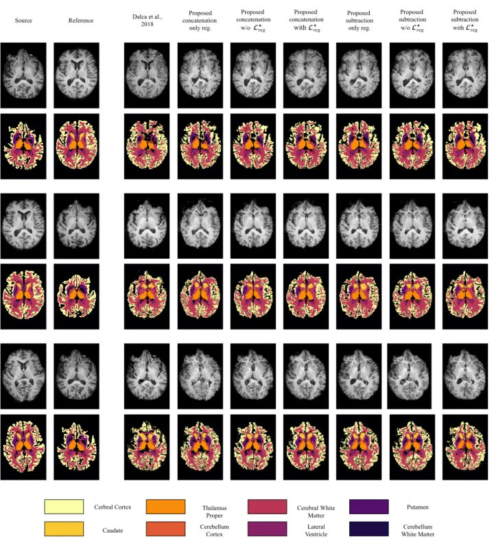
tumor regions, which might produce very high norm due to their texture, but rather focuses on the overall
163
registration task by looking at regions outside the tumor which contain information more pertinent to the
164
alignment of the volumes. Here we should mention that on bSseg we apply the same displacement grid as on
165
S, resulting in Dseg = W( bSseg, G). Further, let bR0seg and D0seg be binary volumes indicating the voxels
166
which are predicted to be outside any segmented regions. Then, the registration loss can be written as
167
where · is the element-wise multiplication, || · ||2 indicates the Frobenius norm, ΨI is the spatial gradient
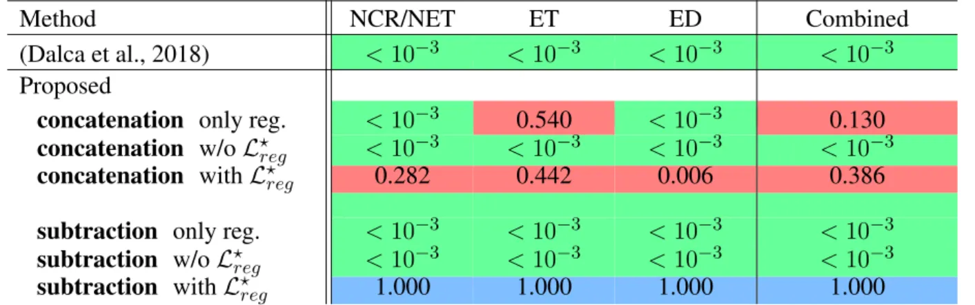
168
of the identity deformation and α is the regularization hyperparameter. The use of regularisation on the
169
displacements Ψ is essential in order to constrain the network to predict smooth deformation grids that are
170
anatomically more meaningful while at the same time regularize the objective function towards avoiding
171
local minimum.
172
Finally the final optimisation of the framework is performed by the joint optimisation of the segmentation
173
and registration loss functions
174
L = Lreg+ βLseg
where β is a weight that indicates the influence of each of the components on the joint optimization of
175
the network and was defined after grid search.
176
For the training process, the initial learning rate was 2 · 10−3 and subdued by a factor of 5 if the
177
performance on the validation set did not improve for 30 epochs. The training procedure stops when there
178
is no improvement for 50 epochs. The regularization weights α and β were set to 10−10and 1 after grid
179
search. As training samples, random pairs among all cases were selected with a batch size limited to 2
180
due to the limited memory resources on the GPU. The performance of the network was evaluated every
181
100 batches, and both proposed models converged after nearly 200 epochs. The overall training time was
182
calculated to ∼ 20 hours, while the time for inference of one pair, using 4 different modalities was ∼ 3 sec,
183
using an NVIDIA GeForce GTX 1080 Ti GPU.
184
2.6 Datasets
185
We evaluated the performance of our method using two publicly available datasets, namely the Brain
186
Tumor Segmentation (BraTS) (Bakas et al., 2018) and Open Access Series of Imaging Studies (OASIS
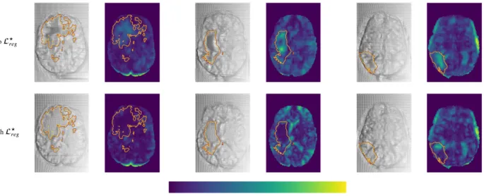
187
3) (Marcus et al., 2010) datasets. BraTS contains multi-institutional pre-operative MRI scans of whole
188
brains with visible gliomas, which are intrinsically heterogeneous in their imaging phenotype (shape and
189
appearance) and histology. The MRIs are all pre-operative and consist of 4 modalities, i.e. 4 3D volumes,
190
namely a) a native T1-weighted scan (T1), b) a post-contrast Gadolinium T1-weighted scan (T1Gd), c) a
191
native T2-weighted scan (T2), and d) a native T2 Fluid Attenuated Inversion Recovery scan (T2-FLAIR).
192
The BraTS MRIs are provided with voxelwise ground-truth annotations for 5 disjoint classes denoting a)
193
the background, b) the necrotic and non-enhancing tumor core (NCR/NET), c) the GD-enhancing tumor
194
(ET), d) the peritumoral edema (ED) as well as invaded tissue, and finally e) the rest of the brain, i.e. brain
195
with no abnormality nor invaded tissue. Each center was responsible for annotating their MRIs, with a
196
central validation by domain experts. We use the original dataset split of BraTS 2018 which contains 285
197
training samples and 66 for validation. In order to perform our experiments, we split this training set into 3
198
parts, i.e. train, validation and test sets (199, 26 and 60 patients, respectively), while we used the 66 unseen
199
cases on the platform to report the performance of the proposed and the benchmarked methods. Moreover,
200
and especially for the registration task, we evaluated the performance of the models trained on BraTS on the
201
OASIS 3 dataset to test the generalisation of the method. This dataset consists of a longitudinal collection
202
of 150 subjects which were characterized as either nondemented or with mild cases of Alzheimer’s disease
203
(AD) using the Clinical Dementia Rating (CDR). Each scan is made of 3 to 4 individual T1-weighted
204
MRIs, which has been intended to reduce the signal-to-noise ratio visible with single images. The scans
205
are also provided with annotations for 47 different structures for left and right side of the brain generated
206
with FreeSurfer. Some samples of both datasets can be seen in Figure 2.
The same pre-processing steps have been applied for both datasets. MRIs were resampled to voxels
208
of volume 1mm3 using trilinear interpolation. Each scan is then centered by automatically translating
209
their barycenter to the center of the volume. Ground-truth masks of training and validation steps were
210
accordingly translated. Each modality of each scan has been standardised, i.e. the values of the voxels of
211
the 3D subscans were of zero mean and of unit variance. This normalization step is done independently for
212
each patient and for each channel in order to equally consider each channel since modalities have voxels
213
values in completely different ranges. Finally, these consequent scans are cropped into (144, 208, 144)
214
sized volumes.
215
2.7 Statistical evaluations
216
Our contributions in the study are threefold: multi-task segmentation and registration, registration with a
217
shared encoder and latent space merge operator, as well as the loss L?reg(Equation 3) that alleviates the
218
registration modifications of tumor tissues in both source and reference patients. Our experiments were
219
intended to weigh the impact of these novelties for both tumor segmentation and registration of MRIs with
220
tumor areas.
221
2.7.1 Methods benchmarked
222
We therefore benchmark multiple versions of our proposed approach with a subset of these novelties to
223
assess their impact on both registration and segmentation. We notably derive 2 variants for both merging
224
operators subtraction and concatenation. The first variant is our fully proposed architecture with a shared
225
encoder for registration and one decoder for segmentation whose tumor predictions are used to implement
226
the proposed loss L?reg. These models are named ”Proposed concatenation with L?reg” and ”Proposed
227
subtraction with L?reg”. The second variant of models does not use the proposed loss, and are identified
228
with ”w/o L?reg”. Finally, we also derive a third variant of our approach, yielding one method per merging
229
operator, by discarding the segmentation decoder. Because the proposed loss use the predicted tumor maps
230
from a segmentation decoder, this variant does not rely on it. These latter methods are named ”Proposed
231
concatenation only reg.” and ”Proposed subtraction only reg.”, and are primarily benchmarked to assess the
232
performance of the segmentation decoder and the loss L?regwith respect to our fully proposed architecture.
233
We also benchmark baseline methods, without any of the proposed contributions. Since our deep learning
234
architecture is derived from the Vnet (Milletari et al., 2016), this model is used as baseline for segmentation.
235
This comparison seems fair since the fully proposed approach can be seen as a Vnet for the task of
236
segmentation: the shared encoder and the proposed loss are primarily designed for registration, and have
237
no direct impact on the segmentation apart from the features learnt in the encoder. For completeness, the
238
top performing results on the BraTS (Bakas et al., 2018) challenge are reported, although we argue that
239
the comparison is unfair since our deep learning architecture is entirely based on the Vnet (Milletari et al.,
240
2016), which is not specifically designed to perform well on the BraTS segmentation task. Finally, we also
241
report the performance of Voxelmorph (Dalca et al., 2018), a well performing brain MRI registration neural
242
network-based approach, although their entire deep learning structure as well as their grid formulation is
243
different.
244
2.7.2 Performance assessment
245
For performance assessment of the segmentation task, we reported the Dice coefficient metric and
246
Hausdorff distance to measure the performance for the tumor classes Tumor Core (TC), Enhancing Tumor
247
(ET) and Whole Tumor (WT) as computed and provided from the BraTS submission website. These classes
248
are the ones used in the BraTS challenge (Bakas et al., 2018), butdiffer from the original ones provided in
the BraTS dataset: TC is the same as the one labelled in the BraTS dataset for necrotic core (NCR/NET),
250
ET is the disjoint union of the original classes NCR/NET and ET, while WT refers to the union of all
251
tumoral and invaded tissues.
252
For the registration, we evaluated the change on the tumor area together with the Dice coefficient
253
metric for the following categories of the OASIS 3 dataset: brain stem (BS), cerebrospinal fluid (CSF),
254
4th ventricle (4V), amygdala (Am), caudate (Ca), cerebellum cortex (CblmC), cerebellum white matter
255
(CblmWM), cerebral cortex (CeblC), cerebral white matter (CeblWM), hippocampus (Hi), lateral ventricle
256
(LV), pallidum (Pa), putamen (Pu), ventral DC (VDC) and 3rd ventricle (3V) categories. Here we should
257
mention that for the experiments with the OASIS 3 dataset, we performed a training only with the
T1-258
weighted MRIs of the BraTS dataset, in order to match the available modalities of the OASIS 3 dataset.
259
This evaluation is important as i) BraTS does not provide anatomical annotations in order to evaluate
260
quantitatively the registration performance and ii) the generalisation of the proposed method on an unseen
261
dataset is evaluated. For the registration of tumor tissues, which might not exist in the source or reference
262
MRIs, we expect the model to register tumor areas while maintaining their geometric properties. In
263
particular, we do not really expect the tumor areas to stay completely unchanged. However, we expect
264
that the volume of the different tumor types would change with a ratio similar to the one that the entire
265
source to the reference volume changes. We calculate this ratio by computing D j seg Ssegj
where j = {0, 1, 2, 3}
266
corresponds to the entire brain and the different tumor classes (NCR/NET, ET and ED). We then assess
267
the change of the tumor by calculating the absolute value of the difference between j = 1 and every other
268
tumor class. Ideally, we expect a model which preserves the tumor geometry and shape during inference to
269
present a zero difference between the entire brain and tumor class ratio. We independently calculate this
270
difference for each tumor class in order to monitor the behavior of each class, but also after merging the
271
entire tumor area.
272
For statistical significance evaluations between any two methods, we compute independent t-tests as
273
presented in (Rouder et al., 2009), defining as null hypothesis the evaluation metrics of the two populations
274
to be equal. We then report the associated p-value, and the Cohen’s d (Rice and Harris, 2005), which we
275
use to measure the effect size. Such statistical significance evaluation is reported in the form (t(n); p; d)
276
where n is the number of samples for each population, t(n) is the t-value, p is the p-value and d is Cohen’s
277
d. We defined the difference of two population means is statistically significant if the associated p-value is
278
lower than 0.005, and consider, as a rule of thumb, that a value of d of 0.20 indicates small effect size, 0.50
279
for medium effect size and 0.80 for large effect size. All of the results in this paper have been computed on
280
unseen testing sets, and the performance of all benchmarked models has been assessed once.
281
For rigor and for each t-test conducted, we ensure the following assumptions are met by the underlying
282
distributions: observations are independent and identically distributed, the outcome variable follows a
283
normal distribution in the population (with (Jarque and Bera, 1980)), and the outcome variable has equal
284
standard deviations in two considered (sub)populations (using Levene’s test (Schultz, 1985)). Finally, when
285
comparing two populations, each made of several subpopulations, we merge such subpopulations into a
286
single set, then compute t-tests on the obtained two gathered-populations.
3
RESULTS
3.1 Evaluation of the Segmentation
288
Segmentation results for the tumor regions are displayed in Table 2 for the caseof the same autoencoder
289
architecture trained only with a segmentation decoder (Baseline segmentation)and the proposed method
290
using different merging operations and with or without L?reg. One can observe that all evaluated methods
291
perform quite similarly with Dice higher than 0.66 for all the classes and models. Thebaseline segmentation
292
model reports slightly better average Dice coefficient and average Haussdorf distance measurements, with
293
an average Dice 0.03 higher, and an average Hausdorff95 distance 0.6 higher than the proposed with
294
concatenation merging operator, although none of these differences are found statistically significant as
295
indicated in Table 5. As an illustration, for Dice, the minimum received p-value was p = 0.24, reported
296
betweenbaseline segmentationand proposed concatenation with L?reg together with an associated Cohen’s
297
d = 0.21 indicating a small size effect. Similarly, for Hausdorff95, the minimum received p-value was
298
p = 0.46, reported this time betweenbaseline segmentationand proposed concatenation w/o L?reg with
299
d = 0.13 also indicating a small size effect, which indicated that the means differences between those two
300
models and any other two models are not statistically significant.This is very promising if we take into
301
account that our proposed model is learning a far more complex architecture addressing both registration
302
and segmentation, with the same volume of training data without significant drop of the segmentation
303
performance.
304
The superiority of thebaseline segmentationseems to be presented mainly due to higher performance for
305
the TC class (baseline segmentationand proposed subtraction with L?reg: t(66) = 1.41; p = 0.16; d = 0.24).
306
Moreover, the concatenation operation seems to perform slightly better for the tumor segmentation than
307
the subtraction, with at least 0.02 improvement for average Dice coefficient, although this improvement
308
is not statistically significant (proposed concatenation with L?reg and proposed subtraction with L?reg:
309
t(66) = 0.62; p = 0.53; d = 0.11).
310
Moreover, even if one of the main goals of our paper is the proper registration of the tumoral regions, we
311
perform a comparison with the two best performing methods presented in BraTS 2018 (Myronenko, 2018;
312
Isensee et al., 2018) evaluated on the validation dataset of BraTS 2018. In particular, the (Myronenko,
313
2018) reports an average dice of 0.82, 0.91 and 0.87 for ET, WT and TC respectively, while (Isensee et al.,
314
2018) reports 0.81, 0.91 and 0.87. Both methods outperform our proposed approach on the validation set
315
of BraTS 2018 by integrating novelties specifically designed to the tumor segmentation task of BraTS
316
2018. In this study, we based our architecture in a relatively simple and widely used 3D fully convolutional
317
network (Milletari et al., 2016) although different architectures with tumor specific components (trained
318
on the evaluated tumor classes), trained on more data (similar to the ones that are used from (Isensee
319
et al., 2018)), or even integrating post processing steps can be easily integrated boosting considerably the
320
performance of our method.
321
Finally, in Figure 3 we represent the ground truth and predicted tumor segmentation maps comparing the
322
baseline segmentationand our proposed method using the different components and merging operators. We
323
present three different cases, two from our custom test set, on which we have the ground truth information
324
and one from the validation set of the BraTS submission page. One can observe that all the methods provide
325
quite accurate segmentation maps for all the three tumor classes.
3.2 Evaluation of the Registration 327
3.2.1 Evaluation on anatomical structures
328
The performance of the registration has been evaluated on an unseen dataset with anatomical information,
329
namely OASIS 3. In Table 3 the mean and standard deviation of the Dice coefficient for the different
330
evaluated methods are presented. With rigid we indicate the Dice coefficient after the translation of the
331
volumes such that the center of the brain mass is placed in the center of the volume. It can be observed that
332
the performance of the evaluated methods are quite similar something which indicates that the additional
333
tumor segmentation decoder does not decrease the performance of the registration. On the other hand, it
334
provides additional information about the areas of tumor in the image. From our experiments, we show
335
that the proposed formulation can provide registration accuracy similar to the recent state-of-the-art deep
336
learning based methods (Dalca et al., 2018) with approximate the same average Dice values, that is 0.50
337
for (Dalca et al., 2018) and 0.49 for all but one of the proposed variants. Moreover, again this difference
338
in the performance between (Dalca et al., 2018) and the proposed method is not statistically significant
339
with t(150) = 0.64; p = 0.52; d = 0.07. From our comparisons, the only significant difference on the
340
evaluation of the registration task was reported between the proposed method concatenation only reg. with
341
an average difference of dice reaching 0.05% and with maximum p-values calculated with concatenation
342
withL?(t(200) = 3, 33; p < 10−3; d = 0, 38). From our experiments we saw that the merging operation
343
affects a lot the performance of the only reg. model, with the concatenation reporting the worst average
344
dice than the rest of the methods.
345
In Figure 4 we present some qualitative evaluation of the registration component, by plotting three
346
different pairs and their registration from all the evaluated models. The first two columns of the figure
347
depict the source and reference volumes together with their tissue annotations. The rest of the columns
348
present the deformed source volume together with the deformed tissue annotations for each of the evaluates
349
methods. Visually, all methods perform well on the overall shape of the brain with the higher errors in the
350
deformed annotations being presented at the cerebral write matter and cerebral cortex classes.
351
Finally, we should also mention that the subjects of the OASIS 3 dataset do not contain regions with
352
tumors. However, our proposed formulation provides tumor masks so that we could evaluate the robustness
353
of the segmentation part. Indeed, our model for all the different combinations of merging operations
354
and loss functions, reported a precision score of more than 0.999, indicating its robustness for the tumor
355
segmentation task.
356
3.2.2 Evaluation on the tumor areas
357
Even if the proposed method reports very similar performance with models that perform only registration,
358
we argue that it addresses better the registration of the tumor areas, maintaining their geometric properties,
359
as can be inferred in Table 4. This statement is also supported by the statistical tests we performed to
360
evaluate the difference in performance between the methods, while registering tumor areas (Table 6). In
361
particular, for each of the tumor classes NCR/NET, ET and ED the difference between the (Dalca et al.,
362
2018) and the proposed method subtraction with L?regwas significant with NCR/NET: t(200) = 10.69;
363
p < 10−3; d = 1.07 — ET: t(200) = 10.51; p < 10−3; d = 1.05 — ED: t(200) = 8.05; p < 10−3;
364
d = 0.81. The similar behavior was obtained when the evaluation was performed by merging all 3 tumor
365
classes into one (denoted Combined). Again, we reported significant differences between (Dalca et al.,
366
2018) and the proposed method: t(200) = 11.38; p < 10−3; d = 1.14.
To evaluate the performance of the different variants of our proposed method, we compared the
368
performance of the proposed subtraction with L?reg and concatenation with L?reg that reported the best
369
performances. Indeed, we did not find significant changes between the two different components except the
370
edema class (t(200) = 2.78; p < 10−3; d = 0.28). Moreover, the proposed concatenation only reg. reports
371
also competitive results without using the segmentation masks. In particular, even if the specific method
372
does not report very good performance on the registration evaluated on anatomical structures (Section
373
3.2.1), it reports very competitive performance on the Combined and the smallest in size tumor class (ET).
374
However, for the other two classes the difference on the performance that it reports in comparison to the
375
proposed variant subtraction with L?reg is significant different: NCR/NET: t(200) = 6, 03; p < 10−3;
376
d = 0, 60 — ED: t(200) = 7, 03; p < 10−3; d = 0, 70). Here we should mention that even though
377
subtraction only reg. works very well for the registration of the anatomical regions (Section 3.2.1), it
378
reports one of the worst results about tumor preservation, with values close to the ones reported by (Dalca
379
et al., 2018). This indicates again that the only reg. model is highly sensitive to the merging operation and
380
it cannot simultaneously provide good performance on tumor areas and registration of the entire volume,
381
proving its inferiority to the proposed method using the with L?reg.
382
Independently of the merging operation with both registration and segmentation tasks, ie with or without
383
L?reg, we find that the proposed approach works significantly better in preserving tumor areas when
384
optimized with L?reg than without (NCR/NET: t(200) = −14.33; p < 0.005; d = 1.43 — ET: t(200) =
385
−9.99; p < 0.005; d = 1.00 — ED: t(200) = −14.17; p < 0.005; d = 1.42 — Combined: t(200) =
386
−10.94; p < 0.005; d = 1.09).
387
Figure 5 presents some qualitative examples from the BraTS 2018 to evaluate the performance of the
388
different methods. The first two columns present the pair of images to be registered and segmented and the
389
rest of the columns the deformed source image with the segmented tumor region superimposed. One can
390
observe that the most of the methods that are based only on registration ((Dalca et al., 2018), proposed
391
concatenation and subtraction only reg.) together with the proposed concatenation and subtraction w/o
392
L?regdo not preserve the geometry of the tumor, tending to significantly reduce the area of tumor after
393
registration, or intermix the different types of tumor. On the other hand the behavior of the proposed with
394
L?
regseems to be much better, with the tumor area properly maintained in the deformed volume.
395
Moreover, in Figure 6 we provide a better visualisation for the displacement grid inside the tumor area,
396
highlighting the importance of Eq. 2. Indeed, one can observe that the displacements inside the tumor area
397
are much smoother and relaxed when we use the information about the tumor segmentation.
398
4
DISCUSSION
In this study, we proposed a novel deep learning based framework to address simultaneously segmentation
399
and registration. The framework combines and generates features, integrating valuable information from
400
both tasks within a bidirectional manner, while it takes advantage of all the available modalities, making
401
it quite robust and generic. The performance of our model indicates highly promising results that are
402
comparable to recent state-of-the-art models that address each of the tasks separately (Dalca et al., 2018).
403
However, we reported a better behavior of the model in the proximity of tumor regions. This behavior has
404
been achieved by training a shared encoder that generates features that are meaningful for both registration
405
and segmentation problems. At the same time, these two problems have been coupled in a joint loss
406
function, enforcing the network to focus on regions that exist in both volumes.
Even if we could not do a proper comparison with (Parisot et al., 2012) which shares similar concepts, our
408
method provides very good improvements. In particular, we train both problems at the same time, without
409
using pre-calculated classification probabilities. The method proposed in (Parisot et al., 2012)) is based on
410
a pre-calculated classifier indicating the tumoral regions. The authors provided their segmentation results
411
by adapting Gentle Adaboost algorithm and using different features including intensity values, texture such
412
as Gabor filters and symmetry. After training the classifier they defined an MRF model to optimise their
413
predictions by taking into account pairwise relations. By adopting this strategy, the used probabilities for
414
the tumoral regions are not optimised simultaneously with the registration, something that it is not the case
415
in our methodology. In particular, by sharing representation between the registration and segmentation
416
tasks we argue that we can create features that are more complex and useful sharing information that comes
417
from both problems. By using a deep learning architecture that is end-to-end trainable, we are able to
418
extract features that are suitable to deal with both problems automatically. Moreover, our implementation
419
is modular and scalable permitting easy integration of multiple modalities, something that is not so
420
straightforward with (Parisot et al., 2012) as it is more complicated to adapt and calculate the different
421
similarity measures and classifiers taking into account all these modalities. Finally, we should mention
422
that our method takes advantage of GPU implementation needing only a few seconds in order to provide
423
segmentation and displacement maps while the method in (Parisot et al., 2012) needs approximately 6
424
minutes.
425
Both qualitative and quantitative evaluations of the proposed architecture highlight the great potentials
426
of the proposed method reporting more than 0.66 Dice coefficient for the segmentation of the different
427
tumor areas, evaluated on the publicly available BraTS 2018 validation set. Our formulation reported
428
similar behaviorthan the model with only the segmentation blockwhich indicates that the joint formulation
429
did not really affect the performance of the tumor segmentation, however, it provides more complex
430
models providingtumor segmentation masks for two images at the same time, predicting simultaneously
431
optimal displacements between them. Moreover, both concatenation and subtraction operators report
432
similar performances, an expected result for the specific segmentation task, since the merging operation is
433
mainly used on the registration decoder, even if it affects the learned parameters of the encoder and thus
434
indirectly the segmentation decoder.
435
Concerning the comparison between top performing tumor segmentation methods, although our
436
formulation underperforms the winning methods of BraTS 2018, we want to highlight two major points.
437
First of all, our formulation is modular in the sense that different network architectures with optimised
438
components for tumor segmentation can be evaluated depending on the application and the goals of the
439
problem. For our experiments we chose a simple VNet architecture (Milletari et al., 2016) proving that
440
the registration components do not significantly hinder the segmentation performance and indicating the
441
soundness of our method however any other encoder decoder architecture can be used and evaluated.
442
Secondly, the main goal of our method was the proper registration and segmentation of the tumoral regions
443
together with the rest of the anatomical structures and that was the main reason we did not optimize
444
our network architecture according to the winning methods of BraTS 2018. However, we demonstrated
445
that with a very simple architecture, we can register properly tumoral and anatomical structures while
446
segmenting with more than 76% of Dice the tumoral regions.
447
Continuing with the evaluation of the registration performance, once more the joint multi task framework
448
reports similar and without statistical difference performance with formulations that address only the
449
registration task evaluated on anatomical regions that exist on both volumes. However, we argue that
450
abnormal regions registration is better addressed both in terms of qualitative and quantitative metrics.
Moreover, from our experiments we observed that subtraction of the coding features of the tumors reports
452
higher performances for the registration of the tumor areas. This indicates that the subtraction can capture
453
and code more informative features for the registration task. What is more, we achieved very good
454
generalization for all the deep learning based registration methods, as they reported very stable performance
455
in a completely unseen dataset (part of the OASIS3).
456
Even if, from our experiments, the competence of our proposed method for both registration and
457
segmentation tasks is indicated, we report a much better performance for the registration of the tumoral
458
regions. In particular, in one joint framework we were able to produce efficiently and accurately tumor
459
segmentation maps for both source and reference images together with their displacement maps that register
460
the source volume to the reference volume space. Our experiments indicated that the proposed method
461
with the L?regvariant register properly the anatomical together with the tumoral regions with statistical
462
significance compare to the rest of the methods for the latter. Both qualitative and quantitative evaluations
463
of the different components indicate the superiority of the with L?regvariant of the proposed method for
464
brain MRI registration with tumor extent preservation. Using such a formulation, the network focus on
465
improving local displacements on tissues anywhere in the common brain space instead of minimizing
466
the loss within the tumoral regions, which are empirically the regions with the highest registration errors.
467
Consequently, the network improves its registration performance on non-tumor regions (as discussed in
468
Section Evaluation on anatomical structures), while also relaxing the obtained displacements inside those
469
predicted tumor regions.
470
Some limitations of our method include the number of parameters that have to be tuned during the
471
training due to the multi task nature of our formulation, namely α and β that affect the performance of the
472
network. Moreover, due to the multimodal nature of the input and the two decoders, the network cannot be
473
very deep due to GPU memory limitations.
474
Although the pipeline was built using different patients for the registration task as a proof of concept,
475
such tool could have numerous applications in clinical practice, especially when applied in different images
476
acquired from the same patient. Regarding the radiotherapy treatment planning, several studies have
477
shown that significant changes of the targeted volumes in the brain occurred during radiotherapy raising
478
the question of replanning treatment to reduce the amount of healthy brain irradiated in case of tumor
479
reduction, or to re-adapt the treatment for brain tumors that grow during radiation (Champ et al., 2012;
480
Yang et al., 2016; Mehta et al., 2018). Since MR-guided linear accelerator will offer the opportunity to
481
acquire daily images during RT treatment, the proposed tool could help with automatic segmentation and
482
image registration for replanning purposes, and it could also allow accurate evaluation of the dose delivered
483
in targeted volumes and healthy tissues by taking into account the different volume changes. Moreover,
484
while changes of imaging features under treatment is known to be associated with treatment outcomes
485
in several cancer diseases (Fave et al., 2017; Vera et al., 2014), the registration grid computed from two
486
same-patient acquisitions realized at different times allows an objective and precise evaluation of the tumor
487
changes.
488
Future work involves a better modeling of the prior knowledge through a more appropriate geometric
489
modeling of tumor proximity that encodes more accurately the registration errors in these areas. This
490
modeling can be integrated into the existing formulation with some additions specific to tumor losses that
491
will further constrain its change. Moreover, we have noticed that the use of Fobenius norm during the
492
training of the registration part is very sensitive to artifacts in the volume, preventing the network process
493
from being completely robust. In the future, we aim to evaluate the performance of the proposed framework
using adversarial losses in order to better address multimodal cases. Finally, means to automatically obtain
495
the training parameters α and β would be investigated.
496
CONFLICT OF INTEREST STATEMENT
The authors declare that the research was conducted in the absence of any commercial or financial
497
relationships that could be construed as a potential conflict of interest.
498
AUTHOR CONTRIBUTIONS
TE, ML, MV, NP, and ED: designed research; TE, ML, and MV: performed research; TE, ML, and MV:
499
analyzed and interpreted data; TE, ML, and MV: wrote the paper; TE, ML, MV, EA, EB, AC, SCha, SChr,
500
MS, RS, CR, HT, NP and ED: revised and approved the paper.
501
FUNDING
This work have been partially funding by the ARC: Grant SIGNIT201801286, the Fondation pour la
502
Recherche M´edicale: Grant DIC20161236437, SIRIC-SOCRATE 2.0, ITMO Cancer, Institut National du
503
Cancer (INCa) and Amazon Web Services (AWS).
504
ACKNOWLEDGMENTS
We would like to acknowledge Y. Boursin, M. Azoulay and GustaveRoussy Cancer Campus DTNSI team
505
for providing the infrastructure resources used in this work as well as Amazon Web Services for their
506
partial support.
507
REFERENCES
Avants, B., Epstein, C., Grossman, M., and Gee, J. (2008). Symmetric diffeomorphic image registration
508
with cross-correlation: Evaluating automated labeling of elderly and neurodegenerative brain. Medical
509
Image Analysis12, 26 – 41. doi:https://doi.org/10.1016/j.media.2007.06.004. Special Issue on The
510
Third International Workshop on Biomedical Image Registration – WBIR 2006
511
Bakas, S., Akbari, H., Sotiras, A., Bilello, M., Rozycki, M., Kirby, J., et al. (2017a). Advancing the cancer
512
genome atlas glioma mri collections with expert segmentation labels and radiomic features. Scientific
513
data4
514
Bakas, S., Akbari, H., Sotiras, A., Bilello, M., Rozycki, M., Kirby, J., et al. (2017b). Segmentation labels
515
and radiomic features for the pre-operative scans of the tcga-gbm collection. The Cancer Imaging
516
Archive
517
Bakas, S., Akbari, H., Sotiras, A., Bilello, M., Rozycki, M., Kirby, J., et al. (2017c). Segmentation labels
518
and radiomic features for the pre-operative scans of the tcga-lgg collection. The Cancer Imaging Archive
519
Bakas, S., Reyes, M., Jakab, A., Bauer, S., Rempfler, M., Crimi, A., et al. (2018). Identifying the best
520
machine learning algorithms for brain tumor segmentation, progression assessment, and overall survival
521
prediction in the brats challenge. arXiv preprint arXiv:1811.02629
522
C. Holland, E. (2002). Progenitor cells and glioma formation 14, 683–8
523
Champ, C. E., Siglin, J., Mishra, M. V., Shen, X., Werner-Wasik, M., Andrews, D. W., et al. (2012).
524
Evaluating changes in radiation treatment volumes from post-operative to same-day planning mri in
525
high-grade gliomas. Radiation Oncology 7, 220
Chandra, S., Vakalopoulou, M., Fidon, L., Battistella, E., Estienne, T., Sun, R., et al. (2019). Context
527
aware 3d cnns for brain tumor segmentation. In Brainlesion: Glioma, Multiple Sclerosis, Stroke and
528
Traumatic Brain Injuries, eds. A. Crimi, S. Bakas, H. Kuijf, F. Keyvan, M. Reyes, and T. van Walsum
529
(Cham: Springer International Publishing), 299–310
530
Christodoulidis, S., Sahasrabudhe, M., Vakalopoulou, M., Chassagnon, G., Revel, M.-P., Mougiakakou, S.,
531
et al. (2018). Linear and deformable image registration with 3d convolutional neural networks. In Image
532
Analysis for Moving Organ, Breast, and Thoracic Images(Springer). 13–22
533
Dalca, A. V., Balakrishnan, G., Guttag, J. V., and Sabuncu, M. R. (2018). Unsupervised learning for fast
534
probabilistic diffeomorphic registration. In MICCAI
535
Fave, X., Zhang, L., Yang, J., Mackin, D., Balter, P., Gomez, D., et al. (2017). Delta-radiomics features for
536
the prediction of patient outcomes in non–small cell lung cancer. Scientific reports 7, 588
537
Glocker, B., Komodakis, N., Navab, N., Tziritas, G., and Paragios, N. (2009). Dense registration with
538
deformation priors. In Information Processing in Medical Imaging, eds. J. L. Prince, D. L. Pham, and
539
K. J. Myers
540
Gooya, A., Biros, G., and Davatzikos, C. (2010). Deformable registration of glioma images using
541
em algorithm and diffusion reaction modeling. IEEE transactions on medical imaging 30, 375–90.
542
doi:10.1109/TMI.2010.2078833
543
Gooya, A., Pohl, K. M., Bilello, M., Cirillo, L., Biros, G., Melhem, E. R., et al. (2012). Glistr: Glioma
544
image segmentation and registration. IEEE Transactions on Medical Imaging 31, 1941–1954
545
Isensee, F., Kickingereder, P., Wick, W., Bendszus, M., and Maier-Hein, K. H. (2017). Brain tumor
546
segmentation and radiomics survival prediction: Contribution to the brats 2017 challenge. In International
547
MICCAI Brainlesion Workshop(Springer), 287–297
548
Isensee, F., Kickingereder, P., Wick, W., Bendszus, M., and Maier-Hein, K. H. (2018). No new-net. In
549
International MICCAI Brainlesion Workshop(Springer), 234–244
550
Jarque, C. M. and Bera, A. K. (1980). Efficient tests for normality, homoscedasticity and serial
551
independence of regression residuals. Economics letters 6, 255–259
552
Kamnitsas, K., Bai, W., Ferrante, E., McDonagh, S., Sinclair, M., Pawlowski, N., et al. (2018). Ensembles
553
of multiple models and architectures for robust brain tumour segmentation. In Brainlesion: Glioma,
554
Multiple Sclerosis, Stroke and Traumatic Brain Injuries, eds. A. Crimi, S. Bakas, H. Kuijf, B. Menze,
555
and M. Reyes (Cham: Springer International Publishing), 450–462
556
Klein, S., Staring, M., Murphy, K., A Viergever, M., and P W Pluim, J. (2009). Elastix: A toolbox
557
for intensity-based medical image registration. IEEE transactions on medical imaging 29, 196–205.
558
doi:10.1109/TMI.2009.2035616
559
Lin, T.-Y., Goyal, P., Girshick, R. B., He, K., and Doll´ar, P. (2017). Focal loss for dense object detection.
560
2017 IEEE International Conference on Computer Vision (ICCV), 2999–3007
561
Marcus, D. S., Fotenos, A. F., Csernansky, J. G., Morris, J. C., and Buckner, R. L. (2010). Open access
562
series of imaging studies: longitudinal mri data in nondemented and demented older adults. Journal of
563
cognitive neuroscience22, 2677–2684
564
McKinley, R., Meier, R., and Wiest, R. (2018). Ensembles of densely-connected cnns with label-uncertainty
565
for brain tumor segmentation. In International MICCAI Brainlesion Workshop (Springer), 456–465
566
Mehta, S., Gajjar, S. R., Padgett, K. R., Asher, D., Stoyanova, R., Ford, J. C., et al. (2018). Daily tracking
567
of glioblastoma resection cavity, cerebral edema, and tumor volume with mri-guided radiation therapy.
568
Cureus10
Menze, B. H., Jakab, A., Bauer, S., Kalpathy-Cramer, J., Farahani, K., Kirby, J., et al. (2015). The
570
multimodal brain tumor image segmentation benchmark (brats). IEEE Transactions on Medical Imaging
571
34
572
Milletari, F., Navab, N., and Ahmadi, S.-A. (2016). V-net: Fully convolutional neural networks for
573
volumetric medical image segmentation. In 2016 Fourth International Conference on 3D Vision (3DV)
574
(IEEE), 565–571
575
Myronenko, A. (2018). 3d MRI brain tumor segmentation using autoencoder regularization. In
576
International MICCAI Brainlesion Workshop(Springer), 311–320
577
Ou, Y., Sotiras, A., Paragios, N., and Davatzikos, C. (2011). Dramms: Deformable registration via
578
attribute matching and mutual-saliency weighting. Medical Image Analysis 15, 622 – 639. doi:https:
579
//doi.org/10.1016/j.media.2010.07.002. Special section on IPMI 2009
580
Parisot, S., Darlix, A., Baumann, C., Zouaoui, S., Yordanova, Y., Blonski, M., et al. (2016). A probabilistic
581
atlas of diffuse who grade ii glioma locations in the brain. PloS one 11, e0144200. doi:10.1371/journal.
582
pone.0144200
583
Parisot, S., Duffau, H., Chemouny, S., and Paragios, N. (2012). Joint tumor segmentation and dense
584
deformable registration of brain mr images. In Medical Image Computing and Computer-Assisted
585
Intervention – MICCAI 2012, eds. N. Ayache, H. Delingette, P. Golland, and K. Mori
586
Postelnicu, G., Zollei, L., and Fischl, B. (2009). Combined volumetric and surface registration. IEEE
587
Transactions on Medical Imaging28
588
Rice, M. E. and Harris, G. T. (2005). Comparing effect sizes in follow-up studies: Roc area, cohen’s d, and
589
r. Law and human behavior 29, 615–620
590
Robinson, E. C., Garcia, K., Glasser, M. F., Chen, Z., Coalson, T. S., Makropoulos, A., et al. (2018).
591
Multimodal surface matching with higher-order smoothness constraints. NeuroImage 167
592
Rouder, J. N., Speckman, P. L., Sun, D., Morey, R. D., and Iverson, G. (2009). Bayesian t tests for
593
accepting and rejecting the null hypothesis. Psychonomic bulletin & review 16, 225–237
594
Schultz, B. B. (1985). Levene’s test for relative variation. Systematic Zoology 34, 449–456
595
Sep´ulveda-S´anchez, J., Langa, J. M., Arr´aez, M., Fuster, J., La´ın, A. H., Reyn´es, G., et al. (2018). Seom
596
clinical guideline of diagnosis and management of low-grade glioma (2017). Clinical and Translational
597
Oncology20, 3–15
598
Shi, W., Jantsch, M., Aljabar, P., Pizarro, L., Bai, W., Wang, H., et al. (2013). Temporal sparse free-form
599
deformations. Medical Image Analysis 17, 779 – 789. doi:https://doi.org/10.1016/j.media.2013.04.010.
600
Special Issue on the 2012 Conference on Medical Image Computing and Computer Assisted Intervention
601
Shu, Z., Sahasrabudhe, M., Riza, A. G., Samaras, D., Paragios, N., and Kokkinos, I. (2018). Deforming
602
autoencoders: Unsupervised disentangling of shape and appearance. In The European Conference on
603
Computer Vision (ECCV)
604
Sotiras, A., Davatzikos, C., and Paragios, N. (2013). Deformable medical image registration: A survey.
605
IEEE Transactions on Medical Imaging32, 1153–1190
606
Stupp, R., Brada, M., Van Den Bent, M., Tonn, J.-C., and Pentheroudakis, G. (2014). High-grade
607
glioma: Esmo clinical practice guidelines for diagnosis, treatment and follow-up. Annals of oncology 25,
608
iii93–iii101
609
Sudre, C. H., Li, W., Vercauteren, T., Ourselin, S., and Jorge Cardoso, M. (2017). Generalised dice
610
overlap as a deep learning loss function for highly unbalanced segmentations. In Deep Learning in
611
Medical Image Analysis and Multimodal Learning for Clinical Decision Support, eds. M. J. Cardoso,
612
T. Arbel, G. Carneiro, T. Syeda-Mahmood, J. M. R. Tavares, M. Moradi, A. Bradley, H. Greenspan,
J. P. Papa, A. Madabhushi, J. C. Nascimento, J. S. Cardoso, V. Belagiannis, and Z. Lu (Cham: Springer
614
International Publishing), 240–248
615
Vera, P., Dubray, B., Palie, O., Buvat, I., Hapdey, S., Modzelewski, R., et al. (2014). Monitoring tumour
616
response during chemo-radiotherapy: a parametric method using fdg-pet/ct images in patients with
617
oesophageal cancer. EJNMMI research 4, 12
618
Wang, G., Li, W., Ourselin, S., and Vercauteren, T. (2017). Automatic brain tumor segmentation using
619
cascaded anisotropic convolutional neural networks. In International MICCAI Brainlesion Workshop
620
(Springer), 178–190
621
Wee, C. W., Sung, W., Kang, H.-C., Cho, K. H., Han, T. J., Jeong, B.-K., et al. (2015). Evaluation of
622
variability in target volume delineation for newly diagnosed glioblastoma: a multi-institutional study from
623
the korean radiation oncology group. Radiation Oncology 10, 137. doi:10.1186/s13014-015-0439-z
624
Yang, Z., Zhang, Z., Wang, X., Hu, Y., Lyu, Z., Huo, L., et al. (2016). Intensity-modulated radiotherapy for
625
gliomas: dosimetric effects of changes in gross tumor volume on organs at risk and healthy brain tissue.
626
OncoTargets and therapy9, 3545
627
Zhao, Z., Yang, G., Lin, Y., Pang, H., and Wang, M. (2018). Automated glioma detection and segmentation
628
using graphical models. PLOS ONE 13, e0200745. doi:10.1371/journal.pone.0200745
629
Zhou, C., Chen, S., Ding, C., and Tao, D. (2018). Learning contextual and attentive information for brain
630
tumor segmentation. In International MICCAI Brainlesion Workshop (Springer), 497–507
631
TABLES
FIGURES
Figure 1. A schematic representation of the proposed framework. The framework is composed by two decoders, one which provides tumor segmentation masks for both S and R images, and one the provides the optimal displacement grid G that will accurately map the S to the R image. The merge bloc will combine the forward signal of the source input and the reference input (which are forwarded independently in the encoder).