HAL Id: hal-02948743
https://hal.inrae.fr/hal-02948743
Submitted on 25 Sep 2020
HAL is a multi-disciplinary open access
archive for the deposit and dissemination of
sci-entific research documents, whether they are
pub-lished or not. The documents may come from
teaching and research institutions in France or
abroad, or from public or private research centers.
L’archive ouverte pluridisciplinaire HAL, est
destinée au dépôt et à la diffusion de documents
scientifiques de niveau recherche, publiés ou non,
émanant des établissements d’enseignement et de
recherche français ou étrangers, des laboratoires
publics ou privés.
Julien Delmas, Richard Bonnet
To cite this version:
Mickael Desvaux, Guillaume Dalmasso, Racha Beyrouthy, Nicolas Barnich, Julien Delmas, et al..
Pathogenicity Factors of Genomic Islands in Intestinal and Extraintestinal Escherichia coli. Frontiers
in Microbiology, Frontiers Media, 2020, 11, �10.3389/fmicb.2020.02065�. �hal-02948743�
doi: 10.3389/fmicb.2020.02065
Edited by: Philippe Lehours, Université Bordeaux Segalen, France Reviewed by: David W. Ussery, University of Arkansas for Medical Sciences, United States Susan M. Bueno, Pontificia Universidad Católica de Chile, Chile *Correspondence: Richard Bonnet rbonnet@chu-clermontferrand.fr
Specialty section: This article was submitted to Evolutionary and Genomic Microbiology, a section of the journal Frontiers in Microbiology Received: 04 May 2020 Accepted: 05 August 2020 Published: 25 September 2020 Citation: Desvaux M, Dalmasso G, Beyrouthy R, Barnich N, Delmas J and Bonnet R (2020) Pathogenicity Factors of Genomic Islands in Intestinal and Extraintestinal Escherichia coli. Front. Microbiol. 11:2065. doi: 10.3389/fmicb.2020.02065
Pathogenicity Factors of Genomic
Islands in Intestinal and
Extraintestinal Escherichia coli
Mickaël Desvaux
1, Guillaume Dalmasso
2, Racha Beyrouthy
2,3, Nicolas Barnich
2,
Julien Delmas
2,3and Richard Bonnet
2,3*
1Université Clermont Auvergne, INRAE, MEDiS, Clermont-Ferrand, France,2UMR Inserm 1071, USC-INRAE 2018, M2iSH,
Université Clermont Auvergne, Clermont-Ferrand, France,3Laboratoire de Bactériologie, CHU Clermont-Ferrand,
Clermont-Ferrand, France
Escherichia coli is a versatile bacterial species that includes both harmless commensal
strains and pathogenic strains found in the gastrointestinal tract in humans and
warm-blooded animals. The growing amount of DNA sequence information generated in
the era of “genomics” has helped to increase our understanding of the factors and
mechanisms involved in the diversification of this bacterial species. The pathogenic
side of E. coli that is afforded through horizontal transfers of genes encoding virulence
factors enables this bacterium to become a highly diverse and adapted pathogen that
is responsible for intestinal or extraintestinal diseases in humans and animals. Many
of the accessory genes acquired by horizontal transfers form syntenic blocks and are
recognized as genomic islands (GIs). These genomic regions contribute to the rapid
evolution, diversification and adaptation of E. coli variants because they are frequently
subject to rearrangements, excision and transfer, as well as to further acquisition of
additional DNA. Here, we review a subgroup of GIs from E. coli termed pathogenicity
islands (PAIs), a concept defined in the late 1980s by Jörg Hacker and colleagues in
Werner Goebel’s group at the University of Würzburg, Würzburg, Germany. As with other
GIs, the PAIs comprise large genomic regions that differ from the rest of the genome by
their G + C content, by their typical insertion within transfer RNA genes, and by their
harboring of direct repeats (at their ends), integrase determinants, or other mobility loci.
The hallmark of PAIs is their contribution to the emergence of virulent bacteria and to
the development of intestinal and extraintestinal diseases. This review summarizes the
current knowledge on the structure and functional features of PAIs, on PAI-encoded
E. coli pathogenicity factors and on the role of PAIs in host–pathogen interactions.
Keywords: Escherichia coli, pathogenicity islands, genomic island, virulence, ExPEC, AIEC, InPEC
INTRODUCTION
Escherichia coli is a versatile bacterial species that has an extensive phylogenetic substructure
comprising eight phylogroups (A, B1, B2, C, D, E, F, and G) that are roughly linked to the
lifestyles of the different strains (
Milkman, 1973
;
Selander et al., 1987
;
Escobar-Páramo et al.,
2004a
;
Clermont et al., 2019
).
E. coli strains colonize the gastrointestinal tract of human infants
and coexist in good health with the host, with strains providing
mutual benefits for decades. These commensal strains rarely
cause diseases in healthy hosts, as they lack specialized virulence
traits; they typically originate from phylogroup A (
Herzer
et al., 1990
;
Escobar-Páramo et al., 2004b
). However, other
E. coli strains acquire virulence attributes that confer them with
the ability to adapt to new niches and cause intestinal and
extraintestinal diseases (
Le Gall et al., 2007
;
Tenaillon et al., 2010
).
Consequently,
E. coli strains can be classified into three groups:
commensal/probiotic strains, intestinal-pathogenic strains and
extraintestinal-pathogenic strains.
Intestinal-pathogenic
E. coli (InPEC), which are rarely
encountered in the fecal flora of healthy hosts, cause
gastroenteritis
or
colitis.
Six
well-described
pathotypes
of
diarrheagenic
E.
coli
(DEC)
have
been
identified:
enteropathogenic
E. coli (EPEC), enterohemorrhagic E. coli
(EHEC), enterotoxigenic
E. coli (ETEC), enteroaggregative E. coli
(EAEC), enteroinvasive
E. coli (EIEC) and diffusely adherent
E. coli (DAEC) (
Nataro and Kaper, 1998
;
Kaper et al., 2004
). The
DEC pathotypes can share certain virulence features; however,
each pathotype possesses a specific combination of virulence
traits that results in a distinctive pathogenic mechanism. The
Abbreviations:A/E, attaching and effacing; AA, aggregative adherence; AAF, aggregative adherence fimbriae; ABU, asymptomatic bacteriuria; aEHEC, atypical
enterohemorrhagic E. coli; aEPEC, atypical enteropathogenic E. coli; Afa,
afimbrial adhesin; Ag43, antigen 43; AIEC, adherent-invasive E. coli; APEC,
avian pathogenic E. coli; AT, autotransporter; CASPR1, contactin-associated
protein 1; CDS, coding sequence; Cdt, cytolethal and distending toxin; CFA, colonization factor antigen; CFTR, cystic fibrosis transmembrane conductance regulator; CLB, colibactin; CNF-1, cytotoxic necrotizing factor-1; DAEC, diffusely adherentE. coli; DEC, diarrheagenic E. coli; Dr, decay-accelerating factor receptor; DRE, downstream regulatory element; DRS, direct repeat sequences; EAEC,
enteroaggregativeE. coli; ECP, E. coli common pili; EHEC, enterohemorrhagic
E. coli; EIEC, enteroinvasive E. coli; EPEC, enteropathogenic E. coli; EspC, E. coli secreted protein C; ETEC, enterotoxigenic E. coli; ExPEC, extraintestinal E. coli; FAK, focal adhesion kinase; GbO3, globotriosylceramide; GbO4, globotetraosylceramide; GbO5, globopentaosylceramide; GI, genomic islands; GS, genome segment; HBMEC, human brain microvascular endothelial cell; Hbp, hemoglobin binding protein or hemoglobin protease; HPI, high-pathogenicity island; Hra, heat-resistant agglutinin; HT, horizontal transfer; HUS, syndrome hemolytic and uremic; IBC, intracellular bacterial community; IBD, inflammatory bowel disease; ICE, integrative and conjugative elements; Iha, iron regulated gene
homolog adhesin; InPEC, intestinal pathogenicE. coli; LA, localized adherence;
LAA, Locus of adhesion and autoaggregation; LAL, localized-adherence like; LEE, locus of enterocyte effacement; Ler, LEE-encoded regulator; LifA, lymphocyte inhibitory factor; LPS, lipopolysaccharide; LT, heat-labile enterotoxin; NlpI, new
lipoprotein I; NMEC, neonatal meningitis E. coli; OmpA, outer membrane
protein A; PagG, PhoP-activated gene C; PAI, pathogenicity islands; Pap,
pyelonephritis associated pili; Pet, plasmid-encoded toxin; PF, Pathogenicity factors; Pic, protein involve in colonization; RDF, recombination directionality factor; RTX, repeat-in-toxin; SAAT, self-assocoiating autotransporter; Sat, secreted
autotransporter toxin; SEPEC, sepsis-associated E. coli; Sha, serine-protease
hemagglutinin autotransporter; ShET1, shigella enterotoxin 1; ShET2, shigella enterotoxin 2; SHI, shigella pathogenicity island; SigA,Shigella immunoglobulin A protease; Sit, Salmonella iron transport; SPATE, serine protease autotransporters of Enterobacteriaceae; SpLE, sakai prophage-like element; SslE, secreted and surface associated lipoprotein; ST, heat-stable enterotoxin; STEC,
shigatoxin-producingE. coli; Stx, shigatoxin; T2SS, type II secretion system; T3aSS, type
III, subtype a, secretion system; T3SS, type III secretion system; T4SS, type IV secretion system; Tag, tandem autotransporter gene; TcpC, TLR domain
containing-protein C; tEHEC, typical enterohemorrhagicE. coli; tEPEC, typical
enteropathogenicE. coli; Tia, toxigenic invasion locus A; TLR, toll-like receptor; Tsh, temperature sensitive hemagglutinin; TTP, thrombotic thrombocytopenic
purpura; UPEC, uropathogenic E. coli; UTI, urinary tract infection; Vat,
vacuolating autotransporter toxin; Ybt, yersioniabactin.
EPEC strains are not specifically classified as a phylogroup,
although some studies preferentially associate them with
phylogroup B1 (
Reid et al., 2000
;
Wang et al., 2013
). The EHEC
are distributed preferentially between phylogroups A and B1
(
Askari Badouei et al., 2015
;
Martins et al., 2015
). However,
the EHEC of serotype O157: H7 belongs to phylogroup E
(
Girardeau et al., 2005
).
Extraintestinal
E. coli (ExPEC) are facultative pathogens
responsible for 80% of urinary tract infections (UTIs) in
outpatients. Infection by ExPEC leads to a large portion of
nosocomial UTIs (50%) and is the leading cause of abscesses,
accounting for 30% of meningitis in neonates (
Gransden et al.,
1990
;
Johnson, 1991
;
Russo and Johnson, 2000
;
Foxman, 2003
;
Nielubowicz and Mobley, 2010
). Bacteremia and septic shock can
accompany infections at any site. ExPEC typically belong to the
phylogroup B2 – an
E. coli genetic background that accumulates
virulence factors – and occasionally to phylogroups D, F or G
(
Tourret and Denamur, 2016
;
Clermont et al., 2019
). Group B2
has the greatest diversity among all
E. coli phylogroups (
Touchon
et al., 2009
), suggesting that it has subspecies status and includes
subgroups correlated with a flexible gene pool (
Le Gall et al.,
2007
;
Lescat et al., 2009
). This flexible gene pool comprises genes
for various combinations of virulence factors, such as adhesins,
iron-acquisition systems, host defense-avoidance mechanisms
and toxins (
Croxen and Finlay, 2010
). The link between the
evolutionary lineages of
E. coli, certain extraintestinal virulence
genes and infection sites has led to the concepts of uropathogenic
E. coli (UPEC), sepsis-associated E. coli (SEPEC), neonatal
meningitis
E. coli (NMEC) and avian pathogenic E. coli (APEC;
which cause extraintestinal infections in poultry). Finally, a
non-diarrheagenic InEC pathotype, called adherent-invasive
E. coli
(AIEC), has been noted for its association with inflammatory
bowel diseases such as Crohn’s disease (
Darfeuille-Michaud et al.,
2004
). AIEC is considered a pathobiont bacterium rather than
a bacterium responsible for acute infection, and it frequently
belongs to the phylogroup B2, like ExPEC, and shares with
ExPEC the most virulence traits.
Whole-genome
E. coli phylogeny suggests an evolution of
ExPEC from commensal
E. coli strains that were originally
devoid of virulence factors but became pathogenic because of
horizontal gene transfers involving transduction, transformation,
and conjugation events (
Lo et al., 2015
). Phages, plasmids and
large parts of the genome, designated as genomic islands (GIs),
were transferred from one bacterium to another (
Benedek and
Schubert, 2007
;
Schubert et al., 2009
;
Schneider et al., 2011
;
Messerer et al., 2017
). These types of GIs carrying
virulence-associated genes were identified in UPEC in the early 1980s by
Hacker et al. (1983, 1990)
and were designated as pathogenicity
islands (PAIs) (
Blum et al., 1994
). Since then, PAIs have been
described from SEPEC, MNEC and diarrheagenic isolates and
in other species, such as
Salmonella enterica. The species
Escherichia coli and Salmonella enterica share 70% of their
genome (
McClelland et al., 2001
) and have recently diverged
(
Doolittle et al., 1996
). The divergence of both species has
been, in large part, due the acquisition of specific PAIs and
has resulted in very different lifestyles (
Nieto et al., 2016
). This
review summarizes the current knowledge on the structure and
functional features of PAIs, on
E. coli PAI-encoded pathogenicity
factors and on the role of PAIs in host-pathogen interactions.
STRUCTURAL FEATURES OF
PATHOGENICITY ISLANDS
Pathogenicity islands are a group of large (>10 kb) integrative
elements that encode one or more virulence genes that are absent
from the genomes of non-pathogenic representative bacteria
of the same species or of closely related species (
Blum et al.,
1994
; Hacker and
Carniel, 2001
;
Dobrindt et al., 2004
;
Schmidt
and Hensel, 2004
). In contrast to other integrative elements,
such as bacteriophages, plasmids or integrative and conjugative
elements (ICEs), PAIs are non-replicative and lack the ability
to self-mobilize.
Comparison of the genomic region of PAIs and the remaining
parts of the host genome shows that PAIs have their own
genomic characteristics, which is strong evidence of their foreign
origin and horizontal acquisition. The G + C contents (i.e.,
the percentage of guanine and cytosine bases), the frequency
of dinucleotides or high-order oligonucleotides and the codon
usage in PAIs often differ from those of the host organisms
(
Groisman and Ochman, 1996
;
Karlin, 2001
;
Dobrindt et al.,
2004
). For instance, the G + C content of the 536 UPEC core
genome is 50%, while the G + C content is 41% in the PAIs
I
536, II
536, IV
536, and V
536. In the EPEC genome, the G + C
content of the LEE (locus of enterocyte effacement) PAI is only
39%, while the G + C content of the
E. coli core genome is ∼50%.
However, the donor and recipient organisms may possibly have a
similar G + C sequence composition, thereby complicating the
extraction of PAIs from the core genome. Even for donor and
recipient organisms with different sequence compositions, the
PAI region can be “ameliorated” throughout evolution, making
the sequence composition or codon usage of the PAI region
similar to that of the core genome (
Lawrence and Ochman, 1997
).
The divergences in GC content can therefore reflect a recent
acquisition and/or an evolutionary mechanism that maintains a
divergence in GC content. This divergence can have a functional
role, since curved and AT-rich PAIs are preferential targets of
the global gene silencer H-NS (
Lucchini et al., 2006
;
Navarre
et al., 2006
;
Oshima et al., 2006
). The H-NS-mediated silencing
prevents the uncontrolled transcription of genes within PAIs to
ensure that bacterial fitness is maintained, and it may also have
evolutionary consequences by influencing the acquisition and
maintenance of foreign DNA (
Lucchini et al., 2006
;
Navarre et al.,
2006
;
Oshima et al., 2006
).
Nonetheless, most GIs contain a recombination module
(Figure 1A) that is also observed in other integrative elements,
such as phages, integrons, conjugative transposons and ICEs. The
module consists of three parts: (i) an integrase of the tyrosine
recombinase family; (ii) two flanking attachment sites forming
direct repeats resembling the
attR and attL sites of prophages;
and, in some cases, and (iii) a recombination directionality
factor (RDF). This recombination module is characteristic of
the integrative elements that insert a circular intermediate into
the host genome. However, island-encoded integrases form a
separate clade within the tyrosine recombinase family (
Boyd
et al., 2009
). Therefore, PAIs are a distinct class of integrative
elements and are not degenerate remnants of other mobile
elements (
Fogg et al., 2011
).
Most island-related integrases are inserted adjacent to tRNA
or transfer-messenger RNA (tmRNA) loci (Figure 1A). However,
the number of specific tRNA genes used is limited. Within the
E. coli genome, ∼87 tRNA genes have been annotated, and
integrative genetic elements use only a handful of these sites.
Fifteen tDNAs are hotspots of integration, with
asnT, aspV, leuX,
metV, pheV, and thrW as the most frequently targeted genes
(
Reiter et al., 1989
;
Cheetham and Katz, 1995
;
Williams, 2003
).
The tDNA pattern targeted by insertions may differ in B2 and
A/B1
E. coli strains, potentially influencing their ability to acquire
and lose PAIs (
Escobar-Páramo et al., 2004a
;
Germon et al., 2007
).
Pathogenicity islands often include mobile elements (or
fragments thereof), such as bacteriophages, plasmids and
insertion sequences. These mobile elements play an important
role in recombination, resulting in genetic rearrangements,
insertions, deletions and, therefore, variation of PAIs. The
elements contribute to the formation of mosaic-like structures,
another hallmark of PAIs that promotes the accretion of traits
into islands (
Dobrindt et al., 2010
).
A number of
E. coli PAIs have the PAI features mentioned
above; however, some lack one, two or even more features,
making the detection of PAIs from sequenced genomes a
challenge. The detection methods usually use the following
indicative features of the horizontal origin of GIs and
PAIs: (i) biased sequence composition, (ii) gene or motif
content (i.e., tRNA/tmRNA, direct repeats, integrases,
mobility-related genes, high prevalence of hypothetical proteins and
virulence/metabolic/antibiotic resistance genes to subclassify
the GIs), and (iii) sporadic phylogenetic distribution assessed
by the identification of regions only present in a subset of
genomes and/or containing genes usually found in PAIs, such as
virulence genes, transposase, integrases or genes coding unknown
functions (
Langille et al., 2010
;
Lu and Leong, 2016
;
Bertelli
et al., 2019
). Composite detection methods, such as IslandViewer
(
Bertelli et al., 2017
) and GIHunter (
Han Wang, 2014
), are the
most sensitive methods (
Bertelli et al., 2019
). The data with the
highest accuracy are provided by tools such as Islander, which
can detect tRNA sequence direct repeats (
Hudson et al., 2015
).
Using these
in silico approaches, the databases of predicted or
curated GIs have been developed from publicly available genome
sequences and provide a large sampling of structural variations
and gene content in PAIs (
Bi et al., 2012
;
Hudson et al., 2015
;
Yoon et al., 2015
;
Bertelli et al., 2017
;
Li et al., 2018
).
INSTABILITY AND MOTILIY OF
PATHOGENICITY ISLANDS
Pathogenicity islands in ExPEC and InPEC can undergo deletion
at frequencies ranging from 5 × 10
−3to 1 × 10
−6(
Turner et al.,
2001
;
Tauschek et al., 2002
;
Middendorf et al., 2004
;
Bielaszewska
et al., 2011
). In many cases, excision and integration seem to
be mediated by PAI-encoded integrases (
Hochhut et al., 2006
).
FIGURE 1 | Structure and motility functions of PAIs. (A) Insertion and excision processes of PAIs. PAIs are chromosomal fragments of pathogenic bacteria that encode biological functions involved in virulence. Their insertion in the chromosome is due to the presence of att sites at a chromosomal acceptor site (attB) and in the episomal PAI (attP). They are recognized by integrases, which catalyze a recombination of att sites. It results in the insertion of the episomal element at the attB site and the formation of direct repeated sequences (DRS) also named attL (left DRS) and attR (right DRS) in the ends of the inserted PAI. The excision of the PAI results from recombination between the direct repeats attL and attR. Catalyzed by integrases and recombination directionality factors (RDFs) also called excisionases, it generates an episomal element that contains one of the att sites (attP), while the other att site remains in the chromosome (attB). (B) Horizontal transfer of PAIs via conjugative plasmids, ICEs, and phages harboring att sites. Episomal PAIs can be inserted at att sites in conjugative plasmids, ICEs and phages as described above and then transferred into a bacterial recipient via conjugation for ICE-type and plasmid-type navettes or via transduction for phage-type navettes.
These harbor a highly conserved C-terminal domain involved
in recombination and a more divergent N-terminal domain
that specifically recognizes the integration site
attB (Figure 1A).
During the acquisition, the PAI-specific attachment site
attP
is frequently integrated adjacent to the 3’ end of tDNAs, as
observed for phage integration. The recombination results in
the direct duplication of the attachment site
attB, which forms
16 to 130 bp direct repeats (attL and attR) that flank the
PAIs. If multiple isoacceptor tDNAs exist, chromosomal insertion
may occur at all available loci, as observed for the
high-pathogenicity island (HPI), LEE PAI or PAI II
J 96(
Buchrieser
et al., 1998
;
Tauschek et al., 2002
;
Bidet et al., 2005
). Integrase
also excises PAIs from the genome as circular non-replicative
intermediates (Figure 1A) using a site-specific recombination
process (
Rajanna et al., 2003
;
Hochhut et al., 2006
;
Wilde et al.,
2008
). In UPEC strain 536, excision of PAIs I
536, II
536and
III
536depends on their own integrases. However, PAI V
536undergoes excision even in the absence of its integrase, which
can be substituted by the integrase of PAI V
536, suggesting
crosstalk between PAIs (
Hochhut et al., 2006
). Enzymes called
excisionases or recombination directionality factors (RDFs) can
assist integrases (
Lewis and Hatfull, 2001
;
Sakellaris et al.,
2004
). A bioinformatic analysis showed that each PAI in
UPEC 536 contained its own cognate putative RDF (
Napolitano
et al., 2011
). RDFs act as positive or negative integrase
transcriptional regulators and offer stability to their integrase
protein partners at the excision site (
Numrych et al., 1992
;
Panis et al., 2010
). The mobility of PAI can also be independent
of the recombination module and involve homologous DNA
recombination (
Schubert et al., 2009
).
Horizontal transfer (HT) is another aspect of PAI mobility
(Figure 1B). Because most islands do not contain an origin
of replication and are not able to self-mobilize, HT of
excised PAIs has been hypothesized to occur with the help
of bacteriophages, ICEs or conjugative plasmids (
Middendorf
et al., 2004
). The presence of phage-related sequences on
most PAIs suggests that phages have a key role in HT (
Boyd
et al., 2001
;
O’Shea and Boyd, 2002
). Alternatively, PAIs can
be transferred by conjugation in the presence of an
attB-presenting helper replicon and accessory transfer genes (
Schubert
et al., 2009
;
Schneider et al., 2011
). An alternative mechanism,
independent of the
att site, is homologous DNA recombination,
which involves sequences shared by plasmids, a PAI or its
environment, and the recipient genome (
Schubert et al., 2009
).
The stabilization of beneficial genetic information localized
on mobile genetic elements can then be achieved by the
selective loss of transfer or mobilization functions encoded
by these elements.
NON-DIARRHEAGENIC ESCHERICHIA
coli
Physiopathology of ExPEC and AIEC
Infections
The non-diarrheagenic
E. coli are opportunistic pathogens.
ExPEC take advantage of host behavior and susceptibility by
employing virulence factors (Figure 2) to colonize the digestive
tract and then move on to the bladder, where they cause
cystitis. The cystitis infection can ascend through the ureters to
the kidneys, eventually causing pyelonephritis, and potentially
reaching the blood compartment to cause sepsis (
Johnson, 1991
;
Nielubowicz and Mobley, 2010
). ExPEC use cell-surface adhesins
to adhere to the host’s epithelial cells (
Croxen and Finlay, 2010
).
Adhesin-receptor interactions, invasion factors and bacterial
engulfment-enhancing toxins stimulate bacterial internalization
into apical uroepithelium cells, leading to invading bacteria that
are endocytosed into membrane vesicles (
Martinez et al., 2000
;
Eto et al., 2007
). The ExPEC then escape from the endocytic
vesicles (
Mulvey et al., 1998, 2001
;
Anderson et al., 2003
;
Eto et al.,
2007
;
Rosen et al., 2007
;
Flores-Mireles et al., 2015
), proliferate
inside the cellular cytoplasm, and form intracellular bacterial
communities (IBCs) (
Mulvey et al., 1998, 2000
;
Wright et al.,
2007
;
Flores-Mireles et al., 2015
).
Intracellular bacterial communities constitute biofilm-like
quiescent reservoirs that preserves ExPEC viability and can
lead to recurrent UTIs (
Eto et al., 2006
). Cytosolic
E. coli also
induce cell lysis and exfoliation (
Mulvey et al., 1998, 2000
).
The innate immune response initiated by infected uroepithelium
cells and exfoliation clears many bacteria from the urinary
tract with the flow of urine, but this also leaves the underlying
layers of immature bladder epithelial cells exposed and more
susceptible to infection (
Mulvey et al., 1998, 2000, 2001
;
Bower
et al., 2005
;
Wiles et al., 2008
). In a small proportion of
UTIs, ExPEC will decrease the expression of bladder-targeting
adhesins and enhance the expression of both kidney-targeting
adhesins and bacterial motility (
Flores-Mireles et al., 2015
).
Adhesion and mobility allow ExPEC to ascend through the
ureters to the kidneys, where they cause acute pyelonephritis
(
Svanborg et al., 2001
).
Asymptomatic bacteriuria (ABU) occurs in up to 6% of
healthy individuals and in 20% of elderly individuals.
ABU-associated
E. coli persist for months or years without evoking a
destructive mucosal host response (
Kunin and Halmagyi, 1962
;
Lindberg, 1975
).
E. coli strains lacking virulence genes and
a defect in TLR4 signaling lead to the asymptomatic carrier
state (
Andersson et al., 1991
;
Hull et al., 1999
;
Frendéus et al.,
2001
;
Svanborg et al., 2001
;
Bergsten et al., 2005
;
Fischer et al.,
2006
;
Klemm et al., 2006
;
Ragnarsdóttir et al., 2008
;
Grönberg-Hernández et al., 2011
).
Bacterial destabilization of the renal epithelium and
subsequent inflammation result in renal scarring, which acts as a
conduit to the bloodstream. One study showed that 91% of
E. coli
strains isolated from the blood and urine of the same hospitalized
patients were closely related, suggesting that UPEC is the main
origin of septicemias and SEPEC (
Vollmerhausen et al., 2014
).
The strains originating from UTIs can also be found in the guts
of the patients (
Moreno et al., 2008
). Accordingly, overgrowing
intestinal ExPEC strains can also translocate through the gut
epithelium and survive in mesenteric lymph nodes to enter the
bloodstream (
Bark et al., 1995
;
MacFie et al., 1999
;
Ljungdahl
et al., 2000
;
Owrangi et al., 2018
). Throughout their progression,
ExPEC use numerous capture systems to find the necessary
elements, particularly iron, to aid their growth (
Braun, 2003
). In
the bloodstream, SEPEC protect themselves from the immune
system by surface structures and effectors which allow them to
escape the bactericidal activity of complement and phagocytosis
(
Cross et al., 1984
;
Kim et al., 1992
;
Kim, 2016
). This resistance
is a factor in the induction of a high degree of bacteremia
(
Kim, 2016
).
Several
in vivo studies on NMEC have pointed out a
relationship between the magnitude of bacteremia and the
development of meningitis (
Kim, 2016
).
E. coli binding and
the invasion of human brain microvascular endothelial cells
(HBMECs) mediated by
E. coli pathogenicity factors is also
a prerequisite (
Huang et al., 1995, 1999
;
Prasadarao et al.,
1996
;
Wang et al., 1999
;
Hoffman et al., 2000
;
Zhao et al.,
2018
). The vacuoles containing NMEC that result from HBMEC
invasion evolve as endosomes without fusion with lysosomes,
thereby allowing
E. coli to cross the blood-brain barrier alive
(
Kim et al., 2003
).
Avian pathogenic
E. coli strains cause avian colibacillosis, a
poultry extraintestinal infection. Extensive genetic similarity has
been documented between APEC and ExPEC strains (
Rodriguez-Siek et al., 2005
;
Moulin-Schouleur et al., 2006
;
Johnson et al.,
2007, 2012
;
Mora et al., 2009, 2012
). APEC can cause diseases in
mammalian infection models, and these diseases mimic human
ExPEC infections (
Tivendale et al., 2010
). Conversely, human
sources of ExPEC can cause diseases in avian species. Human
ExPEC and APEC therefore share similar pathogenic potential,
and some human-associated ExPEC may have evolved from
APEC, and vice versa (
Skyberg et al., 2006
).
Adherent-invasive
E. coli strains have an increased prevalence
in inflammatory bowel disease (IBD). AIEC strains harbor
genetic similarity with ExPEC in terms of phylogenetic origin
and pathogenicity genotype. However, only 6.3% of ExPEC
strains exhibit AIEC phenotypic features, suggesting that the
AIEC pathotype is disease specific and contributes to the
pathophysiology of chronic IBD (
Palmela et al., 2018
). AIEC
bacteria interact with the intestinal mucosa and can (i) cross the
mucus layer and resist antimicrobial peptides, (ii) adhere to the
surface of the mucosa and increase intestinal permeability, and
(iii) penetrate the lamina propria, persist in macrophage vacuoles
and chronically stimulate the immune system. Apart from having
a high genotypic variability, AIEC belong mainly to phylogroup
B2 and resemble ExPEC in terms of virulence factor content.
However, AIEC could have selected an original regulatory system
and/or pathoadaptive mutations to enable better adaptation to
intestinal conditions.
PAIs in ExPEC and AIEC
Experimental and epidemiological investigations of ExPEC
pathogenicity factors (PFs) show that no PF is limited to, and
FIGURE 2 | Nesting distribution of pathogenicity factors in ExPEC pathotypes and their role in the physiopathology of infections.
no single clone is required for, any particular extraintestinal
infection. Statistical analyses of PF content and the resulting
lethality in an animal model suggested that the virulence of
ExPEC is more likely linked to the number of PFs than to the
genetic background (
Picard et al., 1999
;
Johnson and Kuskowski,
2000
). In addition, the success of pathogens at various host
sites can be due to multiple combinations of PFs, and virulence
potential probably exists as a continuum that depends on the
number and type of PFs and the inoculum size (
Russo and
Johnson, 2000
). The prevalence of PFs in ExPEC infections is
reported in Table 1.
Most ExPEC PFs are encoded by PAIs (Table 2), and the
encoded functions are involved in critical steps of the infectious
process. The cooperative effect of PAIs has been experimentally
confirmed with the prototype strains 536, CFT073 and RS218
(
Silver et al., 1980
;
Hacker et al., 1983
;
Brzuszkiewicz et al.,
2006
) by monitoring UTI, septicemia and meningitis mouse
models using single-PAI and/or multiple-PAI deletion mutants
(
Schubert et al., 2002
;
Xie et al., 2006
;
Lloyd et al., 2009a
;
Tourret
et al., 2010
). These studies showed an additive contribution
of PAIs to extraintestinal virulence, but redundancies (i.e.,
iron uptake) were also evident. The involvement of PAIs in
virulence also depends on the genetic background and on
the infection model used to test them, suggesting a complex
network of pathogenetic possibilities resulting from multiple and
independent acquisitions of functions without an unequivocal
goal. Given the functional redundancies in PAIs, and to provide
an overview of the pathogenicity functions encoded by ExPEC
PAIs, their PF content is presented here by PF type and the main
structural features of corresponding PAIs is reported in Table 2
and Figure 3 for reference strains.
Adhesins Encoded or Regulated by PAIs
The capacity of
E. coli to adhere to host cells plays a fundamental
role in the colonization of any site. Adhesion is mediated by
proteins called adhesins, which specifically recognize receptors
and are often contain antigenic sugars that are present on
the surface of host cells. Adhesins are exposed directly to the
surface of bacteria or are carried by filamentous structures called
“fimbriae” or “pili” (Table 1). Most adhesins are encoded by
PAIs, except for conserved fimbriae, such as type 1 fimbriae,
curly fibers, outer membrane protein A (OmpA), chitin-binding
domain of chiA and new lipoprotein I (NlpI) that are involved in
the adhesion of AIEC and NMEC to enterocytes and HBMECs
(
Kim, 2003, 2008
).
Type 1 fimbriae are present in more than 80% of
E. coli
strains and other Enterobacteriales (
Abraham et al., 1988
).
However, they are major players in ExPEC virulence, and their
expression is regulated by PAIs. The type 1 fimbria adhesin
FimH binds specifically to
α-D-mannose residues attached to
membrane glycoproteins exposed at the surface of bladder cells,
enterocytes and the endothelial cells of brain capillaries (
Keith
et al., 1986
;
Teng et al., 2005
). The role of type 1 pili in
cystitis has been demonstrated by mutagenesis and in animal
models (
Keith et al., 1986
;
Johnson, 1991
;
Connell et al., 1996
).
Their adhesin FimH binds to glycosylated uroplakin and
α1β3
TABLE 1 | Prevalence of virulence factors in ExPEC according to the source (Korhonen et al., 1985;Guyer et al., 2000,?;Russo et al., 2001;Bingen-Bidois et al., 2002;
Johnson et al., 2002, 2004, 2008;Marrs et al., 2002;Bonacorsi et al., 2003;Russo, 2003;Srinivasan et al., 2003;Buckles et al., 2004;Bonacorsi and Bingen, 2005;
Parham et al., 2005a;Rendón et al., 2007;Restieri et al., 2007;Ananias and Yano, 2008;Henderson et al., 2009;Schubert et al., 2010;Ejrnæs et al., 2011;
Mahjoub-Messai et al., 2011;Vejborg et al., 2011;Lehti et al., 2012;Qin et al., 2013;Tarchouna et al., 2013;Firoozeh et al., 2014;Basmaci et al., 2015;Nagarjuna et al., 2015;Wijetunge et al., 2015;Beck et al., 2016;Cordeiro et al., 2016;Lee and Lee, 2018;Daga et al., 2019;Nojoomi and Ghasemian, 2019;Raimondi et al., 2019;Sarowska et al., 2019).
DESIGNATION ADHESIN FUNCTION AND ASSOCIATED INFECTIONS Feces (%) Cystitis (%) APN (%) Blood (%) LCS (%) PATHOTYPE
Type 1 fimbriae fimH Colonization factor and biofilm formation in EIs, especially cystitis and meningitidis, by binding receptor D-Mannose
68 to 99 62 to 100 88 to 99
90 to 98 92 UPEC, SEPEC, MNEC, APEC
ECP fimbriae Mat Meningitis associated and temperature regulated fimbriae
48 91 to 98 92 100 96 NMEC
Curli fimbriae csgAB Attachment and invasion of host cells, interaction with host proteins, activation of the immune system, bacterial aggregation and biofilm formation
34 to 36 44 to 100 24 53 to 92 NF UPEC, SEPEC, APEC
P fimbriae papGII Proinflammatory activity and colonization factor in EIs especially pyelonephritis by binding GbO4 and GbO3 17 to 23 13 64 to 67 48 to 68 20 to 50 UPEC, SEPEC, MNEC, APEC
papGIII Proinflammatory activity and colonization factor in EIs especially cystitis by binding receptors GbO5 and GloboA
8 to 9 22 19 to 35
14 to 27 1 to 6 UPEC, SEPEC, APEC
S fimbriae sfaS Adhesion to intestinal and urinary tract cells and facilitate penetration into the tissues by binding receptors NeuNAc (α2-3)Gal in UTIs, meningitidis
7 to 32 13 to 34 88 to 99 39 to 97 28 to 59 UPEC, SEPEC, NMEC
F1C fimbriae focG Adhesion to epithelial and endothelial cells in the bladder and kidney
3 to 6 14 to 39 35 21 1 to 4 UPEC, SEPEC
G fimbriae gafD Binding GlcNAc receptor 5 0 to 4 0 to 18 0 3
Auf fimbriae auf Auf fimbriae 21 to 27 67 NF NF 62 UPEC, NMEC
RTX protein TosA tosA Adhesion to host cells derived from the upper urinary tract, enhances survival in disseminated infections and lethality during sepsis
11 16 29 UPEC
Dr fimbriae dra Binding to the receptors DAF of epithelial cells, type IV collagen and CEACAMs. Promotes host cells invasion
6 0 to 4 6 to 12 5 NF UPEC
Afimbrial adhesin afa Binding to receptor DAF on the epithelial cells and hemagglutination capacity
2 to 17 19 to 81 16 49 9 UPEC
IrgA homolog adhesin iha Iron-regulated-gene-homolog adhesin 14 to 24 22 41 33 to 40 19 UPEC, SEPEC DESIGNATION INVASIN FUCNTION AND ASSOCIATED
INFECTIONS Feces (%) Cystitis (%) APN (%) Blood (%) LCS (%) PATHOTYPE
hemagglutinin hek/hra Promote autoaggregation,
hemagglutination, epithelial cell invasion and is associated with bacteremia in newborns 14 to 28 28 33 to 66 45 11 UPEC, SEPEC, NMEC, APEC Endothelial brain invasion
ibeA Cell invasion into the host tissues such as endothelial cell by binding Vimentine and Caspr1 to promote penetration of the blood–brain barrier
1 to 15 13 to 23 29 4 to 11 32 to 38
UPEC, NMEC, SEPEC, APEC
Arylsulfatase aslA Contributes to brain microvascular endothelial cell invasion
46 NF NF NF 87 to
89
NMEC
DESIGNATION TOXINS FUCNTION AND ASSOCIATED INFECTIONS Feces (%) Cystitis (%) APN (%) Blood (%) LCS (%) PATHOTYPE
α-hemolysin hly Pore-forming RTX toxin responsible for kidney injury and inflammation
4 to 15 15 to 43 38 to 47
34 to 77 9 to 30 UPEC, NMEC
TABLE 1 | Continued
DESIGNATION ADHESIN FUNCTION AND ASSOCIATED INFECTIONS Feces (%) Cystitis (%) APN (%) Blood (%) LCS (%) PATHOTYPE Cytotoxic necrotizing factor 1
cnf1 activation of Rho GTPases, engaging in cell necrosis and promoting resistance to phagocytosis, inflammation, recurrent UTIs and blood-brain barrier penetration 3 to 13 13 to 39 28 to 47 17 to 49 6 to 27 UPEC, SEPEC, NMEC Cytolethal distending toxin
cdt DNAse inducing apoptosis and cellular senescence resulting in cell distention
0 to 1 9 to 17 12 0 to10 46 SEPEC
Colibactin clb PK-NRP compounds inducing DNA damage resulting preferentially in apoptosis in immune cells and premature cellular senescence in epithelial cells.
11 to 32 33 44 18 to 58 75 UPEC, SEPEC, NMEC, APEC
DESIGNATION ATs FUCNTION AND ASSOCIATED INFECTIONS Feces (%) Cystitis (%) APN (%) Blood (%) LCS (%) PATHOTYPE
Antigen43 agn43 Autotransporter involved in auto-aggregation, adhesion to host cells, biofilm development and persistence
41 29 to 94 65 69 NF UPEC
Sap sap Autotransporter adhesin NF 1,5 NF NF NF
Secreted
autotransporter toxin
Sat Autotransporter having serine protease activity inducing cytotoxic effect and kidney injury 14 to 24 23 to 56 55 to 68 26 to 39 49 UPEC, NMEC Vacuolating autotransporter toxin
Vat Autotransporter having serine protease activity affecting mucins, facilitating epithelium colonization and inducing host cell vacuolization
0 to 49 42 to 68 44 12 to 71 51 to 76 UPEC, APEC, NMEC Temperature-sensitive hemagglutinin tsh Autotransporter having temperature-sensitive hemagglutinin activity 1 to 5 4 to 8 18 20 11 APEC, NMEC Serin protease autotransporter
pic Autotransporter exhibiting serine protease activity affecting mucins, facilitating epithelium colonization and damaging host cell membrane
0 to 23 21 to 34 14 to 40
0 to 36 NF UPEC
Secreted and surface associated lipoprotein
sslE/yghJ Cytotoxic metalloprotease exihiting mucinase and proinflammatory activity
NF NF NF NF NF NMEC,IPEC
Contact-dependent growth inibition
cdiAB Induce the growth inhibition of the bacterial target
NF NF NF NF NF UPEC
DESIGNATION IRON UPTAKE FUNCTION AND ASSOCIATED INFECTIONS Feces (%) Cystitis (%) APN (%) Blood (%) LCS (%) PATHOTYPE
Enterobactin ent, fepA Acquisition of siderophore Enterobactin-Fe3 +
100 100 * * * UPEC, SEPEC,
MNEC, APEC Aerobactin aer, iuc, iut Acquisition of siderophore
Aerobactin-Fe ions in the host involved in UTI 16 to 41 24 ot 52 38 to 77 54 to 80 61 to 88 UPEC, APEC, NMEC
Salmochelin iroN Acquisition of siderophore Salmochelin-Fe ions in the host involved in UTI and meningitidis
5 to 26 33 to 74 76 to 78 44 to 63 38 to 75 UPEC, NMEC, SEPEC APEC
Yersiniabactin irp, fuyA Acquisition of siderophore Yersiniabactin-Fe ions in the host involved in UTI and meningitidis
23 to 70 63 to 96 97 82 90 to 99
NMEC
ChuA, Hma chu, hma Acquisition of Fe from hemoglobin in the host system
53 to 59 66 to 80 66 to 71
48 to 73 89 UPEC, SEPEC
Sit sitDCBA Transportation of Fe and Mn 25 44 56 96 92 UPEC, APEC
IreA ireA Iron-regulated homolog of siderophore receptors involved in UTI
7 to 19 13 47 32 NF UPEC
Fec fecARI Acquisition of citrate-Fe3 + NF NF NF NF NF
Fbp fbpABCD Fe acquisition NF 58 59 45 NF
TABLE 1 | Continued
DESIGNATION PROTECTIN FUNCTION AND ASSOCIATED INFECTIONS Feces (%) Cystitis (%) APN (%) Blood (%) LCS (%) PATHOTYPE
K1 capsule kpsMI-neuA Enable resistance to complement activation and phagocytosis, immunological tolerance and intracellular survival promoting both bacteremia and meningitis
0 to 27 35 13 to 47
8 to 82 75 to 90
SEPEC, NMEC
TcpC tcpC Impairs the innate immune response by inhibiting Toll-like receptor and MyD88-specific signaling
8 22 42 32 to 39 0 UPEC, SEPEC
D-serine deaminase dsdCXA Promotes bacterial growth by using D-serine as carbon, nitrogen, and energy source, and prevent the bacteriostatic activity of D-serine in the urine and brain.
0 to 4 20 to 22 NF 6 73 to 87
UPEC, NMEC
TrapT traT Inhibition of the classical pathway of complement activity and serum survival
43 to 88 36 to 68 49 to 81
50 to 81 NF UPEC, SEPEC
Iss Resistance to complement and serum survival
4 to 20 13 35 10 to 34 25 UPEC, SEPEC, NMEC Outer membrane
protein T
OmpT Resistance to protamine and urine survival
15 to 68 70 88 to 95
21 to 81 96 UPEC, SEPEC, NMEC NF, data not found,∗
Present in almost all Escherichia coli genomes.
integrins covering the apical surface of uroepithelium cells,
thereby promoting the invasion of host cells and the development
of IBCs (
Connell et al., 1996
;
Martinez et al., 2000
;
Gunther et al.,
2002
;
Mysorekar and Hultgren, 2006
;
Eto et al., 2007
;
Flores-Mireles et al., 2015
). Notably, in some AIEC, UPEC, MNEC
and APEC strains, the protein sequence of FimH adhesin has
polymorphisms that confer a greater ability to interact with
D-mannose.
The expression of type 1 pili depends on a promoter encoded
by the phase-variation invertible element
fimS. The orientation
of
fimS can be reversed by the recombinases FimB and FimE
encoded within the operon
Fim, and by recombinases FimX,
IpuA and IpbA encoded by PAIs (
Klemm, 1986
;
McClain et al.,
1991
;
Bryan et al., 2006
;
Hannan et al., 2008
). IpbA is ubiquitous,
as it is found approximately half the time in both commensal
fecal and uropathogenic isolates of
E. coli (
Bryan et al., 2006
).
FimX-encoded PAI is observed in more than 80% of UPEC
and is more prevalent in the UPEC of a lower urinary tract
origin (87.5%) than of upper urinary tract origin (74%) or of
commensal isolates (36%) (
Bryan et al., 2006
;
Bateman et al.,
2013
). IpuA-encoding PAIs also tend to occur more frequently
among UPEC strains than in commensal fecal isolates (
Bateman
et al., 2013
). This observation is consistent with the hypothesis
that FimW and IpuA recombinases play a role in the regulation
of
E. coli uropathogenesis. FimX, IpuA and IpbA promote
inversion of
fimS from OFF to ON and probably promote bladder
colonization, while FimE inverts the promoter from ON to OFF
and could therefore promote the release of bacteria from the
bladder epithelium and the infection of the higher tract. The
latter might be enhanced by a coordinated upregulation of both
bacterial mobility and other adhesins, such as P fimbriae (
Klemm,
1986
;
McClain et al., 1991
;
Snyder et al., 2005
;
Bryan et al., 2006
;
Hannan et al., 2008
).
FimX also regulates the predicted LuxR-like response
regulator HyxR, which is encoded from the same PAI and
is a negative regulator of the nitrosative stress response and
intracellular macrophage survival (
Bateman and Seed, 2012b,a
).
IpuA and the associated recombinase IpuB, which lacks activity
on
fimS, regulate the orientation of a phase-variable invertible
element
ipuS, which is located in the same PAI, proximal to
ipuA and ipuB (
Battaglioli et al., 2018
). The orientation of
ipuS
drives the transcription of a two-gene operon containing
ipuR
(a predicted LuxR-type regulator) and
upaE (an autotransporter
involved in biofilm formation and extracellular matrix adhesion)
(
Battaglioli et al., 2018
). Consistent with this phenotype, the
ipuS orientation ON results in (i) defective swimming motility,
(ii) an increase in adhesion to human kidney epithelial cells,
and (iii) promotion of kidney colonization in experimental UTI
mouse models (
Battaglioli et al., 2018
). Overall, therefore, PAIs
not only encode PFs, but they are also involved in virulence
regulation networks.
P fimbriae are encoded by the
pap operon (pyelonephritis
associated pili), which is associated with different PAIs (
Lane
and Mobley, 2007
). The non-structural gene
papX of the pap
operon is another example of the PAI gene that is involved
in virulence regulation. The protein PapX binds directly to
the
flhDC promoter, thereby inhibiting the transcription
of the master regulator of flagellar biosynthesis, motility
and chemotaxis (
Li et al., 2001
;
Simms and Mobley, 2008
;
Reiss and Mobley, 2011
;
Reiss et al., 2012
). The fimbrial-tip
adhesin of the P fimbria PapG (
Källenius et al., 1980
) binds
to glycosphingolipids containing the digalactoside Gal(
α1-4β)
Gal moieties found in the renal epithelium, P blood-group
antigen and gut epithelium (
Antão et al., 2009
). Three major
alleles of the
papG locus (GI to III) have been described, with
alleles GII and GIII being the most frequently encountered in
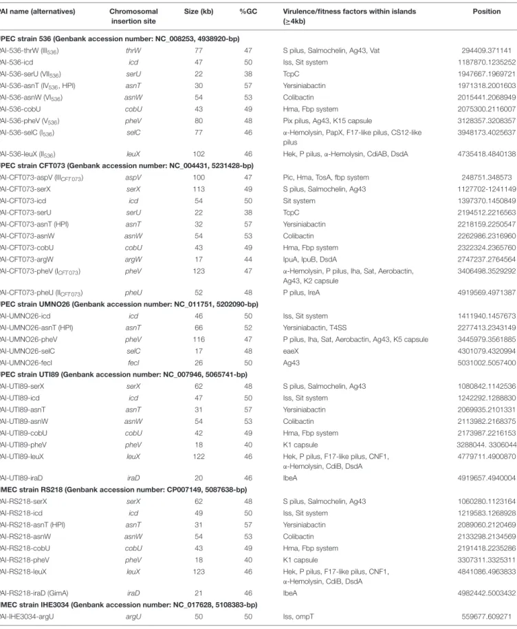
TABLE 2 | Islands predicted by IslandViewer and/or genomic comparison to E. coli K12 from sequenced strains belonging to pathotypes UPEC (536, CFT073, UMNO26, and UTI89), NMEC (RS218, IHE3034, CE10, and S88), APEC (APECO1) and AIEC (LF82, NRG857C, and UM146) and commensal strains (ED1a and HS).
PAI name (alternatives) Chromosomal insertion site
Size (kb) %GC Virulence/fitness factors within islands (≥4kb)
Position
UPEC strain 536 (Genbank accession number: NC_008253, 4938920-bp)
PAI-536-thrW (III536) thrW 77 47 S pilus, Salmochelin, Ag43, Vat 294409.371141
PAI-536-icd icd 47 50 Iss, Sit system 1187870.1235252
PAI-536-serU (VII536) serU 22 38 TcpC 1947667.1969721
PAI-536-asnT (IV536, HPI) asnT 30 57 Yersiniabactin 1971318.2001603
PAI-536-asnW (VI536) asnW 54 53 Colibactin 2015441.2068949
PAI-536-cobU cobU 43 49 Hma, Fbp system 2075300.2116007
PAI-536-pheV (V536) pheV 80 48 Pix pilus, Ag43, K15 capsule 3128357.3208357
PAI-536-selC (I536) selC 77 46 α-Hemolysin, PapX, F17-like pilus, CS12-like
pilus
3948173.4025637
PAI-536-leuX (II536) leuX 102 46 Hek, P pilus,α-Hemolysin, CdiAB, DsdA 4735418.4840138
UPEC strain CFT073 (Genbank accession number: NC_004431, 5231428-bp)
PAI-CFT073-aspV (IIICFT 073) aspV 100 47 Pic, Hma, TosA, fbp system 248751.348573
PAI-CFT073-serX serX 113 49 S pilus, Salmochelin, Ag43 1127702-1241149
PAI-CFT073-icd icd 54 50 Sit system 1397370.1450849
PAI-CFT073-serU serU 22 38 TcpC 2194512.2216563
PAI-CFT073-asnT (HPI) asnT 32 57 Yersiniabactin 2218159.2250547
PAI-CFT073-asnW asnW 54 53 Colibactin 2262986.2316960
PAI-CFT073-cobU cobU 43 49 Hma, Fbp system 2322324.2365760
PAI-CFT073-argW argW 17 44 IpuA, IpuB, DsdA 2747237.2764564
PAI-CFT073-pheV (ICFT 073) pheV 123 47 α-Hemolysin, P pilus, Iha, Sat, Aerobactin,
Ag43, K2 capsule
3406498.3529292
PAI-CFT073-pheU (IICFT 073) pheU 52 48 P pilus, IreA 4919569.4971387
UPEC strain UMNO26 (Genbank accession number: NC_011751, 5202090-bp)
PAI-UMNO26-icd icd 46 50 Iss, Sit system 1411940.1457673
PAI-UMNO26-asnT (HPI) asnT 66 52 Yersiniabactin, T4SS 2277413.2343149 PAI-UMNO26-pheV pheV 116 47 P pilus, Iha, Sat, Aerobactin, Ag43, K5 capsule 3445979.3561885
PAI-UMNO26-selC selC 17 48 eaeX 4301079.4320994
PAI-UMNO26-fecI fecI 26 50 Ag43 5031002.5057400
UPEC strain UTI89 (Genbank accession number: NC_007946, 5065741-bp)
PAI-UTI89-serX serX 62 48 S pilus, Salmochelin, Ag43 1080842.1142536
PAI-UTI89-icd icd 47 50 Iss, Sit system 1242292.1288830
PAI-UTI89-asnT asnT 31 57 Yersiniabactin 2069935.2101331
PAI-UTI89-asnW asnW 54 53 Colibactin 2113982.2168375
PAI-UTI89-cobU cobU 42 49 Hma, Fbp system 2173987.2216153
PAI-UTI89-pheV pheV 18 40 K1 capsule 3288044. 3306044
PAI-UTI89-leuX leuX 122 46 Hek, P pilus, F17-like pilus, CNF1, α-Hemolysin, CdiB, DsdA
4779711.4900870
PAI-UTI89-iraD iraD 20 46 IbeA 4919657.4940004
NMEC strain RS218 (Genbank accession number: CP007149, 5087638-bp)
PAI-RS218-serX serX 62 48 S pilus, Salmochelin, Ag43 1060280.1123164
PAI-RS218-icd icd 49 50 Iss, Sit system 1219583.1268928
PAI-RS218-asnT (HPI) asnT 31 57 Yersiniabactin 2089060.2120469
PAI-RS218-asnW asnW 54 53 Colibactin 2133298.2134569
PAI-RS218-cobU cobU 43 49 Hma, Fbp system 2191418.2235286
PAI-RS218-pheV pheV 18 40 K1 capsule 3307311.3325311
PAI-RS218-leuX leuX 123 46 Hek, P pilus, F17-like pilus, CNF1, α-Hemolysin, CdiB, DsdA
4841086.4963833
PAI-RS218-iraD (GimA) iraD 21 46 IbeA 4982442.5003432
NMEC strain IHE3034 (Genbank accession number: NC_017628, 5108383-bp)
PAI-IHE3034-argU argU 50 50 Iss, ompT 559677.609271
TABLE 2 | Continued
PAI name (alternatives) Chromosomal insertion site
Size (kb) %GC Virulence/fitness factors within islands (≥4kb)
Position
PAI-IHE3034-serX serX 62 48 S pilus, Salmochelin, Ag43 1123943.1185632
PAI-IHE3034-icd icd 48 50 Iss, Sit system 1285251.1331958
PAI-IHE3034-asnT (HPI) asnT 31 57 Yersiniabactin 2148149.2179443
PAI-IHE3034-cobU cobU 43 49 Hma, Fbp system 2252158.2294325
PAI-IHE3034-pheV pheV 18 46 K1 capsule 3441108.3459812
PAI-IHE3034-iraD (GimA) iraD 21 46 IbeA 4964058 4984404
NMEC strain CE10 (Genbank accession number: NC_017646, 5313531-bp)
PAI-CE10-ybcQ ybcQ 32 52 Iss 574012.606099
PAI-CE10-icd icd 56 52 Sit system 1285000.1341381
PAI-CE10-asnT (HPI) asnT 31 57 Yersiniabactin 2259714.2291113
PAI-CE10-cobU cobU 27 47 Hma 2309373.2336831
PAI-CE10-pheV pheV 63 46 P pilus, Aerobactin, Sat, Iha, K1 capsule 3487305.3550576
PAI-CE10-selC argW 10 42 IpuA, IpuB (DsdA) 4303582.4357800
PAI-CE10-pheU pheU 20 47 Hek, P pilus 4961804.4982152
PAI-CE10-leuX leuX 42 49 Fec, VirK, MsbB2 5127122.5169094
NMEC strain S88 (Genbank accession number: NC_011742, 5032268 bp-bp)
PAI-S88-argU argU 38 52 ompT 574349.612629
PAI-S88-icd icd 48 50 Iss, Sit system 1183142.1229738
PAI-S88-asnT (HPI) asnT 31 57 Yersiniabactin 1998347.2029756
PAI-S88-pheV pheV 74 45 Hek, P pilus, IreA, K1 capsule 3219875.3293447
PAI-S88-pheU pheU 64 48 PapX 4621378.4684940
PAI-S88-leuX leuX 39 50 Fec, Ag43 4821000.4862061
APEC strain APECO1 (Genbank accession number: NC_008563, 5082025-bp)
PAI-APECO1-thrW thrW 20 41 Tsh 295124.315041
PAI-APCEO1-icd icd 49 50 Iss, Sit system 1179544.1226126
PAI-APECO1-asnT asnT 31 57 Yersiniabactin 2090794.2122085
PAI-APECO1-pheV pheV 73 44 Hek, P pilus, IreA, K1 capsule 3305091.3378655
PAI-APECO1-iraD (GimA) iraD 21 46 IbeA 4935886.4956233
AIEC strain LF82 (Genbank accession number: NC_011993, 4773108-bp)
PAI_LF82-icd icd 49 50 Iss, Sit system 1174302 1223227
PAI-LF82-asnT asnT 31 57 Yersiniabactin 2007750 2039203
PAI-LF82-pheV pheV 18 42 K5 capsule 3110301 3128705
PAI-LF82-iraD (GimA) iraD 21 46 IbeA 4673091 4693405
AIEC strain NRG857C (Genbank accession number: NC_017634, 4747819-bp)
PAI-NRG857C-icd Icd 61 50 Iss, Sit system 1171022 1231985
PAI-NRG857C-asnT asnT 31 57 Yersiniabactin 2014814 2046266
PAI-NRG857C-pheV pheV 18 42 K5 capsule 3084523 3102935
PAI-NRG857C-iraD (GimA) iraD 21 46 IbeA 4648143 4668457
AIEC strain UM146 (Genbank accession number: NC_017632, 4993013-bp)
PAI-UM146-pheV pheV 28 46 K1 capsule 326687 354390
PAI-UM146-cobU cobU 42 49 Hma, Fbp system 1426242 1468411
PAI-UM146-asnW asnW 54 53 Colibactin 1474024 1529464
PAI-UM146-asnT asnT 31 57 Yersiniabactin 1541958 1573422
PAI-UM146-icd icd 29 53 Sit system 2353562 2382356
PAI-UM146-serX serX 62 48 S pilus, Salmochelin, Ag43 2499854 2561497
PAI-UM146-thrW thrW 20 41 Tsh 3301638 3321999
PAI-UM146-leuX leuX 121 46 Hek, P pilus, F17-like pilus, CNF-1, α-Hemolysin, CdiA, DsdA
4520979 4642133
PAI-UM146-iraD (GimA) iraD 20 46 IbeA 4660918 4681265
Commensal strain ED1a (Genbank accession number: NC_011745, 5209548-bp)
PAI-ED1a-icd icd 12 49 Sit system 1282697.1294467
PAI-ED1a-asnT (HPI) asnT 31 57 Yersiniabactin 2164139.2222325
TABLE 2 | Continued
PAI name (alternatives) Chromosomal insertion site
Size (kb) %GC Virulence/fitness factors within islands (≥4kb)
Position
PAI-ED1a-pheU pheU 44 48 Iha, Aerobactin 4837580 4881090
PAI-ED1a-leuX leuX 57 50 MsbB2, VirK, fec, Ag43 5065910.5122732
Commensal strain HS (Genbank accession number: NC_009800, 4643538-bp)
PAI-HS-hisF hisI 39 47 Klebsiella-related LPS and capsule determinants
2143083.2181967
FIGURE 3 | The position of PAI insertion sites in the non-diarrheagenic E. coli pathotypes. The name of genetic features associated with the insertion is indicated with the name of bacterial strains harboring PAI at the corresponding site. The position of the insertion site is indicated in pink for tRNA genes and in blue for coding genes. The numbers reported in the inner ring are the position (megabase) in the genome of the non-pathogenic E. coli strain MG1565 used as a reference.