Carcinogenesis vol.8 no.9 pp.1337-1341, 1987
DNA methylation in rat tissues by a series of homologous aliphatic
nitrosamines ranging from iV-nitrosodimethylamine to
A^-nitrosomethyldodecylamine
Eric von H o f eu, Ivo Schmerold1, William Lyinsky3,
Willy Jeltsch1 and Paul Kleihues1-4
'Laboratory of Neuropathology, Institute of Pathology, University of Zurich, CH-8091 Zurich, Switzerland, and 3LB1-Basic Research Program, National Cancer Institute—Frederick Cancer Research Facility, Frederick, MD 21701, USA
^Present address: Laboratory of Radiobiology, Harvard School of Public Health, 665 Huntington Ave., Boston, MS 02115, USA
4To whom reprint requests should be sent
Aliphatic iV-nitrosomethylalkylamines exhibit a remarkable organ specificity in rats, the principal targets for tumour in-duction being liver, oesophagus, urinary bladder and lung. We have determined the extent of DNA methylation in these tissues following a single oral dose (0.1 mmol/kg; 6 h sur-vival) of each of 12 homologues, ranging from N-nitro-sodimethyiamlne (Cl) to A4utrosomethyldodecylamine (C12). Methylpurines (7- and O*-methylguanine) were determined by cation exchange HPLC with fluorescence detection. Highest levels of hepatic DNA methylation were found with A'-nkrosodimethjiainine (Cl) and A'-nitrosornethylethylamine (C2), the most potent bepatocarcinogens in this series. Con-centrations of methylpurines in liver DNA decreased with in-creasing chain length for C 1 - C 5 . Administration of the higher homologues (C6-C12) caused levels of DNA methyla-tion which by themselves were considered too low to account for their hepatocarcinogenicity. In rat oesophagus, DNA methylation closely paralleled carcinogenkity, the butyl and pentyl derivatives (C4, C5) being most effective. In rat lung, the extent of DNA methylation was generally lower and there was no apparent correlation with carcinogenicity. Methyla-tion of kidney DNA also decreased with increasing chain length and was only detectable for C1-C5. In urinary blad-der DNA, methylpurines were below or close to the limit of detection. It is concluded that the initiation of malignant transformation by DNA methylation alone (through hydrox-ylation at the methylene a-carbon) could be operative for Cl in kidney and lung, for Cl and C2 in liver, and C 3 - C 5 in oesophagus. For the higher homologues, the extent of DNA methylation seems insufficient to explain the complex pattern of tissue specificity, suggesting that DNA modification other than, or in addition to, methylation may be responsible.
Introduction
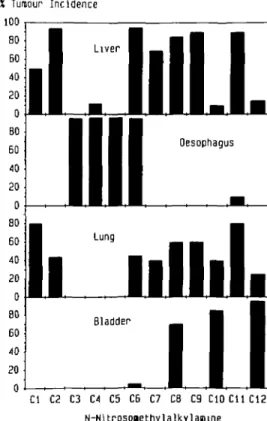
Early investigations by Druckrey and co-workers (1) on the carcinogenicity of asymmetric nitrosamines in rats revealed a high degree of organ-specificity. Short-chain N-nitrosomethyl-alkylamines (Cl, C2*) induced predominantly rumours of liver and lung whereas the higher asymmetric homologues (C3-C6) produced exclusively or preferentially oesophageal neoplasms. •Abbreviations: A'-Nrtrosornethylalkylamines were designated according to the length of the alkyl chain opposite the methyl moiety, i.e. Cl (W-nitrosodimethyl-amine) to C12 (JV-nitrosomethyldodecyl(W-nitrosodimethyl-amine).
Subsequent work by Lijinsky and co-workers on A/-nitrosomethyl-alkylamines containing an alkyl group with more than five car-bon atoms showed a striking alternating pattern, with nitrosamines containing an even-numbered carbon chain (C8, CIO, C12) pro-ducing urinary bladder tumours and those with an odd-numbered carbon chain (C7, C9, Cl 1) inducing liver and lung tumours (2,3). Investigations into the biological basis of this correlation between chemical structure and organ-specific carcinogenicity (Figure 1) have in the past focused on AZ-nitrosomethylbenzyl-amine (4), A/-nitrosomethylethylAZ-nitrosomethylbenzyl-amine (5) and JV-nitrosomethyl-pentylamine (6), and have led to the assumption that the induction of oesophageal carcinomas is related to a preferential bioactiva-tion of these nitrosamines by a P-450 isozyme in die oesophageal mucosa with high substrate specificity for asymmetric nitrosamines (4,5,7). In addition, mere was evidence that the adverse biological effects of these agents is mediated by a mediylation of cellular macromolecules. DNA modifications resulting from an initial hydroxylation of the methyl group were either not detectable (benzylation) or occurred at a considerably
X Tumour I n c i d e n c e 100 BO 60 40 20 0 B0 60 40 20 0 80 60 40 20 0 80 60 40 20 0
I
1
Liver OesophagusI
Lung Hi mill.
Bladder Cl C2 C3 C4 C5 C6 C7 C8 C9 C10C11C12 N-NitrosoaethylalkylasnneFig. 1. Maximum reported tumour incidence in various tissues of male rats exposed to /V-nitrosomethylalkylamines ranging from JV-nitrosodimethylamine (Cl) to /V-nitrosomethyldodecylamine (C12). Carcinogens were given twice weekly by gavage at a dose of 0.014 (C3), 0.021 (C4), 0.025 (Cl, C2), 0.04 (C6), 0.05 (C12) and 0.11 (C7-C11) mmol/kg. JV-Nitrosomethylpropyl-amine (C3) and /V-nitrosomethylpentylJV-Nitrosomethylpropyl-amine (C5) were administered in the drinking water at concentrations of 14 and 30 mg/1 respectively. Compounds Cl - C 4 and C6-C12 were given to Fischer 344 rats, and C5 to nus of the Donryu strain. Data were compiled from refs (3,6,10,28).
E.von Hofe et al.
lower extent (ethylation, hydroxyethylation) than did DNA methylation. This prompted us to determine the extent of DNA methylation in target and non target rat tissues produced by an equimolar dose of homologous A'-nitrosomethylalkylamines, ranging from N-iiitrosodimethylamine (Cl) to A'-nitrosomethyl-dodecylamine (C12). The study was designed to elucidate the structural requirements for bioactivation leading to DNA methyla-tion and to correlate this with the results of long-term car-cinogenicity studies.
Materials and methods
Chemicals
Nitrosamines were prepared as described previously (2,8 — 10). All nitrosamines were found to be pure and authentic as judged by n.m.r. spectroscopy. The samples were also checked by h.p.l.c. on RP-18 columns (Shandon, ODS Hypersil, 4.6 x 250 mm) with u.v.-detection at 230 nm. Using isocratic methanol/water eluents as mobile phase (10—85% methanol, depending on the hydrophobicity of the homologue), all samples eluted as a major peak followed by a smaller second peak representing the respective z-isomer.
Animals and treatment
Young male Fischer 344 rats (120 — 150 g body wt) were obtained from Charles River Wiga GmbH (FRG) and maintained on a standard laboratory diet with water ad libitum. Nitrosamines were dissolved in sterile-filtered tap water containing 0.25% Cremophor EL sdubilizer (Sigma, St. Louis, MO) and subjected to mild bath sonican'on prior to use. Carcinogens were administered by gavage in a volume of 1 ml/100 g body wt.
The time course of formation and persistence of 7-methylguanine in hepatic DNA was determined in groups of two animals receiving a single dose of 0.1 mmol/kg of C3, C6 or C12. Survival times were 3, 6, 9 and 12 h. In a further experiment, groups of 10 animals received a single dose of 0.1 mmol/lcg of each nhrosamine of the series from C1 to C12 and were killed 6 h later. Tissues were rapidly removed, frozen in liquid nitrogen and stored at - 7 0 ° C until analysis. DNA isolation and analysis of adducts
DNA was isolated by phenolic extraction and adsorption onto hydroxylapatite as previously described (5). Following mild acid hydrolysis (0.1 M HC1 at 37°C for 20 h), the amounts of 7-methylguanine and O^-methylguanine were deter-mined by h.p.l.c. using a modification of the procedure of Swenberg and Bedell (11), as previously described (12). Briefly, purine bases were separated on a strong cation exchange column (Partisil SCX, 0.46 x 250 mm), eluted at 2 ml/min with -50 mM N H ^ P O * , pH 2 (7-methylguanine), or with the same buffer contain-ing 10% (vol/vol) methanol (C^-methylguanine). Under these conditions, 7-methylguanine eluted at 9.5 min and O^-methylguanine at 8.7 min. Quantifica-tion of methylpurines was carried out with a Shimadzu spectrofluorophotometer (RF-540), set at 295 nm for excitation and 370 nm for emission. Calibration of the fluorescence signal was performed by injecting radiolabelled methylpurines and determining both radioactivity and fluorescence.
Since the amount of DNA obtained from most tissues was too small to allow for individual analyses, organs from 10 animals were pooled before DNA isola-tion. Duplicate or triplicate analyses were carried out whenever possible and were usually found to differ by less than 10%.
Results
To estimate the most suitable survival time, i.e. when >90% of the nitrosamine would be metabolized, the time course of methylpurine formation in hepatic DNA was determined for some representative A^-nitrosomethylalkylamines (C3, C6 and C12). Concentrations of 7-methylguanine produced in hepatic DNA by a single oral dose (0.1 mmol/kg) were highest between 3 —6 h. At longer time intervals, up to 12 h, there was no significant change in the amout of 7-methylguanine (Figure 2). It was, therefore, decided that 6 h would be an appropriate time inter-val for determining the extent of DNA methylation by a large series of N-nitrosomethylalkylamines. The results of the latter experiment, in which groups of 10 rats received a single oral dose of 0.1 mmol/kg, are shown in Figure 3. The greatest ex-tent of DNA methylation was observed in liver following ad-ministration of C l , with 7-methylguanine and C^-methylguanine concentrations amounting to 3290 and 330 /imol/mol guanine
uaol/iol 1?00 800 400 n ( G C3 •— C6 •- C12«-Liver ^ — — -5 Survival __ -• • 10 Ihuurs
Fig. 2. Concentration of 7-methylguanine in hepatic DNA of male Fischer
344 rats at different time intervals following a single oral dose (0.1 mmolAg) of N-nitrosomethylpropylamine (C3), Af-nitrosomethylhexylamine (C6) and A'-nitrosomethyldodecylamine (C12). Data from pooled livers of three animals for 3, 9 and 12 h and from 10 animals for 6 h. G, guanine.
unol/nol G 4000 1000 750 500 250 0 250 200 150 100 50 0 250 200 150 100 50 1—1 Oesophagus
D
LungnDD
• DD
KidneyOn
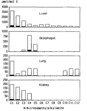
Cl C2 C3 C4 C5 C6 C7 C8 C9 CIO Cll C12 N-NitrosomethylalkylajiineFig. 3. DNA methylation in liver, oesophagus, lung and kidney of male
F344 rats following a single oral dose (0.1 mmol/kg each) of homologous A'-nitrosomethylalkylamines ranging from A/-nitrosodimethylamine (Cl) to A<-nitrosomethyldodccylamine (C12). Concentrations of 7-methylguanine (open columns) and (Amethylguanine (filled columns) are expressed as fimol/mol guanine.
respectively. Hepatic DNA methylation decreased with increas-ing length of the alkyl chain, the lowest value beincreas-ing observed for A'-nitrosomethyldodecylamine (C12). The amount of 7-methylguanine produced by C12 comprised only 5% of that produced by an equimolar dose of N-nitrosodimethylamine (Cl). Long-chain /V-nitrosomethylalkylamines (C6-C12) all produc-ed a low extent of hepatic DNA methylation (200-600 /imol 7-methylguanine/mol guanine).
In contrast, concentrations of 7-methylguanine in oesophageal DNA increased from values below the level of detection (Cl and C2) to 800 and 370 /unol 7-methylguanine/mol guanine for N-nitrosomethylbutyl- and -methylpentylamine, respectively. On-ly with C4 was the extent of methylation in the oesophagus higher than in liver. Following administration of long chain homologues
DNA methj latkm in rat tissues
(C6-C12), 7-methylguanine was not found in oesophagus. The detection limit for C^-methylguanine was somewhat lower due to its strong fluorescence, and traces of this promutagenic base ( < 5 /*mol/mol guanine) in oesophagus were detectable with all compounds with the exception of C2. The highest level of pulmonary DNA methylation (190 /tmol 7-methylguanine/mol guanine) was found after administration of /V-nitrosodimethyl-amine (Cl), but this was still 15 times lower than in rat liver. 7-Methylguanine concentrations in lung ranging from 30 to 100 /tmol/mol guanine were found with C 2 - C 5 and C10-C12, whereas equimolar doses of C6 - C9 only produced trace amounts of C^-methylguanine ( < 2 /imol/mol guanine). In kidney DNA, methylpurines were only detectable after administration of Cl — C4. As observed in rat liver, there was a tendency for a decrease in alkylation with increasing chain length. In the urinary bladder, trace amounts of O^-methylguanine were detectable after administration of C6, C9, CIO ( < 2 /imol/mol guanine) and C12 ( < 5 /tmol/mol guanine).
Discussion
Carcinogenic A^-nitroso compounds are characterized by their capacity to selectively induce a high incidence of malignant tumours in a wide spectrum of target tissues (1,3,13). The biological basis of organ-specific carcinogenesis is not yet fully understood, but several factors have been implicated; including distribution of the parent carcinogen, tissue- and cell-specific bioactivation, cell turnover and DNA repair (for review see refs 14—20). The objective of the present study was to determine the extent to which aliphatic /V-nitrosomethylalkylamines methylate cellular DNA in various rat tissues. Since car-cinogenicity data are available for all of these compounds, it was possible to correlate organ specific tumour induction with levels of methylation in target and non target tissues. We realize that for each tissue, the extent of DNA modification resulting from chronic exposure may differ significantly from that found in single dose experiments, mainly due to different rates of cell turnover and DNA repair. It is also evident that enzymic metabolism of A^-nitrosomethylalkylamines is very complex and generation of a methylating intermediate is only one of several possible end-points of bioactivation. However, we feel that despite these limita-tions, the results allow some tentative conclusions and hypotheses which may be further tested in a more comprehensive study of DNA modifications resulting from exposure to long-chain asym-metric nitrosamines.
DNA methylation is most likely to result from a single hydrox-ylation step at the a-C position of the alkyl chain opposite the methyl moiety. Metabolism can occur at other carbon atoms prior to conversion to a methylating agent but the more polar in-termediates so formed are stable and often more easily excreted than the parent nitrosamine. It is most probable that the pathway for the production of methyl adducts is initial alkyl a-C hydrox-ylation, since the resulting intermediate is very short-lived and will react rapidly with available nucleophiles. Following incuba-tion of rat oesophagus with N-nitrosomethylpentylamine (C5) in
vitro, Mirvish et al. (7) identified four hydroxy derivates, but
only those resulting from attack at C-atoms 2—5. It was con-cluded that the a-hydroxy intermediate was too short-lived for chemical detection. This view is also supported by recent studies on /V-nitrosomethylethylamine (C2) in our laboratory. Although there was evidence of extensive hydroxylation at the /3-carbon of the ethyl group, this led to only a very small amount of DNA hydroxyethylation (5). Accordingly, one would expect a high
ex-tent of methylation only with those A'-nitrosomethylalkylamines which are exclusively or predominantely bioactivated by initial a-C hydroxylation at the alkyl group opposite the methyl moie-ty. The results shown in Figure 3 suggest that this occurs for Cl and C2 in rat liver, and for C4 and C5 in the oesophagus. For the homologous series from /V-nitrosodimethylamine to N-nitrosomethylpentylamine (C1-C5), DNA methylation in rat liver was found to decrease with increasing length of the alkyl chain (Figure 3). The generally low levels of methylation pro-duced by C5 - C 1 2 indicate that the liver does not possess P-450 isozymes capable of efficiendy a-hydroxylating nitrosomethyl-alkylamines with chain lengths greater than four.
Comparing the present results widi those of long-term car-cinogenicity studies, we conclude that initiation of hepato-carcinogenesis by DNA mediylation alone can only be assumed for nitrosodimethylamine (Cl). With A'-nitrosomethylethylamine (C2), methylation is by far the primary type of adduct formed (5,21), but ethylation has also been shown to occur, although at levels four times less than would be expected if this compound acted as a true hybrid between Cl and N-nitrosodiethylamine (12). The possibility must be considered that methylation may func-tion to saturate the C^-alkylguanine-DNA alkyltransferase and thereby potentiate the effects of more minor adducts. Both Cl and C2 are strong hepatocarcinogens and produced 7-methyl-guanine and C^-methyl7-methyl-guanine concentrations of more than 2000 and 200 /imol/mol guanine/0.1 mmol nitrosamine/kg respectively. /V-Nitrosomethylpropylamine (C3) produced less than 100 /imol C^-methylguanine/mol guanine at this dose and is not known to induce liver cancer in rats. All higher homologues (C4—C12) caused substantially less DNA methylation than does C3, although several of these (C6-C9 and Cl 1) induce a high incidence of hepatic carcinomas following chronic oral ad-ministration. This would suggest that the initiation of liver car-cinogenesis by these agents requires DNA modifications other than, or in addition to, methylation.
In their pioneering work on the structure-activity relation-ships of carcinogenic nitroso compounds, Druckrey and co-workers (1) established that the most powerful oesophageal car-cinogens are asymmetric dialkylnitrosamines with a methyl group as one of the alkyl moieties. The present study on the complete series of 12 homologous aliphatic methylalkylnitrosamines of-fers closer insight into the structural requirements for oesophageal tumour induction. Methylation of oesophageal DNA by nitro-sodimethylamine was below the level of quantitative detection and this corresponds with the observation that this agent has never produced tumours in this organ, irrespective of dose and route of administration (1,22). While the next higher homologue (C2) produced very low levels of alkylation in rat oesophagus, previous radiochromatographic analyses established that following a single i.p. or p.o. dose, concentrations of 7-methylguanine in rat liver are 100 times higher than in the oesophagus (5). Nevertheless, N-nitrosomethylethylamine does produce a low incidence of oesophageal carcinomas in addition to hepatic neoplasms when administered in the drinking water (9). The present data (Figure 3) show a considerable extent of oesophageal DNA methylation (7-methylguanine/guanine) by the next higher homologues, with oesophagus/liver ratios increasing from 0.069 (C3) to 1.28 (C4) and 0.98 (C5). These three compounds are powerful oesophageal carcinogens, producing highly malignant squamous carcinomas after an induction time of only 4—6 months (3,6,10). The other compounds studied produce neither oesophageal tumours nor ex-tensive oesophageal DNA methylation, except for A/-nitroso-methylhexylamine (C6) which is a fairly potent carcinogen in
E.von Hofe et at.
this organ (10) even though its capacity to methylate target organ DNA was found to be low (Figure 3). In conclusion, the present study shows that (with the exception of C6) DNA methylation in rat oesophagus closely parallels carcinogenicity. In conjunc-tion with earlier studies (4,7,23,24), this suggests that the oesophageal mucosa of rats contains a P-450 isozyme capable of metabolizing asymmetric nitrosamines at the a-methylene posi-tion, with highest substrate specificities for those with a chain length of 3—5 C-atoms. Our data also reveal that the amount of DNA methylation required to initiate liver carcinogenesis is considerably higher than that required to produce a similar in-cidence of oesophageal cancer. The results obtained suggest that any agent which in rat oesophagus produces 2r 10 /imol Cr-methylguanine/mol guanine/0.1 mmol nitrosamine/kg is a strong and often selective oesophageal carcinogen, i.e. at levels 10—20 times lower than observed in liver DNA following ad-ministration of an equimolar dose of a potent hepatocarcinogen. Lung tumours are induced by long-term exposure to N-nitrosomethylalkylamines at incidences ranging from 30 to 80% (Figure 1), with the exception of C 3 - C 5 . The lack of pulmonary response to the latter may be due to the fact that these agents very rapidly produce fatal oesophageal carcinomas. The extent of DNA methylation following a single exposure (Figure 3) was generally low and there was no correlation with long-term car-cinogenicity. In particular, no methylpurines were detectable after administration of C6 —C8, i.e. compounds known to induce a 40—60% incidence of pulmonary neoplasms after chronic ex-posure.
Kidney DNA, too, showed levels of DNA methylation ~ 15 times lower than in liver, but shares with the latter a tendency for decreasing levels of methylpurines with increasing chain length for Cl - C 4 . All higher homologues (C5-C12) failed to produce detectable amounts of 7- and C^-methylguanine,* in-dicating a lack of P-450 enzymes capable of a-C hydroxylation if the number of C-atoms exceeds four. In carcinogenicity studies employing administration by gavage (C2 and C4) and in drink-ing water (Cl— C5), kidney tumours were not reported. The reason for this discrepancy is not known but there seems to be a general tendency for rat kidney to undergo malignant transfor-mation following pulsed doses rather than chronic exposure in drinking water (25).
The present data on DNA methylation do not explain the in-duction of liver and urinary bladder carcinomas by long-chain odd- and even-numbered N-nitrosomethylalkylamines respective-ly. We did detect small amounts ( ~ 5 junol/mol guanine) of O*-methylguanine in bladder DNA following administration of N-nitrosomethyldodecylamine (C12) and trace amounts after ex-posure to C6, C9 and C10. Since this does not correlate with the induction of bladder cancer by these agents, we assume that adducts other than methylpurines are responsible and/or the levels of methylation change after chronic administration. It is known, however, that methylation alone is sufficient to transform blad-der epithelium of rats (26). We agree with the hypothesis put forward by Okada (27) that tissue specificity depending on even or odd chain length can best be explained by the Knoop type of /3-oxidation which, in the case of even-numbered AZ-nitroso-methylalkylamines, would eventually yield TV-nitrosomethyl-carboxypropylamine, a potent bladder carcinogen in rats. If this were true, one would expect bladder tumours also after administration of A/-nitrosornethylhexylamine and A/-nitroso-methylbutylamine (C4), which should similarly be oxidized to form A/-nitrosomethylcaiboxypropylamine. The low incidence of bladder tumours produced by C6 (9) and the lack of a
carcino-genic response of this organ to C4 is probably due to the very rapid and fatal induction of oesophageal carcinomas.
Acknowledgements
We wish to thank Ms Isabelle Cackett for valuable technical assistance. This work was supported by the Swiss National Science Foundation.
References
l.Druckrey.H., Preussmann.R., Ivankovic.S. and Schmahl.D. (1967) Organotrope carcinogene Wirkungen bei 65 verschiedenen A'-Nitroso-Verbindungen an BD-Ratten. Z Krebsforsch., 69, 103-201.
2.Ujinsky,W., Saavedra,J.E. and Reuber.M.D. (1981) Induction of car-cinogenesis in Fischer rats by methylalkylnitrosamines. Cancer Res., 41,
1288-1292.
3. Lijinsky.W. (1984) Structure-activity relations in carcinogenesis by A'-nitroso compounds. In Rao.T.K., Lijinsky.W. and Epler.J. (eds) Cenotoxicology of N-Nitroso Compounds. Plenum Press, New York, pp. 189-231. 4. Hodgson,R.M., Wiessler.M. and Kleihues.P. (1980) Preferential
methyla-tion of target organ DNA by the oesophageal carcinogen A'-nitrosomethyl-benzylamine. Carcinogenesis, 1, 861-866.
5. von Hofe.E., Grahmann.F., Keefer.L.K., Lijinsky.W.; Nelson,V. and Kleihues.P. (1986) Methylation versus ethylation of DNA in target and non-target tissues of Fischer 344 rats treated with yV-nitrosometnylethylamine. Cancer Res., 46, 1038-1042.
6. Iizuka.T., Ichimura.S. and Kawachi.T. (1980). Esophageal carcinoma in rats induced by A'-amyl-A'-methylnitrosamine. Gann, 71, 94—99.
7. Mirvish,S.S., Wang,M.-Y., SmithJ.W., Deshpande.A.D., Makary,M.H. and lssenberg.P. (1985) /S- to w-Hydroxylalion of the esophageal carcinogen methyl-n-amylnitrosamine by the rat esophagus and related tissues. Cancer Res., 45, 577-583.
8. Lijinsky.W., Taylor.H.W., Mangino.M. and Singer.G.M. (1978) Car-cinogenesis of nitrosomethylundecylamine in Fischer rats. Cancer Lett., 5, 209-213.
9. Lijinsky.W. and Reuber.M.D. (1981) Comparative carcinogenesis by some aliphatic nitrosamines in Fischer rats. Cancer Lett., 14, 297—302. 10. Lijinsky.W., Reuber.M.D. and Singer.G.M. (1983) Induction of.tumors of
the esophagus in rats by nitrosomethylalkylamines. /. Cancer Res. Clin. Oncol., 106, 171-175.
ll.SwenbergJ.A. and Bedell.M.A. (1982) Cell-specific DNA alkylation and repair: application of new fluorometric techniques to detect adducts. In Magee,P.N. (eds), Banbury Report 13: Indicators of Gtnotoxxc Exposure. Cold Spring Harbor Laboratory, New York, pp. 205-220.
12. von Hofe.E. and Kleihues.P. (1986) Comparative studies on hepatic DNA alkylation in rats by /V-nitrosomethylethylamine and Af-nitrosodimethylamine plus A'-nitrosodiethylamine. J. Cancer Res. Clin. Oncol., 112, 205-209. 13. Shank.R.C. and Magee.P.N. (1981) Toxicity and carcinogenicity of N-nitroso compounds, in Shank.R.C. (ed.), Mycotaxins and Nitroso compounds: Environmental Risks. CRC Press, Boca Raton, FL, pp. 185-217. 14. Preussmann.R. and Stewart,B.W. (1984) Af-Nitroso carcinogens. In
Searle.C.E. (ed.), Chemical Carcinogens. American Chemical Society, Washington, DC, ACS Monograph 182, Volume 2, pp. 643-828. 15. Kleihues.P. and Rajewsky.M.F. (1984) Chemical neuro-oncogenesis: role
of structural DNA modifications, DNA repair and neural target cell popula-tion. Prog. Exp. Tumor Res., 27, 1-16.
16. SwenbergJ.A., Dyroff.M.C, Bedell.M.A., PoppJ.A., Huh.N., Kirstein,A. and Rajewsky.M.F. (1984) (/-EmyMeoxythymidine, but not ^-ethyldeoxy-guanosine, accumulates in DNA of hepatocytes of rats exposed continuously to diethylnitrosamine. Proc. Nail. Acad Sci. (USA), 81, 1692-1695. 17. SwenbergJ.A., Richardson,F.C, BoucheronJ.A. and Dyroff.M.C. (1985)
Relationships between DNA adduct formation and carcinogenesis. Environ. Health Perspea., 62, 177-183.
18. Saffhill.R., Margison.G.P. and O'Connor.P.J. (1985) Mechanisms of car-cinogenesis induced by alkylating agents. Biochim, Biophys. Ada, 823,
111 -145.
19. Kleihues.P. and Wicstler.O.D. (1986) Structural DNA modifications and DNA repair in organ-specific tumor induction. In Cohen,G.M. (ed.), Target Organ Toxicity. CRC Press, Boca Raton, FL, Vol. II, pp. 159-180.
20. Belinsky.S.A., White.C.M., BoucheronJ.A., Richardson,F.C, Swenberg, J.A. and Anderson,M. (1986) Accumulation and persistence of DNA adducts in respiratory tissue of rats following multiple administrations of the tobacco specific carcinogen 4-<N-rneuiyl-A'-nitrosamino)-l-(3-pyridyl)-l-butanone. Cancer Res., 46, 1280-1284.
DNA methytation in rat tissues
21. von Hofe,E., KIeihues,P. and Keefer.L.K. (1986) Extent of DNA 2-hydroxyethylatkm by A'-nitrosomethylethylamine and A'-nitrosodiethylamine in vivo. Cardnogenesis, 1, 1335 — 1337.
22. Peto.R., Gray.R., Bramom.P. and Grasso.P. (1984) Nitrosamine car-cinogenesis in 5120 rodents: chronic administration of sixteen different con-centrations of NDEA, NDMA, NPYR and NPIP in the water of 4440 inbred rats, with parallel studies on NDEA alone of the effect of age of starting (3, 6 or 20) weeks and of species (rats, mice or hamsters). In O'Neill,I.K., von Borstel.R.C, Miller.C.T. and Bartsch, H. (eds), N-Nitroso Compounds: Occurrence, Biological Effects and Relevance to Human Cancer. IARC, Lyon, IARC Scientific Publications no. 57, pp. 627-665.
23. Labuc.G.E. and Archer.M.C. (1982) Esophageal and hepatic microsomal metabolism of W-nitrosomethylbenzylamine and A'-nitrosodimethylamine in the rat. Cancer Res., 42, 3181-3186.
24. Barch.D.H. and Kuemmerle.S.C. (1967) Esophageal microsomal metabolism of A'-nitrosomethylbenzylamine in the zinc-deficient rat. Cancer Res., 44, 5629-5633.
25. Swann.P.F., Kaufman,D.G., Magee.P.N. and Mace,R. (1980) Induction of kidney tumours by a single dose of dimethylnitrosamine: dose response and influence of diet and benzo(a]pyrene treatment. Br. J. Cancer, 41, 285-294. 26. Hicks.R.M. (1980) Multistage cardnogenesis in the urinary bladder. Br. Med
Bull., 36, 3 9 - 4 6 .
27. Okada,M. (1984) Comparative metabolism of A'-nitrosamines in relation to their organ and species specificity. In O'Neill,I.K., von Borstel.R.C., Miller.C.T., LongJ. and Bartsch,H. (eds), N-Nitroso Compounds: Occur-rence, Biological Effects and Relevance to Human Cancer. IARC, Lyon, IARC Scientific Publications no. 57, pp. 401 -409.
28. Lijinsky.W., Reuber.M.D., SaavedraJ.E. and Singer.G.M. (1983) Car-cinogcnesis in F344 rats by A'-nitrosomethyl-n-propylamine derivatives. J. Nail. Cancer Inst., 70, 959-963.