HAL Id: tel-01314225
https://tel.archives-ouvertes.fr/tel-01314225
Submitted on 11 May 2016HAL is a multi-disciplinary open access archive for the deposit and dissemination of sci-entific research documents, whether they are pub-lished or not. The documents may come from teaching and research institutions in France or abroad, or from public or private research centers.
L’archive ouverte pluridisciplinaire HAL, est destinée au dépôt et à la diffusion de documents scientifiques de niveau recherche, publiés ou non, émanant des établissements d’enseignement et de recherche français ou étrangers, des laboratoires publics ou privés.
Behavioral and neural correlates of spontaneous
attentional decoupling : towards an understanding of
mind wandering
Rodrigo Antonio Henriquez Chaparro
To cite this version:
Rodrigo Antonio Henriquez Chaparro. Behavioral and neural correlates of spontaneous attentional decoupling : towards an understanding of mind wandering. Cognitive Sciences. Université Pierre et Marie Curie - Paris VI, 2015. English. �NNT : 2015PA066463�. �tel-01314225�
Université Pierre et Marie Curie
Ecole Doctorale Cerveau Cognition Comportement
Institut du Cerveau et de la Moelle Épinière & Physiological investigation of clinically normal & impaired cognition team.
Rodrigo A. HENRÍQUEZ. Ch.
Dissertation submitted for the degree of Doctor in Neuroscience.
Supervised by Prof. Paolo BARTOLOMEO
and co-supervised by Prof. Ana B. CHICA.
Publicly defended on the 9
thof November 2015
Jury:Fabrizio DORICCHI Sapienza - Università di Roma Rapporteur Juan LUPIAÑEZ University of Granada Rapporteur Michel LE VAN QUYEN Université Pierre & Marie Curie Examiner Paolo BARTOLOMEO Université Pierre & Marie Curie Examiner
Ana CHICA University of Granada Examiner
Alessandro FARNÉ Université Claude Bernard Examiner
BEHAVIORAL AND NEURAL CORRELATES OF
SPONTANEOUS ATTENTIONAL DECOUPLING:
TOWARDS AN UNDERSTANDING OF MIND
WANDERING.
ABSTRACT
Spontaneous psychological activity substantially contributes to our everyday experience of the external world. This thesis focuses on spontaneous attenuation of sensory processing in the human brain, an important determinant of attentional decoupling, which results in mind-wandering experience. Mind mind-wandering is characterized by the spontaneous rise of thoughts unrelated with the ongoing task, which varies over time and interferes with the current cognitive processes. Some sensory decoupling processes arise very early after an event, whereas others, such as selective attention processes, take place later on. Previous research mainly focused on the general effects of mind-wandering on cognitive processing, but little is known about the early sensory processes during mind wandering and about their temporal dynamics. This thesis focuses on the study of the temporal development of early sensory attenuation previous to mind-wandering experiences. Specifically, we addressed whether this sensory attenuation is a process that develops gradually or whether it is an abrupt decoupling event that consequently triggers a global cognitive change towards a mind-wandering experience. We developed a new experimental approach, inspired by the classic sustained attention to response task (SART), based on the continuous, online assessment of individual psychophysical performance. Probe questions were asked whenever response times (RTs) exceeded 2 SD from the participant’s average RT. Results showed that mind-wandering reports were generally preceded by slower RTs, as compared to trials preceding on-task reports. Specifically, we could reliably predict mind-wandering episodes based on the response time difference between the last and the second-to-last trials. Thus, mind-wandering follow an abrupt increase in behavioral variability, lasting between 2.5 and 10 seconds. In a second study, we addressed the electrophysiological correlates of the dynamics of the sensory attenuation prior to mind-wandering experiences during the performance of a modified version of the RT task. We described the modulation of the brain activity in different regions of the visual cortex during mind-wandering episodes. We found modulations of components related to early sensory processing (increase of P1 and decrease of N1), shortly before participants reported mind-wandering episodes. This suggests a decrease of neural resources related with the early visual processing of the sensory input. Mind wandering also increased the amplitude of a late component peaking around 300-500 ms, perhaps because of increased preparation of response. Overall, this thesis provides for the first time a clear description of the early temporal organization of the sensory decoupling prior to mind-wandering experiences. In particular, mind wandering appears to be strongly linked to early, abrupt sensory attenuation. Thus, short-lasting sensory decoupling (a few hundreds ms) can thus determine a prolonged and dramatic consciousness state such as mind wandering.
Keywords: Mind-wandering, sensory decoupling, event-related brain response, behavioral variability, thought sampling questions.
ACKNOWLEDGEMENTS
I am delighted to acknowledge all those who have been with me along this journey and contributed to make this thesis posible. First, I am sincerely grateful to my advisor, Dr. Paolo Bartolomeo for giving me the opportunity to start this adventure three years ago and for his invaluable support and guidance that he showed me throughout my PhD. I especially thank also to my co-advisor Dr. Ana Chica, for her invaluable assistance at all critical stages of this project with her useful and critical insights during all my PhD. I would like also to thank the committee members: Dr. Fabrizio Doricchi, Dr. Juan Lupiañez, Dr. Alessandro Farné and Dr. Michel le Van Quyen, for their kind and friendly acceptance to my request of being part of the committee. I want to express my gratefulness also to Dr. Andreas Kleinschmidt for his invaluable support in the beginning of my career. His fresh knowledge and insight have accompanied me throughout my scientific journey.
I would like to thank to Dr. Lionel Naccache and Dr. Laurent Cohen for the wonderful and fecund feedback during the "lab meeting times", their words really pushed my work in a good direction. Thank you Dimitri Bayle and Pablo Billeke for your invaluable help and for be part of this project. I would like to thank my colleagues from the PICNIC-LAB, particularly "La Nonna Stefania de Vito", Tal Seidel and Kasia for being always there for sharing with me great moments in this adventure and for becoming precious friends. I would like to extend my thanks to all current and former lab-mates: Mariam, Cami, Jaco, Florence, Sami, Clémence, Athena, J-R King, Moti and Imen for sharing scientific and extra-scientific thoughts, for the joy and for their invaluable assistance and support. I would
like to extend also my thanks to all to my close mates for the talks and laughs, particularly Julià Amengual, Toni Valero, Dimitri, José Luis, Marine Lunven, Brenda Rocamonte, Lorena Chanes and Marine Vernet. I owe sincere and earnest thankfulness to the "CIMCYC people" from Granada in particularly Ana, Juan, Joaquin, Javier, Paola, Conchi, Maria, Sonia, Carlos, Javi, Julian and Itsaso for the joy and for their invaluable assistance and support during my staying there. Thank you also to Isabel for all "the administrative things" and for your invaluable help. Overall, thank you to all the members of the ICM community. You’ve been wonderful for me in these years. I would like to thank my Chilean, Argentinian and French friends (in particularly Fernando, Nidia, Kari, Coto, Catherina, Bertsy, Pablo, Belen, Victor, Loic and Paula) for being there and my first lab in Chile (Lab de Neurociencia del Prof. Francisco Aboitiz). Finally, I would like to thank my family Ana, Pedro, Fernando and our little Simona for all love, support and for always being there. I would like to thank also Nancy and Myriam for all their invaluable love and support .
In particularly, I would like to thank to Francisca my love, for her continued support, love, patience, understanding during all our journey. Definitely this aventure without you would have been very different. Thank you.
Lastly, I would like to thank the Comisión Nacional de Ciencia y Tecnología (CONICYT) from Chile for funding my PhD project and all the persons that participated in my studies.
To my Fran,
For her love, support and strength,
To Pedro, Ana, Fernando, Nancy and Simona
TABLE
OF
CONTENTS
ABSTRACT ... 3
ACKNOWLEDGEMENTS ... 5
TABLE OF CONTENTS ... 10
TABLE OF FIGURES ... 12
CHAPTER 1. LITERATURE REVIEW ... 13
INTRODUCTION ... 14
1.1. MINDWANDERING:CONCEPTUALANDTHEORETICALISSUES. ... 14
1.1.1. Theoretical framework of mind-wandering ... 14
1.1.2. Terminology of mind-wandering ... 17
1.2. STUDYOFBRAINSIGNALSWITHELECTROENCEPHALOGRAPHY(EEG). ... 19
1.2.1. Event-related potentials ... 24
1.3. EXPERIMENTALMETHODSTOSTUDYMIND-WANDERING ... 28
1.3.1. Neurophenomenological measures ... 28
1.3.2. Behavioral measures ... 30
1.3.3. Neurocognitive measures ... 32
1.4. THENEUROANATOMICALSUBSTRATESOFMIND-WANDERING ... 34
1.4.1. Dorsal and ventral attention networks ... 34
1.4.2. Default mode network and mind-wandering ... 37
1.5. TOWARDFINDINGTHENEURALCORRELATESOFMIND-WANDERING ... 44
1.5.1. Task-evoked responses and mind-wandering ... 44
1.5.2. Brain rhythms and mind-wandering ... 47
CHAPTER 2. OUTSTANDING QUESTIONS ... 49
2.1. AIMSOFTHETHESIS ... 50
CHAPTER 3. EXPERIMENTAL CONTRIBUTION. ... 53
INTRODUCTION TO THE FIRST ARTICLE ... 54
3.1. FLUCTUATINGMIND:SPONTANEOUSPSYCHOPHYSICALVARIABILITY DURINGMIND-WANDERING. ... 55
3.1.1. Abstract ... 55
3.1.3. Materials and methods. ... 58
3.1.4. Results. ... 61
3.1.5. Discussion. ... 68
3.1.6. Author Contributions. ... 70
3.1.7. References. ... 71
INTRODUCTION TO THE SECOND ARTICLE ... 76
3.2. BEHAVIORALANDNEURALCORRELATESOFSENSORYATTENUATION:AN ERPSTUDYOFNEURALEVENTSLEADINGTOMINDWANDERING. ... 77
3.2.1. Abstract ... 77
3.2.2. Introduction ... 78
3.2.3. Materials and methods ... 81
3.2.4. Results. ... 88
3.2.5. Discussion. ... 95
3.2.6. Author contributions. ... 99
3.2.7. References. ... 100
CHAPTER 4. ... 109
GENERAL DISCUSSION AND CONCLUSION ... 109
4.1. GENERAL DISCUSSION. ... 110
4.1.1. Synthesis of the main results. ... 111
4.1.2. Behavioral variability of mind wandering. ... 112
4.1.3. Neural correlates of mind wandering. ... 114
4.1.4. Mind wandering and executive control. ... 116
4.1.5. Mind wandering and attentional regulation. ... 118
4.1.6. Mind wandering and the information processing model. ... 120
4.2. LIMITATIONS AND PERSPECTIVES FORFUTURE RESEARCH. ... 122
4.2.1. General limitations. ... 122
4.2.2. Implications for future research. ... 123
4.3. CONCLUSIONS. ... 124
Table of figures
Figure 1 Hans Berger and his early EEG recording from the 1930s. ________________ 19 Figure 2 The origins of the EEG signal in the human visual cortex. ________________ 20 Figure 3 The acquisition of the EEG signal with electrodes on scalp. ________________ 22 Figure 4 Fronto-parietal networks in human right hemisphere. _____________________ 35 Figure 5 Cortical representation of the six RSNs. _______________________________ 41 Figure 7 The hypothetical functional architecture proposed for mind-wandering. ______ 42 Figure 8 Schematic depiction of the experimetal paradigm. _______________________ 60 Figure 9Distribution of slow RTs (> 2SD) across blocks. _________________________ 63 Figure 10 Distribution of mean RTs for the five trials before TSQ. _________________ 64 Figure 11Log-log power spectra of time series of RT variation. ____________________ 68 Figure 12. Distribution of mean RTs for the five trials before TSQ. ________________ 75 Figure 13 Schematic depiction of the EEG experimental paradigm. _________________ 83 Figure 14 Mean RTs for off-tak and ont-task conditions for the last 5 trials prior to the TSQ. __________________________________________________________________ 89 Figure 15 Grand average ERP waveform, for all trial position (from N-1 to N-5), for the off-task and on-task condition in the selected ROI. ______________________________ 91 Figure 16 Average ERP waveform and topographic power distribution for trial position N-1 and N-2 for on-task and off-task condition in the selected ROI. ____________________ 92 Figure 17 Average ERP waveform and topographic power distribution for trial position N-3 and N-5 for off-task and on-task condition in the selected ROI. ____________________ 94
Table 1 Percentages of responses reported by participants in the TSQs. ______________ 61 Table 2 Mean RT and standard deviation for trials N-1 to N-5 before the TSQ. ________ 65 Table 3 Logistic regression analysis of off-task and on-task condition for the last five trials before TSQ. ____________________________________________________________ 66 Table 4 Logistic regression analysis for off-task and on-task conditions for the last five trials before the TSQ. _____________________________________________________ 90
INTRODUCTION
1.1. MIND WANDERING: CONCEPTUAL AND
THEORETICAL ISSUES.
1.1.1.
Theoretical framework of mind-wandering
One of the most prominent features of ongoing perceptual activity is the fact that it varies over time spontaneously (Busch & VanRullen, 2010; Dehaene, Changeux, Naccache, Sackur, & Sergent, 2006; Fox, Corbetta, Snyder, Vincent, & Raichle, 2006; Hesselmann, Kell, Eger, & Kleinschmidt, 2008; Kleinschmidt, Büchel, Zeki, & Frackowiak, 1998; Laufs et al., 2003; Sadaghiani, Hesselmann, & Kleinschmidt, 2009). When we pay attention to this text, as well as when we watch a movie or read a newspaper, our mental activity constantly fluctuates in a spontaneous way. However, only sometimes we are able to be aware of the internal streams of our thought, detecting that our minds have wandered (Christoff, Gordon, Smallwood, Smith, & Schooler, 2009; Schooler et al., 2011; Smallwood, Baracaia, Lowe, & Obonsawin, 2003). This particular experience, referred to as mind wandering, is defined as a spontaneous mental event that arises without any external precedent, which often interferes with the online processing of sensory information. It is associated with the attentional decoupling from the environment and lower level of alertness (Braboszcz & Delorme, 2011; Schooler et al., 2011; Smallwood & Schooler, 2006).
This spontaneous feature of the mental phenomenon of mind wandering indicates that fluctuations in human perception, which could affect many cognitive processes such as reasoning, decision-making, selective attention, and emotional processes, among others, is composed of two fundamental aspects (Schooler et al., 2011). The first aspect is the ability to be aware of the contents of consciousness, also known as meta-awareness. The second important aspect is the capacity to disengage attention from perception, known as perceptual decoupling (Smallwood & Schooler, 2015; Smith et al., 2006).This perceptual decoupling is related to the internal spontaneous thoughts that interfere mainly with the sensorial encoding of external information (Smallwood et al., 2003).
The classical research on mind wandering has mainly focused on the meta-awareness aspects of this phenomenon (Christoff et al., 2009). Consequently, the second aspect of mind wandering, i.e. the perceptual decoupling, is less understood. There are only a few empirical studies on the behavioral and neural bases of the emergence of spontaneous attentional shifts towards an internal train of thought.
Previous research on perceptual decoupling has demonstrated that the interference in the coupling between sensory processing and conscious perception is commonly associated with mental events that arise without any external precedent (Christoff et al., 2009;Mason et al., 2007). The research has focused on the neural-correlates underlying cognitive activity in the absence of an explicit task or related to what is known in the mind-wandering research as stimulus-independent thought (SIT i.e streams of thoughts and images unrelated to immediate sensory input) by using resting state paradigms (Christoff et al., 2009; Christoff, 2012; Gilbert, Frith, & Burgess, 2005; Mason et al., 2007; Teasdale et al., 1995). These neurophysiological findings suggested that internally generated thoughts and its
associated neural processes are negatively correlated with concurrent perceptual input processing (Schooler et al., 2011). In other words, the attentional decoupling activity related to internal spontaneous thoughts interferes with the encoding of external information (Smallwood et al., 2003).
For instance, studies of reading comprehension (Reichle, Reineberg, & Schooler, 2010; Reichle, 2006;Smallwood, 2011) indicate that if attention is decoupled from perception by spontaneous independent thoughts, then reading comprehension will be affected. Indeed, a negative correlation was observed between mind wandering and comprehension accuracy (Reichle et al., 2010; Schooler et al., 2011).
In support of this evidence, recent studies showed that spontaneous independent thoughts also affect patterns of gaze duration during reading (Reichle et al., 2010). In a reading comprehension study, researchers recorded participants’ eye movements while they were reading a book. They identified intervals of normal reading using variables that are known to modulate fixation durations (e.g., word frequency) and observed that during episodes of stimulus independent thoughts (mind-wandering), patterns of gaze were affected. These results suggested that the coupling between the mind and the text during normal reading breaks down during mind-wandering episodes.
1.1.2.
Terminology of mind-wandering
Until today, mind-wandering seems to be an ill-defined concept. A clear terminology is missing concerning the conceptual bases of self-generated thoughts, and their distinction from task-unrelated thoughts and goal-directed tasks.
In the last decades, many different terms have been used to describe this phenomenon, such as temporary attentional lapses (Weissman, Roberts, Visscher, & Woldorff, 2006), stimulus independent thought (Teasdale et al., 1995), incidental self-processing (Gilbert et al., 2005), task-unrelated images (Giambra, 1989), inner speech (Morin, 2009), task-unrelated thoughts (Smallwood et al., 2003), spontaneous thoughts (Christoff et al., 2009), and mind wandering (Mason et al., 2007), among others.
More recently, some researchers have addressed this lack of accuracy in the definition of mind wandering and have proposed a definitory criteria to mainly distinguish between the most common terms for this phenomenon, such as spontaneous thoughts, stimulus independent thoughts, and mind wandering (Christoff, 2012). The criteria helping to define more precisely this spontaneous phenomenon are: intentionality, relation to the task, and current sensory information.
However, the different categories still remain fuzzy given that the mind-wandering episode can be triggered by an environmental stimulus or by stimulus independent thoughts (spontaneous internal thoughts). Thus, a clear taxonomy that would allow defining the mind wandering experience is still lacking. Consideration of the context and consequences
of mind-wandering episodes may help reaching a more precise definition of these phenomena.
For example, several studies (Killingsworth & Gilbert, 2010; Poerio, Totterdell, & Miles, 2013; Smallwood, Fitzgerald, Miles, & Phillips, 2009) have shown that mind-wandering experiences are accompanied by changes in mood such as feelings of dissatisfaction related to the unaccomplished goals of the moment (Poerio et al., 2013; Smallwood et al., 2009; Smallwood, O’Connor, Sudbery, & Obonsawin, 2007). This can be explained by the fact that mind wandering occurs only when performing a task and consequently may affect the individual’s perception of his/her own performance (Smallwood & Schooler, 2006).
1.2. STUDY OF BRAIN SIGNALS WITH
ELECTROENCEPHALOGRAPHY (EEG).
Hans Berger developed the electroencephalography (EEG) technique in 1929 and obtained the first evidence for the oscillatory nature of brain activity (Karbowski, 2002; Tudor, Tudor, & Tudor, 2005) (Figure 1). Since then, this oscillatory neural activity increasingly gained relevance and nowadays is considered an essential neural mechanism, that underlies all aspects of human cognition, be it in its pathological or healthy states.
Figure 1 Hans Berger and his early EEG recording from the 1930s. (From Wikipedia)
Since these earliest observations, neuroscientists have aimed at understanding how the human brain coordinates the dynamical neural interactions that are essential for the emergence of cognition. Developments in human neuropshysiology such as EEG and magnetoencephalography (MEG), have enabled to measure neural activity directly on the scalp at a millisecond time scale, thereby allowing the temporal precision required to access the fine neural mechanisms of the human brain.
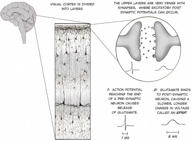
Using this methodology, brain signals can be acquired non-invasively through electrodes to measure neural activity directly on the scalp. These methods have the best possible temporal resolution for non-invasive recordings, and provide the most direct correlate of on-line brain processing. The brain electrical activity recorded comes from the summed activation of millions of excitatory post-synaptic potentials (EPSP) at dendrites of superficial cortical layers of the brain, see Figure 2 (Caspers, Speckmann, & Lehmenkühler, 1980).
Figure 2 The origins of the EEG signal in the human visual cortex. (from the Backyard Brains website).
Action potentials, that last a few milliseconds, trigger the release of neurotransmitters that bind to the post synapse, thereby eliciting these EPSPs (Creutzfeldt, Watanabe, & Lux, 1966). Specifically, the measured post-synaptic currents come from apical dendrites of pyramidal cells, typically active for a few milliseconds after the onset of the synaptic input (Figure 2). These physiological components are the main contribution to EEG and MEG signals (Buzsáki, 2006). These generators of the EEG and MEG signals are known as voltage gradient or primary currents. It causes secondary currents in the surrounding conducting medium. Thus, the EEG signals result from secondary currents propagation, in contrast to the MEG signal (magnetic fields) that is produced by both primary and secondary currents (Buzsáki, 2006) .
Therefore, if many EPSPs are occurring at the same time and in same area, such as when our eyes are stimulated by a visual stimulus and the visual cortex neurons respond to it in synchrony, we can observe the summation of these EPSPs in the EEG signal (Figure 3).
Figure 3 The acquisition of the EEG signal with electrodes on scalp. Taken from the Backyard Brains website.
The brain response after the presentation of a visual stimulus is usually too weak to be detected, making the results obtained using a single event debatable. Thus, to optimize this weak measured brain signal and to obtain an estimate of the brain response, it is necessary to average over many similar trials performed, a method known as event-related potential (ERP) (Luck, 2005). However, even using ERPs, EEG still has several main limitations related to its low spatial resolution.
EEG does not offer direct information about the location of the brain sources underlying the potentials, mainly due to the distance of the sources that generate the potentials from the measuring electrodes. This spatial resolution also depends on the density of the measuring electrodes on the scalp, which is in the order of 1 electrode per few centimeters (Kaiser, 2005; Lopes da Silva, Gonçalves, & De Munck, 2010). Therefore, this method clearly
offers a lower spatial resolution compared with other neuroimaging techniques, such as functional MRI.
Moreover, there are other limitations, related to the fact that ERP generated by different and distant brain areas can be recorded by a single electrode. Therefore, the final signal recorded from the array of electrodes mounted is a mixture of the electrical activity originated in many possible brain areas, each one with their own dynamics (Buzsáki, 2006). Additionally, there is a variability in the electrical properties of the brain layers that separate the cortical sources and the electrodes, which further contribute to the variability of the recorded signal (Buzsáki, 2006; Buzsáki & Draguhn, 2004). Furthermore, there are many additional sources of noise external to brain activity, such as the contraction of facial muscles, heartbeat, and eye movements that can affect the EEG data.
This is mainly due to the EEG signal’s high sensitivity to noise, which necessitates a meticulous approach to data processing, as well as a considerable amount of trials in order to extract only the parts of the signal related to brain activity (Luck, 2005). Thus, data are usually cleaned and divided in segments where time zero is defined habitually as the onset of the stimulus of interest. These time segments are averaged together, either across stimuli or in sub-groups of stimuli that will be compared to each other.
Therefore, the brain responses are time-locked to the presentation of the stimulus and thus can be detected in the average as an ERP.
1.2.1.
Event-related potentials
The ERP waveform consists of a series of positive and negative voltage deflections, each one called “component”, which are labeled depending on their orientation -positive or negative - and latency (100ms as (1)- 200ms as (2)- 300ms as (3), etc).
One of the first reported ERPs, described by Kornhuber and Deecke (1965), was the readiness potential (RP), also named Bereitschaftpotential (BP), associated to the preparation and execution of self-paced movements (Kornhuber & Deecke, 1965; Luck, 2005).
However, before I start describing the remainder of ERP components, it is important to clarify how their orientation is labeled (positive or negative). For instance, when using ERP labels such as P1 and N1, one must be careful not to assume that these labels are linked somehow to the nature of the underlying brain activity. This means that sensory ERPs from different modalities that have the same label are not usually related in any functional manner. For example, the auditory P1 and N1 components bear no particular relationship to the visual P1 and N1 components. Furthermore, even the modality-independent component P3 has modality-specific subcomponents (Luck et al., 1994). This problem even exists within a single modality, as noted by Steven Luck (Luck, 2005): “a component labeled N2
in one experiment may not be the same as a component labeled N2 in another experiment”.
Keeping that in mind, in the visual sensory domain, the first ERP component that is commonly reported in cognitive neuroscience is the C1 (Clark, Fan, & Hillyard, 1995; Jeffreys & Axford, 1972). However, unlike most other components, it is not labeled with a P or an N because its polarity can vary. This early ERP component appears typically around
40–60 ms poststimulus (after stimulus onset), peaks 80–100 ms poststimulus, and is probably generated in area V1 (primary visual cortex mainly in the calcarine fissure) (Luck, 2005). The C1 component is highly sensitive to stimulus parameters, such as contrast and spatial frequency.
The orientation of C1 seems to depend on the retinotopic position of the stimulus in the visual field. If the stimulus is presented in the lower visual field, activating parts of V1 above the calcarine fissure, the C1 will be positive. Similarly, C1 will be negative for stimuli presented in the upper visual field (Luck, 2005). However, if the stimuli are presented on the horizontal midline of the visual field, the C1 component will be small or positive causing it to summate with the P1 into a single component.
The C1 component is followed by the P1 component that is sensitive to variations in stimulus parameters, the direction of spatial attention, and to the subject’s state of arousal (Hillyard, Vogel, & Luck, 1998). The P1 latency is typically around 60–90 ms post-stimulus, with a peak between 100–130 ms after the onset of stimuli. It is typically observed at lateral occipital electrode sites, as would be expected given its likely origins in extrastriate visual cortex (Luck, 2005).
The P1 is followed by the N1 component. However, there are several visual N1 subcomponents: One N1 subcomponent with a peak 100–150 ms post-stimulus in anterior electrode sites, and two posterior N1 components, one from parietal cortex and the second from lateral occipital cortex around 150–200 ms. This lateral occipital N1 subcomponent is larger when subjects are performing discrimination tasks than when they are performing detection tasks, which has led to the proposal that this subcomponent reflects discriminative
processing of some sort (Hopf, Vogel, Woodman, Heinze, & Luck, 2002; Vogel & Luck, 2000).
Two other relevant negative visual components, which are seen in a latency of 150 and 200 ms at central midline sites, are the commonly called N170 and the vertex negative potential (Jeffreys, 1989). These components are related to faces and non-face stimuli processing, respectively. However, more recent studies have found that faces elicit a more negative potential than non-face stimuli at lateral occipital electrode sites, especially over the right hemisphere, with a peak at approximately 170 ms (Bentin, Allison, Puce, Perez, & McCarthy, 1996; Rossion et al., 1999).These results suggest that the N170 and the vertex positive potential are just the opposite sides of the same dipole.
A later component is the P2 component, localized in anterior and central scalp sites. This ERP component occurs only when the target is defined by fairly simple stimulus features, and when the targets are relatively infrequent (Luck et al., 1994). Nevertheless at posterior sites, the P2 wave is often difficult to distinguish from the overlapping wave of the N1 and P3 components. Consequently, not much is known about the posterior P2 component (Luck, 2005).
One of the most prominent ERP components is the called P3, which peaks around 300 ms in auditory modality and at around 400 ms in visual modality. The P3 component is a complex of 2 components, a frontal P3a component and a parietal P3b component. Both components are elicited by unpredictable, infrequent, unexpected, unusual stimuli, shifts in tone or pitch, or surprising task-irrelevant stimuli within an attended stimulus stream. The P3b component is present only when the change is task-relevant but it is not clear whether
this response is related to the P3a component. Other studies have shown that an unexpected, unusual, or surprising task-irrelevant stimulus within an attended stimulus train will elicit a frontal P3a (Courchesne, Hillyard, & Galambos, 1975; Polich & Comerchero, 2003; Soltani & Knight, 2000).Still, several studies provided evidence that the P3b component is observed for targets that are infrequent but are in some sense expected, whereas the frontal P3a wave is elicited by stimuli that are truly unexpected or surprising (Luck, 2005).
Finally, there’s a component called the contingent negative variation (CNV), related to motor preparation (Walter, Cooper, Aldridge, McCallum, & Winter, 1964). It is characterized by a broad negative deflection between a warning stimulus and a target stimulus around 500 or 1000 ms, and then returns to the baseline.
Finally it is noteworthy that in the literature other components have been also described for the different perceptual modalities, which are not reported in the present manuscript. The fundamental reason for this decision is because for this thesis, one of the experimental aims is study those components, particularly relevant for the sensory visual processing.
1.3. EXPERIMENTAL METHODS TO STUDY
MIND-WANDERING
1.3.1.
Neurophenomenological measures
The relevance of considering the constitution of “human experience from the point
of view of the subject himself ” (Varela 1999d, pag 327), as a scientific observation, is key
to try to delimit and correlate any brain activity change with introspection. This consideration helps conceptualizing the relationship between physical phenomena, such as brain processes, and self experience of mental states or phenomenal consciousness (Brockman & Varela, 1995; Thompson, 2004; Varela, 1999; Varela, 1996).
From this point view, in the mid-1990s, Francisco J. Varela initiated a scientific program called Neurophenomenology, which aimed at addressing the integration of first-person data with neuroimaging data (Christoff, Cosmelli, Legrand, & Thompson, 2011; Lutz, Lachaux, Martinerie, & Varela, 2002; Lutz & Thompson, 2003; Rosch, 2003; Rudrauf, Lutz, Cosmelli, Lachaux, & Le Van Quyen, 2003; Varela, 1997; Varela, 1999; Varela, 1996).
This proposal is oriented to solve the explanatory gap between neurobiological and phenomenological activity of consciousness (Rudrauf et al., 2003) such as can be try to understand the biological foundation of mind wandering experience. In other words, this method is a heuristic strategy for describing and quantifying the physiological processes relevant to consciousness (Lutz & Thompson, 2003). Neurophenomenology provides a
rigorous methodology in the acquisition of first-person data underlying any mental experience, related to our brain activity.
Therefore, at a methodological level, this approach intends to obtain richer first-person data through systematic phenomenological explorations of experience, for instance via experience sampling questions (Kahneman, Krueger, Schkade, Schwarz, & Stone, 2004). Furthermore, it is also oriented to use this original first-person data to uncover new third-person data about the physiological processes that are crucial for consciousness (Lutz & Thompson, 2003; Lutz, 2002).
Thus, it is necessary to define phenomenological experience with the use of “first-person methods” for increasing participants’ sensitivity to their own lived experience, i.e improve
the ability to be aware of yourself (Depraz, Varela, & Vermersch, 2003;Varela & Shear,
1999). In this way, the “phenomenal invariants” may be described at a phenomenological level, the “lived experience” (such as perception, action, memory, mental imagery, emotion, attention, empathy, self-consciousness, contemplative states, dreaming, among others). This experience can be verbally articulated and identified in first-person reports (Varela & Shear, 1999).
Therefore, a possible approach to quantify the first-person data, related with mind wandering, is to combine self-catching measures of the mind-wandering phenomenon with experience sampling probes across time, for instance by asking participants thought sampling questions (TSQs) about their state of mind, while performing a sustained attention task (Robertson, Manly, Andrade, Baddeley, & Yiend, 1997; Schooler et al., 2011; Smallwood, Baracaia, Lowe, & Obonsawin, 2003). This measure provides a
straightforward assessment of the number of mind-wandering episodes through first-person reports. Therefore with this methodology, we have the possibility to objectively quantify the relative amount of mind wandering that the participants are aware of.
1.3.2.
Behavioral measures
Several lines of research suggest that mind-wandering episodes are associated with behavioral costs, such as a particularly variable task performance (Kam et al., 2012; Seli, Cheyne, & Smilek, 2013; Smallwood, McSpadden, Luus, & Schooler, 2008; Smallwood et al., 2003; Smallwood & Schooler, 2015; Smilek, Carriere, & Cheyne, 2010; Weissman, Roberts, Visscher, & Woldorff, 2006). Behavioral variability can relate to mind wandering because mind wandering reflects the removal of executive control resources from the task at hand (Cheyne, Solman, Carriere, & Smilek, 2009; Christoff, Gordon, Smallwood, Smith, & Schooler, 2009; Smallwood, Baracaia, Lowe, & Obonsawin, 2003; Smallwood & Schooler, 2006, 2015; Teasdale et al., 1995).
For instance, recent findings showed that brain areas associated with executive control (i.e., the anterior cingulate cortex and the dorsolateral prefrontal cortex) show a negative
correlation between off-task thought and executive control abilities, suggesting that
executive resources are disengaged from the current task during mind-wandering (Christoff et al., 2009; Mason et al., 2007; Smallwood, Brown, Baird, & Schooler, 2012). Removal of executive resources could lead to increased behavioral variability, particularly during situations in which executive resources are needed to maintain a consistent mode of responding. Consequently, a considerable number of studies have emerged in order to
explore the behavioral costs of mind-wandering, as the absent-minded episodes
(Smallwood et al., 2007).
In many mind wandering studies, the behavioral measure of the failures in the ability to maintain online discrimination of the stimuli has been the most used method to assess these absent-minded episodes as mind-wandering. This assessment is used, for instance, while performing a Go/No-go task, known as the Sustained Attention to Respond Task (SART) (Robertson et al., 1997).
In the classic SART, participants are instructed to respond to a sequential series of digits (e.g, 1 to 9) and to withhold a response when an infrequent NO-GO digit appears (e.g., “3”). Classic mind-wandering studies evaluate the failure to inhibit infrequent responses, related to first-person mind-wandering reports.
Another commonly used behavioral measurement of mind wandering is the recording of eye movements (Schoole, Reichle, Halpern, 2004). For example, studies of reading comprehension (Reichle et al., 2010) have revealed that mind wandering affects both patterns of gaze movements and fixation durations (Reichle, 2006). Thus, behavioral measurements can provide objective tools to quantify and measure spontaneous mind wandering.
1.3.3.
Neurocognitive measures
The neurocognitive measures explore the link between mind wandering episodes and the amplitude of neural responses that occur in response to task events. These measures focus specially on amplitude variations of task-evoked responses (Schooler et al., 2011).
Although the mind-wandering research should not restricted to the description of fluctuations in the amplitude of the ERPs, also is possible explore the mind-wandering effect in latency, connectivity, frequency, among other measures.
Evidence shows that attention to an external task maximizes the amplitude of event related potentials (ERPs) as compared to moments of mind wandering (Schooler et al., 2011; Smallwood & Schooler, 2015), which it is characterized by a decrease ERP amplitude (Barron, Riby, Greer, & Smallwood, 2011; Braboszcz & Delorme, 2011).
For instance, during SART (Robertson et al., 1997), ERPs related to mind-wandering episodes (“off-task” responses) show a smaller amplitude in a visual component around 300 ms after stimulus onset (P3), than during “on-task” episodes (Barron et al., 2011; Smallwood, Beach, Schooler, & Handy, 2008). Likewise the amplitude of ERPs associated with sensory-level processing of auditory information (auditory N1) is reduced during mind wandering episodes (Braboszcz & Delorme, 2011; Kam et al., 2011). However, little is known about the temporal distribution of mind wandering in visual sensory processing, because the mind-wandering studies mainly have focused on the effects of mind wandering
on cognitive processing, such as decreased subjective awareness related to amplitude modulation in P3 component (Smallwood, Beach, Schooler, & Handy, 2008).
These observations are compatible with the modulations observed in the so-called “default mode network” (DMN, including the posterior parietal cingulate, the medial prefrontal cortex and the medial temporal lobes), which exhibits high neural activity when individuals do not have to perform, any demanding tasks (i.e, in rest condition) (Christoff et al., 2009; Mason et al., 2007; Smallwood et al., 2012). Likewise, a causal role of the dorsolateral prefrontal cortex has also been proposed for the development of mind wandering (Axelrod, Rees, Lavidor, & Bar, 2015). This region has been related to control functions in the production of wandering mind.
Combined evidence from these three sources, self-report, behavioral measures, and neurocognitive measures, can thus be used to make inferences about mental states, and is hopefully advancing our understanding of the stream of consciousness (Smallwood & Schooler, 2015).
1.4. THE NEUROANATOMICAL SUBSTRATES
OF MIND-WANDERING
1.4.1.
Dorsal and ventral attention networks
Mind wandering is a ubiquitous phenomenon of our mental life that is related to perceptual decoupling of the immediate external environment. In another words, mind-wandering episodes disconnect the perception of external stimulus events in a regular and periodic way (Kam & Handy, 2013). Thus in the cognitive neuroscience has been growing an big interest in understanding the impact of this perceptual decoupling related to mind wandering in the processing to stimuli in the external environment (Barron et al., 2011; O’Connell et al., 2009; Smallwood et al., 2008; Stawarczyk, Majerus, Maquet, & D’argembeau, 2011). Mind wandering is now being recognized as a new expresion of attentional selection, due to this specific feature to disconnected the attention the outside world, such that we no longer select the external stimuli for sensory processing. Therefore, in the last few decades, great progress has been made in the understanding of the neural underpinnings of spatial attention related to mind wandering. In humans, functional MRI (fMRI) has revealed that spatial attention is comprised of two fronto-parietal attention networks (Corbetta, Patel, & Shulman, 2008; Corbetta & Shulman, 2002). A dorsal attentional network (DAN) includes the intra-parietal sulcus (IPS)/superior parietal lobule and the frontal eye field (FEF)/dorsolateral prefrontal cortex. The ventral attention network
(VAN) comprises the temporal-parietal junction (TPJ) and the ventral prefrontal region (inferior and middle frontal gyrus). Importantly, the DAN is thought to be bilateral and symmetric, whereas the VAN is strongly lateralized to the right hemisphere (Corbetta et al., 2008). These fMRI results are based on variants of the classic Posner location-cueing paradigm (Posner, Walker, Friedrich, & Rafal, 1984), often used in the study of spatial attention. In this paradigm, the presentation of a visual target is preceded by a cue, and participants are required to respond to the target and not to the cue (which can or cannot be predictive of one or more of the targets features).
It is also important to highlight, that the DAN is active during the orienting period between cue and target, while the VAN shows an increase response when participants have to respond to uncued (and thereby sometimes unexpected) targets (Corbetta et al., 2008). (Figure 4).
Figure 4 Fronto-parietal networks in human right hemisphere. a) Attentional networks in the right hemisphere, according to Corbetta and Shulman (Corbetta & Shulman, 2002). b) Long range axonal connections between the attention networks (SLF 2) and within them (SLF 1 & SLF 3) (Thiebaut de Schotten et al., 2011).
Nevertheless, the exact division of labor between DAN and VAN and their dynamics in humans is still under debate (Macaluso & Doricchi, 2013; Vossel, Geng, & Fink, 2014). An influential hypothesis stipulates that the DAN operates when attention is oriented voluntarily towards a stimulus, while the VAN modulates DAN activity when an unexpected but relevant stimulus appears and grabs attention (Corbetta et al., 2008).
However, despite of this detailed description of the anatomical and functional architecture of attentional processing, the role of this set of anatomical regions during mind-wandering is still far from being clear. This is mainly because the wandering mind besides being linked to a disruption of selective attention has also been associated with the default mode network (DMN) (Chirstoff et al., 2009; Raichle et al., 2001).
1.4.2.
Default mode network and mind-wandering
In the last decade, one of the focuses of interest for neuroscientists have been study the human brain intrinsic properties to generate and sustain an internal stream of thoughts unrelated to the external world specifically the interest has focused on the neuroanatomy related to mind-wandering. Prior studies have reported increased activity in a coordinated system of brain regions, later dubbed as the “default mode network” (DMN), occurring when individuals do not have to perform demanding perceptual tasks (Raichle et al., 2001).
The brain areas of this network include the ventral medial prefrontal cortex (vMPFC), the posterior cingulate cortex (PCC), the inferior parietal lobule (IPL), the dorsal medial prefrontal cortex (dMPFC), and the hippocampal formation (HF) (for a review see Buckner, Andrews-Hanna, & Schacter, 2008). Evidence also suggests that the DMN is a coherent system, because this set of brain regions shows an intrinsic functional correlation between each other, and are connected via direct and indirect anatomic projections. Therefore two subsystems, organized as hubs of functional conection, have been proposed within the DMN: 1) the PCC, and 2) the MPFC. Moreover, the dMPFC and HF are also both correlated with other regions of the DMN. It is suggested that the HF and dMPFC are two distinct subsystems connected to the two hubs of a larger DMN (Buckner et al., 2008). However, despite the DMN being associated with stimulus-independent thoughts (Buckner et al., 2008), it is still not clear the specific role of DMN during mind-wandering. This is probably because DMN activity cannot be attributed solely to spontaneous thought (Raichle, 2009; Raichle & Snyder, 2007). For instance, seizure episodes in epileptic
patients also coincide with a sudden increase in the activity of the DMN, suggesting a possible link between this network and the onset of crises (Broyd et al., 2009; Ossandon et al., 2011).
Furthermore, spontaneous activity measured with blood oxygen level-dependent (BOLD) during fMRI in the resting awake or anesthetized brain, is organized in multiple highly specific functional anatomical networks, called resting state networks, RSNs (Biswal et al., 2010; Mantini, Perrucci, Del Gratta, Romani, & Corbetta, 2007). These RSNs show fluctuations at frequencies between 0.01 and 0.1 Hz, and exhibit strong patterns of coherence within known brain systems (Raichle & Snyder, 2007). For example, it has been shown that RSNs patterns have a similar anatomical connectivity in both the animal (Vincent et al., 2007) and human brain (Zhang et al., 2008).
Therefore, evidence suggests the existence of six resting state networks (RSNs) (Mantini et al., 2007) with many functions related to mind-wandering processes (Figure 5):
RSN 1: This network classically correspond to the DMN, specifically associated with internal processing, and composed by a set of brain regions such as the posterior cingulate/precuneus, medial frontal gyrus, and bilateral inferior parietal lobule (Buckner & Carroll, 2007; Greicius, Krasnow, Reiss, & Menon, 2003; Raichle et al., 2001).
RSN 2: Corresponds to the dorsal attention network, which includes a set of brain regions such as the intersection of the precentral and superior frontal sulcus, the intraparietal sulcus (bilateral), and the middle frontal gyrus (dorsolateral prefrontal cortex, DLPFC) (Corbetta et al., 2008; Mantini et al., 2007).
RSN 3: A set of brain regions corresponding to the visual posterior network, involving the retinotopic occipital cortex and the temporal-parietal regions including human MT. (Lowe, Mock, & Sorenson, 1998; Mantini et al., 2007).
RSN 4: A network corresponding to the auditory-phonological system, which involves the bilateral superior temporal cortex (Biswal, Van Kylen, & Hyde, 1997).
RSN 5: A motor resting state network, which impinges upon the primary sensory-motor cortex, supplementary motor area and postcentral, precentral and medial frontal gyrus (Biswal, Yetkin, Haughton, & Hyde, 1995; Mantini et al., 2007).
RSN 6: Associated with self-referential mental activity, involving the pregenual anterior cingulate, ventral-medial prefrontal cortex, hypothalamus, and also the cerebellum (D’Argembeau et al., 2005).
Figure 5 Cortical representation of the six RSNs. (Left) Lateral and medial views of left hemisphere. (Center) Dorsal view. (Right) Lateral and medial views of right hemisphere. From Mantini et al. (2007).
It is worth mentioning that the RSN 1 has been described as a task-negative network, demonstrating a negative correlation between its activation and task performance. RSN 2 has been defined as a task-positive network, because it is associated with increased alertness and response preparation and selection (Broyd et al., 2009; Fox et al., 2005, 2006; Sonuga-Barke & Castellanos, 2007).
There seems to be a strong relationship between the role of these two RSN and mind wandering. This close link is mainly due to the fact that mind-wandering processes are negatively correlated with brain regions engaged by external sensory processes. Therefore wandering mind can be best described by the reciprocal interaction between a network related to DMN (RSN 1) and another network related to selective attention processes (RSN 2) (Vincent et al., 2006).
However, this evidence does not imply that both networks (i.e RSN 1 and 2) reflect the same psychological process (Gilbert, Dumontheil, Simons, Frith, & Burgess, 2007). For example, recent findings (Christoff et al., 2009) show that activity within the DMN during mind wandering episodes is greater when participants do not report meta-awareness than when participants report meta-awareness (see Figure 6).
Figure 6 Effect of meta-awareness on mind-wandering. a) Mind wandering with an absence of meta-awareness reports. b) Mind wandering with a presence of meta-awareness reports (From Christoff et al., 2009).
These results have proved that modulations in the DMN may also depend on participants’ ability to be aware of their mind-wandering episodes. On the basis of this evidence, neuroscientists have proposed a hypothetical functional architecture (Smallwood et al., 2012) (see Figure 7) underlying mind wandering that could work in conjunction with frontoparietal regions such as the dorsal attention networks.
Figure 7 The hypothetical functional architecture proposed for mind-wandering.
The upper panel presents an example of the proposed architecture for both internally and externally generated thoughts. The lower left hand panel describes patterns of neural activation, which occur when attention focuses on an internal train of thought. The lower right hand panel presents neural activations that occur during an external train of thoughts. The frontal parietal network (FPN) is represented in green; the default mode network
(DMN) is represented in blue, and the dorsal attention network (DAN) in red. From Smallwood et al., 2012.
This hypothetical functional architecture suggest that periods of internally guided thought (mind wandering) are accompanied by a process of “perceptual decoupling” in which The frontal parietal network (FPN) and DMN compete in the sensory processing of external and internal events. This reciprocal relationship between these brain networks, results in that sensory information processing from DAN is blocked for brain areas corresponding to a frontoparietal network (FPN), including the rostrolateral prefrontal cortex (rlPFC), middle frontal gyrus (mFG), anterior insula/frontal operculum (AI/FO), dorsal anterior cingulate cortex (dACC), precuneus (PCU), the caudate nucleus (CN), the dorsolateral prefrontal cortex (dLPFC), and the anterior inferior parietal lobule (aIPL) (Corbetta, Patel, & Shulman, 2008; Posner & Dehaene, 1994; Vincent, Kahn, Snyder, Raichle, & Buckner, 2008).
1.5. TOWARD FINDING THE NEURAL
CORRELATES OF MIND-WANDERING
1.5.1.
Task-evoked responses and mind-wandering
ERP studies have disclosed evidence suggesting that mind wandering is associated with disruptions in a wide range of cognitive responses, including affective processing (Kam, Xu, & Handy, 2014), stimulus evaluation, and categorization (Barron et al., 2011; O’Connell et al., 2009; Smallwood et al., 2008) as well as in performance monitoring (Kam, Dao, Stanciulescu, Tildesley, & Handy, 2013). An important observation to highlight is that in most of these previous studies research was carried out using visual stimuli that were presented typically at central fixation (Barron et al., 2011; Kam et al., 2011; Smallwood et al., 2008).
It is well known that when attention is directed to an external visual goal, it can facilitate action by increasing the processing of the relevant sensory input or by improving response preparation (Posner & Petersen, 1990; Rizzolatti, Luppino, & Matelli, 1998; Rizzolatti, Luppino, & Umana, 2001). By contrast, when the mind wanders to self-generated thoughts, attention disengages from events in the external world, which is reflected in the modulation of the amplitude or latency of several ERPs related to selective attention processes, such as the visual P3, auditory N1, and mismatch negativity component, among others (Braboszcz & Delorme, 2011; Schooler et al., 2011; Smallwood & Schooler, 2015).
One way to characterize and quantify mind wandering is via the effects of this activity in the early stage of sensory processing of external information (Schooler et al., 2011; Smallwood et al., 2008; Smallwood & Schooler, 2015). Evidence shows that brain-evoked responses related to different cognitive processes, such as in selective attention, memory, motor preparation, among others, are modulated when our mind wanders (Barron et al., 2011; Braboszcz & Delorme, 2011; Kam, Mickleborough, Eades, & Handy, 2015; Schooler et al., 2011; Smallwood & Schooler, 2015)
Early ERP evidence on mind-wandering explored the P3 component that occurs approximately 300 milliseconds after task-relevant events (Luck & Kappenman, 2011; Smallwood et al., 2008). These findings have showed that P3 is reduced when high levels of task-unrelated thinking appears during goal-directed activity (Barron et al., 2011; Kam et al., 2014; Macdonald, Mathan, & Yeung, 2011; Smallwood et al., 2008).
Reduction in ERP amplitude during mind wandering is not limited to P3, but it occurs also in other components, related to sensory processing (auditory, visual, tactile), suggesting that changes in early perceptual processes may occur during mind wandering (Kam et al., 2011, 2014). For example, Braboszcz and Delorme (2011) asked subjects to press a button as soon as they realized that their mind was wandering when counting their breaths while listening passively to auditory stimuli in an oddball task. There was a reduced P2 response to auditory stimuli, and reduced ability to identify the oddball auditory stimuli, with smaller N1 during mind wandering (as compared to on-task responses).
During a simple visual discrimination task with irrelevant probes, Kam and collegues (Kam et al., 2011) found that sensory-evoked responses to stimuli were significantly attenuated during mind wandering reports compared with goal-directed task, which mainly affected the amplitude of the visual P1 ERP component. They also found that during mind wandering reports, the sensory attenuation also extends to the auditory domain, as measured by the auditory N1 ERP component.
Additionally, Kam and colleagues (Kam et al., 2012) used in a visuomotor tracking task (Boyd & Linsdell, 2009; Boyd & Winstein, 2004), in which participants continuously tracked a target moving, founding greater tracking error in periods prior to mind wandering compared with on-task reports. They found that just before that participants reported a mind-wandering state, the P3 ERP component was significantly reduced as compared to on-task states (Kam et al., 2012). These effects co-occurred with decreases in the error-related negativity elicited by feedback signals, a direct measure of behavioral feedback assessment (Kam et al., 2012).
1.5.2.
Brain rhythms and mind-wandering
During prolonged attentional tasks, our attention spontaneously fluctuates on a continuum of engaged and disengaged sensory processes (Smallwood & Schooler, 2006). The brain dynamics associated with mind wandering have been studied mainly in the awake resting state, which is not associated with any specific cognitive task and is prone to mind wandering (Gusnard & Raichle, 2001; Mazoyer et al., 2001).
In the literature, these fluctuations seem to underlie two distinct modes of cerebral activity: a mode dominated by fast frequency waves (12- 30Hz, beta), which has been linked to task-related attention (Laufs et al., 2006; Ray & Cole, 1985), and a mode dominated by slow 3-7Hz theta waves oscillations that has been linked to decreased sustained task-related attention and different stages of transition from awake to sleep (Klimesch, Doppelmayr, Russegger, Pachinger, & Schwaiger, 1998; Loomis, Harvey, & Hobart, 1937; Makeig & Inlow, 1993; Smit, Droogleever Fortuyn, Eling, & Coenen, 2005). Therefore, based on these results, it is possible to describe that task-unrelated attentional drifts (i.e. mind wandering) are associated with decreased ability to maintain the attentional focus (i.e decreased vigilance) and increased delta and theta power.
Accordingly, recent findings show that theta (4–7 Hz) and delta (2–3.5 Hz) EEG activity increases during mind wandering as compared to focused attention states, whereas alpha (9–11 Hz) and beta (15–30 Hz) decreased (Braboszcz & Delorme, 2011). Moreover, these findings are consistent with the EEG and fMRI studies (Mantini et al., 2007). Activity in
different EEG frequency bands is spontaneously fluctuating at rest and can be correlated to spontaneous fluctuations of the BOLD signal (Laufs et al., 2006; Mantini et al., 2007).
Thereby more than one brain rhythm has been associated with the same resting state networks (RSN) explained above (see ERP section). For example, RSN 1 (DMN) and 2 (dorsal attention) are strongly characterized by alpha and beta rhythms but in opposite directions. While RSN1 is showing positive, RSN2 showed a negative correlation with alpha and beta rhythms; RSNs 3 (visual) is characterized by all rhythms with the exception of gamma rhythm; RSN 4 (auditory) involves delta, theta, and beta rhythms; RSN 5 (somato-motor) includes beta rhythm; and RSN 6 (self-referential) is related to gamma rhythm (Mantini et al., 2007).
2.1. AIMS OF THE THESIS
The recent theoretical and methodological developments in the exploration of spontaneous brain activity have radically changed the way to experimentally approach spontaneous cognition activity. The current theoretical proposals now provide new ways to describe the organization of the brain activity. Thus, on the one hand, we know that brain activity is highly organized and hierarchical, but on the other hand, we now know it is also a dynamic and stochastic system (Friston, 2010).
Consequently, the current theoretical proposals related to the role of the different resting state networks (Mantini et al., 2007), aim to provide a detailed description of the dynamics of neural mechanisms. Currently, through this theoretical approach, it is considered that each of the processes underlying spontaneous brain activity, such as mind wandering, should be marked by specific patterns of neural activity observable independently of subjects’ ability to behaviorally report their subjective contents.
The ability to discriminate two differents patterns of brain activities, related to transitions between goal-directed attention and spontaneous cognitive activity (mind-wandering), represent a challenges for this research. More specifically, this work has been motivated for the next questions:
• Is the transition from goal-directed activity towards a spontaneous attentional activity (such as mind-wandering), a process that develops gradually, or is it a unique event that triggers a global cognitive change?
• Which neural signatures of sensory processing differentiate the mind-wandering state from goal-directed activity?
The neural signatures of sensory activity related to spontaneous cognition activity, such as mind wandering, have been established through a number of different experimental methods, as discussed in this thesis. However, the extent to which each of these markers can be used to describe the architecture underlying spontaneous attentional activity, as mind wandering, remains unknown. Thus, to address these major questions, two different approaches will be presented in the thesis.
• The first one consists of testing the psychophysical conditions necessary for mind-wandering to occur, through an innovative modification of the current tasks for the assessment of the mind-wandering state.
• The second approach consists of finding neural markers that allow testing the assumption, based on the perceptual-decoupling model, predicting that there are differences between goal-directed and purely mind-wandering activity.
Hitherto, most of the empirical studies utilized a binary experimental model of mind-wandering (on and off-task reports). It is unclear whether the reported various signatures
related to mind-wandering (such as alertness, spontaneous memories, inner speech etc) occur in synergy. Therefore, a key challenge for mind-wandering research is to identify and quantify the ability of each of these neuro-markers to distinguish between goal-directed attention and mind-wandering.
While most studies reviewed above show statistical differences between groups of mind-wandering reports and goal-directed attention states, the sensitivity and the development of each neural signature during mind-wandering is generally not assessed. Hence, a key challenge of this thesis is to quantify the ability of each of these neuro-markers (for example by amplitude differences) to distinguish goal-directed attention activity and mind wandering activity. In this sense, amplitude modulations in specific neural signatures for visual processing, will be the perfect tool to quantify the difference between these two states (mind wandering and goal-directed attention). This approach perhaps will allow us to identify the most discriminative marker that could lead to a single trial detection and thus, open the possibility to monitor attentional fluctuations in real time. Moreover, this approach could help differentiate the processes that are necessary but not sufficient to the development of spontaneous attentional fluctuations, such as mind-wandering, from those that are specific to it. Furthermore, each neural marker could also contain substantially different information about the underlying mind-wandering activity. For instance as the topographic distribution of the components, as well also its temporal distribution across trials.
Therefore, this thesis will propose a novel experimental paradigm for the study of spontaneous attentional activity that could help understanding the development of
mind-wandering activity across trials. Furthermore, this thesis will extend the current explicative models about the underlying architecture of mind wandering.
CHAPTER 3. EXPERIMENTAL
CONTRIBUTION.
INTRODUCTION TO THE FIRST ARTICLE
In this study, an online assessment of individual psychophysical performance with thought sampling questions (TSQs) was used in order to maximize the psychophysical conditions necessary for mind wandering to occur. We aimed to assess whether the transition to mind-wandering is a process that develops gradually, or it is a unique event that triggers a global cognitive change. Thanks to our methodology outlier RTs were automatically detected, and triggered a TSQ. The results revealed that RTs on the five trials preceding a TSQ showed differences between on-task and off-task reports related to mind wandering. Specifically, we observed that off-task reports were generally preceded by slower RTs as compared with trials before on-task responses. Therefore, this method was useful to highlight a feature that is generally left out in traditional behavioral analyzes, i.e. the extent to which a particular process is implemented by the sequential detection of different stimuli. Thus, in this study we were able to characterize the behavioral dynamics associated to mind wandering elicited by a stimulus detection task.3.1. FLUCTUATING MIND: SPONTANEOUS
PSYCHOPHYSICAL VARIABILITY DURING
MIND-WANDERING.
3.1.1.
Abstract
Mind-wandering is the occasional distraction we experience while performing a cognitive task. It arises without any external precedent, varies over time, and interferes with the processing of sensory information. Here, we asked whether the transition from the on-task state to mind-wandering is a gradual process or an abrupt event. We developed a new experimental approach, based on the continuous, online assessment of individual psychophysical performance. Probe questions were asked whenever response times (RTs) exceeded 2 standard deviations from the participant’s average RT. Results showed that mind-wandering reports were generally preceded by slower RTs, as compared to trials preceding on-task reports. Mind-wandering episodes could be reliably predicted from the response time difference between the last and the second-to-last trials. Thus, mind-wandering reports follow an abrupt increase in behavioral variability, lasting between 2.5 and 10 seconds.