HAL Id: hal-01227937
https://hal-amu.archives-ouvertes.fr/hal-01227937
Submitted on 12 Nov 2015
HAL is a multi-disciplinary open access
archive for the deposit and dissemination of sci-entific research documents, whether they are pub-lished or not. The documents may come from teaching and research institutions in France or abroad, or from public or private research centers.
L’archive ouverte pluridisciplinaire HAL, est destinée au dépôt et à la diffusion de documents scientifiques de niveau recherche, publiés ou non, émanant des établissements d’enseignement et de recherche français ou étrangers, des laboratoires publics ou privés.
Amina Yssouf, Lionel Almeras, Jérôme Terras, Cristina Socolovschi, Didier
Raoult, Philippe Parola
To cite this version:
Amina Yssouf, Lionel Almeras, Jérôme Terras, Cristina Socolovschi, Didier Raoult, et al.. Detection of Rickettsia spp in Ticks by MALDI-TOF MS. PLoS Neglected Tropical Diseases, Public Library of Science, 2015, 9 (e0003473), �10.1371/journal.pntd.0003473�. �hal-01227937�
Detection of Rickettsia spp in Ticks by
MALDI-TOF MS
Amina Yssouf1, Lionel Almeras1, Jérôme Terras1, Cristina Socolovschi2, Didier Raoult1, Philippe Parola1*
1 Aix Marseille Université, Unité de Recherche en Maladies Infectieuses et Tropicales Emergentes (URMITE), UM63, CNRS 7278, IRD 198 (Dakar, Sénégal), Inserm 1095, WHO Collaborative Center for Rickettsioses and Other Arthropod-Borne Bacterial Diseases, Faculté de Médecine, Marseille, France, 2 Hôpital Saint Joseph 26, Marseille, France
*philippe.parola@univ-amu.fr
Abstract
Background
Matrix Assisted Laser Desorption/Ionization Time-of-Flight Mass Spectrometry (MALDI-TOF MS) has been shown to be an effective tool for the rapid identification of arthropods, in-cluding tick vectors of human diseases.
Methodology/Principal Findings
The objective of the present study was to evaluate the use of MALDI-TOF MS to identify tick species, and to determine the presence of rickettsia pathogens in the infected Ticks. Rhipi-cephalus sanguineusand Dermacentor marginatus Ticks infected or not by R. conorii con-oriior R. slovaca, respectively, were used as experimental models. The MS profiles generated from protein extracts prepared from tick legs exhibited mass peaks that distin-guished the infected and uninfected Ticks, and successfully discriminated the Rickettsia spp. A blind test was performed using Ticks that were laboratory-reared, collected in the field or removed from patients and infected or not by Rickettsia spp. A query against our in-lab arthropod MS reference database revealed that the species and infection status of all Ticks were correctly identified at the species and infection status levels.
Conclusions/Significance
Taken together, the present work demonstrates the utility of MALDI-TOF MS for a dual iden-tification of tick species and intracellular bacteria. Therefore, MALDI-TOF MS is a relevant tool for the accurate detection of Rickettsia spp in Ticks for both field monitoring and ento-mological diagnosis. The present work offers new perspectives for the monitoring of other vector borne diseases that present public health concerns.
OPEN ACCESS
Citation: Yssouf A, Almeras L, Terras J, Socolovschi C, Raoult D, Parola P (2015) Detection ofRickettsia spp in Ticks by MALDI-TOF MS. PLoS Negl Trop Dis 9(2): e0003473. doi:10.1371/journal.pntd.0003473 Editor: David H Walker, University of Texas Medical Branch, UNITED STATES
Received: July 23, 2014 Accepted: December 12, 2014 Published: February 6, 2015
Copyright: © 2015 Yssouf et al. This is an open access article distributed under the terms of the
Creative Commons Attribution License, which permits unrestricted use, distribution, and reproduction in any medium, provided the original author and source are credited.
Data Availability Statement: All relevant data are within the paper and its Supporting Information files. Funding: The authors received no specific funding for this work.
Competing Interests: The authors have declared that no competing interests exist.
Author Summary
Tick-borne rickettsioses include mild to life-threatening diseases in humans worldwide. When removing an attached tick from the human body, patients and physicians may have two questions: 1) is the tick a known vector of a human infectious disease, and 2) is the tick infected by a pathogenic agent that could have been transmitted during the attach-ment period? The morphological identification of Ticks is difficult, and requires expertise and specific documentation. The use of Matrix Assisted Laser Desorption/Ionization Time-of-Flight Mass Spectrometry (MALDI-TOF MS) has recently emerged as an effec-tive, rapid and inexpensive tool to identify arthropods including Ticks. Here, we show the utility of MALDI-TOF MS for the dual identification of tick species and the rapid detec-tion of Rickettsia spp in Ticks. Such results can be used to guide decisions related to specif-ic patient monitoring or the administration of preventive treatment. Additionally, the low consumable costs, the minimum time required for sample preparation and the rapid avail-ability of the results of MALDI-TOF MS could be useful for epidemiological studies and tick-borne disease monitoring via the dual identification of vectors and the pathogens they carry in one step. These results present new opportunities for the management of other vector-borne diseases that are of importance to public health.
Introduction
Ticks are obligate hematophagous arthropods that parasitize vertebrates in almost all regions of the world and are currently considered to be the second-most important vectors of human infectious diseases worldwide, after mosquitoes [1]. Tick-borne rickettsioses are caused by obli-gate intracellular bacteria belonging to the spotted fever group of the genus Rickettsia. These zoonoses are among the oldest known vector-borne diseases, and include Mediterranean spot-ted fever, which is caused by Rickettsia conorii conorii and transmitspot-ted by the brown dog tick Rhipicephalus sanguineus. Additionally they include most of the emerging tick-borne diseases such as the infection caused by R. slovaca which is transmitted by Dermacentor spp [1,2].
When removing an attached tick from the human body, patients and physicians may have two questions: 1) is the tick a known vector of human infectious disease, and 2) is the tick in-fected by a pathogenic agent? Identifying the species of the tick may alert the physician to the diseases that may appear, and knowledge of the infectious status of the tick is a key to evaluat-ing the risk of disease transmission. Both pieces of information, if obtained quickly may be clin-ically helpful, particularly with regard to decisions about the use of antibiotic prophylactic treatment to prevent tick-borne diseases.
The routine method of identifying Ticks has traditionally been morphological identification using taxonomic keys, entomological expertise and specific documentation [1]. In the past de-cade, molecular tools have been developed to identify Ticks but these techniques also have their limitations including the selection of ideal primers, the requirement for technically time-consuming and expensive of PCR assays, and the availability of gene sequences in GenBank [1,3]. More recently, we implemented the use of Matrix Assisted Laser Desorption/Ionization Time-of-Flight Mass Spectrometry (MALDI-TOF MS) in our laboratory as an effective tool to rapidly identify arthropods including Ticks [4–7]. Furthermore, with the creation of a database of reference spectra MALDI-TOF MS profiling of tick leg protein extracts will allow the rapid, cost-effective and accurate identification of Ticks.
For the detection and identification of Rickettsia species in infected Ticks, the most widely available tools remain molecular methods [1], and several Rickettsia DNA sequences can be
detected and precisely identified in Ticks by different PCR methods [1]. However, to date, no system allows for the rapid and accurate identification of both the tick species and the Rickett-sia spp that the Ticks harbor. Although the MALDI-TOF MS approach has emerged as a rou-tine method for the identification and classification of bacteria for clinical diagnostics [8], no reference spectrum is available for the identification of intra-cellular Rickettsia in the commer-cial reference spectra database.
The aim of the present study was to determine whether it is possible, to simultaneously identify the tick species and the presence of an associated intra-cellular pathogen in a single assay. To test this, Rh. sanguineus and D. marginatus Ticks that were infected or not, by R. c. conorii or R. slovaca, respectively, were used as experimental models.
Materials and Methods
Ticks
Adult laboratory-reared Rh. sanguineus (n = 15) and D. marginatus (n = 20) were used, includ-ing rickettsia free specimens and specimens infected by R. c. conorii and R. slovaca respectively. Rh sanguineus were collected in France and Algeria and maintained at the URMITE laboratory. The Rh. sanguineus infected by R. c. conorii were obtained from specimens collected in the field, which were initially infected naturally by R. c. conorii. The vertical transmission of the Rickettsia in these Ticks during their laboratory rearing maintained the presence of
R. c. conorii in this colony from generations to generation [9]. The presence of R. c. conorii was regularly confirmed by molecular biological analyses. The laboratory specimens were reared in an environmental incubator (19°C for D. marginatus and 25°C for Rh sanguineus with a rela-tive humidity of 80–90%) and successive generations were obtained by allowing the Ticks to feed on rabbits as previously described [10]. The Ticks infected by Rickettsia spp were main-tained in a biosafety level 3 laboratory (BSL-3). D. marginatus Ticks were also collected on dead wild boars killed by hunters in Southern France in order to obtain specimens infected by R. slovaca (see below). They D. marginatus Ticks were morphologically characterized using standard taxonomic keys [11]. For further analysis, each specimen was placed in 1.5 mL micro-centrifuge tubes and immobilized or anesthetized at -20°C for 30 min. Whole Ticks were rinsed once with 70% ethanol for 2 min followed by 2 washes with distillated water. After air-drying, all of the legs were removed and two- to four-legs were used either for DNA extraction or sam-ple preparation for MALDI-TOF MS analysis. Additionally, infected Ticks removed from pa-tients including 2 specimens of Rh. sanguineus infected with R. c. conorii, 1 specimen of Rh. sanguineus infected with R. massiliae and 1 specimen of D. marginatus infected with R. slovaca were used. The presence of Rickettsia spp was previously confirmed by qPCR [4].
Rickettsia
culture and purification
All processing of infectious Rickettsia spp was carried out in a BSL 3 laboratory. R. c. conorii (ATCC N° VR613) and R. slovaca (CSUR N° R154) were grown into the cell line L929 (ATCC N° CCL-1) for approximately 7 days (+/- 2 days) at 32°C as previously described [12].]. To pu-rify each Rickettsia strain, the infected L929 cells were centrifuged at 11650x g for 10 min. The pellets were rinsed twice in 30 mL of phosphate-buffered saline (PBS) (BIOMERIEUX/France) and centrifuged again at 11650x g for 10 min. The pellets were harvested in 18 mL of sterile PBS, vortexed, diluted in 12 mL of 2.5% concentrated Trypsin (Gibco1) and incubated at 37°C for 60 min. The suspensions were vortexed every 15mn and centrifuged at 11650x g for 10 min. This washing step was repeated three times using sterile PBS; the final suspensions were centri-fuged and the pellets were collected in 1 mL of PBS. To eliminate the last cellular debris, two fil-trations were performed using 5μm and 0.8 μm filters (Millipore/France). The purity level and
the quantification of the Rickettsia strains was evaluated by Gimenez staining [13] to detect re-sidual cellular debris and to determine bacteria concentration. After purification, serial dilu-tions of each purified strain was performed in PBS and 10μL of each Rickettsia sample was applied to a 18 Well microscope slide (THERMO Cel-Line Diagnostic 6mm well), fixed by heat during 15min at 100°C, and stained by the Gimenez method [13]. Whole cells or cell debris were stained green and bacteria stained red. The purification rate was determined visually based on the absence of green labelling and the presence of red staining reflecting the individu-al purified bacteria. Bacteria concentration was estimated by counting individu-all the bacteria in 5 dif-ferent fields by well at two dilutions under microscopy.
After purification Rickettsia counting was also performed using flow cytometry (BD Accuri C6). The combination of side scatter (SSC) and forward (FSC) correlates with the cell size and the density of the particles of the sample analyzed. In this manner, a bacterial population can be distinguished according to the differences of its size and density without any fluorescent staining. In addition, flow cytometry allowed us to control for the purity of the bacterial based on the absence of whole cells or cell debris.
Serial dilutions of each purified Rickettsia bacteria strains in PBS buffer were performed to determine the optimal concentration for MALDI-TOF MS analysis. The rickettsial strain sus-pensions were then either immediately used for MALDI-TOF MS analysis or stored overnight at 4°C before MS analysis.
DNA extraction and PCR detection of Rickettsia
DNA extractions were performed with one or two legs of each tick specimen included in the present study (laboratory and field specimens) using the EZ1 DNA Tissue kit (Qiagen, Hilden, Germany). Rickettsial DNA detection was performed by quantitative PCR using a CFX 96 Real Time System (BIO-RAD, Singapore) and the Eurogentec MasterMix Probe PCR kit (Qiagen, Hilden, Germany) following the manufacturer’s instructions. The presence of R. c. conorii and R. slovaca was determined using the primers R_conorii_6967 and R.slo_7128-R, respectively, which target tRNA intergenic spacers as previously described [14,15]. A negative control (ster-ile water containing DNA extracted from uninfected Ticks maintained in laboratory colonies) and a positive control using DNA from R. c. conorii or R. slovaca strains were included in each respective test.
Preparation of samples for MALDI-TOF MS analysis
Ticks. Two to four legs of Rickettsia-infected and uninfected Ticks were homogenized manual-ly in 40μL of 70% formic acid (Sigma, Lyon, France) and 40 μL of 100% acetonitrile (VWR Prolabo) using pellet pestles (Fischer Scientific). All homogenates were centrifuged at 6700 x g for 20 sec and 1μL of each supernatant was spotted onto a steel target plate (Bruker Daltonics) in quadruplicate. Then, 1μL of matrix suspension composed of saturated α-Cyano-4-hydroxy-cinnamic acid (CHCA) (Sigma), 50% acetonitrile, 10% trifluoroacetic acid (Sigma) and HPLC water was directly spotted onto each sample on the target plate. Following the drying of the matrix at room temperature, the target plate was immediately introduced into the MALDI-TOF MS instrument for analysis.
Rickettsia species. For protein extraction from each Rickettsia species, a suspension of 500 μL of purified bacteria was centrifuged for 5 min at 14,000 x g. The supernatant was discarded and the pellet was washed twice in 500μL of pure water, vortexed and centrifuged for 5 min at 14,000 x g. The pellet was then homogenized with 7.5μL of 70% formic acid and 7.5 μL aceto-nitrile; after centrifugation at 14,000 x g for 5 min, 1μL of supernatant was deposited on the target plate in quadruplicate and overlaid with 1μL of CHCA matrix buffer.
L929 cell line. Uninfected cells were treated with 0.05% trypsin (1X), counted with Kova-Slide and washed twice in 10 mL of PBS; the cells were then centrifuged for 10 min at 262 x g. The pellet was homogenized in 1 mL of buffer to obtain a final concentration of 107cells/mL. After a centrifugation at 14,000 x g for 5 min, 1μL of the supernatant was deposited on the tar-get plate in quadruplicate and overlaid with 1μL of CHCA matrix buffer, as described above. The mass spectrometer was calibrated using the Bruker Bacterial Test Standard in the mass range of 2–20 kDa.
Analysis of MS profiles
Protein mass profiles were acquired using a Microflex LT spectrometer (Bruker Daltonics) with Flex Control software (Bruker Daltonics). The spectra were recorded in a linear, positive ion mode with an acceleration voltage of 20 kV, within a mass range of 2,000–20,000 Da. Each spectrum corresponds to an accumulation of 240 laser shots from the same spot in six different positions. To control the loading on the steel target, the matrix quality and the MALDI-TOF apparatus performance, the matrix solution was loaded in duplicate onto each MALDI-TOF plate with or without Bacterial Test Standard (Bruker Protein Calibration Standard I). The spectrum profiles obtained were visualized with Flex analysis v.3.3 software and exported to ClinProTools version v.2.2 and MALDI-Biotyper v.3.0 (Bruker Daltonics, Germany).
Comparisons of the mass spectra of tick specimens infected or not by
Rickettsia spp
MALDI-TOF MS spectra from the leg protein extracts of 9 D. marginatus infected or not by R. slovaca, and 10 Rh. sanguineus infected or not by R. c. conorii were imported into ClinPro-Tools v.2.2 (Bruker Daltonics, Germany) to identify the specific peaks related to the infection status of the tick. The parameters for ClinProTools software analysis were similar to those pre-viously described [4]. An average spectrum was generated for each condition (i.e., tick species infected or not by Rickettsia spp), using the algorithm“average peak list calculation” tool with-in the range of 2–20 kDa. The detection of discrimwith-inatwith-ing peak masses was performed by com-parison of the average spectrum generated between two classes. The Genetic Algorithm (GA) model of the ClinProTools software was then used to automatically display a list of discrimi-nating peak masses. Based on the selected peak masses, the values of Recognition Capability (RC) and Cross Validation (CV) were determined [16,17]. The presence or absence of each discriminating peak masses generated by the model was verified by the comparison of each peak mass contained in the peak report created for each species, with the total average spec-trum created from all the replicates between two classes (i.e., Rickettsia-infected and uninfect-ed) for each tick species. Additionally the peak mass lists of each Rickettsia strain were retrieved from the Flex analysis v.3.3 software.
Blind tests
The accuracy of MALDI-TOF MS for the detection both of the Ticks and pathogens was as-sessed in a validation step involving a blind test using other tick specimens that were infected or not by Rickettsia spp, including Ticks collected in the field or removed from patients. MALDI-TOF MS spectra from the leg protein extracts of 3 uninfected D. marginatus, 3 D. marginatus infected by R. slovaca, 2 uninfected Rh. sanguineus and 4 Rh. sanguineus in-fected with R. c. conorii, were used for a blind test (Blind test 1) with 1 to 4 new specimens per species against our laboratory’s database of reference spectra for (Database 1). This database in-cludes the leg protein spectra of 6 rickettsia free tick species (Amblyomma variegatum infected by R. africae, Rh. sanguineus, Hyalomma marginatum rufipes, Ixodes ricinus, D. marginatus
and D. reticulatus), 30 mosquito species (Anopheles gambiae molecular form M and An. gam-biae molecular form S, An. funestus, An. ziemanni, An. arabiensis, An. wellcomei, An. rufipes, An. pharoensis, An. coustani, An. claviger, An. hyrcanus, An. maculipennis, Culex quinquefas-ciatus, Cx. pipiens, Cx. modestus, Cx. insignis, Cx. neavei, Ae. albopictus, Aedes excrucians, Ae vexans, Ae. rusticus, Ae. dufouri, Ae. cinereus, Ae. fowleri, Ae. aegypti, Ae. caspius, Mansonia uniformis, Orthopodomyia reunionensis, Coquillettidia richiardii and Lutzia tigripes,), and other arthropods including louse (Pediculus humanus corporis), triatomine (Triatoma infes-tans) and bedbugs (Cimex lectularius), as well as the spectra obtained from the bodies (without the abdomens) of 5 flea species (Ctenocephalides felis, Ct. canis, Archaeopsylla erinacei, Xenop-sylla cheopis and Stenoponia tripectinata) [4–7]. Then, MALDI-TOF MS spectra from unin-fected D. marginatus (n = 4), D. marginatus inunin-fected by R. slovaca (n = 4), uninunin-fected Rh. sanguineus (n = 4) and Rh. sanguineus infected with R. c. conorii (n = 5) were added to our da-tabase; this upgraded database is referred to as Database 2. The same specimens of D. margina-tus, D. marginatus infected by R. slovaca, uninfected Rh. sanguineus and Rh. sanguineus infected with R. c. conorii, were tested in a blind test against Database 2 (Blind test 2). Addi-tionally, the spectra from the leg protein extracts of 3 Ticks removed from 3 patients were also tested against Database 2. The presence of Rickettsia spp was previously confirmed by qPCR including 1 specimen of Rh. sanguineus infected with R. c. conorii (Ct = 22), 1 specimen of Rh. sanguineus infected with R. massiliae (Ct = 24), and 1 specimen of D. marginatus infected with R. slovaca (Ct = 19) (Table 1) [4].
The reliability of the identification was estimated based on the Log Score values (LSVs) ex-hibited by the MALDI-Biotyper software, between 0 and 3. These LSVs correspond to the de-gree of homology between the query mass spectra and the reference spectra. An LSV was obtained for each spectrum of the samples tested.
Ethical statement
The maintenance of laboratory colony of Rhipicephalus sanguineus and Dermacentor margina-tus Ticks [18] has been approved by the Institutional Animal Care and Use Committee of the Faculty of Medicine at Aix-Marseille University, France. The collection of Dermacentor mar-ginatus Ticks in the field did not involve privately owned, wildlife, national park or other pro-tected areas and endangered or propro-tected species.
Results
Confirmation of rickettsial infection in Ticks
When the legs of 15 Rh. sanguineus specimens including 8 specimens presumably infected with R. c. conorii and 7 Rickettsia-free specimens from the laboratory colony were tested by qPCR, R. c. conorii DNA was detected in 100% (8/8) of the Rh sanguineus legs predicted to be infected by this bacterium, with a mean Ct ± SD value of 28.76 ±3.27 (Table 1). As expected, R. c. conorii DNA was not detectable in the Rh. sanguineus Rickettsia-free specimens. When the legs of 12 D. marginatus collected in the field were tested by qPCR, 58% (7/12) of the tick legs tested positive for the presence of R. slovaca with a mean Ct ± SD value of 23.93 ± 5.62 (Table 1). Additionally, the absence of R. slovaca from the laboratory reared D. marginatus col-ony was confirmed by quantitative PCR.
Rickettsia
culture and purification
Gimenez straining was performed to determine the purity and concentration of each Rickettsia strain (S1A and S1B Fig.). The absence of green labelling indicated that the purified bacteria
samples were free of cells and cell debris. The purity of the samples was confirmed by flow cy-tometry (BD ACCURI C6 instrument) to detect a homogeneous population of bacteria. Serial dilution of the purified bacteria samples was performed to determine the Rickettsia concentra-tion. Flow cytometry and direct counting on slides by Gimenez labelling led to similar results (S1C and S1D Fig.). The concentration of each purified strain was of 1.6 x10⁷bacteria /mL and 1.35 × 10⁷bacteria /mL for R. c. conorii and for R. slovaca, respectively (S1E Fig.) for the MALDI-TOF MS analysis.
MALDI-TOF MS spectra
Legs from a total of 19 Rickettsia-infected and 13 uninfected specimens belonging to Rh. san-guineus (n = 17) and D. marginatus (n = 15) were subjected to MALDI-TOF MS analysis (Table 1). Although one leg of adult tick was sufficient to generate an accurate MS spectra, to increase the rate of identification, at least two adult tick legs should be included in the prepara-tion for mass spectra analyses (Yssouf et al 2013). Similar MALDI-TOF MS spectra profiles from the leg protein extracts were obtained for each tick species and infectious status. Repre-sentative MS profiles with high intensities peaks in the range of 2–20 kDa are presented in
Fig. 1. Using Flex analysis software, the alignment of the leg MALDI-TOF MS spectra of 2
Table 1. Tick species selected for blind tests against the arthropod MALDI-TOF MS reference databases. Species Source Detection of Rickettsia spp by
specific qPCR (Cycle Threshold)
Identification and higher LSVs against Database 1a
Identification and higher LSVs against Database 2b
D. marginatus Laboratory (-)* D. marginatus(2.431) D. marginatus(2.431) D. marginatus Laboratory (-)* D. marginatus(2.298) D. marginatus(2.298) D. marginatus Laboratory (-)* D. marginatus(2.449) D. marginatus(2.449) D. marginatusinfected
by R. slovaca
Laboratory 19.91 D. marginatus(1.817) D. marginatusinfected by R. slovaca(1.864) D. marginatusinfected
by R. slovaca
Laboratory 17.7 D. marginatus(1.756) D. marginatusinfected by R. slovaca(2.193) D. marginatusinfected
by R. slovaca
Laboratory 21.1 D. marginatus(1.831) D. marginatusinfected by R. slovaca(1.941) D. marginatusinfected
by R. slovaca
Removed from patient
19 D. marginatus(1.793) D. marginatusinfected by R. slovaca(1.857) Rh. sanguineus/ Laboratory (-)* Rh. sanguineus(2.277) Rh. sanguineus(2.277) Rh. sanguineus Laboratory (-)* Rh. sanguineus(2.305) Rh. sanguineus(2.305) Rh. sanguineusinfected
by R. c. conorii
Laboratory 26.69 Rh. sanguineus(2.101) Rh. sanguineusinfected by R. c. conorii(2.243) Rh. sanguineusinfected
by R. c. conorii
Laboratory 26.46 Rh. sanguineus(1.845) Rh. sanguineusinfected by R. c. conorii(2.406) Rh. sanguineusinfected
by R. c. conorii
Laboratory 26.18 Rh. sanguineus(1.92) Rh. sanguineusinfected by R. c. conorii(2.242) Rh. sanguineusinfected
by R. c. conorii
Laboratory 30.09 Rh. sanguineus(1.98) Rh. sanguineusinfected by R. c. conorii(2.047) Rh. sanguineusinfected
by R. c. conorii
Removed from patient
22 Rh. sanguineus(2.119) Rh. sanguineusinfected by R. c. conorii(2.216) Rh. sanguineusinfected by R. massiliae Removed from patient 24 Rh. sanguineus(2.057) Rh. sanguineus(2.057) * Negative qPCR;
aDatabase 1 is composed of 6 tick, 30 mosquito and 5flea species and other arthropods such as a louse (Pediculus humanus corporis), triatomines
(Triatoma infestans) and bedbugs (Cimex lectularius);
bDatabase 2 is composed of Database 1 plus Rickettsia spp infected by Rh. sanguineus and D. marginatus; LSVs, log score values.
uninfected specimens of R. sanguineus and 2 specimens of Rh. sanguineus infected by R. c. conorii, confirmed the reproducibility of the spectra and also revealed changes in the MS pattern according to the infectious status. Comparable results were obtained from MS spectra of D. marginatus specimens infected or not by R. slovaca. Although several protein peaks were conserved in the spectra from specimens belonging to the same species, modifications of the MS patterns were detectable in Rickettsia-infected specimens compared to uninfected speci-mens (Fig. 2). Technical and biological replicates yielded reproducible spectra (Fig. 1). The spectra of at least 4 specimens of each species (infected and uninfected) were added to our ar-thropod database (Database 1) in MALDI-Biotyper 3.0, which was designated as Database 2. In parallel, MALDI-TOF MS spectra of each Rickettsia strains were compared to that of the L929 cell line. The alignment of the spectrum profiles of the strains with the cell line using Flex
Fig 1. Comparison of MALDI-TOF MS profiles of Ticks infected or not by Rickettsia spp. Representative spectra from biological replicates of Rh. sanguineus(A, B), Rh. sanguineus infected by R. conorii conorii (C, D), D. marginatus (E, F) and D. marginatus infected by R. slovaca (G, H) were aligned using Flex analysis 3.3 software. a.u., arbitrary units; m/z, mass-to-charge ratio.
analysis software revealed the absence of peaks with identical masse-to-charge ratios, support-ing the conclusion that Rickettsia strains were not contaminated by L929 cell proteins and that the MS spectra corresponded to the Rickettsia strains.
Singularity of the MS patterns according to tick species and infectious
status
To determine whether the mass spectra data were suitable for the identification of discriminat-ing peaks (m/z-values) accorddiscriminat-ing to the Rickettsia-infectious status, 16 to 20 MS spectra per group were selected for further analysis and loaded into the ClinProTools software. Among the Rh. sanguineus and D. marginatus Ticks that were infected or not, by R. c. conorii or R. slovaca, respectively, 76 spectra from 19 specimens that were selected for the MALDI-Biotyper database were imported into the ClinProTools software. The Genetic Algorithm model displayed the peak masses that discriminate between the Ticks that were infected or not by Rickettsia spp with RC and CV values of 100% for both comparisons. After verification of the peak report in the averaged spectrum of the Rh. sanguineus species, 30 biomarker masses were identified that
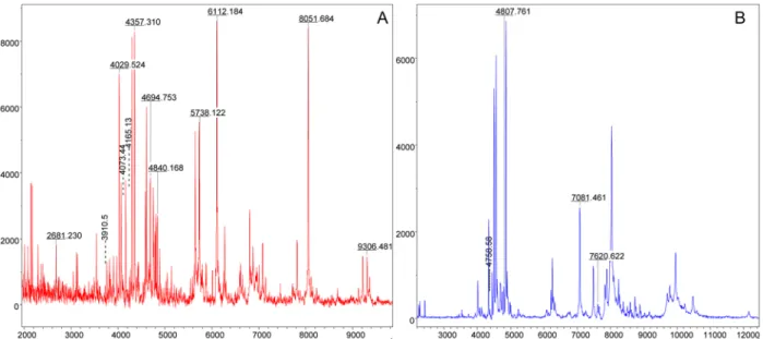
Fig 2. Alignment of MALDI-TOF MS profiles of Rickettsia strains and tick species infected or not by Rickettsia using Flex analysis 3.3 software. Representative spectra of a purified R. conorii conorii strain (A), Rh. sanguineus (B), Rh. sanguineus infected by R. conorii conorii (C), a purified R. slovaca strain (D), D. marginatus (E) and D. marginatus infected by R. slovaca (F) are presented. a.u., arbitrary units; m/z, mass-to-charge ratio.
could distinguish Rh. sanguineus specimens that were infected or not by R. c. conorii (Table 2). Among them, 22 peak masses were observed uniquely in the R. conorii-infected specimens and 8 peak masses were associated with the uninfected Rh. sanguineus specimens (Table 2). To con-firm the specificity of several of these discriminant biomarker masses, a comparison of the MSP between Rh. sanguineus infected by R. c. conorii and the purified R. c. conorii strain was performed (Table 2). Twelve peak masses were common to both samples, and they were local-ized in the spectra of Rh. sanguineus infected by R. c. conorii using Flex analysis software (Fig. 3A). Using a comparable strategy for D. marginatus specimens, 35 discriminating peak masses were identified, among which 21 peak masses were specific to spectra from D. margina-tus infected by R. slovaca (Table 3). Moreover, among these 21 specific peak masses, 4 were shared between D. marginatus infected by R. slovaca and the purified R. slovaca strain. These 4 peak masses were localized on the spectra profiles of infected D. marginatus using the Flex analysis software (Fig. 3B).
Table 2. Peak masses distinguishing uninfected and R. c conorii-infected Rh. sanguineus Ticks and the determination of the peak masses shared with a purified R. c. conorii strain based on statistical analysis with ClinProTools.
Mass (Da) Rh. sanguineus non infected Rh. sanguineus infected by R.c. conorii Strain of R.c. conorii
2148.78 No Yes No 2177.18 No Yes No 2279.34 Yes No No 2304.11 No Yes No 2586.36 Yes No No 2686.07 No Yes Yes 3121.32 No Yes No 3910.5 No Yes Yes 4020.51 Yes No No 4030.21 No Yes Yes 4073.44 No Yes Yes 4165.13 No Yes Yes 4350.22 No Yes Yes 4358.24 No Yes No 4456.14 Yes No No 4584.2 No Yes Yes 4841.34 No Yes Yes 4868.62 Yes No No 5730.84 No Yes Yes 5741.13 No Yes No 6082.26 Yes No No 6108.64 No Yes Yes 6220.67 Yes No No 6811.25 No Yes No 6881.24 No Yes No 7817.31 No Yes No 8042.45 Yes No No 8056.12 No Yes Yes 9307.75 No Yes Yes 12207.38 No Yes No Total 8 22 12 doi:10.1371/journal.pntd.0003473.t002
Blind tests
A total 15 specimens, including uninfected and Rickettsia-infected Ticks, were queried succes-sively against the MS reference Database 1 and Database 2 (i.e., Database 2 = Database 1 plus the spectra from Rickettsia-infected Ticks). Using Database 1, the blind test yielded 100% cor-rect identification at the species level for the specimens tested irrespective of their infectious status and their origin of collection (i.e., Ticks that were laboratory-reared, collected in the field or removed from patients). The LSVs of the first top-ranking hits against Database 1 varied from 1.756 to 2.449 (Table 1). Interestingly, the tick specimens infected by Rickettsia spp had lower LSVs than the uninfected specimens. The same specimens were then tested against Data-base 2, and 100% of the specimens tested possessing a corresponding reference spectrum in Database 2 were correctly identified at the levels of tick species and infectious status (Table 1). Moreover, with the exception of the Rh. sanguineus specimen infected by R. massiliae, only the LSVs from Rickettsia-infected Ticks were increased, and all of these specimens had an LSV larger than 1.85. Interestingly, no association was observed between the cycle threshold value of qPCR and the LSVs. Although no reference spectrum was included in the database for the Rh. sanguineus specimen infected by R. massiliae, it was correctly identified at the level of the tick species as an uninfected Rh. sanguineus specimen, with an LSV greater than 2.
Discussion
After the demonstration that MALDI-TOF MS profiling is an accurate tool to identify arthro-pods [19–23], including vectors of infectious diseases such as Ticks [4,24], the possibility of identifying the presence of microorganisms inside the vectors became evident.
Recently, we showed that the MALDI-TOF MS approach could successfully detect and screen Borrelia spp in their soft tick vectors [25]; the legs of Ticks were used for the dual identi-fication of tick species and the detection of Borrelia relapsing fever [25]. It has also been shown
Fig 3. Location of discriminating peak masses shared between the spectra acquired from Rickettsia-infected specimens and the corresponding Rickettsia strain using Flex analysis software 3.3. The alignment spectra comparing the infected specimen and the corresponding strain spectra are shown in detail. (A) R. conorii conorii shared discriminating peak masses located on the MS profile of Rh. sanguineus infected by R. conorii conorii. (B) R. slovacashared discriminating peak masses located on the MS profile of D. marginatus infected by R. slovaca.
that MALDI-TOF-MS could be employed for the rapid screening of pathogens in tick vectors within the same experiment used for tick identification.
Here, we assess the application of MALDI-TOF MS for the detection of intracellular Rickett-sia bacteria and the identification of their respective tick vectors. The present study revealed that the MALDI-TOF MS spectra obtained from two to four tick leg protein extracts were suffi-cient to accurately identify both the arthropod species and its infectious status. The advantage of performing both of these identifications using only legs is that allows the remaining body parts to be utilized for other analyses. In our study, the infection of Ticks by Rickettsia spp was confirmed by molecular approaches using DNA extracted from the remaining tick legs. In
Table 3. Peak masses distinguishing uninfected and R.slovaca-infected D. marginatus Ticks and the determination of the peak masses shared with a purified R. slovaca strain based on statistical analysis with ClinProTools.
Mass (Da) D.marginatus uninfected D.marginatus infected by R. slovaca Strain of R. slovaca
2043.33 Yes No No 2082.79 Yes No No 2585.65 Yes No No 3493.53 Yes No No 3542.88 Yes No No 3587.51 Yes No No 3960 Yes No No 4136.53 No Yes No 4173.02 Yes No No 4226.84 Yes No No 4305.29 Yes No No 4510.78 No Yes No 4592.4 Yes No No 4629.69 Yes No No 4694.34 No Yes No 4758.58 No Yes Yes 4808.98 No Yes Yes 4906.81 No Yes No 4923.94 Yes No No 6336.71 No Yes No 7082.05 No Yes Yes 7485.1 No Yes No 7626 No Yes Yes 7665 No Yes No 7835.64 No Yes No 7894.93 No Yes No 8241.9 No Yes No 8389.5 No Yes No 8596.3 No Yes No 8738.5 No Yes No 8846.1 No Yes No 9785 No Yes No 9957.33 No Yes No 10483.7 No Yes No 11316.91 Yes No No Total 14 21 4 doi:10.1371/journal.pntd.0003473.t003
addition to validation of the tick infectious status, Rickettsia specific quantitative PCR con-firmed the dissemination of these bacteria in the tick body including the legs.
To evaluate the consequences of Rickettsia infection on the MS profiles of Ticks, we com-pared the spectra produced by Rh. sanguineus and D. marginatus Ticks that were, infected or not by R. c. conorii and R. slovaca, respectively. The alignment of the MS profiles from Rh. san-guineus Ticks that were uninfected or infected by R. c. conorii led to incomplete superimpos-able protein profiles. Similar results were obtained for D. marginatus specimens that were infected or not by R. slovaca. The uniqueness of the MS profiles according to the tick species and infectious status suggests that the detected variations could be attributed to the presence of Rickettsia spp. The analysis of the spectra with ClinProTools revealed the existence of specific discriminating peak masses between infected and uninfected specimens. In total 30 and 35 bio-marker mass sets distinguished the uninfected specimens of Rickettsia spp from the infected specimens of D. marginatus and Rh. sanguineus species, respectively. Interestingly, although the majority of the discriminating peaks appeared in the protein profiles of the infected Ticks, some were not maintained. This loss of some peak masses could be detrimental to the level of significant identification (i.e., LSVs) of Ticks at the species level. Effectively, our blind test ex-periments indicated that the LSVs of the infected specimens were lower than those of the unin-fected Ticks when compared with Database 1, which included only uninunin-fected specimens. In the future, it will be necessary to test the infectious status of a specimen of new species prior to including the results in the reference database. Moreover, the addition of MS spectra from specimens infected with pathogens will improve the identification of arthropod species and the pathogens that they carry.
In addition, among the discriminating peak mass sets found in the infected Ticks, few of them were shared with their respective purified Rickettsia strains. These masses could corre-spond to Rickettsia-specific proteins. Moreover, some discriminating peaks detected uniquely in the Rickettsia-infected Ticks were not present in the spectra peaks of the bacteria strains. These differential peak masses could be attributed either to Rickettsia strains (i.e., variations be-tween laboratory and field strains) [26] or to a response of the Ticks to infection [27]. Comple-mentary experiments are needed to test these hypotheses.
The validity of the databases was established by blind tests using infected and uninfected specimens. A query against Database 2 demonstrated that all the specimens possessing refer-ence spectra in the database were correctly identified at the level of the tick species and the Rickettsia-infectious status. Moreover, 86% (n = 12/14) of these spectra presented LSVs greater than 1.9, which is considered to be reliable score for bacterial species identification [28]. Thus, the spectral variations that are detected following Rickettsia infection are sufficient to avoid cross-recognition between uninfected and infected Ticks. Moreover, the presence of Rickettsia in the Ticks did not mask the protein profiles for unambiguous identification at the species level (e.g., querying the MS spectra against Database 1). These results are in agreement with a previous study showing that these variations do not interfere with species determination [24]. However, the absence of corresponding reference spectra in Database 2 for Rh. sanguineus in-fected by R. massiliae resulted in an incorrect identification of this sample. It is necessary to complete this database with additional tick species infected by Rickettsia strains.
Conclusions
The present study shows that MALDI-TOF MS can be used to reliably identify tick species in-fected or not by Rickettsia spp without the use of a molecular method requiring DNA sequence information. It is important to note that no Rickettsia spp spectrum is available in the Bruker reference database and that this is the first analysis of Rickettsia strain by MALDI-TOF MS.
This work also demonstrated that MALDI-TOF MS could be applied for the rapid detection of Rickettsia spp in Ticks removed from patients. The rapid determination of a tick’s identity and it infectious status should guide decisions related to specific patient monitoring or the adminis-tration of preventive treatment. Additionally, the low consumable costs, minimal time required for sample preparation and rapid availability of the results of MALDI-TOF MS could be useful for epidemiological studies and the monitoring of tick-borne diseases via the dual identification of vectors and their borne pathogen in one step. The main obstacle to the use of the MALDI-TOF MS approach is the cost of acquiring the machine, but its use is cost effective thereafter [29]. These results also open new doors for the monitoring and management of other vector-borne diseases that are of importance for public health in human and veterinary medicine. For example, it would be advantageous to test whether MALDI-TOF MS, which has been shown to be a relevant tool for the identification of mosquito species [5,7,29,30], could be useful for de-tecting the Plasmodium-infectious status of mosquito malaria vectors.
Supporting Information
S1 Fig. Purification and quantification of Rickettsia strains.A 100x magnification image of Rickettsia slovaca grown in L929 cells through an optical microscope before purification (A) and after purification (B). Representative FSC-A vs SSC-A plots of R. conorii (C) and. R. slo-vaca strains in logarithmic scale for counting bacteria. Purified Rickettsia strains were diluted in PBS at 10-1and 10-2and then analyzed on an ACCURI C6 (Medium fluidics speed and Threshold at 10000 for analysis of small particles). (E) Raw quantification data and calculated concentration of each Rickettsia strains are presented.
(TIF)
Acknowledgments
We warmly thank Jean-Michel Berenger and Cristophe Flaudrops for their excellent technical assistance during this work.
Author Contributions
Conceived and designed the experiments: DR PP LA. Performed the experiments: AY JT. Ana-lyzed the data: AY LA. Contributed reagents/materials/analysis tools: CS. Wrote the paper: AY LA DR PP JT CS. First drafted the paper: AY LA.
References
1. Parola P, Raoult D (2001) Ticks and tickborne bacterial diseases in humans: an emerging infectious threat. Clin Infect Dis 32: 897–928. doi:10.1086/319347PMID:11247714
2. Parola P, Paddock CD, Socolovschi C, Labruna MB, Mediannikov O, Kernif T, Fournier PE, Raoult D (2013) Update on Tick-Borne Rickettsioses around the World: a Geographic Approach. Clin Microbiol Rev 26: 657. doi:10.1128/CMR.00032-13PMID:24092850
3. Zhang RL, Zhang B (2014) Prospects of using DNA barcoding for species identification and evaluation of the accuracy of sequence databases for Ticks (Acari: Ixodida). Ticks and Tick-borne Diseases 5: 352–358. doi:10.1016/j.ttbdis.2014.01.001PMID:24656809
4. Yssouf A, Flaudrops C, Drali R, Kernif T, Socolovschi C, Berenger JM, Raoult D, Parola P (2013) Ma-trix-assisted laser desorption ionization-time of flight mass spectrometry for rapid identification of tick vectors. J Clin Microbiol 51: 522–528. doi:10.1128/JCM.02665-12PMID:23224087
5. Yssouf A, Socolovschi C, Flaudrops C, Ndiath MO, Sougoufara S, Dehecq JS, Lacour G, Berenger JM, Sokhna CS, Raoult D, Parola P (2013) Matrix-assisted laser desorption ionization-time of flight mass spectrometry: an emerging tool for the rapid identification of mosquito vectors. PLoS One 8: e72380. doi:10.1371/journal.pone.0072380PMID:23977292
6. Yssouf A, Socolovschi C, Leulmi H, Kernif T, Bitam I, Audoly G, Almeras L, Raoult D, Parola P (2014) Identification of flea species using MALDI-TOF/MS. Comp Immunol Microbiol Infect Dis 37: 153–157. doi:10.1016/j.cimid.2014.05.002PMID:24878069
7. Yssouf A, Parola P, Lindstrom A, Lilja T, L’Ambert G, Bondesson U, Berenger JM, Raoult D, Almeras L (2014) Identification of European mosquito species by MALDI-TOF MS. Parasitol Res 113: 2375–2378. PMID:24737398
8. Seng P, Rolain JM, Fournier PE, La Scola B, Drancourt M, Raoult D (2010) MALDI-TOF-mass spec-trometry applications in clinical microbiology. Future Microbiol 5: 1733–1754.PMID:21133692
9. Socolovschi C, Bitam I, Raoult D, Parola P (2009) Transmission of Rickettsia conorii conorii in naturally infected Rhipicephalus sanguineus. Clin Microbiol Infect 15 Suppl 2: 319–321. doi: 10.1111/j.1469-0691.2008.02257.x
10. Vu Hai V, Almeras L, Audebert S, Pophillat M, Boulanger N, Parola P, Raoult D, Pages F (2013) Identifi-cation of salivary antigenic markers discriminating host exposition between two European Ticks: Rhipi-cephalus sanguineusand Dermacentor reticulatus. Comparative Immunology, Microbiology and Infectious Diseases 36: 39–53. doi:10.1016/j.cimid.2012.09.003PMID:23040662
11. A. Estrada Pena (2004) Ticks of domestic Animals in the Mediterranean Region.
12. Masala G, Chisu V, Satta G, Socolovschi C, Raoult D, Parola P (2012) Rickettsia slovaca from Derma-centor marginatusTicks in Sardinia, Italy. Ticks Tick Borne Dis 3: 393–395.PMID:23140897
13. Gimenez DF (1964) Staining Rickettsiae in yolksack cultures. Staining Technol 39: 135–140. 14. Ogata H, Audic S, Barbe V, Artiguenave F, Fournier E, Raoult D, Claverie J (2000) Selfish DNA in
pro-tein-coding genes of Rickettsia. Science 290: 347–350. doi:10.1126/science.290.5490.347PMID:
11030655
15. Fournier PE, El Karkouri K, Robert C, Medigue C, Raoult D (2012) Complete genome sequence of Rickettsia slovaca, the agent of tick-borne lymphadenitis. J Bacteriol 194: 1612. doi:10.1128/JB. 06625-11PMID:22374949
16. Calderaro A, Gorrini C, Piccolo G, Montecchini S, Buttrini M, Rossi S, Piergianni M, Arcangeletti MC, De Conto F, Chezzi C, Medici MC (2014) Identification of Borrelia species after creation of an in-house MALDI-TOF MS database. PLoS One 9: e88895. doi:10.1371/journal.pone.0088895PMID:24533160
17. Calderaro A, Piccolo G, Gorrini C, Montecchini S, Buttrini M, Rossi S, Piergianni M, De CF, Arcangeletti MC, Chezzi C, Medici MC (2014) Leptospira species and serovars identified by MALDI-TOF mass spectrometry after database implementation. BMC Res Notes 7: 330. doi:10.1186/1756-0500-7-330
PMID:24890024
18. Matsumoto K, Brouqui P, Raoult D, Parola P (2005) Experimental infection models of Ticks of the Rhipi-cephalus sanguineusgroup with Rickettsia conorii. Vector Borne Zoonotic Dis 5: 363–372.PMID:
16417432
19. Campbell PM (2005) Species differentiation of insects and other multicellular organisms using matrix-assisted laser desorption/ ionization time of flight mass spectrometry protein profiling. systematic Ento-mology 30: 186–190. doi:10.1111/j.1365-3113.2004.00279.x
20. Feltens R, Gorner R, Kalkhof S, Groger-Arndt H, Von BM (2010) Discrimination of different species from the genus Drosophila by intact protein profiling using matrix-assisted laser desorption ionization mass spectrometry. BMC Evol Biol 10: 95. doi:10.1186/1471-2148-10-95PMID:20374617
21. Kaufmann C, Schaffner F, Ziegler D, Pfluger V, Mathis A (2012) Identification of field-caught Culicoides biting midges using matrix-assisted laser desorption/ionization time of flight mass spectrometry. Parasi-tology 139: 248–258. doi:10.1017/S0031182011001764PMID:22008297
22. Kaufmann C, Ziegler D, Schaffner F, Carpenter S, Pfluger V, Mathis A (2011) Evaluation of matrix-as-sisted laser desorption/ionization time of flight mass spectrometry for characterization of Culicoides nubeculosusbiting midges. Medical and Veterinary Entomology 25: 32–38.PMID:21118284
23. Modika RP, Flores Vargas RD, Jones MGK (2005) Identification of aphid species using protein profiling and matrix-assisted laser desorption/ionisation time-of-flight mass spectrometry. Entomologia Experi-mentalis et Applicata 117: 243–247.
24. Karger A, Kampen H, Bettin B, Dautel H, Ziller M, Hoffmann B, Suss J, Klaus C (2012) Species determi-nation and characterization of developmental stages of Ticks by whole-animal matrix-assisted laser de-sorption/ionization mass spectrometry. Ticks Tick Borne Dis 3: 78–89. doi:10.1016/j.ttbdis.2011.11. 002PMID:22487425
25. Fotso Fotso A, Mediannikov O, Diatta G, Flaudrops C, Parola P, Drancourt M (2014) MALDI-TOF mass spectrometry detection of relapsing fever Borrelia crocidurae in Ornithodoros sonrai Ticks. PLoS Negl Trop Dis, in press.
26. James A C, El-Hage N, Miller JC, Babb K, Stevenson B (2001) Borrelia burgdorferi RevA Antigen Is a Surface-Exposed Outer Membrane Protein Whose Expression Is Regulated in Response to Environ-mental Temperature and pH. Infect Immun 69: 5286.
27. Antunes S, Galindo RC, Almazan C, Rudenko N, Golovchenko M, Grubhoffer L, Shkap V, do RV, de la Fuente J, Domingos A (2012) Functional genomics studies of Rhipicephalus (Boophilus) annulatus Ticks in response to infection with the cattle protozoan parasite, Babesia bigemina. Int J Parasitol 42: 187–195. doi:10.1016/j.ijpara.2011.12.003PMID:22265898
28. Fournier PE, Couderc C, Buffet S, Flaudrops C, Raoult D (2009) Rapid and cost-effective identification of Bartonella species using mass spectrometry. J Med Microbiol 58: 1154–1159. doi:10.1099/jmm.0. 009647-0PMID:19528172
29. Müller P, Pflüger V, Wittwer M, Ziegler D, Chandre F, Simard F, Lengeler C (2013) Identification of cryp-tic Anopheles mosquito species by molecular protein profiling. PLoS One 8: e57486. doi:10.1371/ journal.pone.0057486PMID:23469000
30. Schaffner F, Kaufmann C, Pfluger V, Mathis A (2014) Rapid protein profiling facilitates surveillance of invasive mosquito species. Parasit Vectors 7: 142. doi:10.1186/1756-3305-7-142PMID:24685094