Fourier Transform Holography: A Lensless Non-destructive Imaging
Technique
M. Saliba, T. Latychevskaia, J.N. Longchamp, H.W. Fink
Physics Institute, University of Zurich, 8057, Zurich, Switzerland
We will discuss the application of Fourier Transform Holography (FTH) [1, 2], a high resolution lensless imaging method [3] based on the use of an extended reference [4], to non-destructively image a specimen of biological or non-biological nature.
The recording is performed in the low-energy electron regime (50-200 eV) where a parallel beam illuminates the specimen and reference object or pinhole. The result of the interference between the wave scattered by the specimen and the reference wave results in an intensity distribution, or holographic diffraction pattern, whose Fourier transform represents the autocorrelation of the transmission function of the specimen under study.
In this energy range, a total dose of 108 electrons/nm2 hardly causes any radiation damage to a single biomolecule for a continuous exposure of more than one hour [5], without the need for staining or cryo-techniques in sample preparation. FTH highlights the most advantageous aspects of both Holography and Coherent Diffraction Imaging, i.e. it is a hybrid technique that naturally encrypts the phase information by the use of an extended reference and yields high
resolution (theoretically 1.8 Å) complex-valued images of the specimen of interest in a single
computational step.
We will briefly discuss the mathematical aspects of FTH theory. We will then introduce the custom preparation of nano-pinholes (5-20nm) serving as extended references, which indispensably requires the use of the Helium Ion Microscope (HIM) [6] that delivers low
radiation damage under a dose of 3x10⁵ ions/nm2. Obtaining the minimal pinhole size (5nm)
with the HIM is crucial to push the resolution limit. Furthermore, we will showcase the implementation of a home-made user-friendly electrostatic micro-lens [7], for collimating the low energy electron beam and minimizing aberrations. Finally, we will display preliminary results of low energy FTH and deliberate the prospect of single-step non-destructive biomolecular imaging.
References:
[1] G. W. Stroke et al. JOSA, Vol. 55, Issue 10, (1965), 1327-1328 [2] I. McNulty et al. Science 256, (1992), 1009
[3] S. Eisebitt et al. Nature 432, (2004)
[4] M. Guizar-Sicairos and J.R. Fienup, Optics Express, Vol. 15 (26), (2007), 17592-17612 [5] M. Germann et al. Phys. Rev. Lett. 104, (2010)
[6] M. Ananth et al. Proc. of SPIE Vol. 8036, (2011) [7] E. Steinwand et al. Ultramicroscopy 110 (9) (2010)
[8] This research was funded by the Swiss National Science Foundation
[9] We kindly acknowledge the assistance and support of the Center for Microscopy and Image Analysis, University of Zurich, with the Tecnai Spirit ® transmission electron microscope.
[10] We kindly acknowledge the assistance of I. Shorubalko and F. Lamattina with the Orion® helium ion microscope at the Swiss Federal Laboratories for materials science and technology, Dübendorf.
564
doi:10.1017/S1431927612004679 © Microscopy Society of America 2012Microsc. Microanal. 18 (Suppl 2), 2012
https://doi.org/10.1017/S1431927612004679
Fig. 1. Schematic of Fourier Transform Holography: From left to right: a divergent beam of low energy electrons impinges on an electrostatic micro-lens which collimates the beam. It is incident on the sample plane that includes a specimen and a pinhole. The corresponding scattered waves interfere to form a holographic diffraction pattern whose Fourier transform results in two twin images of the specimen itself.
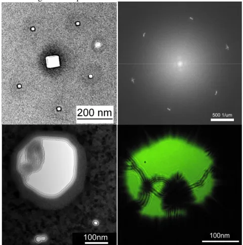
Fig. 2. Top Left: TEM image at 120 KV of 5 pinholes of 20nm size, surrounding a 100nm square hole. Top Right: Fourier Transform of the 5 pinholes showing peaks at positions perpendicular to those of the pinholes. Bottom Left: TEM image at 120KV of 2 sub-50nm holes milled with the HIM, placed next to a bundle of Carbon Nanotubes suspended over a 200nm hole in a Silicon Nitride support. Bottom Right: Low energy electron hologram of another bundle of Carbon Nanotubes exhibiting knots on a 200nm hole, taken with 60eV electrons.
Microsc. Microanal. 18 (Suppl 2), 2012 565
https://doi.org/10.1017/S1431927612004679