HAL Id: hal-02406985
https://hal-amu.archives-ouvertes.fr/hal-02406985
Submitted on 12 Dec 2019
HAL is a multi-disciplinary open access archive for the deposit and dissemination of sci-entific research documents, whether they are pub-lished or not. The documents may come from teaching and research institutions in France or abroad, or from public or private research centers.
L’archive ouverte pluridisciplinaire HAL, est destinée au dépôt et à la diffusion de documents scientifiques de niveau recherche, publiés ou non, émanant des établissements d’enseignement et de recherche français ou étrangers, des laboratoires publics ou privés.
Methylation hotspots evidenced by deep sequencing in
patients with facioscapulohumeral dystrophy and
mosaicism
Stéphane Roche, Camille Dion, Natacha Broucqsault, Camille Laberthonnière,
Marie-Cécile Gaillard, Jérôme Robin, Arnaud Lagarde, Francesca Puppo,
Catherine Vovan, Charlene Chaix, et al.
To cite this version:
Stéphane Roche, Camille Dion, Natacha Broucqsault, Camille Laberthonnière, Marie-Cécile Gaillard, et al.. Methylation hotspots evidenced by deep sequencing in patients with facioscapulohumeral dystrophy and mosaicism. Neurology Genetics, American Academy of Neurology, 2019, 5 (6), pp.e372. �10.1212/NXG.0000000000000372�. �hal-02406985�
ARTICLE OPEN ACCESS
Methylation hotspots evidenced by deep
sequencing in patients with facioscapulohumeral
dystrophy and mosaicism
St´ephane Roche, PhD,* Camille Dion, PhD,* Natacha Broucqsault, PhD,* Camille Laberthonni`ere, MS, Marie-C´ecile Gaillard, PhD, J´erˆome D. Robin, PhD, Arnaud Lagarde, MS, Francesca Puppo, PhD,
Catherine Vovan, BS, Charlene Chaix, BS, Emmanuelle Salort Campana, MD, Shahram Attarian, MD, PhD, Marc Bartoli, PhD, Rafaelle Bernard, MD, Karine Nguyen, MD, PhD,† and Fr´ed´erique Magdinier, PhD† Neurol Genet 2019;5:e372. doi:10.1212/NXG.0000000000000372
Correspondence Dr. Magdinier
frederique.magdinier@univ-amu.fr
Abstract
Objective
To investigate the distribution of cytosine-guanine dinucleotide (CpG) sites with a variable level of DNA methylation of the D4Z4 macrosatellite element in patients with facioscapulo-humeral dystrophy (FSHD).
Methods
By adapting bisulfite modification to deep sequencing, we performed a comprehensive analysis of D4Z4 methylation across D4Z4 repeats and adjacent 4qA sequence in DNA from patients with FSHD1, FSHD2, or mosaicism and controls.
Results
Using hierarchical clustering, we identified clusters with different levels of methylation and separated, thereby the different groups of samples (controls, FSHD1, and FSHD2) based on their respective level of methylation. We further show that deep sequencing–based methylation analysis discriminates mosaic cases for which methylation changes have never been evaluated previously.
Conclusions
Altogether, our approach offers a new high throughput tool for estimation of the D4Z4 methylation level in the different subcategories of patients having FSHD. This methodology allows for a comprehensive and discriminative analysis of different regions along the macro-satellite repeat and identification of focal regions or CpG sites differentially methylated in patients with FSHD1 and FSHD2 but also complex cases such as those presenting mosaicism.
*First coauthors †Last coauthors
From Aix Marseille University, INSERM, MMG (S.R., C.D., N.B., C.L., M.-C.G., J.D.R., A.L., F.P., E.S.C., S.A., M.B., R.B., K.N., F.M.); D´epartement de G´en´etique M´edicale (A.L., C.V., C.C., R.B., K.N.), AP-HM, Hˆopital de la Timone enfants, Marseille; and Centre de r´ef´erence pour les maladies neuromusculaires et la SLA (E.S.C., S.A.), AP-HM, Hˆopital de la Timone, Marseille, France. Go to Neurology.org/NG for full disclosures. Funding information is provided at the end of the article.
The Article Processing Charge was funded by the authors.
This is an open access article distributed under the terms of the Creative Commons Attribution-NonCommercial-NoDerivatives License 4.0 (CC BY-NC-ND), which permits downloading and sharing the work provided it is properly cited. The work cannot be changed in any way or used commercially without permission from the journal.
One of the most frequent genetic diseases associated with epigenetic alterations is facioscapulohumeral dystrophy (FSHD), an autosomal dominant neuromuscular disorder with an estimated prevalence of 1:8,000 to 1:20,000.1,2In 95% of patients (FSHD1, Online Mendelian Inheritance in Man #158900), the dystrophy is linked to deletion of an integral number of repetitive D4Z4 macrosatellites in the sub-telomeric 4q35 locus.3–5In the general population, the number of repeats varies between 11 and 150 units, whereas patients with FSHD1 carry 1 to 10 copies. D4Z4 array shortening occurs de novo in 30% of cases, with a rate of 40% of somatic mosaicism among them (i.e., around 4%–12% of all cases).6
D4Z4 is extremely GC-rich (70%),7 methylated in normal individuals8and decreased by approximately 20% in patients with FSHD1. In 5% of individuals with a typical FSHD phenotype, there is no D4Z4 shortening (type 2 FSHD2; Online Mendelian Inheritance in Man #158901), but these patients display a marked D4Z4 hypomethylation,9–12 asso-ciated with mutation in the SMCHD1 gene.13,14
To further investigate D4Z4 methylation at a high-resolution level, we developed a deep sequencing method after sodium bisulfite DNA modification. By applying this technology to the analysis of 65 blood samples from controls, patients with FSHD1, patients with FSHD2 with SMCHD1 mutation, and mosaic cases, we showed that hypomethylation of the proxi-mal part of the repeat can be evidenced with a strong statistical significance in patients with FSHD1 and FSHD2 in com-parison to healthy donors, but also in patients with different levels of mosaicism.
Methods
Data availability
Values for DNA methylation are provided infile e-1, links.lww. com/NXG/A191. Details on samples used in the study are provided in tables e-2 and e-3, links.lww.com/NXG/A190.
Standard protocol approvals, registrations, and patient consents
All individuals have provided written informed consent for the use of the DNA sample for medical research, and the study was conducted in accordance with the Declaration of Helsinki. Controls are randomly selected individuals or patient’s rela-tives. Controls are neither carrier of any genetic mutation nor affected by any muscular pathology. Controls were selected in the same age range and sex representation as patients. All patients were diagnosed at the Department of Medical Ge-netics, La Timone Hospital, Marseille, either by Southern
blotting or molecular combing.15,16Further details on methods are provided in the supplementary information section.
Results
Deep sequencing for analysis of DNA methylation after sodium
bisulfite modification
Given the importance of epigenetic changes in deciphering the molecular mechanisms of FSHD, we developed a novel strat-egy for in-depth D4Z4 methylation analysis based on sodium bisulfite modification followed by deep sequencing (figure 1). The sodium bisulfite methodology based on the chemical modification of cytosines allowing DNA methylation profiling at a single base and single molecule level is considered as a gold standard for quantitative DNA methylation analysis.17 After deep sequencing, data are analyzed using a specific workflow (figure e-1A, links.lww.com/NXG/A190). Alignment of bisulfite-treated sequences is performed using BiQ Analyzer HiMod that converts aligned methylated cytosine-guanine di-nucleotide (CpG) to 1 (cytosine-guanine [CG]) or 0 for unmethylated CpG (TG).18Unaligned or unsequenced CGs, noted“x”, are not further considered except for determination of the sequencing coverage (figure e-1B, links.lww.com/NXG/ A190). Then, using a custom algorithm in R, we generated the methylation profile of each region CpG by CpG and calculated the methylation level for each sequence (figure 1; figure e-1A, links.lww.com/NXG/A190). Only sequences with >95% of conversion are kept for analysis. We used stringent filters allowing only analysis of highly matching sequences with a minimum of 500 DNA molecules per barcode. Depth of coverage does not affect the accuracy of DNA methylation level determination (figure e-1B, links.lww.com/NXG/A190). Methylation is determined at 3 different levels. The first gives the methylation level for each CpG. The second corresponds to the level of methylation of each DNA fragment (figure e-1C, links. lww.com/NXG/A190), and the third to methylation for each biological sample calculated as the ratio of methylated CG di-vided by the number of CGs analyzed. Homogeneity in the distribution of methylation or presence of sequences with a low or high methylation level is estimated using the mixtools package in R using a random sampling method and confidence intervals calculation to visualize the multimodal distribution of sequenced molecules based on their methylation level (figure 1, figure e-1D, links.lww.com/NXG/A190). When performing the entire assay in triplicate (DNA extraction, bisulfite conversion, PCR, se-quencing, and quantification), we found a reduced variability with an SD of 3.6% for DR1, 6.5% for 5P, 1.6% for primers used to investigate the Middle region of the D4Z4 repeat (MID), and
Glossary
CG= cytosine-guanine; CpG = cytosine-guanine dinucleotide; FSHD = facioscapulohumeral dystrophy; MID = primers used to investigate the middle region of the D4Z4 repeat; PBMC = peripheral blood mononuclear cell.
5.1% for 3P in the calculation of the mean methylation level, validating the performance of the method.
Overall, this methodology requires less DNA than a Southern blot, centered on the analysis of a few CpGs in a single repeat. Compared with bisulfite sequencing requiring cloning and 10–15 colony picking or pyrosequencing, deep sequencing limits potential bias in sample selection, dilution, or masking of the methylation signal of underrepresented sequences be-cause CGs are sequenced several times. Furthermore, direct sequencing allows samples multiplexing and analysis of het-erogeneous biological samples.
Comprehensive analysis of D4Z4 methylation for determination of the D4Z4 methylation pattern at a single-base resolution
Wefirst validated the different primers used for sodium bisulfite sequencing (DR1, 5P, MID, and 3P,10,11table e-1, links.lww. com/NXG/A190) by assessing the power of discrimination between methylated and unmethylated molecules of our next generation sequencing assay and bioinformatics pipeline using an in vitro methylation assay. To this aim, we mixed different proportions of an in vitro methylated plasmid carrying 1 copy of the D4Z4 repeat and unmethylated plasmid DNA (0%–100% of in vitro methylated molecules). After bisulfite
Figure 1Workflow for DNA methylation analysis
Genomic DNA was extracted from blood cells from different donors. After extraction, DNA was modified by sodium bisulfite treatment, PCR amplified, and analyzed by deep sequencing. Specific repetitive elements are amplified using a high-fidelity PCR in bisulfite-treated samples. After preparation of a first equimolar pool of PCR fragments and end repair, adaptors and barcodes are ligated. Samples are collected and cleaned up by magnetic purification. An equimolar pool of all barcoded samples is prepared before Ion Torrent PGM sequencing. Barcoded samples are submitted to an emulsion PCR and loaded onto a 316v2 Ion Torrent chip. After 650 flows of sequencing, barcodes and regions are split, and CpG alignment is performed using the BiQ Analyzer HiMod software.18For each region, the global methylation level and methylation profile are determined. Random sampling method and confidence interval
calculation identified 10 classes of molecules, depending on their methylation level from strongly hypomethylated (0%–10% of methylated CG per molecule) to highly methylated sequences (90%–100% of methylated CpG) allowing identification of subpopulations of DNA molecules with a low (red line) or high methylation (green line) level calculated as the area under each curve. CG = cytosine-guanine; CpG = cytosine-guanine dinucleotide.
conversion, PCR amplification, next generation sequencing, and sequence alignment, we analyzed the methylation profile in the different conditions and observed only low CpG to CpG variations for the different proportions of samples tested (figure e-2, links.lww.com/NXG/A190). Of note, the level of meth-ylation did not reach 100% but 96.2% of global methmeth-ylation indicating a capability of discriminating sequences with very high level of methylation (>95%) or molecules with a low methylation level (<10%). Furthermore, we demonstrated a linear relationship between theoretical and measured meth-ylation, with a correlation of 0.979 for DR1, 0.987 for 5P, 0.998 for MID, and 0.995 for 3P (figure e-2, links.lww.com/NXG/ A190).
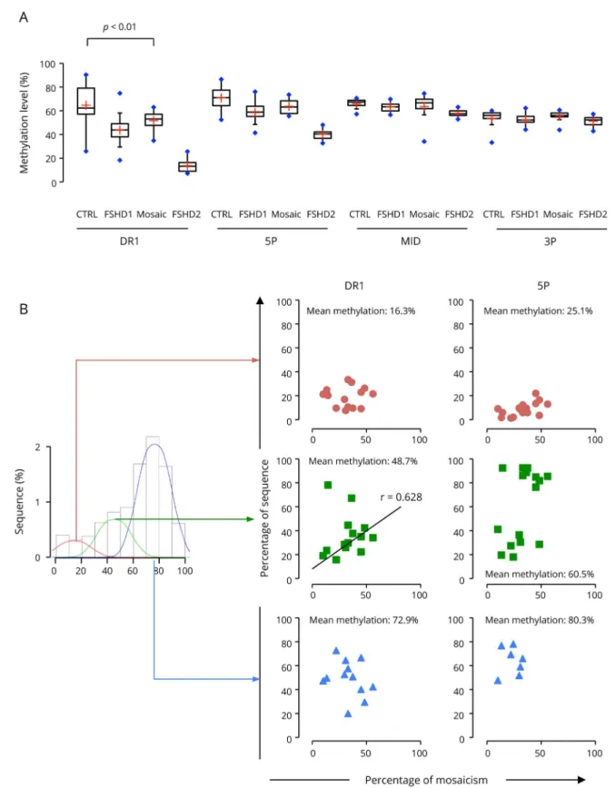
We then applied our methodology to the analysis of D4Z4 methylation of peripheral blood mononuclear cell (PBMC) DNA from healthy donors (controls, n = 10), patients with FSHD1 (n = 29), and patients with FSHD2 mutated for SMCHD1 (n = 10) (figures 2A and B, table e-2, links.lww.com/ NXG/A190; file e-1, links.lww.com/NXG/A191) for the 4 different regions across the macrosatellite (DR1, 5P, MID, and 3P).10,11The DNA methylation level of individual CpG sites is not uniform across D4Z4 (figure 2A). Mean DR1 and 5P methylation is high (66.5% ± 19 and 72.8% ± 10.2) in controls but significantly decreased in FSHD1 (DR1: 46.1% ± 12.5, p = 3.1 × 10⁻⁶and 5P: 59.8% ± 7.8, p = 9.5 × 10⁻⁷, respectively) and FSHD2 (DR1: 12.8% ± 5, p = 1.7 × 10⁻⁵and 5P: 42.7% ± 4.5, p = 9.2 × 10⁻⁵) (figure 2B, file e-1, links.lww.com/NXG/A191). Euclidean correlation and hierarchical clustering analysis re-veal 3 major dendrogram branches that separate samples in 3 groups, from high to low methylation corresponding, re-spectively, to controls, FSHD1, and FSHD2 (figure 2C, figure e-3A, links.lww.com/NXG/A190). The heatmap of the rela-tive methylation level for each individual CpG across the different D4Z4 subregions (figure e-3A, links.lww.com/ NXG/A190) enabled the identification of critical CG hypo-methylated in the disease, mostly clustered at DR1 and 5P (figure e-3B, links.lww.com/NXG/A190). As for the global methylation level (figure 2A, B), methylation of individual CGs is not different for the MID and 3P regions in the dif-ferent groups of samples with exception for the MID-CpG.1, MID-CpG.23, and 3P-CpG.11, less methylated in patients with FSHD1 and FSHD2 compared with controls (figure 2A, figures e-3B, links.lww.com/NXG/A190).
Our primers amplify D4Z4 elements present both at the 4q35 and 10q26 regions (figure e-4, links.lww.com/NXG/A190) but do not amplify D4Z4 homologous elements present elsewhere in the genome and containing numerous polymorphisms.19 Given the absence of significant methylation change at the MID and 3P regions (figure 2D), we used methylation of these regions for normalization of methylation values to the total number of D4Z4 repeats on the 4q and 10q (table e-2, links. lww.com/NXG/A190). We confirmed the significant differ-ences in the methylation ratio for the DR1 and 5P after nor-malization to the MID (figure 3A) or 3P methylation levels
(figure 3B) showing that analyses of different regions across D4Z4 can be used as internal standard for normalization of methylation changes and calculation of a mean methylation ratio for individual samples. When methylation was plotted as a function of the total number of repeats at the 4q and 10q in controls, FSHD1, and FSHD2, (table e-2, links.lww.com/ NXG/A190), methylation for the DR1 and 5P significantly separated samples in the 3 different categories (controls, FSHD1, and FSHD2) but not MID and 3P (figure 2D), in-dicating that deep sequencing of bisulfite-converted DNA can be reliably used for analysis of D4Z4 methylation changes in patients with FSHD.
By analyzing the distribution of hypo- or hypermethylated molecules using random sampling and confidence interval calculation, the number of molecules in each subclass con-firms the variability in D4Z4 methylation. Controls display a low proportion of hypomethylated molecules (mean methylation level <25%, red curves) and a higher proportion of highly methylated molecules (green curves,figure e-5, links. lww.com/NXG/A190). Patients with FSHD1 display an av-erage of 50% of molecules with a low methylation level, whereas the number of hypomethylated molecules is strongly increased in patients with FSHD2.
Hypomethylation is restricted to D4Z4 in patients with FSHD
Repetitive DNA sequences represent the largest proportion of methylated DNA sequences in the human genome.20–23Hence, to assess more globally methylation changes in the different conditions, we included in our analysis primers for interspersed short repetitive sequences such as AluY or the 59 UTR of LINE-1 retrotransposon dispersed throughout the genome, often used as a surrogate marker for global methylation analysis24,25 or additional repetitive elements such as the RS447 macrosatellite or TAR1 subtelomeric element (figure e-6A, links.lww.com/ NXG/A190; table e-1, links.lww.com/NXG/A190).
For AluY, LINE-1, or RS447, we observed a stable level of methylation (figure e-6B; links.lww.com/NXG/A190; file e-1, links.lww.com/NXG/A191) in the different groups of samples. The global methylation level is not modified in patients with FSHD, including patients with FSHD2 mutated for SMCHD1, underlining a specific role for this factor in D4Z4 regulation.26
Of interest, we observed a slight methylation decrease for the TAR1 subtelomeric element in FSHD1 blood samples (41.4% ± 8.3) compared with controls (52.9% ± 11.9, p < 0.0034) and patients with FSHD2 (56.7% ± 7.6%, p < 0.0005), suggesting that besides D4Z4 FSHD patients might be also prone to hypomethylation of other subtelomeres (figure e-6, links.lww. com/NXG/A190;file e-1, links.lww.com/NXG/A191).
Methylation of the DR1 sequence for identification of hypomethylated alleles in patients with mosaicism
FSHD is characterized by a high proportion of somatic mo-saicism. To test whether our methodology enabled estimation
Figure 2D4Z4 methylation profile in blood cells from healthy donors, patients with FSHD1, or patients with FSHD2
(A) For each sample, DNA methylation was determined after sodium bisulfite for 4 regions within D4Z4 (DR1, [254bp; 31 CGs], 5P [275bp, 22 CGs], MID [353bp, 31 CGs], and 3P [171bp, 14 CGs]) after PCR amplification and deep sequencing. Primers were designed by avoiding the presence of CGs in the primers sequence to amplify methylated and unmethylated DNA with the same efficiency. For each sequence, dots above the histograms represent individual CpGs. Histogram bars represent the percentage of methylated (black) or unmethylated (white) CpG for each position. Histograms correspond to the average methylation level of blood cells DNA from 10 healthy donors, 29 patients with FSHD1, and 10 patients with FSHD2. CpGs with a low coverage are indicated by a black arrow and were discarded from the calculation of the mean methylation level. (B) Scatterplot distribution of the D4Z4 methylation level at DR1, 5P, MID, and 3P in control (n = 10), FSHD1 (n = 29), and FSHD2 (n = 10) PBMC DNA samples. Horizontal red lines correspond to the median for each group. Data sets were compared using the Wilcoxon nonparametric test, and brackets identify significantly different groups based on post hoc Dunn comparison and Bonferroni correction. p Values are indicated for each group of samples. (C) Heatmap analysis of the global methylation level in blood from healthy donors (n = 10, purple stars), patients with FSHD1 (n = 29, gray circles), and patients with FSHD2 (n = 10, orange triangles). The dendrogram representation discriminates the different subgroups of patients based on their respective level of methylation. (D) Scatterplot showing the percentage of methylation for the different D4Z4 regions as a function of the cumulative number of repeats from chromosomes 4 and 10 for the different subregions in blood samples. For the DR1 and 5P region, ellipses were used to discriminate the 3 groups (controls, FSHD1, and FSHD2). Methylation levels are not significantly different for the MID and 3P regions with overlap between the different groups of samples. FSHD = facioscapulohumeral dystrophy; PBMC = peripheral blood mononuclear cell.
of the degree of hypomethylation in this subset of complex cases, we analyzed methylation of the DR1, 5P, MID, and 3P regions in 16 patients for which the percentage of mosaicism and size of the different 4q and 10q alleles have been precisely determined by molecular combing16 (table e-3, links.lww. com/NXG/A190). By plotting the mean methylation level in these patients compared with controls, patients with FSHD1, and patients with FSHD2 for the different regions across D4Z4, we show that patients with mosaicism are significantly hypomethylated compared with controls (figure 4A, p value <0.01) with a level of methylation for the DR1 sequence intermediate between the levels in controls and patients with FSHD1. We then compared the distribution of the number of sequence as a function of the percentage of methylation. In 14 of the 16 mosaic cases, we observed a 3 classes distribution
with a proportion of molecules with an intermediate level of methylation for DR1 (figure e-7, links.lww.com/NXG/ A190). The remaining 2 samples correspond to patients with the higher proportion of short D4Z4 allele (42% and 51% of mosaicism, respectively). By plotting the percentage of mol-ecules in the different classes of methylated molmol-ecules to the percentage of short allele for the different patients (figure 4B), we obtained a positive correlation between the level of methylation for DR1 and the percentage of mosaicism. We concluded that deep sequencing of bisulfite-converted DNA discriminates efficiently alleles with a decreased level of methylation from highly methylated ones in patients with mosaicism.
Methylation changes at the distal D4Z4 repeat and adjacent A-type sequence
Taking advantage of published results27,28 and to deepen our approach, we applied the same methodology to investigate the methylation of the most distal D4Z4 unit and adjacent 4qA sequence containing the DUX4 polyadenylation site. We used previously published primers and adapted the initial procedure designed for nested PCR amplification of a 594-bp sequence encompassing 57 CpGs (figure 5A),27,28
subcloning, and Sanger sequencing of clones to deep sequencing. In the different groups of samples, we observed a slight significant difference (p = 0.048) between controls (with a range of 20.5%–40.8% of methylation) and patients with FSHD1 (14.1%–31% range) (figure 5B; file e-1, links.lww.com/NXG/A191).
Of interest, the 59 forward primer located within D4Z4 is al-most identical to our 3P-forward primers. Thus, the 14 proxi-mal CGs of this 4qA bisulfite sequence are identical to those analyzed using our 3P primers (figure e-8, links.lww.com/NXG/ A190). To compare the last D4Z4 repeat carried by 4qA alleles to the most distal part of all sequenced D4Z4, wefirst analyzed separately the most proximal part of the 4qA-specific sequence, which matches the 3P sequence (CpGs 1–14, figure e-8, links. lww.com/NXG/A190; figure 5A). This distal part of the last D4Z4 repeat is not differentially methylated in FSHD1 or FSHD2 compared with controls, as observed when all D4Z4 are considered (3P primers,figure 2).
Hypomethylation seems more pronounced in the region encompassing CGs 15–56 and corresponding to DUX4 exons 2 and 3 and intronic sequences (pLAM and DUX4 39 UTR, p value = 0.027 between controls and FSHD1 blood samples (figure 5B, figure e-9, links.lww.com/NXG/A190) indicating epigenetic changes in the 39 UTR of DUX4.
By analyzing CG sites overlapping with splice junction, no significant difference in methylation was observed, suggesting that alternative splicing of DUX4 39 UTR is not dependent on methylation changes. Hence, by comparing the methylation in the last D4Z4 unit, our approach reveals that most of the hypomethylation occurs in the most proximal part of the repeat in the 3 groups of patients investigated (FSHD1, FSHD2, and mosaic cases).
Figure 3D4Z4 methylation level in the proximal end of the repeat discriminates healthy donors from patients with FSHD1 and FSHD2
(A and B). Our primers amplify D4Z4 elements present on both chromo-somes 4 and 10. Therefore, we used the methylation level determined for the MID and 3P regions to normalize the results obtained in the DR1 and 5P regions in the different groups of samples (controls, n = 10, FSHD1, n = 29, and FSHD2, n = 10). (A) Ratio of methylation in the DR1 and 5P regions normalized to the methylation level of the MID region. (B) Ratio of methyl-ation in the DR1 and 5P regions normalized to the methylmethyl-ation level of the 3P region. Statistical significance was determined using a Mann-Whitney non-parametric test, **p < 0.0001; *p < 0.005. CTRL = control; FSHD = faciosca-pulohumeral dystrophy.
Figure 4Methylation level of the proximal part of D4Z4 discriminates mosaic individuals from patients with FSHD1
(A) Boxplot representation of the mean methylation level for controls (n = 10), patients with FSHD1 (n = 28), patients with FSHD2 (n = 10), and patients with mosaicism (n = 16) for the DR1, 5P, MID, and 3P sequences. Horizontal black lines correspond to the median for each group, and the red cross to the mean for each group. Data sets were compared using the Wilcoxon nonparametric test, and brackets identify significantly different groups based on post hoc Dunn comparison and Bonferroni correction. The p values are indicated. (B) Scatterplot showing for the different patients with mosaicism (n = 16), the percentage of molecule with a low mean methylation, DR1: 16.1%, 5P: 25.1% (upper panel, red circles), intermediate mean methylation, DR1: 48.7%, 5P: 60.5% (middle, green squares), and high mean methylation, DR1: 72.9%, 5P: 80.3% (lower panel, blue triangles) for DR1 and 5P regions as a function of the percentage of mosaicism (or percentage of short D4Z4 4q allele) in blood samples. The correlation was calculated using the“cor” function of the R software and Spearman method. A positive correlation was found between the percentage of DR1 sequence with a medium level of methylation (48.7%) and the percentage of mosaicism (r = 0.628). FSHD = facioscapulohumeral dystrophy.
Discussion
Hypomethylation has been either linked to D4Z4 array shortening or mutation in SMCHD1. We analyzed DNA methylation of this macrosatellite and other repetitive ele-ments in patients with FSHD using a custom deep sequencing method based on sodium bisulfite treatment of genomic DNA. For D4Z4, we combined analysis of regions with var-iable methylation (DR1 and 5P)10,11and regions not differ-entially methylated.10 By comparing different groups of individuals, we demonstrate that changes in the level of methylation in the proximal part of the repeat can be quan-tified by sodium bisulfite and deep sequencing. The decreased methylation consistently observed in patients with FSHD at the DR1 and 5P regions indicates that hypomethylation occurs for the vast majority of sequenced fragments and not only at the distal DUX4-encoding unit27–29 or proximal re-peat.30 We inferred from our results and analysis of other regions that D4Z4 hypomethylation is not uniform but fo-calized in the proximal part of the repeat. We further showed that analysis of regions with a constant methylation level (MID and 3P) can be used as internal standard and for nor-malization to D4Z4 copy number.
Since the discovery of the “FSHD locus”, a decrease in DNA methylation of the shortened D4Z4 array has been
systematically associated with the disease. Several methodol-ogies have been developed for analysis of this epigenetic modification going from Southern blot after digestion with methylation sensitive enzymes to sodium bisulfite sequencing. When probing D4Z4 methylation, the difficulty resides in the comprehensive analysis of the 4q-causing allele. By probing methylation of a single FseI site in the most proximal unit without distinction of the 4q and 10q chromosomes, Lem-mers et al30defined the Delta Score for evaluation of hypo-methylation in patients. Besides, 2 groups recently proposed analyses of the most distal D4Z4 unit and adjacent A-type sequences by sodium bisulfite sequencing using primers that distinguish 4q from 10q alleles27,29and revealed that the distal DUX4-coding sequence (exon 2–3 and intronic sequences) is also hypomethylated.27–29 Nevertheless, in these studies, amplification of both the long and shortened arrays yields a potential bias in the estimation of the methylation level for individuals carrying 2 A-type alleles compared with 4qA/4qB patients carrying only a short 4qA allele.27–29Overall, these observations on the methylation of the most distal repeat raise question of the role of methylation in DUX4 splicing because methylation changes in coding sequences correlate with splicing sites usage than activation of transcription.31 Despite the systematic hypomethylation in patients with FSHD,8–10,32–34the impact of this epigenetic modification
Figure 5Analysis of the methylation pattern of the distal end of the D4Z4 array after sodium bisulfite modification and deep sequencing
We adapted the protocol of bisulfite modification, nested PCR amplification of the distal part of the last D4Z4 repeat and adjacent pLAM sequence, cloning and sequencing of selected clones to deep sequencing described in Ref. 27. (A) The position of the region amplified is indicated by black arrows. The 4qA BSS sequence overlaps with the 3P region, with 14 CpG in common between the 2 regions. (B) Quantification of the mean methylation level of the 4q BSS sequence in DNA extracted from blood cells from controls, patients with FSHD1, and patients with FSHD2. The red line corresponds to the mean value. Data sets were compared using the Wilcoxon nonparametric test. Brackets identify significantly different groups based on post hoc Dunn comparison and Bonferroni correction (p = 0.048 between controls and patients with FSHD1). Quantification of the mean methylation level in the most proximal part of the 4qA BSS sequence, encompassing the 3P region is displayed separately (CpG.1–14, as well as the most distal part of the sequence (CpG 15–56). Histograms display the mean methylation level in DNA from PBMCs from controls (n = 6), patients with FSHD1 (n = 10), and patients with FSHD2 (n = 6). BSS = bisulfite sequencing; CG = cytosine-guanine; CpG = cytosine-guanine dinucleotide; FSHD = facioscapulohumeral dystrophy; PBMC = peripheral blood mononuclear cell.
in FSHD pathomechanism remains poorly understood and its use as a disease biomarker debated. In some cases, meth-ylation might be more reduced than expected,30and there is no correlation between the extend of hypomethylation and DUX4 expression with a >50-fold range of increase in the DUX4-fl level but only small differences in the methylation level of the last D4Z4 unit at 4qA regions as reported.27,28 Furthermore, the marked hypomethylation in SMCHD1-mu-tated patients with FSHD2 is not accompanied with either a more severe phenotype and earlier onset9,11,34 or with a muscle phenotype in patients with bosma arhinia micro-phthalmia syndrome.26,35,36Furthermore, we recently showed that D4Z4 methylation is dynamically regulated in an SMCHD1-dependent manner opening new questions on the regulation of this epigenetic mechanism and DUX4 activation.26 There is no ideal method for evaluation of D4Z4 array meth-ylation in patients with FSHD, given the repetitive nature of the region. Compared with other methods, our approach requiring a limited amount of DNA offers the possibility of analyzing simultaneously different regions across D4Z4 including the distal A-type region.10,11,27It is noteworthy that all statistical assays or normalization methods applied to our data set lead to the same conclusion in the discrimination between controls, FSHD1, and FSHD2 but also mosaic cases. Importantly, we report here for thefirst time the analysis of DNA methylation in patients with mosaicism, accounting for approximately 40% of FSHD1 de novo cases (4%–12% of all FSHD1) with a cor-relation between the level of methylation with the percentage of mosaicism or short allele.
These observations would not be possible if one assume that a single D4Z4 unit is markedly hypomethylated (i.e., either the first unit as probed by Southern blotting or the DUX4 encoding more distal unit), indicating that hypomethylation is not limited to the proximal or distal repeats but occurs within all repeats. The consistency and significance of results obtained using dif-ferent methods of quantification argues against any potential bias in data interpretation and evaluation D4Z4 methylation despite the amplification of 4q- and 10q-derived repeats.
Thus, besides the clinical evaluation and molecular diagnosis, evaluation of D4Z4 methylation appears as an interesting tool for the evaluation of the patients, given the wide diversity in the FSHD-associated genotypes.16,30,37
Acknowledgment
The authors are indebted and thank all patients for participating in this study. They thank Mr. Armand Tasmadjian for technical helps. They acknowledge the Marseille Maladies Rares (MarMaRa) institute for funding.
Disclosure
S. Roche, C. Dion, N. Broucqsault, M.-C. Gaillard, J.D. Robin, F. Puppo, E.S. Campana, S. Attarian, M. Bartoli, R. Bernard, K. Nguyen, F. Magdinier, C. Laberthonni`ere, A. Lagarde, C. Vovan,
and C. Chaix report no disclosures. Go to Neurology.org/NG for full disclosures.
Study funding
This study was funded by“Association Française contre les Myopathies” (AFM; MNHDecrypt and TRIM-RD), Agence Nationale de la Recherche (ANR, FSHDecipher, ANR-13-BSV1-0001). CD was the recipient of a fellowship from the French Ministry of Higher Education and Re-search and FSH Society. CL is the recipient of a fellowship from the French Ministry of Higher Education and Re-search. We acknowledge the Marseille maladie rare in-stitute (MarMaRa) for funding.
Publication history
Received by Neurology: Genetics February 7, 2019. Accepted infinal form October 4, 2019.
AppendixAuthors
Name Location Role Contribution
St´ephane Roche, PhD Aix-Marseille University, INSERM, MMG U1251, Marseille
Author Designed and conceptualized the study; analyzed the data; and drafted the manuscript for intellectual content Camille Dion, PhD Aix-Marseille University, INSERM, MMG U1251, Marseille
Author Major role in the acquisition of data Natacha Broucqsault, PhD Aix-Marseille University, INSERM, MMG U1251, Marseille
Author Major role in the acquisition of data Camille Laberthonni`ere, MS Aix-Marseille University, INSERM, MMG U1251, Marseille
Author Major role in the acquisition of data Marie C´ecile Gaillard, PhD Aix-Marseille University, INSERM, MMG U1251, Marseille
Author Major role in the acquisition of data
J´erˆome D. Robin, PhD
Aix-Marseille University, INSERM, MMG U1251, Marseille
Author Revised the manuscript for intellectual content Arnaud Lagarde, MS Aix-Marseille University, INSERM, MMG U1251, Marseille
Author Designed and conceptualized the study Francesca Puppo, PhD Aix-Marseille University, INSERM, MMG U1251, Marseille
Author Role in the acquisition of data Catherine Vovan, BS Department of Medical Genetics, La Timone Hospital, Marseille
Author Major role in the acquisition of data
References
1. Wang LH, Tawil R. Facioscapulohumeral dystrophy. Curr Neurol Neurosci Rep 2016;16:66.
2. Deenen JC, Arnts H, van der Maarel SM, et al. Population-based incidence and prevalence of facioscapulohumeral dystrophy. Neurology 2014;83:1056–1059. 3. Sarfarazi M, Wijmenga C, Upadhyaya M, et al. Regional mapping of
facioscapulo-humeral muscular dystrophy gene on 4q35: combined analysis of an international consortium. Am J Hum Genet 1992;51:396–403.
4. Upadhyaya M, Lunt P, Sarfarazi M, Broadhead W, Farnham J, Harper PS. The mapping of chromosome 4q markers in relation to Facioscapulohumeral Muscular Dystrophy (FSHD). Am J Hum Genet 1992;51:404–410.
5. Wijmenga C, Hewitt JE, Sandkuijl LA, et al. Chromosome 4q DNA rearrangements associated with facioscapulohumeral muscular dystrophy. Nat Genet 1992;2:26–30. 6. van der Maarel SM, Deidda G, Lemmers RJ, et al. De novo facioscapulohumeral muscular dystrophy: frequent somatic mosaicism, sex-dependent phenotype, and the
role of mitotic transchromosomal repeat interaction between chromosomes 4 and 10. Am J Hum Genet 2000;66:26–35.
7. Lyle R, Wright TJ, Clark LN, Hewitt JE. The FSHD-associated repeat, D4Z4, is a member of a dispersed family of homeobox-containing repeats, subsets of which are clustered on the short arms of the acrocentric chromosomes. Genomics 1995;28: 389–397.
8. van Overveld PG, Lemmers RJ, Sandkuijl LA, et al. Hypomethylation of D4Z4 in 4q-linked and non-4q-4q-linked facioscapulohumeral muscular dystrophy. Nat Genet 2003; 35:315–317.
9. de Greef JC, Lemmers RJ, Camaño P, et al. Clinical features of facioscapulohumeral muscular dystrophy 2. Neurology 2010;75:1548–1554.
10. Gaillard MC, Roche S, Dion C, et al. Differential DNA methylation of the D4Z4 repeat in patients with FSHD and asymptomatic carriers. Neurology 2014, 83: 733–742.
11. Hartweck LM, Anderson LJ, Lemmers RJ, et al. A focal domain of extreme deme-thylation within D4Z4 in FSHD2. Neurology 2013, 80:392–399.
12. Gaillard MC, Puppo F, Roche S, et al. Segregation between SMCHD1 mutation, D4Z4 hypomethylation and Facio-Scapulo-Humeral Dystrophy: a case report. BMC Med Genet 2016;17:66.
13. Lemmers RJ, Tawil R, Petek LM, et al. Digenic inheritance of an SMCHD1 mutation and an FSHD-permissive D4Z4 allele causes facioscapulohumeral muscular dystrophy type 2. Nat Genet 2012;44:1370–1374.
14. Blewitt ME, Vickaryous NK, Hemley SJ, et al. An N-ethyl-N-nitrosourea screen for genes involved in variegation in the mouse. Proc Natl Acad Sci USA 2005;102: 7629–7634.
15. Nguyen K, Walrafen P, Bernard R, et al. Molecular combing reveals allelic combi-nations in facioscapulohumeral dystrophy. Ann Neurol 2011;70:627–633. 16. Nguyen K, Broucqsault N, Chaix C, et al. Deciphering the complexity of the 4q and
10q subtelomeres by molecular combing in healthy individuals and patients with facioscapulohumeral dystrophy. J Med Genet 2019;56:590–601.
17. Frommer M, McDonald LE, Millar DS, et al. A genomic sequencing protocol that yields a positive display of 5-methylcytosine residues in individual DNA strands. Proc Natl Acad Sci USA 1992;89:1827–1831.
18. Bock C, Reither S, Mikeska T, Paulsen M, Walter J, Lengauer T. BiQ Analyzer: visualization and quality control for DNA methylation data from bisulfite sequencing. Bioinformatics 2005;21:4067–4068.
19. Zeng W, de Greef JC, Chen YY, et al. Specific loss of histone H3 lysine 9 trimethy-lation and HP1gamma/cohesin binding at D4Z4 repeats is associated with faciosca-pulohumeral dystrophy (FSHD). PLoS Genet 2009;5:e1000559.
20. Charlesworth B, Sniegowski P, Stephan W. The evolutionary dynamics of repetitive DNA in eukaryotes. Nature 1994;371:215–220.
21. Jurka J, Kapitonov VV, Kohany O, Jurka MV. Repetitive sequences in complex genomes: structure and evolution. Annu Rev Genomics Hum Genet 2007;8:241–259. 22. Rollins RA, Haghighi F, Edwards JR, et al. Large-scale structure of genomic
methyl-ation patterns. Genome Res 2006;16:157–163.
23. Eden A, Gaudet F, Waghmare A, Jaenisch R. Chromosomal instability and tumors promoted by DNA hypomethylation. Science 2003;300:455.
24. Lisanti S, Omar WA, Tomaszewski B, et al. Comparison of methods for quantifi-cation of global DNA methylation in human cells and tissues. PLoS One 2013;8: e79044.
25. Horard B, Eymery A, Fourel G, et al. Global analysis of DNA methylation and transcription of human repetitive sequences. Epigenetics 2009;4:339–350. 26. Dion C, Roche S, Laberthonni`ere C, et al. SMCHD1 is involved in de novo
meth-ylation of the DUX4-encoding D4Z4 macrosatellite. Nucleic Acids Res 2019, 47: 2822–2839.
27. Jones TI, Yan C, Sapp PC, et al. Identifying diagnostic DNA methylation profiles for facioscapulohumeral muscular dystrophy in blood and saliva using bisulfite se-quencing. Clin Epigenetics 2014;6:23.
28. Jones TI, King OD, Himeda CL, et al. Individual epigenetic status of the pathogenic D4Z4 macrosatellite correlates with disease in facioscapulohumeral muscular dys-trophy. Clin Epigenetics 2015;7:37.
29. Calandra P, Cascino I, Lemmers RJ, et al. Allele-specific DNA hypomethylation characterises FSHD1 and FSHD2. J Med Genet 2016;53:348–355.
30. Lemmers RJ, Goeman JJ, van der Vliet PJ, et al. Inter-individual differences in CpG methylation at D4Z4 correlate with clinical variability in FSHD1 and FSHD2. Hum Mol Genet 2015;24:659–669.
31. Lev Maor G, Yearim A, Ast G. The alternative role of DNA methylation in splicing regulation. Trends Genet 2015;31:274–280.
32. Sacconi S, Camaño P, de Greef JC, et al. Patients with a phenotype consistent with facioscapulohumeral muscular dystrophy display genetic and epigenetic heteroge-neity. J Med Genet 2012;49:41–46.
33. van Overveld PG, Enthoven L, Ricci E, et al. Variable hypomethylation of D4Z4 in facioscapulohumeral muscular dystrophy. Ann Neurol 2005;58:569–576. 34. de Greef JC, Lemmers RJ, van Engelen BG, et al. Common epigenetic changes of
D4Z4 in contraction-dependent and contraction-independent FSHD. Hum Mutat 2009;30:1449–1459.
35. Gordon CT, Xue S, Yigit G, et al. De novo mutations in SMCHD1 cause Bosma arhinia microphthalmia syndrome and abrogate nasal development. Nat Genet 2017;49:249–255. 36. Shaw ND, Brand H, Kupchinsky ZA, et al. SMCHD1 mutations associated with a rare muscular dystrophy can also cause isolated arhinia and Bosma arhinia microphthalmia syndrome. Nat Genet 2017;49:238–248.
37. Nguyen K, Puppo F, Roche S, et al. Molecular combing reveals complex 4q35 rear-rangements in Facioscapulohumeral dystrophy. Hum Mutat 2017;38:1432–1441.
Appendix (continued)
Name Location Role Contribution
Charl`ene Chaix, BS
Department of Medical Genetics, La Timone Hospital, Marseille
Author Major role in the acquisition of data Emmanuelle Salort Campana, MD Aix-Marseille University, INSERM, MMG U1251, Marseille Centre de reference pour les maladies neuromusculaires et la SLA, Hˆopital de la Timone, Marseille
Author Major role in the acquisition of samples Shahram Attarian, MD, PhD Aix-Marseille University, INSERM, MMG U1251, Marseille Centre de reference pour les maladies neuromusculaires et la SLA, Hˆopital de la Timone, Marseille
Author Major role in the acquisition of samples Marc Bartoli, PhD Aix-Marseille University, INSERM, MMG U1251, Marseille
Author Role in the acquisition of data Rafaelle Bernard, MD Aix-Marseille University, INSERM, MMG U1251, Marseille Department of Medical Genetics, La Timone Hospital, Marseille
Author Major role in the acquisition of data Fr´ed´erique Magdinier, PhD Aix-Marseille University, INSERM, MMG U1251, Marseille
Author Designed and conceptualized the study; analyzed the data; and drafted, wrote, and revised the manuscript for intellectual content Karine Nguyen, MD, PhD Aix-Marseille University, INSERM, MMG U1251, Marseille Department of Medical Genetics, La Timone Hospital, Marseille
Author Designed and conceptualized the study; major role in the acquisition of data; and revised the manuscript for intellectual content
DOI 10.1212/NXG.0000000000000372
2019;5;
Neurol Genet
Stéphane Roche, Camille Dion, Natacha Broucqsault, et al.
facioscapulohumeral dystrophy and mosaicism
Methylation hotspots evidenced by deep sequencing in patients with
This information is current as of November 14, 2019
Services
Updated Information &
http://ng.neurology.org/content/5/6/e372.full.html
including high resolution figures, can be found at:
References
http://ng.neurology.org/content/5/6/e372.full.html##ref-list-1
This article cites 37 articles, 7 of which you can access for free at:
Subspecialty Collections
http://ng.neurology.org//cgi/collection/muscle_disease
Muscle disease
http://ng.neurology.org//cgi/collection/gene_expression_studies
Gene expression studies
http://ng.neurology.org//cgi/collection/all_genetics
All Genetics
following collection(s):
This article, along with others on similar topics, appears in the
Permissions & Licensing
http://ng.neurology.org/misc/about.xhtml#permissions
its entirety can be found online at:
Information about reproducing this article in parts (figures,tables) or in
Reprints
http://ng.neurology.org/misc/addir.xhtml#reprintsus
Information about ordering reprints can be found online:
reserved. Online ISSN: 2376-7839.
Published by Wolters Kluwer Health, Inc. on behalf of the American Academy of Neurology.. All rights an open-access, online-only, continuous publication journal. Copyright Copyright © 2019 The Author(s).
is an official journal of the American Academy of Neurology. Published since April 2015, it is