HAL Id: tel-00645820
https://tel.archives-ouvertes.fr/tel-00645820
Submitted on 28 Nov 2011HAL is a multi-disciplinary open access
archive for the deposit and dissemination of sci-entific research documents, whether they are pub-lished or not. The documents may come from teaching and research institutions in France or abroad, or from public or private research centers.
L’archive ouverte pluridisciplinaire HAL, est destinée au dépôt et à la diffusion de documents scientifiques de niveau recherche, publiés ou non, émanant des établissements d’enseignement et de recherche français ou étrangers, des laboratoires publics ou privés.
High Order Models in Diffusion MRI and Applications
Aurobrata Ghosh
To cite this version:
Aurobrata Ghosh. High Order Models in Diffusion MRI and Applications. Traitement du signal et de l’image [eess.SP]. Université Nice Sophia Antipolis, 2011. Français. �tel-00645820�
PhD THESIS
prepared at
INRIA Sophia-Antipolis M ´editerran ´ee
and presented at the
University of Nice-Sophia Antipolis
Graduate School of Information and Communication Sciences
A dissertation submitted in partial fulfillment
of the requirements for the degree of
DOCTOR OF SCIENCE
Specialized in Control, Signal and Image Processing
High Order Models in Diffusion MRI
and Applications
Aurobrata GHOSH
PhD thesis supervised by Dr. Rachid Deriche
Reviewers Dr. Peter J Basser STBB, NICHD, Bethesda, USA
Dr. Baba C Vemuri University of Florida, Gainesville, USA Examiners Dr. Habib Benali INSERM / Piti´e-Salpˆetri`ere, Paris, France
Dr. Pierre Comon I3S / CNRS / University of Nice, France
Dr. Rachid Deriche INRIA Sophia-Antipolis M´editerran´ee, France Dr. Nikos Paragios ECP / INRIA Saclay Ile-de-France, France
UNIVERSIT ´
E NICE-SOPHIA ANTIPOLIS - UFR Sciences
´
Ecole Doctorale STIC
(Sciences et Technologies de l’Information et de la Communication)
TH `
ESE
pour obtenir le titre de
DOCTEUR EN SCIENCES
de l’UNIVERSIT ´E de Nice-Sophia Antipolis
Discipline: Automatique, Traitement du Signal et des Images
pr´esent´ee et soutenue par
Aurobrata GHOSH
Mod `eles d’ordre sup ´erieur en IRM de
Diffusion et applications
Th`ese dirig´ee parDr. Rachid DERICHE
Date de soutenance le 11 avril 2011
Composition du jury:
Rapporteurs Dr. Peter J Basser STBB, NICHD, Bethesda, USA
Dr. Baba C Vemuri University of Florida, Gainesville, USA
Examinateurs Dr. Habib Benali INSERM / Piti´e-Salpˆetri`ere, Paris, France Dr. Pierre Comon I3S / CNRS / University of Nice, France Dr. Rachid Deriche INRIA Sophia-Antipolis M´editerran´ee,
France
Abstract
Diffusion MRI (dMRI) is a powerful tool for inferring the architecture of the cerebral white matter in-vivo and non-invasively. Based on model assumptions, reconstructed diffusion functions can provide sub-voxel resolution microstructural information of the white matter superior to the resolution of the raw diffusion images. In the com-monly used Diffusion Tensor Imaging (DTI) the diffusion function is modelled by a second order tensor. However, since it is limited in regions with fiber inhomogeneity, recent research has produced numerous reconstruction techniques that infer the fiber layout in the underlying white matter with greater accuracy. These techniques rep-resent the diffusion function by complex shaped spherical functions. In this thesis we address two problems – first we examine one such reconstruction technique closely, and second we propose a generic way of extracting geometric features from a wide class of reconstructed diffusion spherical functions to characterize the white matter.
In this thesis we make a number of contributions. First we examine, Generalized DTI, which uses Cartesian tensors of order higher than two to model the diffusion profile (the diffusion coefficient along multiple spatial directions) in regions with fiber inhomogeneity. We propose two independent methods for estimating fourth order dif-fusion tensors with a positive difdif-fusion profile, which is an important consideration since negative diffusion is non-physical. Then we propose an analytical approxima-tion for estimating the diffusion propagator (the probability density funcapproxima-tion describ-ing the diffusion process) from tensors of order higher than two, which allows us to measure both a modified diffusion profile and the propagator, which contain comple-mentary information, from regions with fiber inhomogeneity. The analytical formu-lation allows us to efficiently estimate the propagator which is needed to infer the underlying fiber layout. Finally we propose a generic method for extracting the max-ima from a wide class of diffusion spherical functions (functions on a sphere), since these indicate the fiber layout in the white matter. We also extract other geomet-ric features from these complex shaped sphegeomet-rical diffusion functions to propose new biomarkers for characterizing the white matter. To illustrate the maxima-extraction, we also propose extensions to well known deterministic tractography methods where we apply maxima extraction to complex shaped orientation distribution functions (ODFs) to trace fibers through regions with fiber crossings.
R ´esum ´e
Cette th`ese traite du probl`eme de la mod´elisation du signal en IRM de Diffusion et `a son exploitation pour caract´eriser au mieux le r´eseau de fibres de la mati`ere blanche c´er´ebrale. Nous examinons d’abord l’IRM du Tenseur de Diffusion G´en´eralis´e, qui utilise des tenseurs Cart´esiens d’ordre sup´erieur `a deux pour mod´eliser le signal de Diffusion. Nous proposons deux m´ethodes ind´ependantes pour estimer des tenseurs d’ordre 4 avec un profil de diffusion positive. Ensuite, nous proposons une approxi-mation analytique du propagateur de diffusion de tenseurs d’ordre sup´erieur `a deux, ce qui nous permet de mesurer `a la fois un profile de diffusion modifi´e et le propaga-teur, qui contiennent des informations compl´ementaires. Finalement, nous proposons une m´ethode permettant d’extraire les maxima d’une large classe de fonctions de dif-fusion sph´eriques, que l’on peut obtenir `a partir du propagateur reconstruit. Ces fonctions sont utilis´ees pour nous indiquer finement les directions des fibres dans la substance blanche. Nous analysons et extrayons les caract´eristiques g´eom´etriques de ces fonctions sph´eriques et exploitons ces r´esultats pour proposer de nouveaux biomarqueurs pour la caract´erisation de la substance blanche c´er´ebrale. Enfin, nous exploitons l’extraction des maxima de ces fonctions pour g´en´eraliser deux m´ethodes de tractographie d´eterministe afin de permettre la gestion des singularit´es comme celles qui correspondent aux fibres qui se croisent.
Acknowledgments
First and foremost I would like to express my heartfelt thanks to my supervisor Dr. Rachid Deriche. I am grateful to him for his patient and endless efforts in carving out a semblance of a researcher in me. His calm and balanced leadership skills have always inspired me. I would like to thank Dr. Olivier Faugeras who in the days of Odyss´ee had received me in his team. Needless to say I would also like to thank the other permanent researchers of the Odyss´ee/Athena project-teams – Maureen, Th´eo, Pierre and Bruno. Two other people who have constantly made it possible to live the complex administrative part of the PhD life smoothly without making their presences ever being felt – almost being invisible themselves, have also my gratitude. They are our secretaries Marie-C´ecile and Claire. I would like to also acknowledge my institute INRIA, Sophia Antipolis – M´editerran´ee, France, for hosting me in an environment with excellent work facilities and a charming campus. My PhD work was partially funded by the Association France Parkinson, and by the French National Research Agency (ANR, Neurodegenerative and Psychiatric Diseases).
I have been indeed fortunate to have had the opportunity to work with a number of eminent collaborators from both abroad and inside INRIA. Dr. Peter Basser, who has almost been a second mentor to me, Evren and Guan from the NIH, USA, have all three spent numerous hours with me explaining to me the intricate details of the physics of diffusion MRI. This has been crucial to my understanding of the core subject matter of this thesis. It was great to have been able to work together with Drs. Bernard Mourrain and Pierre Comon, and Elias of the INRIA project-team GALAAD. I would like to thank the reviewers and the members of the jury of my PhD for their detailed and selfless efforts.
I have had excellent colleagues at Odyss´ee/Athena whom I’d like to acknowledge too. Maxime who was my constant guide and helper when I had just arrived and who helped me wade through complex and endless codes, thank you for helping me out and for the great discussions we had together. I would like to thank Demian, with whom I finally had the opportunity to collaborate and whose incredible energy is an inspiration to anyone! Helping out Jian and learning from the discussions with him has been an invigorating and dynamic experience. I hope they can happen again. I would also like to acknowledge Emmanuel, Sylvain, Anne-Charlotte, Joan, Maria-Jose, Greg, James and the rest of the Odyss´ee/Athena family!
Then there were those who were both colleagues and friends, who moved in and out of both my professional life and my personal life – close friends who have made living here so far from home such a wonderful experience. I would like to men-tion the ever smiling “legend” Adan, Horacio, ´Emilien, Dani`ele (and now their “petit Olivou”), ´Emil, Dana, Juan-Carlos, Diana and Manolo. There is my “tea-partner” and “political-advisor” Arijit and of course Dr. Kaushik Majumdar or simply Kaushik-da who has been a constant help. Steady links to home, Pondicherry, were pro-vided by Samarth and Ahuti in Paris, Champak with his little family in the idyl-lic Cevennes and now Abhinav right here. There are also my good friends and ex-flatmates Manuel, Brice and Henri. One of the best things that happened to me during my PhD life here was during the final four months when I was writing my thesis. I moved into the FJT and met a wonderful group of friends who looked after me during a difficult phase. They were Dheeraj, Sadish, Manoj, Christian, Sudar-shan, Anne, Franzi and of course Ra·mo·na. Just for laughs – even my gmail account recognizes them!
A PhD is not a moment’s effort in time. It is a process of growth that spans perhaps my entire life. Although it is not possible to acknowledge them individually here, I would like to remember and thank all my teachers who nudged me along this entire process and have helped me grow until this point.
Last but most importantly I would like to mention my parents. In the crazy pro-cess of life although I have diffused further and further away from home and my source and even though my return to origin probability has dipped towards null at times, my parents have been my constant reference. In a world with ever shifting and changing coordinates they have held my foundations steady and have shown me again and again that there is more to life than simply its professional and social as-pects. It was their wish that I dedicate this thesis to The Mother and Sri Aurobindo. Thank you.
Sincerely, Auro
Contents
1 Introduction 1
2 The Brain 7
2.1 Introduction . . . 8
2.2 The Nervous System . . . 8
2.2.1 The Brain: Anatomy . . . 9
2.2.2 Building Blocks: The Neuron . . . 11
2.2.3 Grey Matter . . . 13
2.2.4 White Matter . . . 14
2.3 Summary . . . 17
3 Diffusion NMR and MRI 19 3.1 Introduction . . . 20
3.2 Nuclear Magnetic Resonance . . . 22
3.2.1 Spin Echo . . . 26
3.3 Diffusion NMR . . . 28
3.3.1 Diffusion . . . 28
3.3.2 Diffusion from NMR: A Phenomenological Approach . . . 33
3.3.3 q-space Formalism: A Random Walk Approach . . . 36
3.4 Diffusion MRI . . . 40
3.4.1 Diffusion Tensor Imaging . . . 41
3.4.2 ADC & Generalized DTI . . . 45
3.4.3 Diffusion Spectrum Imaging . . . 49
3.4.4 Other Methods . . . 50
3.4.5 EAP Estimation . . . 54
3.5 Summary . . . 56
4 High Order Symmetric Tensors and Positive ADC Modelling 59 4.1 Introduction . . . 60
4.2 A Riemannian Approach for Symmetric Positive Definite 4th Order Dif-fusion Tensors . . . 62
4.2.1 Algebra of 2nd Order Tensors . . . 63
4.2.3 Riemannian Framework for Symmetric Positive Definite
Matrices . . . 68
4.2.4 Estimating a SPD 4th Order Diffusion Tensor . . . 70
4.2.5 Experiments and Results . . . 72
4.2.6 Discussion . . . 78
4.3 A Ternary Quartic Approach for Symmetric Positive Semi-Definite 4th Order Diffusion Tensors . . . 80
4.3.1 Estimating a SPSD 4th Order Diffusion Tensor . . . 84
4.3.2 Experiments and Results . . . 88
4.3.3 Discussion . . . 95
4.4 Discussion and Conclusion . . . 97
5 Fast & Analytical EAP Approximation from a 4th Order Tensor 99 5.1 Introduction . . . 100
5.2 Materials and Methods . . . 101
5.3 Experiments and Results . . . 105
5.4 Discussion and Conclusion . . . 109
6 Geometric Features from Spherical Diffusion Functions and Tractog-raphy 111 6.1 Introduction . . . 112
6.2 Maxima Extraction . . . 115
6.2.1 From SHs to Tensors or Homogeneous Polynomials . . . 115
6.2.2 Solving a Polynomial System . . . 117
6.2.3 Categorizing the Extrema . . . 120
6.2.4 Experiments and Results . . . 124
6.2.5 Discussion . . . 128
6.3 Peak Fractional Anisotropy . . . 128
6.3.1 Materials and Methods . . . 129
6.3.2 Experiments and Results . . . 132
6.3.3 Discussion . . . 136
6.4 Tractography . . . 137
6.4.1 Materials and Methods . . . 138
6.4.2 Experiments and Results . . . 140
6.4.3 Discussion . . . 145
6.5 Conclusion . . . 145
7 Conclusion 147 A Datasets 151 A.0.1 Synthetic dataset . . . 151
A.0.3 In-vivo Human dataset, Max Planck Institute (MPI),
Leipzig, Germany . . . 152 A.0.4 In-vivo Human dataset, NeuroSpin/CEA, Paris, France . . . 152
Appendix 151
B Publications of the Author 153
C
HAPTER1
I
NTRODUCTION
C
ONTEXTDiffusion MRI (dMRI) with its ability to measure the translational diffusion of water molecules is a powerful tool for inferring the architecture of the cerebral white matter in-vivo and non-invasively. Diffusion weighted images (DWIs) measure the diffusion of water molecules along particular directions and dMRI reconstruction algorithms incorporate this partial directional information into integrated diffusion functions. As the length scale of the Brownian motion of water molecules contained in the brain is comparable to the scale of the coherent structures of the axons constituting the white matter, diffusion is a sensitive probe of the white matter’s microstructure and reconstructed diffusion functions can therefore reveal microstructural details superior to the resolution of the raw DWIs. However, since the current resolution achieved by DWIs is orders of magnitude coarser than the true scale of an axon, the reconstructed diffusion functions are affected by partial voluming effects from sub-voxel resolution axonal microstructures and can at best represent the dominant axon fiber structure. Nonetheless, where invasive methods such as dissection, injection of tracers and indirect observation of changes in response due to sustained injuries or pathologies used to be the only way of studying the brain in the past, today dMRI from its ability to quantify the anisotropic diffusion of water molecules presents a unique non-invasive, although indirect, perception into the fibrous cerebral white matter. Diffusion Tensor Imaging (DTI) is the most commonly used dMRI reconstruction tech-nique, where the reconstructed diffusion function is represented by an ellipsoid pa-rameterized by a second order Cartesian tensor, called the diffusion tensor. DTI’s popularity can be attributed to its simple mathematical formulation and acquisition requirements and to its elegant physical interpretation of its diffusion function for inferring the microstructure of the cerebral white matter. However, due to its as-sumption that the underlying microstructure is homogeneous, DTI is inaccurate in regions with microstructural heterogeneity.
To overcome this limitation of DTI, other dMRI reconstruction techniques have been proposed recently, which attempt to infer heterogeneous white matter microstruc-tures with greater accuracy. These “high order models” stem from the mathematical and physical models of the diffusion process, namely the phenomenological approach of Fick and the random walk model proposed by Einstein. These have given rise to methods that estimate the apparent diffusion coefficient (ADC) with greater accuracy and the q-space formalism, which relates the diffusion signal to the diffusion prop-agator or the probability density function by a Fourier Transform. While the high order ADC models measure the inherent diffusion coefficient and capture the effects of microstructural heterogeneity in the q-space, the high order diffusion propagator models capture these effects in the real space.
In this thesis, we address a number of important problems related to these high order dMRI reconstruction techniques. The starting point of this thesis is the Gen-eralized DTI (GDTI) reconstruction approach, which uses Cartesian tensors of order higher than two (higher order tensors: HOTs) to model the ADC in regions with mi-crostructural heterogeneity with greater accuracy than DTI. We study the problem of estimating HOTs with positive diffusion profiles or ADCs, since negative diffusion is non-physical. We show the relevance of the problem and the usefulness of the positiv-ity constraint from the improved results of two independent solutions for estimating HOTs with positive diffusion profiles.
Next we address the important problem of estimating the high order diffusion prop-agator from HOTs, since the ADC represents the effects of microstructural hetero-geneity in the q-space. To infer the microstructure of the cerebral white matter, it is therefore necessary to estimate the diffusion propagator, which quantifies the effects of the microstructural heterogeneity in the real space. We propose an analytical ap-proximation of the propagator from a modified GDTI model HOTs, and we show that it is possible to infer the underlying microstructure, such as axon fiber directions, from the angular structure of the approximations.
Finally, we address the pertinent problem of extracting the complex information re-covered by the high order models, which is contained in their reconstructed diffusion functions. This is a significant problem since geometric features of a wide class of dif-fusion functions have physical interpretations, such as for inferring the microstruc-ture, or for characterizing the anisotropy of the cerebral white matter. We propose a complete method for the exact detection of all the maxima of a wide class of spherical diffusion functions, which we show can be used for correctly inferring the axon fiber directions and therefore also for tracing fibers. We also propose a sub-voxel resolution anisotropy measure for this class of spherical diffusion functions, to characterize the cerebral white matter in a detail commensurate with the information revealed by the high order models.
The underlying mathematical framework connecting the contributions of this the-sis is the symmetric high order Cartesian tensor. The spherical projection of the Cartesian tensor or the projection of the tensor’s multilinear form on a sphere, pro-vides a substitute framework for the Spherical Harmonic basis, which has come into almost standard use in current high order dMRI reconstruction techniques. There-fore, this thesis with its predisposition towards Cartesian tensors, provides a signifi-cant alternate outlook and approach through Cartesian coordinates to the popularly used spherical coordinates in dMRI. This is an important consideration, since just as the spherical coordinates simplify the computation of angular characteristics of the underlying white matter, such as the angular moments of the diffusion propagator, which reveal the axon fiber directions, the Cartesian coordinates simplify the compu-tation of Cartesian characteristics, such as the Cartesian moments or cumulants of the diffusion propagator, which can be used to quantify the anisotropy of the white matter.
O
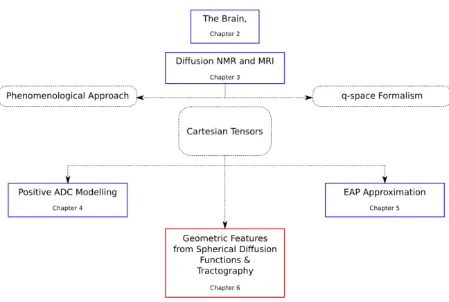
UTLINEThis thesis begins by exploring the physical context of the problem – the brain. Then it presents the principles of the non-invasive modality of dMRI. Finally it presents the problems related to high order dMRI reconstruction techniques that we have ad-dressed and our solutions. This thesis contains the following chapters. An illustration of the flow of the chapters is presented in Fig-1.1.
Chapter-2 The introductory chapter is dedicated to the brain, which forms the phys-ical basis of our problem of study. It presents an overview of the brain’s anatomy, rel-evant microscopic neural tissue, and important macroscopic cerebral tissue organiza-tion, namely the major white matter pathways. These are central for understanding the diffusion of water molecules in the brain, and for understanding the usefulness of dMRI in studying the brain.
Chapter-3 This chapter presents the fundamentals of nuclear magnetic resonance (NMR), diffusion NMR (dNMR) and reviews a number of reconstruction techniques in dMRI that are used to infer the microstructure of the cerebral white matter. A major contribution of this chapter is in its observation of how the ways of modelling dif-fusion has influenced the reconstruction techniques in dMRI. This chapter presents the two ways of modelling diffusion, namely Fick’s phenomenological approach and Einstein’s random walk approach, and develops on these to show how they lead to the two ways for modelling the dNMR signal, namely the Stejskal-Tanner formula-tion and the q-space formalism. These two ways of modelling the dNMR signal play pivotal roles in dMRI, since almost all dMRI reconstruction techniques employ either of these signal models, or both, for reconstructing a diffusion function from the signal to infer the microstructure of the white matter. The dMRI reconstruction techniques
Diffusion NMR and MRI
Chapter 3
Phenomenological Approach q-space Formalism
Cartesian Tensors
EAP Approximation
Chapter 5
Geometric Features from Spherical Diffusion
Functions & Tractography
Chapter 6
Positive ADC Modelling
Chapter 4
The Brain,
Chapter 2
Figure 1.1: Organization of the thesis.
that are reviewed in the final part of this chapter are therefore presented under this light. Finally the review of techniques reveals the current trend towards spherical co-ordinates and the Spherical Harmonic basis, which motivates the work in this thesis with an alternate Cartesian coordinate approach to dMRI.
Chapter-4This chapter addresses the problem of estimating a 4th order Cartesian tensor from the GDTI model with a positive diffusion profile since negative diffu-sion is non-physical. We propose two independent methods, namely a Riemannian approach based on the algebra of 4th order tensors, and a polynomial parameteriza-tion approach based on Hilbert’s theorem on ternary quartics (TQ). We show that the Riemannian approach guarantees positive definite diffusion but solves a more con-strained problem than implied by the GDTI model, while the TQ approach solves the correct problem but guarantees only non-negativity. However, in practice the results of the Riemannian approach are similar to the TQ approach and due to numerical computations we never find zero diffusion from the TQ approach. The relevance of the problem is motivated from the improved results.
Chapter-5 This chapter addresses the problem of estimating the higher order dif-fusion propagator from HOTs. Although the GDTI HOTs estimate the ADC with greater accuracy than DTI, since the ADC describes the effects of microstructural heterogeneity in the signal domain or the q-space, it is necessary to compute the
dif-fusion propagator, which describes the effects of the microstructural heterogeneity in the real space, to infer the geometry of the underlying microstructure. Therefore, while the GDTI model is based on the Stejskal-Tanner formulation of the signal, in this chapter we also employ the q-space formalism to leverage the Fourier Transform relationship between the diffusion signal and the diffusion propagator, and we pro-pose an analytical approximation of the diffusion propagator from a modified GDTI model using 4th order tensors. We show that the approximation converges well to the true diffusion propagator and since it is analytical it is fast and can be implemented efficiently. From the results, we show that it is possible to infer the microstructure from the angular structure of the approximate diffusion propagator.
Chapter-6 In this chapter we address the problem of extracting the geometric in-formation recovered by the higher order models. In DTI the diffusion inin-formation is represented by a diffusion ellipsoid or a 2nd order tensor, where it is straightforward to interpret its geometry from the eigen-decomposition of the tensor. However, in the higher order models, the diffusion information is generally represented by a generic function on the sphere. We propose a complete method for detecting the maxima of a wide class of spherical functions, since the maxima often represent underlying fiber directions. We show, therefore that this method can be used for tracing fibers through regions with fiber crossings by extending the standard Streamline tractography and also the Tensorline tractography to work with generic spherical diffusion functions. We also extract further geometric features from these spherical functions and propose a sub-voxel resolution anisotropy measure to characterize the cerebral white matter.
Chapter-7The last chapter concludes the thesis by summarizing the problems we have addressed and their solutions we have proposed. In this chapter we also present perspectives and directions for developing future research, namely in further explor-ing the problems that can be addressed and solved by the Cartesian coordinate ap-proach to dMRI.
C
HAPTER2
T
HE
B
RAIN
Contents
2.1 Introduction . . . . 8 2.2 The Nervous System . . . . 8
2.2.1 The Brain: Anatomy . . . 9 2.2.2 Building Blocks: The Neuron . . . 11 2.2.3 Grey Matter . . . 13 2.2.4 White Matter . . . 14
2.1
I
NTRODUCTIONThe seat of consciousness and intelligence, the brain has tenaciously de-sisted generations of probing investigators, jealously guarding its secrets over cen-turies from inquisitive minds. Only bit by bit has it revealed the mysteries on “how it functions”, and “how it is constructed”. Twofold are the difficulties in investigating the brain – its sheer complexity, its sophisticated design, and the permanent damage that a direct investigation would cause to the control-center regulating the function-ing of the body and hostfunction-ing the perception of consciousness, resultfunction-ing almost certainly in an irreversible modification of cognition or even in the death of the subject.
Our current knowledge about the brain is the result of enduring and accumulated research, primarily from dissections of the post-mortem brain – both human and an-imal and from experiments on anan-imal brains, since only in these cases are invasive investigations possible or ethically accepted. A great deal about the functional char-acteristics of the brain has also been learned by observing animals and humans who have sustained injuries to their brains, either through accidents or diseases. This is changing with the advent of modern non-invasive and in-vivo imaging techniques of the human body such as Magnetic Resonance Imaging (MRI). While Functional MRI (fMRI), a specialized modality of MRI, addresses the brain on “how it functions”, Dif-fusion MRI (dMRI), another specialized modality of MRI and the central topic of this thesis, is well suited for addressing “how it is connected”.
This chapter is dedicated to a quick perusal of the brain – its general structure and organization, the tissues constituting it, and in highlight, it’s major neuronal pathways interconnecting its various regions. This chapter aims to provide a con-text for understanding the general physical problem dMRI attempts to solve. The contents of this chapter have been collected from the following various sources [1, 2, 3, 4, 5, 6, 7, 8, 9, 10], and from the Internet.
2.2
T
HEN
ERVOUSS
YSTEMThe role of the human nervous system is to coordinate the functions of the body, both basic or unconscious, and intricate and deliberate. It gathers information on the internal and external environment from its sensory receptors, integrates the information to determine the appropriate response, and sends out signals to muscles and glands to actuate the response. At the most fundamental level, the nervous sys-tem propagates signals from one cell to others. This is done chiefly in two ways. Nerve cells can either transmit electrochemical waves or action potentials along wire-like axons interconnecting them to send out signals to specific target cells from one point to another, or they can engage the endocrine system to activate glands that release hormones into the internal circulation that diffuse to distant cells like a broadcast system. At the global level, the nervous system not only regulates voluntary and
in-voluntary bodily functions such as breathing, the beating of the heart, blood pressure, body temperature, movement of muscles and the limbs, but also makes possible more advanced and elaborate modes of perception and communication such as awareness, emotions, languages, ideas and abstraction of concepts, transmission of cultures and other expressions of cognition, behaviour and features of society.
The nervous system is subdivided anatomically into the central nervous system (CNS) and the peripheral nervous system (PNS), and functionally into the somatic nervous
system(SNS) and the autonomic nervous system (ANS). The CNS is comprised of the brain and the spinal cord and is responsible for all the central processing. The PNS is an extension of the CNS, and consists of cranial and spinal nerves emerging from the brain and the spinal cord respectively, that connect the CNS to sensory receptors, muscles and glands. Functionally the SNS is comprised of all the structures of the CNS and the PNS that convey sensory or afferent signals consciously or unconsciously from extremities to the CNS such as vision, pain, touch, muscle tone, etc., and those that convey motor control or efferent signals from the CNS to voluntary or striated muscles. The ANS on the other hand is comprised of structures of the CNS and the PNS that convey afferent input from internal organs to the CNS, and those that convey efferent signals from the CNS to involuntary or smooth muscles, such as the cardiac muscles, and glands.
2.2.1 The Brain: Anatomy
The brain is the primary building block of the nervous system and its central process-ing unit. The adult human brain on an average weighs 1500g. It is a soft and delicate organ that is encased within the thick bones of the cranium and is wrapped in three layers of membranes known as the meninges. The brain floats in the cerebrospinal
fluid(CSF), a transparent colourless fluid, which also fills the open chambers in the brain or the ventricular system, and spaces in and around the brain. The CSF brings nutrients to the brain, removes wastes, provides an immunological protection, and supports the brain mechanically through buoyancy. It acts as a “water-jacket” or a “shock-absorber” cushioning the brain against sudden jolts and head movements. The buoyancy also reduces the weight of the brain sixty-fold to about 25g, thus protecting its soft tissue from being crushed under its own weight.
Described simply, the brain is anatomically subdivided into the cerebrum, the
brain-stem, and the cerebellum. The cerebrum, the top-most part of the brain, is constituted of two large lateral hemispheres known as the cerebral hemispheres, and the
dien-cephalon. The surface of the cerebral hemispheres is constituted of grey matter and is topographically highly folded or wrinkled and is marked by the formation of slit-like fissures or valleys known as sulci and raised ridges between these fissures known as gyri. The mid-line or the longitudinal fissure separates the two hemispheres. The
Figure 2.1: (a) The divisions of the brain, adapted from [7]. (b) The four major lobes of the cerebral cortex [6].
cerebral hemispheres are each divided into four lobes roughly separated by important sulci. These are the frontal lobe, the parietal lobe, the occipital lobe, and the temporal
lobe. The frontal lobe is involved in high level thinking, planning, decision-making, and execution of movements. The parietal lobe is important in sensory perceptions such as touch and pressure, and also helps in spatial orientation and information processing. The occipital lobe is the visual centre of the brain and processes the infor-mation from the eyes. The temporal plays a key role in auditory processing and the storing of memory.
The diencephalon is mainly constituted by the thalamus and the hypothalamus, which are deep brain structures located just below the cerebral hemispheres and above the midbrain which is a part of the brainstem. The thalamus is a crucial centre for integrating and relaying motor and sensory information to the cerebral hemispheres for higher processing. It is critical for cognition and awareness. The hy-pothalamus regulates functions such as hunger, thirst, pain, and pleasure. One of its most important functions is to link the CNS to the endocrine system via the pituitary gland. Its influence is widespread and is even involved with emotions and behavior. The brainstem is the bottom-most part of the brain and connects the brain to the spinal cord. It has three major parts – the midbrain, the pons, and the medulla
oblongata. The brainstem regulates the most basic functions of the body such as consciousness, the sleep-wake cycle, and respiratory and cardiovascular control. The cerebellum is located at the back of the head, between the cerebrum and the brainstem, behind the medulla and the pons. Its role is in controlling balance, pos-ture, storing learned movements, and in synchronizing contractions of muscles to smooth out responses by regulating muscle tensions.
Functionally speaking, the top-most part of the brain is the most complex and has the greatest influence on conscious action. Moving lower into the brain, the parts
become increasingly primitive and are responsible for more basic functions that re-quire less conscious control. However, all these parts have to intercommunicate to coordinate their actions for the correct overall functional responses. This communi-cation is possible due to a network of connectivities with highly organized structures that constitute the so called white matter of the brain. Inferring the structure of this white matter non-invasively is the central problem that dMRI attempts to resolve.
2.2.2 Building Blocks: The Neuron
At a microscopic scale the brain is a network of neurons containing on an average 100 billion(1011) neurons, each connected to about 1000 other neurons, totalling ap-proximately to 100 trillion (1014) synaptic connections, wired together by 150,000 –
180,000 km of nerve fibers with diameters in the range of 0.3µm – 10µm. These humongous numbers can be fully comprehended when compared to other similarly humongous numbers. It is estimated (very roughly) that the number of atoms in the
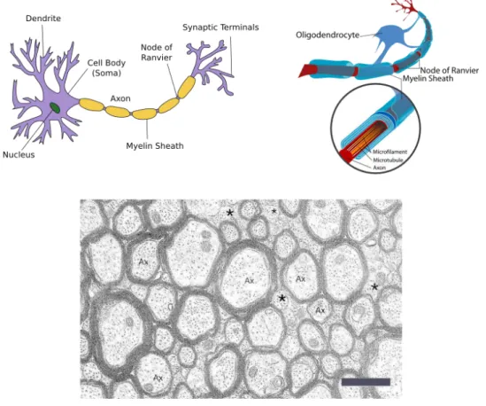
Figure 2.2: Top left: the neuron. Adapted from Wikipedia (Neuron). Top right: an oligodendrocyte creating myelin sheath to wrap the axon. Adapted from Wikipedia (Oligodendrocyte). Bottom: Electron-micrograph of axons from the Corpus Callosum. Cross section view. Myelin sheaths appear as dark bands around paler axons. Ax: axon, *: small axons that aren’t myelinated, scale-bar: 1µm. Adapted from [5].
observable universe is of the order of1080, the number of stars in the observable uni-verse is of the order of1020, and the number of stars in our galaxy, the Milky Way, is
of the order of 1011. Indeed, with the number of neurons comparable to the number of stars in our galaxy, interconnected by microscopic wiring with a total length of a hundred thousand kilometers, the brain is highly a complex and sophisticated organ. The neuron is an electrically excitable cell capable of processing and transmitting information by electrochemical signalling. It is the fundamental processing unit of the brain. The brain is primarily constituted of neurons and glial cells which support and maintain neurons. The neurons have a cell body containing a nucleus, dendrites, which are short filaments attached to the body that branch out forming a tree-like structure, and a single long axon, a cable-like fiber extending from the neuronal body and ending in branched out synaptic terminals. The dendrites receive electrochemical stimuli from adjacent neurons via the synaptic terminals of their axons, and act as input sources for the neuronal cell body. The cell body processes this information and is then capable of retransmitting a signal via the axon. The signal travels along the axon and is transmitted via synapses at its terminals to dendrites of adjacent and connected neurons.
Axons extend over distances that can be exceedingly long when compared to their diameter of a few microns. In the human brain this can reach up to a meter, which is a difference of six orders of magnitude. However, bare axons are poor transmitters of electrochemical signals due to leakage. Special glial cells called oligodendrocytes wrap the axons in a fatty substance known as myelin to considerably improve both the efficiency and the speed of the transmission. The insulation or myelin sheaths are created in regular lengths and are separated by tiny gaps known as nodes of Ran-vier. Such axons are known as myelinated axons. A loss of myelin or demyelination disrupts signal transmission and can result in functional pathologies of the brain. Myelin, which is whitish in colour, imparts a whitish tinge to myelinated axons. Neurons differ from other cells by the fact that they don’t replicate themselves. Therefore, not only do injuries to the brain and degenerative pathologies result in a permanent loss of affected neurons, but even the process of aging results in a pro-gressive loss of neurons and axons. Between the age of twenty years and ninety years there is a loss of 9.5% of the total neurons and a loss of 40 – 50% of the total length of all the myelinated axons.
dMRI can non-invasively infer the highly organized structures formed by axons traversing the brain interconnecting its various regions. Therefore, dMRI is a suit-able tool for studying the demyelination and degeneration of axons due to injuries, pathologies and aging.
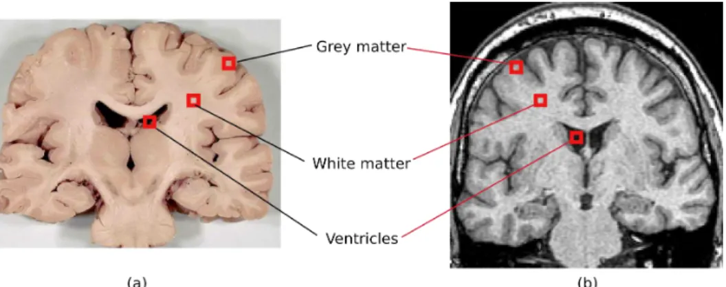
Figure 2.3: Grey & white matter and the ventricles in a coronal slice. (a) adapted from www.healcentral.org. (b) T1 image adapted from the Whole Brain Atlas.
2.2.3 Grey Matter
At a macroscopic scale the brain is made up of grey matter and white matter. Grey matter is constituted of neuron cell-bodies, dendrites, synapses, and glial cells. It is grey brown in colour due to the capillary blood vessels and the neuronal cell-bodies. White matter is constituted of myelinated axons and oligodendrocytes. Since grey matter is constituted mainly of neuronal cells, grey matter regions are the processing centres of the brain. The topographies of both grey matter and white matter are highly organized.
The largest, most important and most complex formation of grey matter is the surface of the cerebral hemispheres known as the cerebral cortex. The extensive wrinkling or folding of this surface increases the area of the cortex threefold and about two thirds of the grey matter of the cerebral cortex is buried in the sulci of the cerebral hemispheres. The cerebral cortex handles the most complex or highest processes of the brain. Evolutionarily speaking it is the most recently developed part of the brain, and its topographic complexity has increased over time. For example in rodents and small mammals the cerebral cortex is smooth, and it develops sulci in primates and larger mammals.
Other important regions of grey matter include the basal ganglia which are located deep within the cerebrum, and connected to the cerebral cortex and the thalamus. The various grey matter nuclei of the basal ganglia – the caudate nucleus, putamen,
globus pallidus, and substantia nigra, are involved in voluntary motor control, eye movements, cognition and emotions. There exist other important grey matter regions such as nuclei in the thalamus and the hypothalamus, the surface of the cerebellum or the cerebellar cortex, etc.
Figure 2.4: Commissural fibers. (a) top view of the Corpus Callosum (CC), adapted from [11]. (b) Divisions of the CC, adapted from www.healcentral.org. (c) The CC and the anterior commissure, adapted from www.humannervoussystem.info.
2.2.4 White Matter
While the grey matter is made up of the neuronal cell bodies, the myelinated ax-ons originating from these neurax-ons cax-onstitute the white matter, which gets its colour and name from the colour of the myelin sheaths. The myelinated axons interconnect various grey matter structures, and traverse the brain in highly organized conglom-erations or pathways. While the surface of the cerebral hemispheres is the cerebral cortex, a grey matter structure, much of the volume beneath is formed by these white matter pathways interconnecting different regions of the cortex, and connecting the cortex to other grey matter structures such as deep brain nuclei, the cerebellum, and the spinal cord. These pathways give white matter a coherent and fibrous qual-ity from the microscopic to the macroscopic scale. This forms the physical basis of dMRI’s usefulness and suitability for studying the connectivity of the brain.
Figure 2.5: Short and long association fibers. Short or “U”-fibers, the cingulum, the superior longitudinal fasciculus (SLF), and the inferior longitudinal fasciculus (ILF). (a) Adapted from [6]. (b) Adapted from [11].
The abundantly present water molecules in the white matter diffuse in a very par-ticular fashion within this medium. The coherent fibrous structure of white matter hinders diffusion perpendicular to its filaments and fibers, while the diffusion paral-lel to these structures is left relatively unaffected. In other words as water molecules diffuse in white matter due to thermally driven Brownian motion, they probe its fi-brous structure. Since dMRI is sensitive to the diffusion of water molecules, it can be used to measure this constrained or anisotropic diffusion in white matter, which makes it possible to indirectly perceive the fine microstructure of the medium and to non-invasively infer the white matter pathways.
The major white matter pathways can be classified into three groups.
Commissural Fibers The commissural or transverse fibers (Fig-2.4) connect mir-roring and different sites between the two cerebral hemispheres. The most important commissural structure is the corpus callosum (CC), which is also the largest white matter structure. It contains about three hundred million fibers. The CC is divided into the rostrum, the genu, the body, and the splenium from anterior to posterior. Commissural fibers traversing the CC in the genu and curving into the frontal lobe make up the forceps anterior (minor). Similarly fibers traversing in the splenium and curving into the occipital lobe make up the forceps posterior (major). Another impor-tant commissural structure is the anterior commissure (AC). The CC and the AC are responsible for inter-hemispheral communication.
Association Fibers The association fibers (Fig-2.5) connect regions of the cerebral cortex within the same hemisphere. Short association fibers, also known as “U”-fibers connect adjacent gyri, going around the sulcus separating them. These lie immediately beneath the grey matter of the cerebral cortex. Long association fibers
Figure 2.6: Projection fibers. (a) The corona radiata (CR), the anterior thalamic ra-diation (ATR), and the corticospinal tract (CST). Adapted from [11]. (b) The effer-ent/motor (red: CST) fibers and the afferent/sensory (blue) fibers. Adapted from [6].
Figure 2.7: Region with three fiber bundles crossing. CST: vertically in the plane. CC: diagonally in the plane. SLF: perpendicular to the plane. Figures adapted from, left: www.healcentral.org, right: www.humannervoussystem.info.
travel greater distances to connect distant cortical regions. Some of such important association fibers are the cingulum, the superior longitudinal fasciculus (SLF), and the inferior longitudinal fasciculus (ILF).
Projection Fibers The projection fibers (Fig-2.6) consist of efferent and afferent fibers connecting the cerebral cortex to the cerebellum, the spine and subcortical grey matter areas such as the thalamus, and the basal ganglia. Functionally, the main efferent fibers are the motor fibers originating from the pyramidal motor cells of the cerebral cortex, that run down and converge in the internal capsule and travel further down the brainstem to the spine. These comprise the important corticospinal
tract(CST) also known as the pyramidal tract. Structurally, the projection fibers are widely diffused just under the cerebral cortex before converging in the internal cap-sule. This important fanning structure is known as the corona radiata (CR). The CR is comprised of both efferent and afferent fibers, and handles nearly all the traffic to and from the cerebral cortex. Structures like the anterior thalamic radiation (ATR) connect the nuclei in the thalamus to cerebral cortex of the frontal lobe.
It must be noted that these white matter pathways or fiber tracts or simply fibers often criss-cross, resulting in geometrically complex microstructures at a scale much finer than the resolution of dMRI. This is therefore an important physical consider-ation in dMRI, and correctly inferring the geometry in regions with fiber-crossings is addressed as a central problem. In this respect, Fig-2.7 presents a region with fiber crossings that will be often scrutinized in this thesis. The region of interest highlighted in the coronal slice contains crossing fibers from three major pathways. Descending vertically in the plane is the CST, traversing diagonally in the plane from above the ventricles to the cerebral cortex are the lateral radiations of the CC, and within the central region of interest is the SLF perpendicular to the plane.
2.3
S
UMMARYIn this introductory chapter we briefly presented the brain with the goal of providing a physical context of the problem dMRI attempts to solve. We presented an overview of the brain’s anatomy, relevant microscopic neural tissue, and important macroscopic cerebral tissue organization, namely the major white matter pathways. These are central for understanding the diffusion of water molecules in the brain, and for understanding the usefulness of dMRI in studying the brain.
At a macroscopic scale, the brain is primarily constituted of grey matter and white matter. While the grey matter contains the neuronal cell bodies and forms the main information processing centers of the brain, white matter is made up of the axons originating from the neurons, which interconnect various grey matter regions and form information carrying pathways with highly coherent fibrous structures from the microscopic to the macroscopic scale. These fibrous microstructures of the white matter form the physical basis for dMRI’s usefulness in studying the brain. The Brownian motion of the water molecules contained in the brain is hindered in the white matter by these fibrous structures in a particular fashion. While the diffusion of water molecules is greatly hindered perpendicular to these structures, the diffusion parallel to these structures is relatively less affected. In other words the diffusing water molecules probe the white matter’s microstructure. Therefore, since dMRI is sensitive to the diffusion of water molecules, it is used to measure the constrained or anisotropic diffusion of water molecules in the white matter, to infer its major axon fiber bundles non-invasively.
However, since the white matter axon fibers criss-cross in many regions, the cerebral white matter often contains complex structures constituted of multiple coherent mi-crostructures at a scale that is orders of magnitude finer than the resolution achieved by current dMRI. Although the coherence or homogeneity of the multiple microstruc-tures in such regions is visible at the fine length scale of the axons themselves, the homogeneity is lost at the resolution achieved by dMRI and such regions appear to contain heterogeneous microstructures. Therefore, the problem of accurately infer-ring the geometry of the microstructure in regions with fiber crossings is of central importance in dMRI. This problem is also at the heart of this thesis.
C
HAPTER3
D
IFFUSION
NMR
AND
MRI
Contents
3.1 Introduction . . . . 20 3.2 Nuclear Magnetic Resonance . . . . 22
3.2.1 Spin Echo . . . 26
3.3 Diffusion NMR . . . . 28
3.3.1 Diffusion . . . 28 3.3.2 Diffusion from NMR: A Phenomenological Approach . . . 33 3.3.3 q-space Formalism: A Random Walk Approach . . . 36
3.4 Diffusion MRI . . . . 40
3.4.1 Diffusion Tensor Imaging . . . 41 3.4.2 ADC & Generalized DTI . . . 45 3.4.3 Diffusion Spectrum Imaging . . . 49 3.4.4 Other Methods . . . 50 3.4.5 EAP Estimation . . . 54
3.1
I
NTRODUCTIONOnly in the last century, has physics, mathematics, and technology pro-gressed enough to be en par with the complex and involved problem of understand-ing the brain that has perplexed people so far. New and non-invasive approaches now provide an unprecedented power to see within this delicate organ while it functions and without harming the process of life.
Magnetic Resonance Imaging (MRI) is one such sophisticated technique, which allows to examine the brain (and also the entire body) non-invasively. At the core of MRI is the phenomenon of nuclear magnetic resonance (NMR). The NMR experiment can detect a number of different properties of a sample. For example it can detect the Brownian motion or the diffusion of the particles in the sample. This sensitivity to diffusion and the measurement of the diffusion properties from an NMR experiment is at the heart of diffusion MRI, which allows one to infer from the diffusion proper-ties, the microstructure of the brain’s white matter in-vivo and non-invasively. This chapter is focussed on the fundamentals of NMR and diffusion NMR, and on diffusion MRI reconstruction algorithms, which are employed to infer the cerebral white matter’s microstructure. This chapter opens with the NMR phenomenon and the ingenious spin-echo experiment, which forms the foundation of diffusion NMR. Then it presents diffusion NMR to show how diffusion can be measured from NMR, with an emphasis on the two ways of modelling diffusion, namely the phenomenolog-ical approach of Fick and the random walk model proposed by Einstein, since these have greatly influenced the development of diffusion NMR. These have given rise to two complementary approaches for modelling the diffusion NMR signal, namely the Stejskal-Tanner signal model, and the q-space formalism. Finally this chapter presents a number of diffusion MRI reconstruction algorithms, starting from the clas-sical technique of diffusion tensor imaging, and then exploring various other “higher order” approaches, which attempt to describe the microstructure of the cerebral white matter with greater accuracy.
A brief history of NMR and MRI The scientific heritage of NMR and MRI is re-flected by the list of Nobel laureates who contributed to their developments. The theoretical underpinnings that made NMR possible were proposed in 1924 by Wolf-gang Pauli who suggested a new quantum degree of freedom that later came to be known as spin. He formulated the mathematical theory by 1927, and was awarded the Nobel prize in physics in 1945 for his contributions. The concept of spin implies that atomic nuclei bearing spins exhibit magnetic moments. The fact that protons exhibit magnetic moments had already been discovered in 1922 by Otto Stern prior to the concept of spin. Stern was awarded the Nobel prize in physics in 1943. Pauli’s theory was verified in 1938 by Isidor Rabi in molecular beams. From his experiments Rabi was able to both detect the effects of spin and measure the gyromagnetic ratio
that is the characteristic signature of an atomic nucleus due to its spin. His experi-ments also established the concept and the technique of NMR for manipulating spins. Rabi was awarded the Nobel prize in physics in 1944.
In 1946 Felix Bloch [12] and Edward Mills Purcell [13] independently extended the techniques established by Rabi. They successfully demonstrated the magnetic reso-nance effect in liquids and solids. Bloch and Purcell shared the Nobel prize of 1952 in physics, and NMR was established. In his seminal paper of 1950 [14] Erwin Hahn proposed the spin echo experiment, which used a combination of 90o and 180o elec-tromagnetic or radio frequency pulses to filter out effects of magnetic field inhomo-geneities in the measurement of the transverse signal. Further works of Herman Carr and Purcell in 1954 [15] led to the full development of the radio frequency pulse technique introduced by Hahn. This formed the foundations of NMR.
It must be noted at this point that both the papers of Hahn [14] and Carr & Pur-cell [15] critically point out the observed effects of diffusion of the spin bearing nu-clei in magnetic resonance experiments with a succession of radio frequency pulses. Although these papers generally perceive the diffusion effect as an unfortunate phe-nomenon resulting in a loss of signal, Carr & Purcell [15] in fact demonstrate that diffusion can be directly measured from NMR and go on to actually measure the dif-fusion constant of water at 25oC. This forms the corner-stone of diffusion NMR. Although NMR became a well established technique for studying various materials, it took almost three decades since the experiments of Bloch and Purcell in 1946, for MRI to be invented. NMR by itself is capable of examining a single spin ensemble or a tiny region of a sample, but it can’t image the whole sample to recreate a 2D slice or a 3D volumetric image necessary to study entire biological samples like the human body. Paul Lauterbur in 1973 [16] proposed the use of magnetic gradient fields to spatially encode the positions or voxel regions of the spin ensembles. This was a remarkable invention, which made it possible to reconstruct entire slice or volumetric images from NMR data. Spatial encoding was improved in terms of frequency encoding by Richard Ernst in 1978, and phase encoding by Bill Edelstein in 1980 using pulsed gradients. In 1977 Peter Mansfield [17] developed the mathematical framework for rapidly switching gradients for spatial encoding, greatly speeding up the process of reconstructing images of an entire biological sample. This is known as echo planar
imaging (EPI). Lauterbur and Mansfield were jointly awarded the Nobel prize in medicine in 2003 for making MRI possible. Thus modern MRI was developed from the phenomenon of NMR coupled with the method of spatial encoding.
O
UTLINEThis chapter is divided into three parts dedicated respectively to NMR, diffusion NMR and diffusion MRI. Section-3.2 presents the semi-classical description of NMR
and the spin-echo experiment proposed by Hahn (section-3.2.1), which forms the cor-ner stone for the diffusion NMR experiment. Section-3.3 presents diffusion NMR and shows how diffusion can be measured from the NMR experiment. Section-3.3.1 discusses the two main approaches for modelling diffusion mathematically, namely Fick’s phenomenological approach and Einstein’s random walk model. Sections-3.3.3 & 3.3.2 attempt to show how these two complementary approaches to diffusion, one macroscopic and one microscopic, have influenced the development of diffusion NMR. Finally, section-3.4 presents modern diffusion MRI reconstruction techniques where, using the diffusion NMR experiment, the diffusion measured from anisotropic me-dia is employed to infer the microstructure of the meme-dia. A number of approaches are presented, starting from the classical diffusion tensor imaging, to more recent state-of-the-art algorithms that make use of complex models and functional bases to improve the results of diffusion tensor imaging.
3.2
N
UCLEARM
AGNETICR
ESONANCEThe principles of NMR are based on spin, a fundamental quantum charac-teristic possessed by electrons, protons, and neutrons, like electrical charge or mass. Spins come in multiples of1/2 and can be positive or negative. In grouped particles, e.g. atomic nuclei, opposite spin-signs can pair up to eliminate the total spin of the group. But the net spin of unpaired particles or atomic nuclei imparts a magnetic dipole moment. In other words such particles or such atomic nuclei can be influenced by an external magnetic field. In the presence of a strong magnetic field B0 with
magnitudeB0, the magnetic dipole moment vector or the spin vector of the particle
or nucleus aligns itself with B0 and precesses around it with an angular frequency
known as the Larmor frequency:
ω0= γB0,
whereγ is the gyromagnetic ratio, characteristic of the particle or the nucleus. How-ever, this cannot be observed for individual particles or nuclei. The effect is detectable when it becomes pronounced in the presence of an ample collection of spin bearing particles or nuclei with the same gyromagnetic ratio. From a macroscopic perspective, when such a collection is subjected to a magnetic field, the randomly oriented individ-ual magnetic dipole moment vectors align themselves along B0, to form a resultant
ensemble magnetic dipole moment vector M, which phenomenologically satisfies: dM
dt = γM × B0. (3.1)
Although spin is a quantum characteristic, the macroscopic perspective with the re-sultant magnetic dipole moment vector M, is a semi-classical description.
Of particular interest is the hydrogen nucleus 1H, which is found abundantly in na-ture, accounts for 99.98% of all hydrogen atoms, and also constitutes water. 60% of
the human body and 78% of the brain is water. Therefore,1H is a natural spin bear-ing nucleus of choice for MRI.1H is an unpaired proton with a net spin of 1/2, and
has a gyromagnetic ratio ofγ = 42.58 MHz/T.
A macroscopic scale or an ensemble perspective is adopted to describe MRI instead of the quantum formulation because of its simplicity. From this point of view, MRI is explained in terms of the resultant ensemble magnetization vector M. Under the influence of the external magnetic field B0, the initially randomly distributed
indi-vidual spins in the ensemble, align themselves along B0. There are two available
energy configurations or states. These are the low and high energy states depending upon whether the individual spin magnetization vectors point along the magnetic field or opposite to it. Laws of thermodynamics ensure that the number of spins N+ in the low energy configuration slightly outnumber the number of spins N− in the
high energy configuration. However, this implies that the magnitude of the net mag-netization vector M is only a fraction of what it would have been had all the spins been pointing along the same direction. This natural distribution of spins along or opposite B0 is known as the thermal equilibrium state.
The NMR signal is generated by exposing the ensemble of spins precessing along B0
to an oscillating magnetic field or an electro-magnetic (radio-frequency: RF) pulse. This is known as the excitation phase. The energy absorbed by the low energy config-uration spins from this pulse tilts the magnetization vector M away from B0towards
the high energy configuration. The oscillation of the secondary magnetic field ensures that the spins (and hence M) continue to precess around B0 even tilted away from
it – along the surface of a cone. Once the RF pulse is switched off, the spins begin to recover their alignment with the main magnetic field B0, and to return to their
low energy configuration or the thermal equilibrium. This is known as the relaxation phase. The signal is created as the spins precess tilted away from B0, and it decays
as the spins relax, dissipating the absorbed energy.
This process can be better understood and put in a mathematical framework by con-sidering the external magnetic field B0 to be aligned with the Z-axis. The XY-plane
is then known as the transverse plane, and the net magnetization vector M can be separated into the longitudinal component Mz, along the Z-axis (or B0), and the
transversecomponent Mxy, in the transverse plane. Or the net magnetization vector
Mcan be written in terms of its components[Mx, My, Mz]T. In this framework the low
energy configuration is along the positive Z-axis, while the high energy configuration is along the negative Z-axis. {X,Y,Z} is known as the laboratory frame of reference and is considered fixed. Under the influence of B0, the initial net magnetization
vec-tor M has magnitude M0, and is equal to its longitudinal component Mz, while its
transverse component Mxy is null.
Under the influence of B0, as the spins precess at the Larmor frequency, it is possible
to envisage a rotating frame of reference {X’, Y’, Z’ (= Z)}, with the Z’-axis aligned with the Z-axis of the fixed frame of reference. This rotating frame is taken to rotate clockwise with the spins at the Larmor frequency. In other words in the rotating frame of reference, the precessing spins appear static. Applying a secondary magnetic field in the rotating frame of reference along the X’-axis, in the transverse plane, has the effect of tilting the net magnetization vector M away from B0 and towards the
transverse plane, which is half way to the high energy configuration. In the fixed frame of reference this is seen as the net magnetization vector M spiralling down towards the transverse plane while precessing at the Larmor frequency. In either of the reference frames this implies that the magnitude of the longitudinal component Mz decreases, while the magnitude of the transverse component Mxy increases.
In practice, this can be achieved by passing an alternating current through a coil aligned with the X-axis of the fixed frame of reference. When the current in the coil alternates at the Larmor frequencyω0, the coil generates an oscillating magnetic field
along the X-axis that appears static to the precessing spins. Or the precessing spins perceive the oscillating magnetic field as a static magnetic field in the rotating frame of reference. This secondary magnetic field is known as the B1 magnetic field or the
RF pulse. The angle of the tilt experienced by the net magnetization vector M due to B1 depends on the magnitude and time of exposure of the RF pulse. A 90o RF pulse
tilts M into the transverse plane and zeros out its longitudinal component. A 180o
RF pulse flips M completely around and points it along its opposite direction.
The relaxation phase begins when the RF pulse or the secondary magnetization field is turned off. The RF pulse has the effect of disturbing the thermal equilibrium in-duced by the primary magnetization vector B0. As it is turned off the spins begin to
relax and to return to their thermal equilibrium. The relaxation can also be seen in terms of its longitudinal and transverse components. However, these are governed by different phenomena and are characterized by different time signatures.
T1 Relaxation The longitudinal relaxation is known as the T1 relaxation since it is described using a time signature denoted T1. The T1 relaxation occurs as the spin ensemble radiates the energy it had absorbed from the RF pulse to the surround-ing thermal reservoir or lattice and regains its thermal equilibrium with the lattice. Therefore, the T1 relaxation is also known as the spin-lattice relaxation. In this pro-cess the spins realign themselves with B0. In terms of the net magnetization vector
M, this implies that the longitudinal component Mz progressively regains its
ini-tial magnitude M0, while the transverse component Mxy progressively becomes null
again. Phenomenologically this is described by: dMz
dt = −
(Mz− M0)
T1
whereT1 is a time constant representing the time signature T1.
T2 Relaxation The transverse relaxation involves the phenomenon of the spins regaining their thermal equilibrium amongst themselves, and is characterized by the time signature T2. Therefore it is also known as the spin-spin relaxation or the T2 relaxation. In the transverse plane this is interpreted by the spins losing their initial coherence. From an initial coherent transverse magnetization vector Mxy, they
progressively dephase as they radiate the energy they had absorbed to neighbouring spins. Phenomenologically this is described by:
dMxy
dt = − Mxy
T2
, (3.3)
where T2 is a time constant that differs from T1 and represents the time signature
T2. Transverse relaxation is, however, a complex phenomenon. Although theoret-ically B0 is supposed homogeneous, in reality minor inhomogeneities exist. These
inhomogeneities are relevant enough to also contribute to spins dephasing in the transverse plane, though this is not a true relaxation. Transverse relaxation is there-fore a combination of spin-spin relaxation and field inhomogeneity dephasing. The pure spin-spin relaxation time is known as T2. The combined transverse relaxation time is known as T2∗.
Bloch Equations The Bloch equations are a coupled set of three differential equa-tions that combine the effects of NMR and describe the evolution of the net magne-tization vector M over time. These are macroscopic and phenomenological equations that include the effects of Larmor precession and T1 and T2 relaxations. Combining Eqs-3.1, 3.2, and 3.3, they are written in the fixed frame of reference in terms of the relaxation time constants as:
dMx(t) dt = (γM(t) × B(t))x− Mx(t) T2 (3.4) dMy(t) dt = (γM(t) × B(t))y− My(t) T2 dMz(t) dt = (γM(t) × B(t))z− Mz(t) − M0 T1 ,
where B(t) = B0 + B1(t) is the total external magnetic field. These can also be
rewritten in a vector form as: dM(t) dt = γM(t) × B(t) + −T12 0 0 0 −T1 2 0 0 0 −T1 1 M(t) + 0 0 M0 T1 . (3.5)
The signal is generated in a receiver coil in the transverse plane when the spins precess tilted away from the Z-axis. When the magnetic moment vectors of the spins
are tilted to say the transverse plane and precess around the Z-axis, they generate an alternating current in the receiver coil at the Larmor frequency. This can be seen as the converse of the spin excitation phase where a coil in the transverse plane was used with a current alternating at the Larmor frequency to tilt the spins. The frequency of the signal current is ω0 and its amplitude reflects the amount of magnetization
of the spin ensemble. Hence, since the 1H nucleus is chosen for imaging in MRI, the excitation RF pulse is generated at the Larmor frequency of the 1H nucleus.
Therefore, the magnitude of the signal generated from the NMR experiment reflects the density of the1H nucleus, or the amount of water in the tissue.
3.2.1 Spin Echo
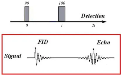
Diffusion NMR is derived from the spin echo experiment that was conceived by Er-win L Hahn [14]. It combines a pair of RF pulses of different amplitudes to remove the effects of field inhomogeneities or T2∗ from the signal. Hahn put forth the idea that following a 90o RF pulse that tilts the net magnetization vector to the trans-verse plane, the dephasing that follows caused by the field inhomogeneities, could be refocused using a second RF pulse of 180o, thus removing the effects of the field inhomogeneities.
Again this can be understood simply in the rotating frame of reference. In this frame of reference, after the 90o RF pulse, the spins precessing in the transverse plane should appear static. However, due to field inhomogeneities, as the spins begin to dephase, some would appear to speed up (or move ahead clockwise in the rotating frame of reference), while some would slow down (or fall back anti-clockwise in the
Figure 3.1: Spin Echo pulse sequence showing the Free Induction Decay (FID) af-ter the 90o RF pulse and the Echo signal after the 180o RF pulse. Adapted from Wikipedia (Spin Echo Signal).
![Figure 2.4: Commissural fibers. (a) top view of the Corpus Callosum (CC), adapted from [11]](https://thumb-eu.123doks.com/thumbv2/123doknet/14697071.746350/31.893.114.737.161.361/figure-commissural-fibers-view-corpus-callosum-cc-adapted.webp)