HAL Id: tel-01726966
https://tel.archives-ouvertes.fr/tel-01726966
Submitted on 8 Mar 2018
HAL is a multi-disciplinary open access
archive for the deposit and dissemination of sci-entific research documents, whether they are pub-lished or not. The documents may come from teaching and research institutions in France or abroad, or from public or private research centers.
L’archive ouverte pluridisciplinaire HAL, est destinée au dépôt et à la diffusion de documents scientifiques de niveau recherche, publiés ou non, émanant des établissements d’enseignement et de recherche français ou étrangers, des laboratoires publics ou privés.
Molecular mechanisms of exocytosis-endocytosis
coupling in neuroendocrine cells : role of Scramblase-1
and Oligophrenin-1 proteins
Catherine Estay Ahumada
To cite this version:
Catherine Estay Ahumada. Molecular mechanisms of exocytosis-endocytosis coupling in neuroen-docrine cells : role of Scramblase-1 and Oligophrenin-1 proteins. Neurobiology. Université de Stras-bourg, 2016. English. �NNT : 2016STRAJ088�. �tel-01726966�
UNIVERSITÉ DE STRASBOURG
ÉCOLE DOCTORALE DES SCIENCES DE LA VIE ET DE LA SANTE
THÈSE
présentée par :
Catherine Estay Ahumada
soutenue le : 2 Décembre 2016
pour obtenir le grade de :
Docteur de l’Université de Strasbourg
Discipline:
Science du vivantSpécialité : Aspects moléculaires et cellulaires de la biologie
THÈSE dirigée par :
GASMAN Stéphane Directeur de recherche INSERM, Université de Strasbourg
RAPPORTEURS :
DANGLOT Lydia Chargé de recherche INSERM, Université de Paris Diderot
BENHAMOU Marc Chargé de recherche INSERM, Université de Paris Diderot
AUTRE MEMBRE DU JURY :
Professeur Université de Strasbourg
LELIEVRE Vincent
Mécanismes moléculaires du couplage exocytose-endocytose
dans les cellules neuroendocrines : rôle des protéines
Scramblase-1 et Oligophrénine-1
INVITE :
UNIVERSITÉ DE STRASBOURG
ÉCOLE DOCTORALE DES SCIENCES DE LA VIE ET DE LA SANTE
THÈSE
présentée par :
Catherine Estay Ahumada
soutenue le : 2 Décembre 2016
pour obtenir le grade de :
Docteur de l’Université de Strasbourg
Discipline:
Science du vivantSpécialité : Aspects moléculaires et cellulaires de la biologie
Soutenue publiquement le 2 Décembre 2016 devant le jury composé de : Dr. DANGLOT Lydia (Rapporteur externe)
Dr. BENHAMOU Marc (Rapporteur externe) Dr. LELIEVRE Vincent (Examinateur interne) Dr. GASMAN Stéphane (Directeur de thèse) Dr. ORY Stéphane (membre invité)
Mécanismes moléculaires du couplage exocytose-endocytose dans les
cellules neuroendocrines : rôle des protéines Scramblase-1 et
Acknowledgements
First of all I would like to thank the jury members for accepting the proposition to judge my thesis. It is a pleasure for me tho know that my work is evaluated by such experts as Dr. Lydia Danglot specialist in cellular neurobiology and membrane trafficking; Dr. Marc Benhamou specialist in signaling and phospholipid scramblase 1 and Dr. Vincent Lelièvre specialist in neuroscience.
I would like particularly to thank my supervisor, Dr. Stéphane Gasman for allowing me to make my thesis in this laboratory, give me the opportunity to perform science and help me in the writing of this manuscript thesis.
Especially I would like to thank Dr. Stéphane Ory, for which I feel a great admiration, thank you for all your help during the realization of my thesis, design of experiments, analysis and also for all the advice on the writing of this manuscript thesis... it was not easy. For me it was a great honor to meet you and work with you, because you are a brilliant and creative professional, available at all times to discuss science. Thank you very much for everything Stheph.
Thank you very much to all the Gasman–Vital group, all the present and the past member for very interesting Monday scientific discussions. Thank you to Dr. Nicolas Vitale, Dr. Sylvette Chasserot-Golaz, you were always in a good disposition to give me advice and explanations. Thank you to Dr. Nancy Grant it was very nice to share the office with you, I thank you for your scientific and personal advice they were always very helpful. For me it was a pleasure to work near a rigorous and methodical person like you.
Many thanks to Tam Thahouly, for her assistance with molecular biology and of course all those beautiful cell cultures. I also want to thank you for those long conversations in the culture room and your joy. It was a pleasure working with such an efficient and effective professional thank you very much for everything.
Special enormous thank at all my friends, Mili for all your help and support during the writing of the thesis (eres un sol),Nuria… mi guapa, the perfect woman Aurea-Maria, Juli.. tan linda,Marie… mi bella, Alvaro (mi querido marido de la semana), Fernado (bebote), Fer
(chilango), Orkan, Romain, Ivan for being my chosen family, thank you for sharing incredible and unforgettable moments.
A mega thank you to my Très belle famille, especially Nathalie and Jean Claude for all your help. They have been incredible all the time, without you this period would have been very difficult.
Thank you for the support of my parents, siblings and nieces regardless independently the distance.
And the most especial and and sincere thank to my guapos boys. Dr.Houy for his support, comprehension, and help during the thesis writing, the work we do and endless scientific discussions, thanks your Grace. And finally Hector (my mitochondria), my guapo bello thank you for all your comprehension, patience and sacrifices that you had to endure during my doctorate... Thank you very much bebé.
Table of contents
TABLE OF CONTENTS ... 1
AVANT-PROPOS ... 3
LIST OF ABBREVIATIONS ... 6
I. GENERAL INTRODUCTION ... 9
1. THE ADRENAL GLAND ... 9
2. CALCIUM REGULATED EXOCYTOSIS IN CHROMAFFIN CELLS: FROM THE BIOGENESIS TO THE RELEASE ... 11
2.1 Biogenesis of secretory granules ... 12
2.2 Transport of secretory granules ... 14
2.3 Pools of secretory granules ... 16
2.4 Late phases of exocytosis: from docking to fusion ... 17
2.4.2 Priming ... 21
2.4.3 Fusion ... 23
3. ROLE OF CALCIUM SENSORS IN REGULATED EXOCYTOSIS ... 25
3.1 SYNAPTOTAGMIN ... 25
3.2 MUNC PROTEINS ... 26
3.3 DOC2 PROTEINS ... 26
4. THE RHO-FAMILY PROTEINS IN CA2+-REGULATED EXOCYTOSIS ... 27
5. ROLE OF LIPIDS IN CA2+-REGULATED EXOCYTOSIS ... 30
5.1 Anionic lipids ... 30
5.2 Cholesterol ... 31
6. COMPENSATORY ENDOCYTOSIS IN NEUROENDOCRINE CELLS ... 32
6.1 Clathrin-dependent endocytosis ... 34
6.2 PROTEINS AND LIPIDS IN COMPENSATORY ENDOCYTOSIS ... 36
6.2.1 SNARE proteins in endocytosis... 36
6.2.2 BAR domain proteins in endocytosis ... 37
6.2.3 Role of lipids in endocytosis ... 39
6.3 PROTEINS WITH DUAL FUNCTIONS ... 40
6.3.1 Intersectin ... 40
6.3.2 OPHN1 ... 42
7. REGULATION OF LIPID DYNAMICS ... 44
7.1 SCRAMBLASES ... 45
7.1.1 TMEM16 and XKR protein ... 45
7.1.2 Phospholipid Scramblases (PLSCR)... 46
II. RESEARCH AIMS OF THE THESIS ... 51
III. RESULT ... 52 1. PLSCR1 RESULTS ... 52 1.1 RESEARCH CONTEXT ... 52 IV. PLSCR1 DISCUSSION ... 54 1. REGULATION OF PLSCR1 ACTIVITY ... 54 1.1 Calcium or no calcium? ... 54
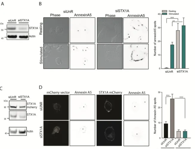
1.2 SNARE proteins as interacting partners for PLSCR1 ... 55
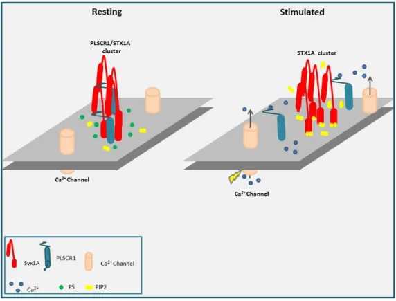
1.3 What is the role of syntaxin1A in PLSCR1 scrambling? ... 56
2
V. OPHN RESULT ... 71
1. RESEARCH CONTEXT ... 71
VI. OPHN DISCUSSION ... 73
1. OLIGOPHRENIN-1: A MOLECULAR SWITCH BETWEEN EXOCYTOSIS AND ENDOCYTOSIS OF SECRETORY GRANULES ... 73
1.1 HOW OPHN1 REGULATES CALCIUM-REGULATED EXOCYTOSIS ? ... 73
1.2 How OPHN1 switches from exocytosis to endocytosis ? ... 77
1. CELL CULTURE AND TRANSFECTION ... 80
1.1 Primary culture bovine Chromaffin cells ... 80
1.2 Primary culture mouse Chromaffin cells ... 80
1.3 Culture of PC12 cells ... 81
2. TRANSFECTION ... 81
2.1 Transfection of bovine chromaffin cells ... 81
2.2 Transfection of PC12 cells ... 81
3. MOLECULAR BIOLOGY ... 82
3.1 DNA Construct ... 82
3.2 REAL-TIME QUANTITATIVE PCR ... 84
3.3 Transformation and culture competent bacteria ... 84
3.4 Plasmid purification ... 85
3.5 Recombinant protein synthesis ... 86
4. BIOCHEMICAL TECHNIQUES ... 87
4.1 PROTEIN EXTRACTION ... 87
4.2 Pull down assay ... 87
4.3 Co-immunoprecipitation with GFP-Trap A system ... 88
4.4 Subcellular fractionation ... 89
4.5 Rho GTPase activity assays ... 89
4.6 Western blotting ... 90
4.7 Induction of Apoptosis by Staurosporine ... 90
4.8 Catecholamine measurement assay ... 91
5. IMMUNOFLUORESCENCE AND MICROSCOPY ... 91
5.1 Antibodies ... 91
5.2 IMMUNOCYTOCHEMISTRY ... 92
5.3 DBH ASSAY ... 92
5.4 PS STAINING ... 93
5.5 Confocal microscopy ... 93
5.6 Transmission electron microscopy ... 93
6. AMPEROMETRY ... 94
IX. REFERENCES ... 95
1. PUBLICATION ... 120
Avant-Propos
Après un Master de Neuroscience à l’Université de Valparaiso au Chili, j’ai obtenu une bourse du ministère Chilien de l’éducation dans le cadre de l’appel d’offre « advanced human capital program scholarships » (Becas Chile-CONICYT), pour effectuer un doctorat à l’Université de Strasbourg dans l’équipe du Dr. Stéphane Gasman situé à l’Institut des Neurosciences Cellulaires et Intégratives (INCI, CNRS UPR3212). L’équipe de Stéphane Gasman s’attache depuis de plusieurs années à comprendre les mécanismes cellulaires et moléculaires qui contrôlent la sécrétion neuroendocrine.
Le système neuroendocrinien se compose des organes, tissus et cellules spécialisés qui libèrent des hormones et des neuropeptides dans la circulation sanguine par un processus d’exocytose vésiculaire régulée par le calcium. Ce processus est finement régulé par les protéines SNARE (Soluble NSF Attachment protein REceptor), qui permettent la fusion de la membrane des vésicules avec la membrane plasmique, étape ultime de l'exocytose, aboutissant à la libération du contenu vésiculaire. Les mécanismes qui régulent l'exocytose et la fusion membranaire sont étudiés de façon intensive. En revanche, les mécanismes permettant de préserver l’intégrité physique des membranes plasmique et vésiculaire après fusion membranaire, et par conséquent de maintenir l’équilibre fonctionnel de la cellule, ne sont pas connus et restent peu explorés aujourd’hui. Les travaux de l’équipe réalisés dans les cellules chromaffines de la glande surrénale suggèrent que la libération du contenu intra-granulaire est couplée de façon spatiale et temporelle à un processus d’endocytose compensatrice qui permet la recapture de la membrane du granule. Ainsi, nous émettons l’hypothèse selon laquelle la membrane granulaire préserverait son intégrité au sein de la membrane plasmique après l’exocytose avant d’être spécifiquement recapturée avec l’ensemble de ses composés. Cependant, les mécanismes à la base de cette activité d’endocytose compensatrice ne sont pas connus dans les cellules neuroendocrines. Dans ce contexte, le but général de ma thèse fut d’apporter de nouveaux éléments permettant de comprendre comment l'endocytose compensatrice est-elle déclenchée et régulée dans les cellules neuroendocrines et par quels mécanismes est-elle couplée à l'exocytose.
A mon arrivée en doctorat, le Dr Stéphane Ory (qui fût mon encadrant pendant ces trois années et demi) venait de montrer qu’au cours de l'exocytose, la proteine PLSCR1 (Phospholipid Scramblase-1) est capable de redistribuer les phospholipides d’un feuillet à l’autre de la membrane plasmique, perturbant ainsi de façon transitoire l'asymétrie membranaire au niveau des sites d’exocytose. De façon intéressante, Stéphane Ory montre élégamment que cette perturbation membranaire n’empêche pas la sécrétion mais bloque significativement l’endocytose compensatrice des granules de sécrétion. La PLSCR1 m’est alors apparue comme un candidat idéal pour contrôler le couplage entre l’exocytose et l’endocytose. Ainsi, l’un des buts premiers de mon doctorat fut d’essayer de comprendre comment l’activité de la PLSCR1 est régulée et pourquoi un mélange de phospholipides est préalable à la recapture des granules de sécrétion.
En parallèle, je me suis intéressée aux mécanismes de régulation de la sécrétion par une protéine appelée oligophrénine-1 (OPHN1). Cette protéine est particulièrement intéressante. Impliquée dans l’endocytose des vésicules synaptiques, elle possède un domaine « BAR » (Bin, Amphiphysin, Rvs) qui est un senseur de courbure membranaire ainsi qu’un domaine GAP permettant l’inactivation des protéines Rho, une famille de GTPases largement impliquée dans les processus d'exo- et d'endocytose. Au cours de ma première année de thèse, Sébastien Houy un doctorant de l’équipe montrait, en utilisant des souris invalidées pour le gène Ophn1 que l’oligophrénine participe à la fois à la formation du pore de fusion et à l’endocytose compensatrice de la membrane granulaire. J’ai activement participé à ce projet en essayant notemment de comprendre comment OPHN1 pouvait coordonner son rôle sur l’exocytose avec un rôle dans l’endocytose.
Ce manuscrit fait la synthèse de l’ensemble de mes travaux et s’articule en quatre grandes parties. La première partie introduit de façon générale les connaissances actuelles concernant l'exocytose régulée et l'endocytose compensatrice dans les cellules neuroendocrines. J'y présente le modèle de la cellule chromaffine que nous utilisons au laboratoire et j'y détaille le cycle complet de la vie d’un granule de sécrétion, depuis sa biogénèse jusqu’aux mécanismes permettant son recyclage au cours du processus d’endocytose compensatrice. J’insiste également sur l’implication des protéines et des lipides qui ont été au cœur de mes problématiques de thèse.
La seconde partie est dédiée à mes données sur la régulation de l’activité et le rôle de la PLSCR1 au cours des processus d’exocytose et d’endocytose dans les cellules neuroendocrines tandis que la troisième partie du manuscrit se focalise sur l’implication de la protéine Oligophrénine1. Ces deux parties sont organisées de la même façon. Après un bref rappel du contexte scientifique et des problématiques posées, les données sont exposées sous forme d’article (une ébauche d’article en préparation pour la partie PLSCR1 et un article publié dans Journal of Neuroscience pour la partie sur l’oligophrénine). Je tente ensuite de prendre un peu de recul et de discuter mes données de façon plus globale afin d’élaborer quelques concepts mécanistiques.
Enfin une dernière partie présente les détails des matériels et méthodes utilisés pour mener à bien mes expériences. En annexe, vous trouverez l'ensemble des articles auxquels j'ai pu contribuer de près ou de loin lors de mon doctorat.
L’objectif de ce manuscrit est d’apporter une vision globale des mécanismes régulant la sécrétion neuroendocrine tout en mettant en exergue l’implication des protéines scramblase-1 et oligophrénine-1. Le Français n’étant pas ma langue maternelle, j’ai préféré rédiger ce manuscrit en anglais. J’en profite pour remercier Stéphane Gasman de m’avoir aide à traduire ce prologue.
List of abbreviations
AP2: Adaptor protein2 BAR: BIN/Amphiphysin/Rvs BoNT : Botulinum neurotoxin Ca2+: Calcium
CME: Clathrin-mediated endocytosis DAG: Diacylglycerol
DBH: Dopamine β-hydroxylase ER: Endoplasmic reticulum F-BARs: Fes/CIP4 homology-BAR GDI: Gunanine nucleotide Dissociation Inhibitor GDP: Guanosine Di-Phosphate GEF: Guanine-nucleotide Exchange Factor GTP: Guanosine Tri-Phosphate
hPLSCR: Human Phospholipid scramblase HSCP: Highly Sensitive Calcium-Pool Inverse-BAR: I-BARs
IRP: Immediately Releasable Pool ITSN: Intersectin
JMD: Juxta membrane domain LDCV: large dense core vesicle L-DOPA: 3,4dihydroxyphenylalanine Munc13-interacting domains: MID Na+ : Sodium
N-terminal amphipathic helix-BAR: N-BAR N-WASP: Wiskott-Aldrich syndrome protein
Oligophrenin-1: OPHN1 PC: Phosphatidylcholine PH: Pleckstrin homology
Phenylethanolamine N-methyltransferase: PNMT Phosphatidic acid: PA
PIP2: Phosphatidylinositol 4,5-bisphosphate PKC: Protein Kinase C PLC: Phospholipase C PLD1: Phospholipase D1 PLSCR: Phospholipid scramblase PM: Plasma membrane PS: Phosphatidylserine PX: Phox homology
RRP: Readily Releasable Pool SH3: Src homology 3
SM: Sec1/munc18-like
SNAP: Synaptosomal-associated protein
SNARE: Soluble N-éthylmaleimide-sensitive-factor attachment receptor SRP: Releasable Pool
STX: Syntaxin
TGN: Trans-Golgi network TH: tyrosine hydroxylase
TIRF: Total internal reflection florescence TM: Transmembrane
TMD: Transmembrane domain t-SNAREs: Target-SNAREs
unc18: uncoordinated18 UPP: Unprimed Pool VAMP: Synaptobrevin
VGCCs: Voltage-gated Ca2+ channels v- SNARE: Vesicular-SNAREs
I.
General introduction
1. The adrenal gland
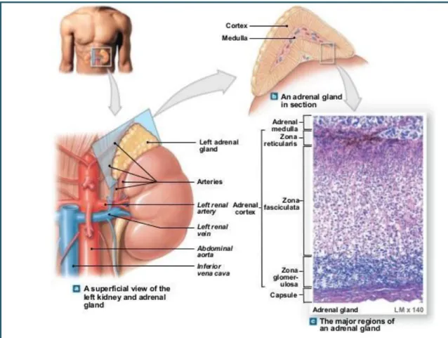
The adrenal glands are two pyramidal structures located in the upper pole of both kidneys. They produce hormones that help the body to control blood sugar, burn protein and fat, react to stressors like a major illness or injury, and regulate blood pressure.
The adrenal gland is formed by two well defined structures covered by a capsule of connective tissue with different functions and embryological origins: the adrenal cortex derived from the intermediate mesoderm which surround the medulla derived from neural crest cells. The adrenal cortex is devoted to production of steroid hormones, namely aldosterone, cortisol, and androgens. The adrenal medulla is constituted by chromaffin cells, which are able to release hormones and neuropeptides into the bloodstream.
Adrenal gland disorders can be caused by an imbalance in the hormone secretion. For example, Cushing syndrome due to cortex adrenal tumor is caused by an overproduction of cortisol. The Cushing syndrome can lead to diabetes, high blood pressure, and osteoporosis, and other health issues. On the contrary the adrenal insufficiency occurs when the adrenal glands do not make enough cortisol, and aldosterone. The characteristic symptoms include fatigue, muscle weakness, decreased appetite, and weight loss. Another disease associated with this organ is a tumor that arises from the adrenal medulla (Pheochromocytomas). This tumor produces over-secretion of the adrenaline causing of the severe elevation in blood pressure.
Fig 1: Adrenal gland. A) Representative scheme of the location of the adrenal gland. B) Representative scheme of the adrenal gland section, with the cortex at the periphery and the medulla in the center. C) Amplification of adrenal gland section. (http://www.slideshare.net/gwrandall/163-ch-10lecturepresentation)
2. Calcium regulated exocytosis in chromaffin cells: From the biogenesis
to the release
Chromaffin cells are a widely used model to study calcium-regulated exocytosis. They share with neurons the same embryonic origin (neural crest) and both release hormones and neurotransmitters stored in vesicles, by Ca2+-regulated exocytosis
In the adrenal medulla, the splanchnic nerve establishes cholinergic synapse with chromaffin cells. Upon stimulation, acetylcholine is released from presynaptic terminals and nicotinic receptors located on the membrane of chromaffin cells are activated. Nicotinic receptors are cation channels and once activated, they trigger sodium (Na+) entry in the cells. This ionic input induces membrane depolarization, activating voltage-dependent calcium (Ca2+) channels which increase the cytosolic Ca2+ concentration, and in turn leads to the release of catecholamine from chromaffin cells.
Fig2: Adrenal gland innervation. The splanchnic nerve fibers innervate chromaffin cells, stimulating the release of catecholamine (Colomer et al. 2012).
The main hormones synthesized and released by chromaffin cells are catecholamine (dopamine, noradrenalin and adrenaline). Two types of chromaffin cells are found in the adrenal medulla: adrenergic chromaffin cells secreting adrenaline (or epinephrine) which represent 80% of the chromaffin cells and noradrenergic cells which secrete noradrenalin (or norepinephrine) and represent the remaining 20%.(Kobayashi and Coupland 1993). Chatecolamines are synthesized following a multistep enzymatic cascade. Dopamine is the end product of two enzymatic reactions: hydroxylation of the tyrosine amino acid into 3,4- dihydroxyphenylalanine (L-DOPA) by tyrosine hydroxylase (TH), an enzyme found in dopaminergic cells, and decarboxylation of L-DOPA. These reactions occur in the cytoplasm and dopamine is rapidly transported into specialized organelles, the large dense core vesicle or secretory granule by nonspecific vesicular monoamine transporters (Wimalasena and Wimalasena 2004). Dopamine can be further transformed into norepinephrine (or noradrenalin) by the Dopamine β-hydroxylase (DBH) and epinephrine (or adrenalin) by phenylethanolamine N-methyltransferase (PNMT) (Kuhar et al. 1999).
2.1 Biogenesis of secretory granules
Secretory granules or large dense core vesicles (LDCV) are the main storage unit of chromaffin cells. Although catecholamines are loaded into LDCV once vesicles are formed, proteins found in the matrix of LDCV are involved in the biogenesis of secretory granules (proteins of the granin family) (Kim et al. 2005). Together with the lipid composition of the trans Golgi network (TGN) membranes, they determine the sorting of vesicular components intended either to the constitutive or regulated secretory pathway.
Mechanistically, it has been proposed that the regulated secretory pathway requires a driving force of protein (chromogranin A , B, secretogranin II–IV ) and lipid like diacylglycerol, phosphatidic acid, and cholesterol to be generated (Kim et al. 2006). Diacylglycerol (DAG) and phosphatidylcholine can be converted in phosphatidic acid (PA) via DAG kinase and phospholipase D1 respectively. Phosphatidic acid accumulation rather than DAG is a key step in regulating budding of secretory vesicles from the TGN in mammalian cells (Siddhanta and Shields 1998). PA may induce negative curvature of membranes to favor
the budding of granules occurs (Dhanvantari and Loh 2000). On the other hand, high Ca2+ concentration and acidic pH conditions encountered in the TGN promotes aggregation of granins which associate directly or indirectly with lipid rafts to induce budding and formation of the immature secretory granules.(Laurent Taupenot et al.2003; Elias et al. 2012). Different domains of chromogranin have been shown to be important for LDCV formation. For example, a motif consisting of an intramolecular disulfide loop domain bounded by cysteine residues and containing a number of aliphatic hydrophobic residues are necessary and sufficient for the association of the chromogranin B to the TGN membrane and, hence, sorting of chromogranin B to the regulated secretory pathway in the PC12 neuroendocrine cell line (Glombik et al. 1999). This loop is also present in Chromogranin A but does not appear to be necessary for sorting of chromogranin A to the regulated secretory pathway in PC12 cells (Taupenot et al. 2002). It may therefore require additional domain like the region 77-115 which is necessary but not sufficient for trafficking of this protein to the regulated secretory pathway (Hosaka et al. 2002). Conversely, deletion of the segment 48-111 of the chromograninA resulted in missorting of CgA to the constitutive pathway in pituitary (AtT- 20) and β-pancreatic (INS-1) cell lines. In addition to their direct role in granule biogenesis, Chromogranins may also help pro-hormones to be sorted into LDCV. Pro-vasopressin, oxytocin and pro-opiomelanocortin are also able to aggregate in condition of high calcium concentration and acidic pH conditions. As they can interact with granin, they participate into sprouting and formation of immature granules (Beuret et al. 2004). This context suggests that the association of granins and prohormone aggregates at lipid rafts is essential to provide the driving force for granule budding at the TGN, whereas lipid components such as DAG, PA, and cholesterol facilitate formation of membrane curvature.
At this stage, secretory granules are immature and may contain proteins not intended to enter the regulated secretory pathway since segregation is not completely efficient. A maturation process takes place which permit the vesicles to sort proteins not supposed to take the regulated pathway. Immature secretory granules (ISG) undergo several steps necessary to convert them into mature granule. The first one is the homotypic fusion of immature granules involving SNARE proteins (Urbé et al.1998; wendler et al. 2001). This granule fusion will induce the increase of their size and also their enrichment in protein necessary for the next steps of maturation like the V-ATPase proton pumps. These latter will
permit the acidification of immature granule and then modify the intragranular pH from 7 to 5 (mature granule). Acidification of the intra-granule content will allow the activation of pro- hormone convertase and carboxypeptidases necessary to the regulated secretory pathway and this acidification is done throughout the transport of the vesicle to the plasma membrane (Wu et al. 2001).
The second step is the removal of lysosomal enzymes, and some membrane proteins that are co-packaged (Kuliawat et al. 1997). This process occurs by budding off of constitutive-like vesicles from the immature granule by a clathrin-dependent mechanism (Kim et al. 2006). It is well known that during this process of maturation, the clathrin coat is removed from the granule. However, the mechanisms regulating this uncoating are still poorly understood (Orci et al. 1985; Tooze and Tooze 1986). Finally, cargo molecules in maturing granules undergo condensation which requires acidification and removal of water by the lipid microdomain-associated aquaporin along with the efflux of Na+, K+, Cl (Arnaoutova et al. 2008).
2.2 Transport of secretory granules
Like most intracellular vesicles, secretory granules distribution into the cytoplasm relies on actin filaments and microtubules. Long range transport from the Golgi to the plasma membrane is mediated by microtubules and associated motor proteins whereas short range movement occurring close to the plasma membrane are rather mediated by actin filament and myosins. Although less is known about the role of microtubules in the transport of LDCV compared to actin, interfering with the dynamics of actin or microtubules impair the motility and eventually the release of catecholamine (Neco et al. 2003; Maucort et al. 2014). Interestingly, some evidences suggest that transport of vesicles from the regulated pathway may rely on a subset of proteins represented by SNARE proteins, small GTPases from the Rab family and their regulators. For example, in neuronal cells, Rab27 is required for kinesin1-dependent anterograde movement of Trk-containing (Arimura et al. 2009). In the hippocampal neurite, the SNARE VAMP7 binds to Varp, an activator of Rab21
(Burgo et al. 2012). In chromaffin cells, although the molecular mechanisms remained to be determined, a subset of proteins from the SNARE and Rab family (VAMP2, Rab3a, Rab27) is also required for efficient transport of secretory granules and fusion at the plasma membrane. It suggests that a general mechanism could be preserved in long range transport of vesicles intended to regulated exocytosis in secretory cells
The actin-based transport of secretory granules has been better studied in chromaffin cells. The first studies proposed that the cortical actin filaments (F-actin) forms a physical barrier that restricts the secretory vesicle access to the plasma membrane. This model was based on the fact that actin depolymerization was observed after stimulation and before massive exocytosis (Trifaró et al. 2000) (Aunis and Bader 1988; M. L. Vitale et al. 1991; Gil et al. 2000) (Nakata and Hirokawa 1992). However, depending on the concentration of actin polymerization inhibitors, both increase (low concentration) or decrease (high concentration) of exocytosis was observed suggesting that actin function was more complex than acting as a physical barrier. Actin can indeed act as a transporter in combination with molecular motors and help directly in the fusion process.
One of this motor, the MyosinVa, plays a crucial role in the control of F-actin dynamics and vesicle displacement. The GTPase Rab27A, located on the granule, interacts with MyRIP (Myosin and Rab interacting protein) and MyosinVa, both bound to actin cytoskeleton. They constitute therefore a link between granules and the actin cytoskeleton (Desnos et al. 2003). Inhibition of MyosinVa function by specific antibody decreases the secretory response in chromaffin cells (Rosé et al. 2003) and the use of MyosinVa dominant negative mutant blocks the traffic of granules at the vicinity of the plasma membrane when granules are entrapped in the actin cytoskeleton in PC12 and β-pancreatic cells (Rudolf et al. 2003; Varadi et al. 2005). This suggests that MyosinVa plays an important role in the granule trafficking and actin is passively required to form “track” for vesicle displacements.
But actin has also an active role by polymerizing at specific site. For example, the actin nucleation promoting factor neural Wiskott-Aldrich syndrome protein (N-WASP) is recruited to plasma membrane upon stimulation and mediate the secretory response in PC12 cells (Gasman S et al. 2004). F-actin forms trails that favor the secretory vesicle motion to the plasma membrane. Forces generated by actin can regulates the expansion of the
fusion pore(Giner et al. 2005; Berberian et al. 2009). Recently, electron microscopy tomography showed that actin filaments can be seen attached to the secretory granule and the plasma membrane to stabilize secretory granule likely in a “ready to fuse” state. Actin filaments have therefore both passive and active function in the transport of vesicle transport and in the regulation of the regulated exocytotic in the chromaffin cells.
2.3 Pools of secretory granules
Biogenesis and maturation of LDCV is a continuous process which supposes that, depending on the maturation step and their distance from the plasma membrane, some LDCV will be ready for fusion with the plasma membrane or not. In chromaffin cells (and in neurons), 4 pools of secretory granule have been described on the basis of their kinetic of release. The Readily Releasable Pool (RRP) in which the vesicles are fusing with the plasma membrane with a time constant 20-40ms after stimulation. The Slowly Releasable Pool (SRP) with a time constant of approximately 200ms. Note that although this pool is made available in the presence of high intracellular Ca2+concentration, a physiological stimulation only generates the release of RRP. Recent studies have shown that RRP can be subdivided into two subgroups: A) The IRP (Immediately Releasable Pool) corresponding to about 25% of the RRP in which the granules are located in the vicinity immediate to the calcium channel (Yang et al., 2002) and B) the HSCP (Highly Sensitive Calcium-Pool) which can be released at lower Ca2+ concentrations than the IRP and the RRP. Additionally, another pool was detected in chromaffin cells, the UPP (unprimed Pool). This pool corresponds to the granules located at a maximum distance of 200 nm and which do not belong to the RRP and IRP. The last granule population is the reserve pool where the granules are located at more than 200nm away
2.4 Late phases of exocytosis: from docking to fusion
The exocytosis is the process by which stored neurotransmitters and hormones are released by the fusion of secretory granule with the plasma membrane. This process is dynamic, rapid and spatially restricted in the cells. Exocytosis involved multiple steps including granule trafficking, tethering, docking, priming and eventually fusion. Structural, biochemical and functional studies have allowed the identification of multiple factors and proteins implicated in the exocytosis.
In neurons and neuroendocrine cells, calcium regulated exocytosis is divided in four main steps which are first the tethering of LDCV at the plasma membrane and second, the docking at exocytic sites. At this step, LDCV have to be matured (third step called priming) to be competent for the final and fourth step consisting of fusion and release of intragranular contents (Figure 3).
Fig3: Step of exocytosis in the chromaffin cells.
The exocytosis process starts by the recruitment of the granule at the plasma membrane (tethering) and the subsequent docking where Synaptobrevin (a vesicular SNARE) and Syntaxin/SNAP25 (plasma membrane SNARE proteins) interact. During the priming, SNARE proteins are coiled prompting the approach of the granule membrane with the plasma membrane.Finally, the granules fuse with the plasma membrane, releasing their contents.
2.4.1 Docking
Ca2+-triggered release of neurotransmitters and hormones depends on soluble N- ethylmaleimide-sensitive factor attachment protein receptors (SNARE). The SNARE-complex constitutes the minimal machinery needed for the fusion of the secretory granule with the plasma membrane. Within the SNARE proteins involved in regulated exocytosis, we can find the t-SNAREs in the PM and the v-SNAREs located in the vesicular membrane. The SNARE- complex is made up of three SNARE proteins; Syntaxin1 (STX1), synaptosomal-associated protein (SNAP-25) and synaptobrevin (VAMP-2).
Syntaxins were first described as two synaptotagmin-interacting proteins (STX1A and 1B) with a molecular weight of 35 kilo Dalton (kDa) and 84% identical amino acid sequence (Bennett et al. 1992). In adrenal chromaffin cells, STX1A was found to be localized to the membrane as the dominant isoform (Baltazar et al. 2003). This protein present a carboxy- terminal transmembrane domain, SNARE domain (termed H3), a long coiled-coil α-helical structure, and an additional Habc domain with an N-terminal domain containing a short N- peptide (Weimbs et al. 1997). This domain is a linker region, which connects Habc to the H3 domain. The Habc domain (amino acid residues 28-144) is made up of three α helices with high sequence conservation. Habc domain interacts with the H3 domain of the protein which maintains STX1A in a ‘closed conformation’, preventing its interaction with other SNAREs partner (Misura et al.2001). The closed conformation of STX1A needs to be released to participate in the assembly of the SNARE complex. Point mutations in the linker region of the Habc domain (L165A/E166A) are sufficient to release the interaction between Habc and H3 and bring STX1A in an open conformation able to interact with other partner of the SNARE machinery.
Tight regulation of STX proteins function has to be set up to prevent unexpected release of LDCV contents. The Sec1/Munc-18 like (SM) protein family plays critical function in STX1 regulation. Munc18 was identified as a binding partner to STX and plays an essential role in vesicle fusion from yeast to mammals (Burkhardt et al. 2008), (Südhof and Rothman 2009; Verhage et al. 2000). Munc18 is encoded by the mammalian homologue of the C. elegans gene, uncoordinated18 (unc18) (Hata et al. 1993). In mammals, three isoforms of Munc18 are known: Munc18-1, Munc18-2 and Munc18-3. Knocking out Munc18-1 in mice
leads to deficiency in transmitter release and death at birth due to respiratory defects (Verhage et al. 2000). Munc18, which consists of three D domains (Misura et al.2000), with the third domain divided in D3a and D3b , adopts the form of a horseshoe. D1 and D3a form the bottom part of the horseshoe and D2 and D3b form the upper part. Munc18 binds STX through its central groove and keeps it in a ‘closed conformation’, preventing STX from binding to SNAP-25 and VAMP2 (Misura et al. 2000). Munc18-1 may stabilize a half-closed conformation of STX1 (Zilly et al. 2006), and forms STX1:Munc18 dimers, serving as stimulators of docking (Gandasi and Barg 2014). Munc18 has a dual function by acting as a chaperone and translocates STX1 to the plasma membrane (Rickman et al. 2007) and as an inhibitor of STX1 activation by maintaining it in a closed conformation (Toonen et al., 2005).
The critical role of Munc18 in docking has been shown in chromaffin cells from Munc18 -/-mice where 90% of LDCV are not docked (Voets et al., 2001). But whether it is a direct role of Munc18 or its function as chaperone has to be defined.
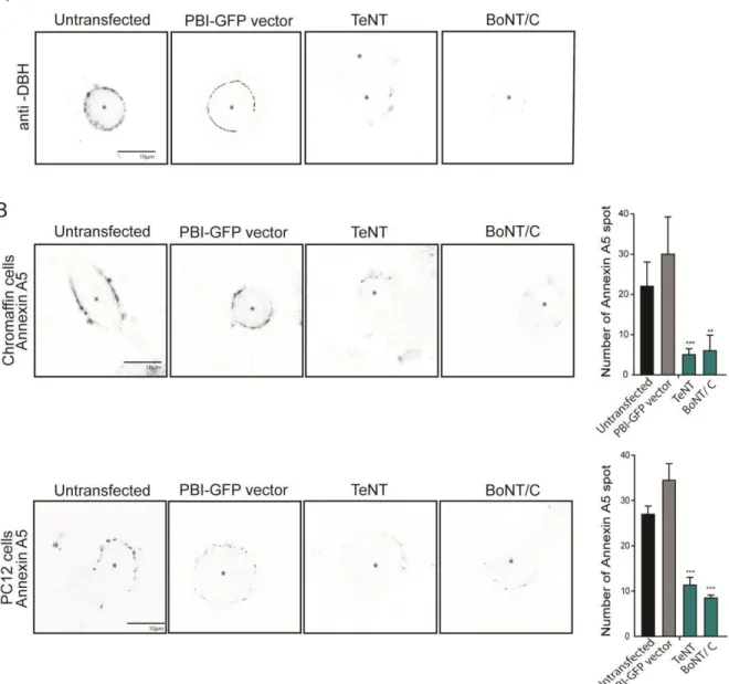
Docking of LDCV implies that STX1 is engaged at least temporarily in the SNARE complex which is formed by the association with SNAP25 localized to the plasma membrane and VAMP2 (or Synaptobrevin2) found at the LDCV membrane. SNAP-25 proteins (SNAP-25a and SNAP-25b) has a molecular weight of 25 kDa and is targeted to the plasma membrane thanks to palmitoylation sites (Bark et al. 1995) (Greaves et al. 2010). In adrenal chromaffin cells, the SNAP-25a isoform is predominating, but both isoforms are expressed (Grant et al. 1999). The role of SNAP25 in docking has been shown with the use of bacterial toxins that cleaves SNAP-25 (Botulinum neurotoxin type A (BoNT/A), C (BoNT/C) or E (BoNT/E)). Treatment of chromaffin cells with these toxins led to reduction of LDCV docking (de Wit et al. 2009) and impairs exocytosis in PC12 cells (Gerona et al. 2000).
Finally the v-SNARE VAMP2 is a protein of 18kDa which is inserted in the LDCV membrane by a single transmembrane (TM) domain. VAMP2 protein presents a juxtamembrane domain able to bridge the negatively charged phospholipid of the plasma membrane. This interaction facilitates the function of the secretory granules with the plasma membrane (Williams et al. 2009).
The structure of the SNARE complex has been resolved in 1998 (Sutton et al. 1998). It is a tightly packed four-helical parallel structure with leucine zipper resemblance. The interaction consists in the carboxyl terminal H3 domain of STX1A (9 kDa) (blue), the cytoplasmic domain of VAMP2 (11 kDa) (green) and the N- and C-terminal portions of SNAP- 25B (9 and 10 kDa) (orange), is a cable of four intertwined α-helices with their N-termini at one end and their C-termini at the other protein, as illustrated in figure 4. (Sutton et al. 1998).
2.4.2 Priming
Once the SNARE complex has formed and docks the LDCV, the priming step will allow these proteins to be wound correctly to bring closer enough the membranes and initiate the formation of the fusion pore after cell stimulation. The molecular details that distinguish the docking from priming is however incomplete but SNARE zippering that could bring LDCV close to the plasma membrane is favored during priming. This is an important step since it will determine the number of vesicles ready to be released when the future intracellular calcium increase will occur.
Munc proteins play also a function in priming. Members of the Munc13 protein family consisting of Munc13-1, -2, -3, and -4 were found to be absolutely required for this priming process (Stevens et al. 2005). During the priming, the SNARE STX1 switch from a closed conformation that binds Munc18-1 tightly to an open conformation within the highly stable SNARE complex. NMR and fluorescence experiments have shown that the Munc13-1 MUN domain, markedly accelerates the transition from the STX1–Munc18-1 complex to the SNARE complex. This activity depends on weak interactions of the MUN domain with the STX1 SNARE motif, and probably with Munc18-1 (Fig5) (Ma et al. 2011).
Fig5: Proposed model whereby the MUN domain (purple clear) promotes the transition from the STX1–Munc18-1 complex to the SNARE complex through its weak interactions with the STX1 SNARE motif (from Ma et al. 2011).
Structural and biochemical studies have shown that Munc13 interact with SNARE proteins by a calcium-dependent process because Munc13 present calcium binding C2 domains (Jahn and Fasshauer 2012).
Another protein involved in this process is the Ca2+-dependent Activator Protein for Secretion, aka CADPS (CAPS). CAPS is a regulator of SNARE complex assembly by creating direct interactions with membrane-associated SNARE STX1 and SNAP25 (Daily et al. 2010). The activity of CAPS in promoting SNARE complex formation was also demonstrated in studies of SNARE-dependent liposome fusion where CAPS markedly increased the rate and extent of fusion between donor VAMP2 liposomes and STX1/SNAP-25 acceptor liposomes (James et al. 2009). Additionally recent work showed that PIP2, STX1 and SNAP25 interactions stabilize the CAPS dimers. The role of the CAPS C2 domain (calcium sensor) in mediating homodimerization was revealed by studies of the mammalian homologous unc- 31C2 domain mutants and a C2 domain deletion that exhibits altered dimer formation and loss-of-function in vesicle exocytosis. Lastly, a study suggested that CAPS dimerizes similarly to Munc13-1/2 using conserved homodimerization residues in its C2 domain (Petrie et al. 2016).
The Rab3A, a small G-protein of the Rab family is involved in the priming steps of exocytosis. Recent work showed that Rab3A is essential for the performance of Munc13-1 during vesicle priming. Munc18-1 interacts with Rab3A and promotes Rab3A dissociation from the vesicle membrane; this is downstream of the Rab3A/Munc13-1interaction that regulates vesicle priming (Huang et al. 2011).
After finishing this step, the SNARE complex is mature and the granule is ready to fuse with the plasma membrane.
2.4.3 Fusion
Following stimulation, LDCV either fill up the reserve pool or are recruited to the plasma membrane as a part of the readily releasable pool. Docked LDCV fuse with the plasma membrane. Different release mechanisms have been described in neuroendocrine cells. The full fusion corresponds to the complete flattening out of the LDCV into the plasma membrane which led to the total release of intra-vesicular contents. Despite the insertion of the LDCV membrane into the plasma membrane, mechanisms exit to preserve the integrity of the LDCV membrane into the plasma membrane since no intermixing has been observed (Ceridono et al. 2011)(Bittner et al., 2013). The Kiss-and-Run corresponds to the formation of a narrow fusion pore between the plasma membrane and the LDCV which allows the release of small compounds of the LDCV like catecholamines. The majority of the granular content is retained and lipids do not intermingle (Gandhi and Stevens 2003). Finally, an intermediate mode of fusion has been described. The Cavicapture (cavity recapture or granule recapture) corresponds to partial expansion of the fusion pore, releasing catecholamines and small neuropeptides according to their molecular weight. Like kiss and run, the omega shape of the fusing granule is preserved (Henkel and Almers 1996). (Figure6).
Fig6: Release models in neuroendocrine cells. (Figure modified from Houy et al. 2013) Mature granules either fill up the reserve pool or are recruited to the plasma membrane as a part of the readily releasable pool. Large proteins (blue dots), small neuropeptides (black dots), and small molecules like catecholamines (red dots) can be released differentially according to the exo-endocytosis mode. During “kiss-and-run” mode, only small molecules are released through a narrow fusion pore, whereas cavicapture (vesicle cavity capture) allows the partial release of small neuropeptides. Note that for these two modes, retrieval of intact granules is easily conceived as the granule shape remains almost intact. During full fusion exocytosis, the intra-granular contents are all released and the granule membrane collapses into the plasma membrane.
3. Role of calcium sensors in regulated exocytosis
In neuroendocrine cells and neurons, the SNAREs proteins are not directly responsible for sensing the Ca2+ after stimulation. Important numbers of evidence show that synaptotagmin is the main calcium sensor involved in exocytosis. However, numerous other proteins have been characterized possessing a calcium binding domain and able to regulate the exocytosis in neuroendocrine cells and neurons.
3.1 Synaptotagmin
The synaptotagmin-1 is a granular protein possessing a short N-terminal followed by a transmembrane domain (TMD) and two calcium binding C2 domains (C2A and C2B) (Perin et al. 1991; Herrick et al. 2006). In addition to the Ca2+-binding loops, the C2B domain contains a polybasic region, enriched with lysine residues, that interacts with PIP2 and phosphatidylserine (PS). These interactions are essential for the exocytosis (Araç et al. 2006; Wang et al. 2011) (Honigmann et al. 2013). By interacting with PIP2 clusters formed upon intracellular Ca2+ increase following stimulation, C2 domains reinforced LDCV anchorage. Using various muntants of C2A and C2B domains, it has been proposed that both domains cooperate to bring LDCV membrane and plasma membrane as close as 2 nm distance by interacting with PIP2 at the plasma membrane and PS of the LDCV (Honigmann et al., 2013). In addition to calcium sensing, Synaptotagmin-1 may mechanically promote SNARE zippering to end up with membrane merging (Park et al. 2015).
Synaptotagmin-1 was also involved in docking and priming of vesicles to the plasma membrane in chromaffin cells. Pull down experiments have revealed a possible interaction of Synaptotagmin-1 with SNAP25 and Syntaxin (Mohrmann et al. 2013).
3.2 Munc proteins
As mentioned before, in addition to its function as STX regulators, SM proteins may act as calcium sensors during exocytosis. This is the case of Munc13 (not Munc18). Munc13 proteins have a Ca2+-binding C2-domains (Lipstein et al. 2012). It has been reported that the central C2-domain in Munc13-1 (C2B) bind phospholipids like PI(4,5)P2 during granules recruitment and release (Shin et al. 2010) suggesting that in addition to help in opening up the so-called “closed” STX1 within STX1/Munc18-1 dimers to allow SNARE complex formation, it may cooperate with Synaptotagmin to bring closer LDCV and plasma membrane. In chromaffin cells, the isoform Munc13-2 plays a fundamental role in the exocytosis (Man et al. 2015). The absence of Munc13-2 decreases significantly the readily releasable pool size and catecholamine release.
3.3 Doc2 proteins
Double C2-domain protein (Doc2) is another calcium-sensing protein. This protein plays an important role in Ca2+-triggered exocytosis. Four members of the protein family that bind Ca2+ are Doc2α , Doc2β, Doc2γ and raphilin-3A. Doc2 proteins possess two C2 domains (C2A, C2B) separated by a linker region, and a N-terminal Munc13-interacting domains (MID) (Sato et al. 2010). The C2 domains have structural similarities, but C2B binds Ca2+ with higher affinity that C2A (Giladi et al. 2013). Doc2 proteins were reported as Ca2+ sensors in neurotransmission (Groffen et al. 2010) and able to interact with other exocytotic proteins like Munc18-Munc13 and STX. These interactions have been reported in different cell model including β-cells, chromaffin cells and neurons (Ke et al. 2007) (Orita et al. 1997). In neurons, Doc2 is able to translocate to the plasma membrane after stimulation, and the disruption of interaction between Munc13 and Doc2 causes a slow activity in the synaptic transmission (Mochida et al. 1998). In chromaffin cells, Doc2A and Doc2B are recruited to the plasma membrane promoting the increase of granule priming (Voets et al. 2001) suggesting that Doc2 has a regulating role in the priming step.
4. The Rho-family proteins in Ca
2+-regulated exocytosis
The Rho-family proteins constitute a major branch of the Ras superfamily of small GTPases. They are of small size (190-250 residues, 21 kDa) and consist in a highly conserved GTPase domain, short N- and C-terminal. The GTP-binding domain has a strong affinity for GDP (Guanosine Di-Phosphate) and GTP (Guanosine Tri-Phosphate) but they are thought to cycle between inactive, GDP-bound and active GTP-bound states. The switch between the two states is catalyzed by regulatory proteins namely Rho GEF (Guanine-nucleotide Exchange Factor) and Rho GAP (GTPase activating) proteins. Despite it stronger affinity for GTP and a lipid anchor, Rho GTPases are mostly found inactive, bound to GDP, and cytoplasmic thanks to Rho GDI (Gunanine nucleotide Dissociation Inhibitor). Upon GDI release, Rho GTPases translocate to membranes where Rho GEF catalyses the exchange of GDP into GTP leading to conformational changes of the Rho proteins and subsequent binding to their effectors. Rho GAP quickly inactivates Rho proteins by increasing their low intrinsic GTPase activity. Rho GDI further “extracts” Rho proteins from membranes. The most studied members of Rho-family proteins are RhoA, Rac1 and Cdc42 and constitute the so-called canonical Rho GTPases as compared to atypical Rho GTPases which mode of regulation depends on degradation or post-translational modification in addition to nucleotide binding (Wennerberg and Der 2004; Aspenström et al 2007).
Fig7: The GTPase activation-desactivation cycle. RAS-family proteins are low-molecular- weight guanine-nucleotide-binding proteins. They are inactive when bound to GDP and active when bound to GTP. Regulation of this molecular switch mechanism occurs through a GDP–GTP cycle that is controlled by the opposing activities of guanine nucleotide-exchange factors (GEFs), which catalyze the exchange of GDP for GTP, and GTPase-activating proteins (GAPs), then increase the rate of GTP hydrolysis to GDP (see diagram). In the case of Rho proteins, another layer of regulation is provided by Rho–GDP-dissociation inhibitors (Rho- GDI), which sequester Rho away from the GDP–GTP cycle. GTPases interact with various effector proteins, which influence the activity and/or localization of these effectors; this ultimately influences cell-cycle progression (Coleman et al. 2004).
RhoA, Rac1 and Cdc42 are well-known regulators of actin dynamics and phosphoinositide production in different cell systems (Croisé et al. 2014). As actin play an important role in controlling different phases of exocytosis (LDCV transport, access to the exocytotic site of and expansion of the fusion pore), Rho GTPases appeared as likely regulator of Ca2+- regulated exocytosis. Indeed, depolarization by high concentration of potassium led to the activation of Rac1 and Cdc42, but only Cdc42 was found to induce N-WASP-dependent actin polymerization at the plasma membrane (Frantz et al 2002; Stéphane Gasman et al. 2004; Momboisse et al. 2009). Rac1 instead induced the production of phosphatidic acid (PA), a fusogenic lipid involved in the last step of granule fusion by activating the phospholipase D1 (PLD1). A complex formed by the scaffold protein Scrib and the exchange factor for Rac1 βPIX are crucial to activate Rac1 at the plasma membrane in response to depolarization (Momboisse et al. 2009). RhoA is also involved in that process but unlike Rac1 and Cdc42, RhoA is localized to secretory granules and it has been proposed to regulate PI4Kinase on secretory granules. RhoA inactivation could be a prerequisite to favor exocytosis since, in contrast to Rac1 and Cdc42, the expression of a constitutive mutant inhibit exocytosis in response to depolarization (Gasman S et al. 1999; Gasman S et al. 2004). The precise spatial and temporal regulation of RhoA, Rac1 and Cdc42 have still to be unraveled, but Rho GTPases appeared to act on two fundamental regulators of exocytosis, actin dynamics and lipids biosynthesis.
5. Role of lipids in Ca
2+-regulated exocytosis
The secretion in the neuroendocrine cells involve the increase of the intracellular Ca2+and the assembly of SNARE complex. Fusion implies merging of two membranes of different origin and it is not so surprising that lipids play an important role in the regulation of exocytosis. Several lipids have been found at exocytotic sites or involved in the regulation of the fusion process which are exemplified below.
5.1 Anionic lipids
The first lipid identified as a regulator of exocytosis is the phosphatidylinositol 4,5- bisphosphate (PIP2) (Martin 2001). Under appropriate conditions, PIP2 can engage in strong electrostatic–based interactions with positively charged molecules (McLaughlin and Murray 2005), and can cluster in small membrane microdomains (Slochower et al. 2014). Phosphoinositide binding proteins can be recruited from the cytosol to cluster of PIP2 to fulfill their function. PIP2 microdomains are required for the docking and fusion of LDCV. In PC12 cells, a spatial correlation between PIP2 microdomains and exocytotic machinery, in particular with syntaxin clusters was observed (Aoyagi et al. 2005). In neurons, loss of plasma membrane PIP2 leads to a decrease in exocytosis and changes in electrical excitability. Restoration of PIP2 levels after phospholipase C (PLC) inactivation which prevent hydrolysis of phospholipid PIP2 into diacylglycerol DAG and the soluble inositol triphosphate (IP3), is therefore essential for a return to basal neuronal activity (Kruse et al. 2016).
Other lipids have been proposed to regulate exocytosis. For example is the DAG, which binds to Munc13-1 through its C1-domain (Rhee et al. 2002) or the Protein Kinase C (PKC), which phosphorylates SNAP25 (Nagy et al. 2002), Munc18-1 (Wierda et al. 2007; Genc et al. 2014) and Synaptotagmin-1 (de Jong et al. 2016), all promoting exocytosis.
In chromaffin cells, the production of Phosphatidic Acid (PA) by phospholipase D1 (PLD1) at exocytotic site is necessary to LDCV (Bader and Vitale 2009) (Zeniou-Meyer et al. 2007) (Vitale et al. 2001). Although we cannot exclude that increased concentration of PA
my also recruit essential proteins for fusion, structural properties of this small lipid which induce negative curvature is privileged and may help in merging membranes.
5.2 Cholesterol
Cholesterol is an abundant lipid in cells and its accumulation at specific sites modify the biophysical properties of the membrane by reducing membrane fluidity and increasing resistance to membrane deformation. These characteristics have led to hypothesize that the cholesterol regulates exocytosis, by modulating membrane curvature, where the spontaneous negative curvature of cholesterol is believed to favor negative curvature regions of the membrane such as the cytosolic side of the fusion pore as illustrated in the Figure 8 (Yang et al. 2016).
Fig8: cholesterol modulation of the membrane curvature (Figure adapted from S.-T. Yang et al. 2016)
The second role proposed for cholesterol is related to its ability to partition proteins, some proteins accumulating preferentially in cholesterol rich domains. SNARE proteins are preferentially recruited at cholesterol enriched sites in the plasma membrane (Lang 2007; Chasserot-Golaz et al. 2010). During chromaffin cell stimulation, ganglioside GM1/cholesterol/PIP2-enriched lipid microdomains are formed and accumulate at exocytotic site. Cholesterol, PIP2 and GM1 may spatially define the exocytotic sites. Preventing the formation of these microdomains impairs exocytosis. Likewise extraction of cholesterol by methylβ-cyclodextrin treatment affect the actin polymerization (Hissa et al. 2013), alter the molecular organization of synaptic membrane (Chamberlain et al. 2001; Toft-Bertelsen et al. 2016), as well as vesicle motion and docking (Zhang et al. 2009).
To conclude, the lipid distribution and concentration in the exocytotic sites has an important role in the regulation of exocytosis by recruiting, interacting and activating the key proteins to promote the exocytosis in neuroendocrine cells and neurons.
6. Compensatory endocytosis in neuroendocrine cells
Retrieval of membrane proteins or soluble compounds from the extracellular space by endocytosis is a fundamental process critical for cell homeostasis. It is usually separated into two form of endocytosis: the clathrin-dependent or independent pathway. Endocytosis can be either constitutive (best exemplified by the endocytosis of the transferrin receptor for example), or induced by a signal such as the binding of a ligand to its receptor. A third kind of endocytosis can be distinguished: the compensatory endocytosis. This process takes place only when secretory vesicles have fused to the plasma membrane in response to Ca2+ increase. It therefore suggests that a tight coupling should be maintained between regulated exocytosis and compensatory endocytosis to efficiently retrieve transmembrane proteins and lipids coming from the secretory vesicles. Different studies have indeed shown that it is the case in chromaffin cells. Monitoring exocytosis by staining secretory granule membrane components (like dopamineβ-hydroxylase or VMAT2, a transporter of monoamine) inserted into the plasma membrane after full-fusion exocytosis revealed that they remained clustered
2013). Although some diffusion of secretory granule proteins have been reported after exocytosis (Sochacki et al., 2012), clathrin is nonetheless important to restrict diffusion since proteins newly inserted into the plasma membrane concentrated to the nearest clathrin- coated pit. The picture is more complicated in neuron where distinct pool of membrane proteins can be retrieved by clathrin-dependent and independent process depending on the cell type and the stimulation (Cárdenas and Marengo 2016).
Recently, the development of a high-resolution and high-throughput fluorescence imaging approach has maped 78 proteins at individual exocytic and endocytic structures in the cell and identified two core groups of proteins that associate with endocytic structures or exocytic vesicles part of which is illustrated in the Figure 9. This approach permited to describe few shared components and several new associations (Larson et al. 2014) suggesting that there are proteins that act independently in each process and others involved in the two process like synaptotagmin-1 (McAdam et al. 2015). This suggests also that these processes occur in a concerted manner in the cell.
As stated before, lipids play important function in exocytosis either as biochemical intermediates or by their physical properties. In compensatory endocytosis, they may act as a scaffolding system to maintain specific machinery at restricted site of the plasma membrane. For example, the formation of ganglioside- and PIP2-enriched microdomains at the exocytic sites may prevent or limit proteins and lipids diffusion after the granule have fused (Chasserot-Golaz et al. 2005; Umbrecht-Jenck et al. 2010). On the other hand, a loss of plasma membrane asymmetry has been observed following exocytosis. As it occurs at the rim of the exocytotic site, it might restrict diffusion by creating phase separation into the plasma membrane (Ory et al., 2013). But differences in the way secretory vesicle are released might condition the mode of component retrieval.
6.1 Clathrin-dependent endocytosis
A long lasting question in the field of regulated exocytosis is how proteins and lipids are retrieved by endocytosis after being delivered at the plasma membrane, especially after full fusion, when the secretory granule membrane or the synaptic vesicle collapse into the plasma membrane.
Among the basic pathways involved in endocytosis, clathrin-mediated endocytosis is prominent and consists of embedding a piece of plasma membrane in a coat formed by clathrin. Five steps have been identified to generate clathrin coat vesicles. The first one is the nucleation in which the FCHO proteins bind PIP2-rich zones of the plasma membrane and recruit EPS15 and intersectins which in turn recruit adaptor protein 2 (AP2) to initiate clathrin-coated pit formation (Stimpson et al. 2009; Henne et al. 2010). The second step is the AP2-dependent cargo selection where AP2 recruits several classes of receptors directly through its μ-subunit and σ-subunit. Cargo-specific adaptors (stonin, HRB and Numb for example) bind to AP2 appendage domains and recruit specific receptors to the AP2 hub (Collins et al. 2002; Robinson 2004). Next, clathrin triskelia is recruited by the AP2 hub and polymerizes in hexagons and pentagons to form the clathrin coat around the nascent pit (Boucrot et al. 2010). Once the coat has formed, a scission step occurs to generate a clathrin- coated vesicle. The GTPase dynamin is recruited at the neck of the forming vesicle by
Bin/Amphiphysin Rsv (BAR) domain proteins, where it self-polymerizes and, upon GTP hydrolysis, induces membrane scission by twisting around the neck (Roux et al. 2006) (Roux et al., 2006). The actin machinery module can be added at this stage where actin polymerizes at the neck of the pit and help in vesicle production (Ferguson et al. 2009). The final step consists of the uncoating of the vesicle which is regulated by auxilin or cyclin G- associated kinase (GAK) with the help of the ATPase heat shock cognate 70 (HSC70) to disassemble the clathrin coat and generate an endocytic vesicle containing the cargo molecules (Schlossman et al. 1984; Ungewickell et al. 1995). Synaptojanin probably facilitates this step by releasing adaptor proteins from the vesicle membrane through its PtdIns lipid phosphatase activity, able to hydrolyse PIP2, suggesting that PIP2 cycling is important for the endocytosis (Cremona et al. 1999). It has been proposed that Synaptojanin facilitates also the auxilin recruitment (Massol et al. 2006). After this cycle, components of the clathrin machinery are then available for another round of clathrin-coated vesicle formation (Figure 10).
Fig10: Cycle of clathrin-dependent endocytosis. A) Schematic illustrating the five stages of clathrin-dependent endocytosis. The nucleation, cargo selection, coat assembly, section and uncoating. B) The clathrin network. The protein–protein interactions underlying the different stages of vesicle progression are shown. Major hubs are obvious because of their central location in the network and the large number of interacting molecules. They are essential for pathway progression and are denoted by the central colored circles. Possible pathways of progression between hubs are shown with thicker lines.
6.2 Proteins and lipids in compensatory endocytosis
The role of compensatory endocytosis is to refill the vesicle pool that need to be sustained in response to strong and repeated stimulation. As compensatory endocytosis needs exocytosis to occur, proteins participating in both mechanisms would be ideal to couple both. Indeed, in addition to proteins mentioned in the preceding part, the proteins from the exocytic machinery may participate to compensatory endocytosis.
6.2.1 SNARE proteins in endocytosis
Studies using neurons from knock-out animals gave some insight into the role of SNARE proteins in compensatory endocytosis. For example, after depletion of the readily releasable vesicle pool by high K+ solution stimulation, replenishment of the pool is delayed in hippocampal neurons from VAMP2 knockout mice indicating that slow compensatory endocytosis is altered (Deák et al. 2004). Similarly, treating neurons with tetanus toxin which cleaves and inactivate VAMP2 reduces slow endocytosis. The implication of the VAMP2 in the slow endocytosis was also demonstrated in the hippocampal neurons by capacitance assays (Hosoi et al. 2009; Z. Zhang et al. 2013). Interaction between VAMP proteins and adaptors proteins involved in clathrin-mediated endocytosis have been reported including CALM and AP180. These data suggest a interesting mechanism of SNARE motif-dependent endocytic sorting and identify the ANTH domain proteins AP180 and CALM as cargo-specific adaptors for VAMP2 endocytosis in the central nervous system (Koo et al. 2011).
Contradictory results have been reported regarding the implication of SNAP25 in compensatory endocytosis. Recent studies in hippocampal neurons and PC12 cells showed SNAP 25 plays an important role in endocytosis evidenced by capacitance measurement and dye uptake assay (Z. Zhang et al. 2013). Nonetheless the role of SNAP25 has been
questioned after studies used SNAP25 knock-out which does not show any defect in FM dye uptake into hippocampal boutons after stimulation with high K+ solution (Bronk et al. 2007).
Finally, the most promising proteins involved in compensatory is the Synaptotagmin- 1 (SytI). SytI knock-out mice have a strong defect in Ca2+ regulated exocytosis, but the spontaneous activity is sufficient to monitor compensatory endocytosis which is defective in neuron from SytI-/- mice. Reexpression of calcium binding domains of SytI (C2A and C2B) restored compensatory endocytosis in SytI-/- neurons (Yao et al., 2011). Like VAMP2, SytI can bind to regulators of clathrin-mediated endocytosis including AP-2 (Jarousse et al. 2001).
6.2.2 BAR domain proteins in endocytosis
The BIN/Amphiphysin/Rvs (BAR) domain superfamily of proteins is a central player of endocytosis by linking the plasma membrane to the actin cytoskeleton. The BAR protein family is defined by its membrane-binding BAR domain that folds into a dimeric, tightly inter 6-helix bundle with a curved, crescent-like shape (McDonald and Gould 2016). It forms coiled-coils that dimerize into modules with a positively charged surface able to bind membranes (Shimada et al. 2007). Three kind of BAR domains have been described on the basis of the shape they confer and the way they bind membranes: the “classical” BARs, the F-BARs (Fes/CIP4 homology-BAR), and the I-BARs (Inverse-BAR) (Frost et al. 2009).