HAL Id: tel-00881786
https://tel.archives-ouvertes.fr/tel-00881786
Submitted on 9 Nov 2013HAL is a multi-disciplinary open access
archive for the deposit and dissemination of sci-entific research documents, whether they are pub-lished or not. The documents may come from
L’archive ouverte pluridisciplinaire HAL, est destinée au dépôt et à la diffusion de documents scientifiques de niveau recherche, publiés ou non, émanant des établissements d’enseignement et de
Comparative mapping of E2-host interactions unravels
new roles of E2 in human papillomavirus-induced
pathogenesis
Muller Mandy
To cite this version:
Muller Mandy. Comparative mapping of E2-host interactions unravels new roles of E2 in human papillomavirus-induced pathogenesis. Virology. Université Paris-Diderot - Paris VII, 2013. English. �tel-00881786�
UFR Sciences du Vivant Unit´e G´en´etique, Papillomavirus
Bˆatiment Lamarck, Pi`ece RH71 et Cancer Humain
Rue H´el`ene Brion, Institut Pasteur
case courrier 7136 25 rue du Dr Roux
75205 PARIS Cedex 75724 Paris Cedex 15
UNIVERSIT´E DENIS DIDEROT - PARIS VII ´
Ecole Doctorale B3MI – Biochimie, Bioth´erapie, Biologie Mol´eculaire et Infectiologie
Th`
ese de doctorat
Option : Virologie
Comparative mapping of E2-host interactions unravels new
roles of E2 in human papillomavirus-induced pathogenesis
pr´esent´ee par Mandy Muller
pour obtenir le titre de Docteur de l’Universit´e Paris Diderot Th`ese pr´epar´ee sous la direction de Caroline DEMERET
Soutenue le 28 juin 2013 devant le jury compos´e de :
Pierre-Emmanuel Ceccaldi Pr´esident du jury Cheng-Ming Chiang Rapporteur Antoine Touz´e Rapporteur Fran¸coise Bachelerie Examinateur Murielle Masson Examinateur Caroline Demeret Directrice de th`ese
If I have seen farther it is only by standing on the shoulders of giants. I. Newton
La cartographie comparative des interactions E2-hˆote souligne le rˆole de E2 dans la pathog´enie associ´ee aux papillomavirus humain
R´esum´e - Les papillomavirus humains (HPV) sont de petits virus non-envelopp´es contenant un ADN circulaire d’environ 8000 paires de bases. Ces virus sont strictement epith´eliotropes mais peuvent infecter autant la peau que les muqueuses g´enitales ou encore orales, d´emontrant ainsi une grande diversit´e de tropisme. De mˆeme, la diversit´e pathog´enique est tr`es impor-tante puisque les HPV sont connus pour ˆetre `a l’origine d’infection asymptomatiques, de l´esions b´enignes hyperprolif´eratives telles que les verrues, mais aussi de l´esions pouvant progresser vers le d´eveloppement de cancers. Les HPV `a bas-risque sont `a l’origine de l´esions b´enignes tandis que les HPV `a Haut-risque sont ´egalement responsables des l´esions cancereuses. Notamment, ces HPV sont `a l’origine des cancers du col de l’ut´erus, le second cancer de la femme, ainsi que nombreux autres cancers de la sph`ere ano-g´enitale. Les HPV sont ´egalement connus pour ˆetre impliqu´es dans le d´eveloppement de certains cancers de la tˆete et du cou, occupant une place de plus en plus importante dans cette niche depuis la baisse des cancers induits par le tabac. Du cˆot´e de la peau, les HPV sont responsables de cancers chez les personnes immunod´eprim´ees et chez des patients souffrant d’une maladie g´en´etique rare : l’´epidermodysplasie verruciforme. Dans la population g´en´erale, un rˆole dans les cancers de la peau est pour l’heure suspect´e mais fait encore d´ebat.
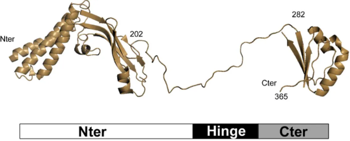
L’organisation g´en´etique est globalement conserv´ee parmi les diff´erents types de HPV. Le g´enome viral peut ainsi ˆetre s´epar´e en trois parties : une r´egion de r´egulation appell´ee LCR (pour Long Control Region) contenant l’origine de r´eplication de mˆeme qu’un certain nombre de s´equences de r´egulation transcriptionelle ; une r´egion tardive codant pour les prot´eines struc-turales L1 et L2 constituant la capside et particuli`erement importantes pour les ´etapes d’entr´ee du virus dans la cellule hˆote ; et une r´egion pr´ecoce codant pour les prot´eines exprim´es dans les premi`eres ´etapes du cycle viral aboutissant `a la r´eplication virale. Parmi les prot´eines pr´ecoces, E6 and E7 jouent un rˆole central dans la d´eregulation du cycle cellulaire et de la prolif´eration, faisant de ces deux prot´eines les oncog`enes viraux majeurs des HPV `a haut-risque. La prot´eine E1 est l’h´elicase virale impliqu´ee dans la r´eplication des g´enomes viraux. La prot´eine E2, qui fait l’objet de cette th`ese, intervient dans de nombreuses ´etapes du cycle viral. E2 est une prot´eine constitu´ee de trois domaines : les domaines N-terminal et C-terminal sont bien conserv´es `a la fois au niveau de la s´equence primaire que de la structure 3D et sont s´epar´es par une r´egion charni`ere flexible non structur´ee et non-conserv´ee. Le domaine N-terminal est un domaine de transactivation tandis que le domaine C-terminal est un domaine de dim´erization et un domaine de liaison `a l’ADN. Par sa capacit´e `a se lier `a l’ADN, E2 est un r´egulateur majeur du cycle viral. En effet, la r´egion de r´egulation du g´enome des HPV contient des s´equences particuli`eres reconnues par les prot´eines E2 et appel´ees E2BS (pour E2 Binding Sites). En se liant `a ces E2BS, E2 empˆeche le recrutement de facteurs de transcription essentiels et r´egule n´egativement l’expression des g`enes viraux pr´ecoces E6 et E7. De mˆeme, en se fixant `a ces E2BS, E2 aide le recrutement et la fixation de E1, l’h´elicase virale, sur l’origine de r´eplication, faisant de E2 un ´element essentiel `a l’initiation de la r´eplication des HPV. Enfin, E2 est aussi connu depuis de nombreuses ann´ees comme ´etant le facteur viral responsable de la s´egr´egation des g´enomes viraux lors de la division cellulaire. En se liant `a la fois `a l’ADN viral et au chromosome de l’hˆote, E2 agit comme un pont et permet ainsi de conserver un r´eservoir de genome HPV dans les cellules prolif´erantes de l’´epithelium. E2 est donc une prot´eine essentielle `a la fois au cycle viral productif et `a la persistance virale, la persistance ´etant un facteur clef dans le risque de d´eveloppement de cancer. De plus, depuis la derni`ere d´ecennie, il ´emerge qu’au del`a de ses
fonc-tions d´ependantes de la liaison `a l’ADN viral, E2 serait aussi capable de moduler directement la cellule hˆote, principalement en ´etablissant des interactions avec des prot´eines cellulaires. Il est ainsi envisag´e que E2 pourrait directement participer `a l’´etablissement de conditions cellu-laires permissives au cycle viral le long de ´epith´elium. De mani`ere int´eressante, certaines de ces fonctions de E2 sont sp´ecifiques des HPV `a haut-risque, tels que l’induction d’arrˆets du cycle cellulaire, menant g´en´eralement `a un certain degr´e d’instabilit´e genomique ou encore l’induction de l’apoptose, un m´ecanisme envisag´e comme contre-balan¸cant les propri´et´es transformantes de E6 et E7. Ceci a men´e `a l’hypoth`ese que E2 pourrait ˆetre directement impliqu´ee dans des m´ecanismes menant au d´eveloppement de cancer.
Depuis 2006, deux vaccins sont disponibles pour lutter contre les infections `a HPV, cepen-dant, la couverture vaccinale reste faible, le coˆut global est ´elev´e et ces vaccins ne sont pas `a vis´ee th´erapeutique. Il semble donc important de d´evelopper de nouvelles mol´ecules et strat´egies anti-virales. De par son rˆole dans de nombreuses ´etapes du cycle et dans la persistance virale, sa bonne conservation de s´equence et le fait qu’elle est exprim´ee pr´ecocement durant l’infection, la prot´eine E2 est envisag´ee comme une bonne cible pour d´evelopper une drogue anti-HPV. Ce-pendant, il semble n´ecessaire de mieux connaitre l’impact de cette prot´eine virale sur la cellule hˆote, en particulier, d’approfondir les notions de sp´ecificit´e de fonction selon le pouvoir oncog`ene du HPV.
Le but de cette th`ese ´etait de mieux cerner le rˆole de E2 dans la pathogen`ese et la carcino-gen`ese associ´ees aux infections par les HPV. ´Etant donn´e que les interactions prot´eine-prot´eine constituent un moyen efficace pour les prot´eines virales d’agir sur la cellule hˆote, nous avons d´ecid´e d’aborder le probl`eme des fonctions de E2 `a travers la cartographie de son r´eseau d’inter-action avec les prot´eines cellulaires. N´eanmoins, contrairement `a la plupart des ´etudes similaires qui se concentrent principalement sur les HPV les plus significatifs d’un point de vue clinique, nous avons choisi de tirer profit de la grande diversit´e de tropisme et de pouvoir pathog`ene de cette famille virale afin d’extraire des sp´ecificit´es d’interaction propres `a un type d’HPV. Nous avons donc s´electionn´e un panel de prot´eines E2 provenant de 12 g´enotypes HPV repr´esentatifs de leur diversit´e : HPV cutan´es (HPV1, 3, 5, 8, 9) ou muqueux (HPV6, 11, 16, 18, 32, 33, 39) ; HPV haut-risque (HPV5, 8, 16, 18, 33, 39) ou bas-risque (HPV1, 3, 5, 8, 9). Ces 12 prot´eines E2 ont ´et´e utilis´ees comme proies dans un criblage par double hybride en levure d’une banque d’ADN compl´ementaires issues de cellules HaCaT, une approche non-biais´ee pour la d´etection d’inter-actions. Environ 200 prot´eines cellulaires ont ´et´e identifi´ees dans ce crible, dont seule une faible proportion interagissait avec plusieurs prot´eines E2. Cependant, en raison des caract´eristiques intrins`eques des approches en levure telles que le taux d’interactions fausse-positives et surtout fausse-n´egatives, il est commun´ement admis que les interactions identifi´ees doivent ˆetre valid´ees par une autre technique. Nous avons donc s´electionn´e une centaine de prot´eines parmi les cibles les plus pertinentes identifi´ees dans le crible auxquelles nous avons ajout´e un certain nombre de contrˆoles positifs, des prot´eines cellulaires connues dans la litt´erature comme ´etant des in-teracteurs de E2. Le tout combin´e, nous avions 121 prot´eines cellulaires `a tester dans notre deuxi`eme ´etape de validation. Afin d’estimer le sp´ecificit´e des interactions vis `a vis des diff´erents g´enotypes, il nous a sembl´e important de re-tester chaque prot´eine cellulaire avec l’ensemble des 12 prot´eines E2. Il nous fallait donc tester plus de 1400 interactions (121 prot´eines cellulaires contre 12 prot´eines E2). Pour cela nous avons utilis´e la technique haut-d´ebit d´evelopp´ee dans notre laboratoire appel´ee HT-GPCA (pour High-Throughput Gaussia princeps Complementa-tion Assay). Dans cette technique utilis´ee en cellule de mammif`ere, les prot´eines de l’interacComplementa-tion `a tester sont chacune fusionn´ees `a une moiti´e de l’enzyme Gaussia luciferase. Si les deux prot´eines interagissent, les fragments de la luciferase sont amen´es `a se rapprocher, ce qui est suffisant pour reconstituer l’activit´e enzymatique. Pour d´etecter une interaction, il suffit donc de mesurer un
signal luciferase. Cette technique a ´et´e montr´ee comme g´en´erant un taux d’interactions fausse-positives particuli`erement bas ainsi qu’un tr`es bon taux de recouvrement d’interactions connues, indiquant un faible taux d’interaction fausses-n´egatives. L’avantage de ce type d’analyse `a large ´echelle est que ce n’est pas seulement des interactions qui sont compar´ees, mais des profiles d’interaction, ce qui permet de faire ressortir des informations g´en´erales sur les prot´eines virales ´etudi´ees. Dans notre cas, l’ensemble des donn´ees a ´et´e trait´e par≪Hierarchical Clustering≫, une m´ethode permettant de regrouper les profiles d’interaction par similarit´e. Un dendrogramme a ´et´e g´en´er´e - un arbre hi´erarchique - classant les profiles d’interaction par ressemblance maximum. Nous avons compar´e cet arbre bas´e sur les profiles d’interaction de E2 `a un arbre phylog´en´etique bas´e sur les s´equences des diff´erentes prot´eines E2. De mani`ere int´eressante, nous avons ainsi pu observer que les deux arbres ´etaient tr`es similaires, avec dans les deux cas, une premi`ere s´egr´egation des E2 bas´ee sur une diff´erence de tropisme, puis un second regroupement bas´e sur le pouvoir pathog`ene des diff´erents HPV. Ceci signifie qu’en regardant simplement les in-teractions des prot´eines E2, il est possible de distinguer un HPV `a haut-risque d’un HPV `a bas-risque, sugg´erant deux hypoth`eses : soit les interactions sont le r´esultat de diff´erence d’in-fection (diff´erentes niches d’ind’in-fection, modification globale du prot´eome de l’hˆote...), soit, par ces interactions, E2 contribue directement au pouvoir pathog`ene des HPV. E2 ´etant de plus en plus associ´e `a des fonctions autonomes pouvant potentiellement influencer la cellule hˆote et le d´eveloppement de cancer, il est tentant de penser que ce r´esultat est plutˆot favorable `a la deuxi`eme hypoth`ese.
En utilisant toutes les interactions d´etect´ees dans cette ´etude, nous avons construit les r´eseaux d’interactions des prot´eines E2. L’analyse des degr´es (nombre d’interaction connues) des cibles de E2 a mis en ´evidence que la prot´eine E2, comme de nombreuses autres prot´eines virales, cible pr´ef´erentiellement des prot´eines hautement connect´ees dans la cellule hˆote. Cibler des prot´eines cellulaires qui sont centrales `a de nombreuses voies de signalisation permettrait aux prot´eines virales d’avoir un effet tr`es large sur la cellule infect´ee en un minimum d’interac-tions. Finalement, pour avoir une vision plus fonctionnelle du r´eseau d’interactions, les cibles de E2 ont ´et´e class´ees en familles fonctionnelles en se basant sur leur classification en GO termes (Gene Ontology). Il a ainsi ´emerg´e que les cibles de E2 peuvent principalement ˆetre regroup´ees en cinq grandes familles fonctionnelles. La premi`ere famille ´emergeant de cette analyse corres-pond `a des prot´eines impliqu´ees dans la r´egulation de la transcription, ce qui corrobore le rˆole principal de E2 comme facteur de transcription viral. Ainsi, en adoptant une approche haut-d´ebit d’identification non-bias´ee d’interaction par double-hybride suivi d’une ´etape de validation comparative en cellule de mammif`ere, la principale fonction de E2 en tant que r´egulateur trans-criptionel ressort en priorit´e, ce qui d´emontre la grande fiabilit´e de l’approche pour d´etecter les interactions importantes des prot´eines E2. Nous avons ´egalement mis en ´evidence un ciblage de prot´eines impliqu´ees dans des m´ecanismes d’apoptose, d’ubiquitination, et de r´egulation des ARN, ce qui avait d´ej`a ´et´e li´e au rˆole de E2 dans la cellule infect´ee, avec ici, un ´elargissement du spectre d’interactions connues. Finalement, nous avons mis en ´evidence un ciblage de prot´eines impliqu´ees dans le transport intra-cellulaire et particuli`erement de v´esicules cytoplasmiques, ce qui correspond `a une fonction potentiellement nouvelle pour les prot´eines E2. Le d´etournement des m´echanismes de transport du syst`eme v´esiculaire de l’hˆote par les HPV a lieu `a deux mo-ments clefs lors de l’infection : lors de l’entr´ee du virus et de la translocation du g´enome viral au noyau, et lors de l’´evasion immunitaire pour pr´evenir l’exposition d’antig`enes viraux `a la surface de la cellule infect´ee. E2 ´etant capable de se lier fortement au g´enome viral et `a L2, une des deux prot´eines de capside, il est possible que E2 soit pr´esent dans la particule virale, ce qui laisserait supposer que E2 pourrait avoir un rˆole dans les m´ecanismes d’entr´ee du virus plutˆot que dans la r´egulation de la pr´esentation d’antig`ene.
Cette approche comparative d’´etude des r´eseaux d’interactions de 12 prot´eines E2 a donc permis d’am´eliorer la compr´ehension globale des fonctions de cette prot´eine virale. L’aspect com-paratif de cette ´etude en fait une approche de choix pour identifier des interactions sp´ecifiques `a un sous type de HPV qui pourraient ˆetre reli´ees `a des caract´eristiques pathologiques. En parti-culier, l’identification d’interactions sp´ecifiques aux HPV `a haut-risque pourrait ˆetre le premier pas vers l’identification de biomarqueurs permettant de d´etecter pr´ecocement l’apparition de cancers. Dans ce contexte, une interaction sp´ecifique parmi celles identifi´ees a particuli`erement attir´e notre attention. Il s’agit de l’interaction entre une prot´eine cellulaire d´enomm´ee CCHCR1 et la prot´eine E2 sp´ecifiquement de HPV16. CCHCR1 est impliqu´ee dans des m´ecanismes de r´egulation de proliferation de k´eratinocytes, les cellules cibles des HPV. ´Etant donn´e l’aspect tr`es important de la r´egulation de la prolif´eration cellulaire pour le d´eveloppement de cancers et le caract`ere tr`es sp´ecifique de cette interaction pour HPV16, le HPV le plus repr´esent´e dans les cancers associ´es aux HPV, nous avons d´ecid´e d’explorer plus en d´etail l’impact potentiel de cette interaction sur la cellule infect´ee. Cette interaction est consid´er´ee comme sp´ecifique puisque d’une part, les autres prot´eines E2 test´ees ne peuvent pas, ou alors que de mani`ere tr`es marginale, interagir avec CCHCR1 et que d’autre part, parmi toutes les interactions test´ees avec la prot´eine E2 de HPV16, l’interaction avec CCHCR1 est de loin la plus forte. Cette interaction avait pr´ec´edemment ´et´e identifi´ee dans une ´etude en levure et les auteurs avait pu d´eterminer que l’interaction d´ependait du domaine N-terminal de E2. Afin de cartographier de mani`ere plus pr´ecise le domaine d’interaction, nous avons introduit des d´el´etions en s´erie des h´elices alpha du domaine N-terminal de E2. D`es que la premi`ere h´elice de E2 est retir´ee, l’interaction avec CCHCR1 est compl`etement abolie, ce qui est similaire `a l’interaction avec BRD4, un partenaire majeur de E2. L’interaction entre E2 et BRD4 est tr`es document´ee et son domaine d’interaction sur E2 recouvre les trois h´elices N-terminales de E2, il est donc envisageable que l’interaction entre CCHCR1 et HPV16 E2 soit similairement d´ependante de ces trois h´elices. Pour d´eterminer quelle surface des h´elices est importante pour l’interaction, nous avons mut´e des acides amin´es dans E2 expos´es d’un cˆot´e ou de l’autre des h´elices. Ici encore, les mutations des mˆemes acides amin´es affectant la fixation de BRD4 `a E2 inhibent aussi l’interaction avec CCHCR1. Pris dans leur ensemble, ces r´esultats de mutagen`ese semblent indiquer que BRD4 et CCHCR1 partagent la mˆeme surface d’interaction au niveau du N-terminal de E2. Nous avons donc voulu d´eterminer s’il y avait comp´etition entre ces deux prot´eines cellulaires pour interagir avec la prot´eine E2 de HPV16. Et en effet, nous avons pu mettre en ´evidence qu’en pr´esence de CCHCR1, l’inter-action entre BRD4 et HPV16 E2 est r´eduite d’un facteur cinq, et que cet effet est le r´esultat de l’interaction directe entre HPV16 E2 et CCHCR1 puisqu’en utilisant une autre prot´eine E2 capable d’interagir avec BRD4 mais non avec CCHCR1, aucune comp´etition n’est observable. Cette comp´etition a ´egalement une r´epercussion fonctionnelle puisqu’en pr´esence de CCHCR1, l’effet activateur de BRD4 sur l’activit´e transcriptionnelle de E2 est r´eduit. Ces r´esultats tendent `
a montrer que l’interaction entre CCHCR1 et HPV16 E2 affecte la liaison de E2 avec BRD4, ce qui pourrait affecter les fonctions de HPV16 E2 en tant que r´egulateur transcriptionel du g´enome viral.
Afin de poursuivre l’´etude de l’interf´erence de CCHCR1 sur les fonctions transcriptionelles de E2, nous avons men´e des ´etudes de fluorescence. Il en ressort que CCHCR1 s’exprime dans le cytoplasme dans de petites structures rondes tandis que E2 d’HPV16 est plutˆot diffus, `a la fois dans le cytoplasme et le noyau, mais principalement dans le noyau. Cependant, lorsque les deux prot´eines sont co-exprim´ees, E2 est quasiment enti`erement relocalis´ee dans les mˆemes structures que CCHCR1, l’emp´echant ainsi d’atteindre le noyau. Le fait que CCHCR1 pi`ege E2 dans le cytoplasme renforce probablement la comp´etition observ´ee avec BRD4 mais doit ´egalement avoir un fort impact sur les autres fonctions nucl´eaires de E2.
Nous nous sommes finalement pench´es sur le lien entre cette interaction sp´ecifique et la r´egulation de la prolif´eration des k´eratinocytes. La r´egulation de la prolif´eration est un m´ecanisme fortement d´etourn´e par les HPV pour leur propre profit. En effet, le site d’initiation de l’infec-tion se situe `a la base des ´epith´elia, au niveau d’une couche de cellules prolif´erantes. Dans le cadre d’un epithelium non infect´e, `a un certain point, une des cellules de cette couche basale va se d´etacher et entrer dans un processus de diff´erenciation qui l’am`enera jusqu’au pˆole op-pos´e de l’´epith´elium o`u elle finira par mourir et ˆetre lib´er´ee dans le milieu environnant. C’est donc une v´eritable balance qui s’´etablit dans l’´epith´elium entre prolif´eration et diff´erenciation et qui va d´eterminer le destin de la cellule. Cependant, en d´epit des m´ecanismes de r´egulation de la diff´erenciation, les HPV ont d´evelopp´e des m´ecanismes capables de maintenir la pro-lif´eration cellulaire dans les diff´erentes couches de l’´epith´elium afin de subvenir `a leur besoin pour la r´eplication virale. Nous avons montr´e que CCHCR1 favorise la prolif´eration, tandis que HPV16 E2 stimule la diff´erenciation pr´ecoce des cellules. Mais lorsque les deux prot´eines sont co-exprim´ees, l’effet de E2 en tant qu’inducteur de la diff´erenciation est fortement r´eduit. E2 est le facteur viral contre-bala¸cant les propri´et´es oncog`eniques de E6 et E7. Or les prot´eines E6 et E7 maintiennent la cellule sous un stimuli permettant la prolif´eration des cellules. Il n’est donc pas ´etonnant de voir E2 s’opposer aux effets prolif´eratif de E6 et E7 en induisant la diff´erenciation cellulaire. Cependant ici, il semblerait que E2 de HPV16, en interagissant avec CCHCR1, ait un d´esavantage dans ce m´ecanisme, ce qui pourrait augmenter le risque de prolif´eration incontrˆol´e stimul´e par E6 et E7.
L’´etude de l’interaction sp´ecifique entre HPV16 E2 et CCHCR1 met donc en exergue un m´ecanisme qui pourrait potentiellement participer `a un processus propre `a HPV16 expliquant sa forte propension `a progresser vers la conversion maligne.
L’ensemble des r´esultats obtenus durant cette th`ese am´eliore la compr´ehension g´en´erale des fonctions de E2 lors d’une infection aux HPV. E2 apparaˆıt comme un facteur critique qui participe au d´etournement des fonctions de l’hˆote pour permettre au virus de se d´evelopper en d´epit des moyens mis en oeuvre par la cellule pour se d´efendre. E2 ´emerge aussi comme un composant viral susceptible d’influencer directement l’issue de l’infection et de prendre part aux ´etapes pr´eliminaires de conversion maligne.
Mots clefs : HPV, E2, R´eseaux, Interaction Virus-Hˆote, D´etournement cellulaire, Diff´erenciation de k´eratinocytes, HT-GPCA
Comparative mapping of E2-host interactions unravels new roles of E2 in human papillomavirus-induced pathogenesis
Abstract - Papillomaviruses are responsible for widespread infections in humans, causing pathegenesis ranging from inapparent infections to benign lesions, hyperplasia or cancers. Given the major public health concern due to HPV-associated cancers, most studies have focused on the early proteins expressed by the most clinically relevant HPVs most frequently found in can-cers. Among the early proteins encoded by HPVs, the E2 protein regulates viral transcription, replication and mitotic segregation of the viral genome, mainly through the recruitment of host factors to the HPV regulatory region. E2 is therefore pivotal for both the viral productive cycle and for viral persistence, which is a major risk factor for cancer development. In addition, the E2 proteins have been shown to engage interactions important to directly modulate the host cell, thereby contributing to create suitable cell conditions for the successive stages of the HPV life cycle. Interestingly, some E2’s roles have been demonstrated to be specific to the oncogenic HPVs, raising the idea that beyond its role in the general HPV regulation, E2 could also directly influence the fate of cancer development.
This thesis aimed at providing an overview of E2’s functions across multiple HPV geno-types and at identifying specific features that distinguish the different HPV pathological traits. We mapped the virus-host interaction networks of the E2 proteins from a panel of 12 HPVs selected to be representative of the HPV diversity. Clustering of E2’s interaction profiles corre-lated with the HPV phylogeny, raising the notion that E2 could directly contribute to the HPV pathogenesis. This work also emphasizes that the E2 proteins, like many other viral proteins, tend to target highly connected cellular proteins (cellular hubs), which is presumed to be an evolutionary way to maximize viral impacts on the host. E2 predominantly targets a subset of key cellular processes, like transcriptional regulation, apoptosis, RNA metabolism, ubiquitina-tion or intracellular transport, which both confirms already known E2’s funcubiquitina-tions and points to potential new functions. In addition, this large-scale comparative approach offers a frame-work to pinpoint interactions that are specifically associated with the most represented HPVs in cancers and therefore can be used as targets for the development of new therapeutics. In particular, we identified a specific interaction between the E2 protein from HPV16 and a cellu-lar protein, CCHCR1, involved in the regulation of keratinocyte proliferation. We determined that CCHCR1’s interaction domain on E2 overlaps with that of BRD4, a major interactor of E2, inducing a physical competition between the two cellular proteins. This competitive binding affects BRD4-mediated enhancement of E2’s transcriptional activity, suggesting that the inter-action with CCHCR1 might have an impact on the role of E2 in the infected cell. In addition, we showed that CCHCR1 induces the docking of HPV16 E2 into the cytoplasm which could further affect E2’s nuclear functions. We also demonstrated that CCHCR1 impairs HPV16 E2’s induction of keratinocytes early differentiation, presumably resulting from the negative effect of CCHCR1 on the nuclear functions of E2. This effect could have drastic consequences on the oncogenic potential of HPV16 and could participate to high prevalence in cancers of HPV16.
Taken together, these results enhance the general understanding of the impact of E2 during HPV infections and highlights its contribution in the HPV pathogenesis. E2 appears as a critical factor that participates in the global hijacking of the host cell to allow the virus to replicate despite the hostile environment. E2 also emerges as a viral component susceptible to directly influence the outcome of an HPV infection and to potentially impact on the preliminary steps of carcinogenic conversion.
Key words: HPV, E2, Network, Virus-host interactions, Cellular Hijacking, Ker-atinocyte differentiation, HT-GPCA.
Acknowledgments
I want to express my gratitude to the member of the jury and especially professor Chen-Ming Chiang for its careful reading of the manuscript and who came all the way from Texas to help me in the final steps of this thesis.
Tout d’abord, je souhaiterais remercier les membres de mon jury qui ont accept´e de juger ce travail de th`ese. Les professeurs Chiang et Touz´e qui, par la lecture minutieuse et critique de ce manuscrit, m’ont permis de l’am´eliorer. J’exprime ´egalement toute ma reconnaissance aux docteurs Masson et Bachelerie pour avoir accept´e d’en ˆetre les examinateurs. Je remercie ´egalement le professeur Ceccaldi d’avoir accept´e de pr´esider ce jury de th`ese.
Je tiens `a remercier toutes les personnes avec qui j’ai partag´e le quotidien pendant ces ann´ees `a l’Institut Pasteur. Tout d’abord Caroline qui a encadr´e mon travail de th`ese, pour son ´eternel soutien, sa force, sa motivation, sa connaissance sans fin des papillomavirus, sa gentillesse et sa g´en´erosit´e. Je la remercie de m’avoir men´e l`a o`u je suis aujourd’hui. Yves ´egalement, pour sa joie de vivre perp´etuelle et son optimisme d´ebordant, et Fran¸coise pour sa grande rigueur et son amiti´e. Je remercie aussi Michel Favre et Sylvie Van Der Werf de m’avoir accueillit dans leurs laboratoires. Je remercie ´egalement Patricia pour son aide tout au long de ce travail de th`ese. Un grand merci aussi toutes les personnes qui ont crois´e mon chemin autour des papillomavirus, Christian, H´el`ene, Laurence, Guillaume, Delphine, Valentine, Caroline. Merci ´egalement `a tous les membres de l’unit´e de G´en´etique Mol´eculaire des Virus ARN qui m’accueillent dans leur laboratoire sur cette derni`ere ligne droite.
Je remercie ´egalement la fondation l’Or´eal pour son soutien financier qui m’a permis de finir cette th`ese sereinement.
Je remercie tout particuli`erement ma famille, mes plus grands fans et fervents sup-porters. Ma m`ere, qui savait qu’`a coup de livres, je franchirais tous les murs ; mon p`ere, qui ne dira plus jamais qu’il n’y a pas de quoi ´ecrire une th`ese ; mon fr`ere et sa fille, ma ni`ece, que j’aime tant ; Cathy et Philippe pour tout l’amour qu’ils m’ont toujours donn´e. Une grosse pens´ee ´egalement pour mon P´ep´e et ma M´em´e, mes oncles et mes tantes, mes cousins et cousines, parce que je suis fi`ere de faire partie de cette grande famille.
Un grand merci `a tous mes amis, particuli`erement Alexandre, C´edric et Wahb qui me font partager leur vie de physiciens mais ´egalement `a celles et ceux qui ont accompagn´e mes premiers pas dans biologie, je pense notamment `a Audrey et C´eline. Un petit clin d’œil `a Newton, parce qu’il m’aura bien fait rire depuis 2 ans ;-)
Mes derniers remerciements vont `a Romain, ma moiti´e, qui m’a soutenu et aid´e tout au long de ces ann´ees, qui a toujours cru en moi et avec qui j’esp`ere pouvoir poursuivre encore longtemps notre bout de chemin, aussi loin que le vent nous portera.
Contents
I . Introduction
3
A -Historic . . . 3 B - General description . . . 3 a . Classification . . . 4 b . HPV infection . . . 5 1 . Mucosal HPV . . . 5 2 . Cutaneous HPV . . . 6 c . Genomic organization . . . 7 1 . LCR . . . 7 2 . Early region . . . 8 3 . Late region . . . 10 d . Viral cycle . . . 12 1 . Non-productive cycle . . . 12Attachment and entry . . . 13
Internalization and uncoating . . . 14
Nuclear import . . . 15
2 . Productive cycle . . . 16
Replication and genome amplification . . . 16
Assembly and virion egress . . . 17
C - Carcinogenesis. . . 18 a . Proliferation . . . 18 b . Genome integration . . . 20 c . Malignant transformation . . . 21 d . Immune evasion . . . 23 D -The E2 proteins . . . 26 a . Structural properties . . . 26 Transactivation domain . . . 26 DNA-binding domain . . . 27 Hinge . . . 27 b . Regulatory roles . . . 27
1 . Viral genome binding-associated activities . . . 28
Transcription . . . 28
Replication . . . 30
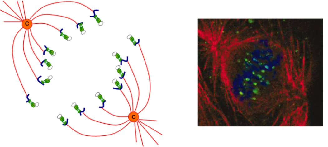
Mitotic segregation . . . 30
2 . Host cell manipulation . . . 33
Apoptosis . . . 33
Ubiquitin-Proteasome System targeting - Cell cycle . . . 35
Ubiquitin-Proteasome System targeting - Stability . . . 36
Cell differentiation and migration . . . 37
II . Results
41
A -Viral interactomic. . . 41a . Context . . . 41
b . The HPV E2 proteins interaction network . . . 43
1 . E2-host protein-protein interaction mapping . . . 44
2 . E2-host interaction network . . . 51
B - Specific HPV16E2-CCHCR1 interaction . . . 61
a . Context . . . 61
b . Characterization . . . 61
c . Consequences on E2’s cellular localization . . . 65
d . Functional impact on keratinocyte differentiation . . . 68
III . Discussion
73
IV . Materials & Methods
81
1 . Cell culture and transfections . . . 812 . Plasmids . . . 81
3 . HT-GPCA . . . 82
4 . Hierarchical clustering and topology . . . 83
5 . Fluorescence . . . 83
6 . Transactivation . . . 84
7 . Differentiation . . . 84
8 . RNA extraction and RT-qPCR. . . 84
Bibliography
87
Presentations
116
I . Introduction
From an evolutionist’s point of view, papillomaviruses are very successful infectious agents. They induce unapparent chronic infections that rarely kill the host and periodi-cally shed infectious virions for their transmission. In humans, certain types of papillo-maviruses (HPV) referred to as High-Risk HPV (HR-HPV), are predominant risk factors for the development of cervical cancers and other epithelial cancers as well. HPVs are the most frequent sexually transmitted agent causing pathologies ranging from asymptomatic infections to benign hyperplasia or cancer and are therefore considered as a major public health concern. Interest has grown over the last decade to better understand the biology of these small DNA viruses.
A - Historic
Genital warts have been known for many centuries but until the 19th century, they were generally considered to be a form of syphilis or gonorrhea. It is only in 1907 that Giuseppe Ciuffo demonstrated the viral nature of human warts after cell filtrates from lesions were shown to transmit the disease [1]. Details on when and how the first strain of HPV was discovered are incomplete, but the papillomavirus was first glimpsed as a disease by R. Shope of Rockefeller University in the 1930s [2]. A rabbit strain of papillomavirus (nowadays referred to as CRPV, Cottontail Rabbit Papillomavirus) often causes horn-like warts on infected rabbits. The cause of these warts was not known at the time, but Shope took samples of the warts and injected them back into healthy rabbits. The healthy rabbits soon developed the same warts. Shope did not identify the exact viral strain, but he correctly deduced that the warts were caused by a virus. In addition to causing benign papillomas, some warts induced by CRPV were observed to undergo malignant progression [3]. As such, the Shope virus became an important model to study viral tumorigenesis. It was not until the 1970s and the advent of molecular cloning that researchers have started to better understand the biology of papillomaviruses. The sequencing of the genome led to the identification of the open reading frames (ORF) as putative viral genes resulting in the characterization of their functions. German virologist Harald zur Hausen proposed in 1976 that HPV was the cause of cervical cancer, a theory that other scientists originally rejected [4]. In 1984, the team of zur Hausen discovered HPV DNA in cervical cancer tumors, proving his theory [5]. In 2008, he received the Nobel Prize for this research.
B - General description
The identification in the early 1980s of HPV16 and HPV18 by the German team provided the field with HPV types present in most cervical cancers. Subsequent studies have sought to understand the natural history of HPV infection, determine the biological
properties of different HPV types, elucidate the role of viruses in pathogenesis and identify non-viral factors that may influence the outcome of an infection.
a . Classification
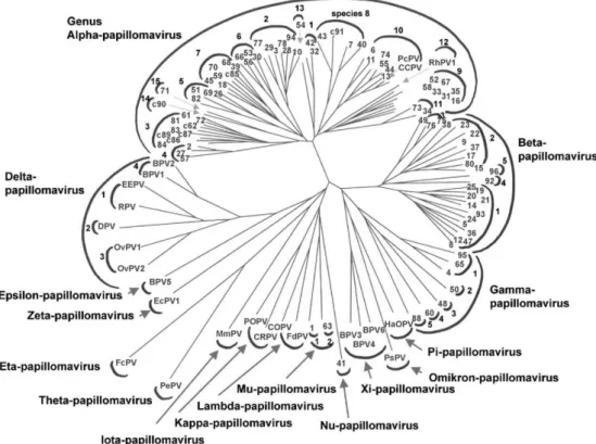
Papillomaviruses (PV) have been isolated in many host species from humans to birds or even reptiles. Given the high species-specificity of PV, there are hundreds of PV types. The most extensively studied are the Human Papillomaviruses (HPVs) with more than 200 different genotypes identified, of which 118 are fully sequenced [6–8]. If originally classified according to their host species, DNA sequencing of the HPV genomes has led to their classification according to the comparison of the L1 ORF, which encodes the HPV major structural protein (Fig I.1).
Figure I.1: Phylogenetic tree of papillomaviruses. Classification obtained by comparison of the L1 ORF sequences. The number at the end of each branch identifies a type of HPV, the other abbreviations refer to animal papillomavirus types. (From deVilliers et al. [7]).
The papillomaviruses are divided into several genera (designated with a greek let-ter) and further subdivided into genotypes (designated by a number for human papillo-maviruses or a letter for animal papillopapillo-maviruses). HPVs are clustered into five genera: alpha, beta, gamma, mu and nu. This classification often correlates with the tropism and pathogenic potential of viruses, demonstrating that viruses and host have co-evolved. The most clinically relevant HPVs (those at the origin of genital cancers) are members of the alpha genus. Most of the human papillomaviruses of the alpha genus infect mucosal
epithelia while members of the other genera primarily infect the skin. The β HPVs in-clude those associated with epidermodysplasia verruciformis (HPV5 and 8), a rare genetic predisposition to widespread non-genital HPV cancerous lesions.
b . HPV infection
HPVs are strictly epitheliotropic viruses that induce in the general population fre-quent asymptomatic infections. The HPV life cycle is tightly linked to the differentiation program of keratinocytes, their target cells. HPVs are implicated in the development of benign lesions of the skin (wart) or of mucous tissues (condylomas) as well as in malignant hyperplasia.
1 . Mucosal HPV
To establish infection, HPVs are believed to infect epithelial cells that possess pro-liferative capacities. As these cells are located at the basal layer of the epithelium, it is commonly accepted that it is microtrauma of the upper layers of the epithelium that provide access for the viruses to the basal cells. Such microtrauma occur frequently dur-ing intercourse and it is thus accepted that the standard transmission process is sexual intercourse. The prevalence of HPV infection is thus usually correlated with measures of sexual promiscuity like the number of lifetime sexual partners, recent change in sexual partners and history of other sexually transmitted infections. Genital HPV infection is the most common sexually transmitted viral infection [9, 10]. Infections by the genital HPVs are widespread and are associated with a broad range of clinical manifestations. HPV types like the HPV6 and 11 are the etiologic agents of benign hyperplasia such as genital warts or Condylomata acuminata. Although these lesions can resolve on their own, they are often recurrent and there is currently no long-term effective treatment. Other HPVs like HPV16, 18, 31, 33, are associated with the development of intra-epithelial neoplasia or pre-cancerous lesions. Most of the lesions regress spontaneously in less than a year [11, 12], however, in some cases, the lesions can persist, which represents a major risk for the development of cancer. HPV16 infections are more likely to persist than infection by other HPV types [13] and this particular HPV account by itself for more than 50% of cervical cancers. Cervical cancer is the second most common cancer in women worldwide, with more than 500,000 newly diagnosed cases each year [14], most of them occurring in developing countries. The disproportionately high burden of cervical cancers in devel-oping countries and in medically underprivileged populations is mainly due to a lack of screening that allows detection of precancerous lesions at early stages [15]. Most cancers occur in the transformation zone of the cervix, at the junction between the endocervix and the stratified squamous epithelium of the exocervix. Histological classification of cervical intraepithelial neoplasia (CIN) grades the lesions from 1 to 3 based on the severity of the dysplasia: CIN1 corresponds to a mild dysplasia , CIN2 to moderate dysplasia, and CIN3 to severe dysplasia or carcinoma.
Infections of the oral mucosa by certain types of HPVs have also been reported for many years [16]. Most of Head and Neck cancers are caused by smoking and/or alcohol consumption but consistent data indicate that a subset of oral cancers, mainly in the upper oral area including tonsils, base of the tongue and soft palette, are attributable
to HPV infection, with HPV16 accounting for about 90% of the HPV-positive tumors [17–19].
2 . Cutaneous HPV
Skin warts are benign papilloma most frequently found on the hands and feet and lesions are usually small dome-shaped papules with a keratotic and verrucous surface [20]. They occur commonly in older children and young adults [21]. Since HPVs need to infect basal cells, maceration of the skin is believed to be a predisposition for infection. There is a good correlation between HPV types and clinical lesions with HPV1, 2, 4, 27, 57 and 65 usually associated with common and plantar warts, whereas HPV3, 10, 28 and 41 are mostly associated with flat warts. The lesions tend to be self resolving, but complete clearance may take up to several years. Regression of lesions is most likely due to an effective immune response and the low incidence of warts in older individuals might suggest that immune mechanisms have rendered them resistant to infection.
Patients with a rare genetic disorder, Epidermodysplasia verruciformis (EV), have a unique susceptibility to cutaneous HPV infections [22–24]. Starting during childhood, warts quickly spread over the body, tend to persist, and may even progress to squamous cell carcinomas. About a third of EV patients develop skin cancers and if most of the lesions remain local, distant metastases may appear. Patients with epidermodysplasia verruciformis are usually infected with multiple types of HPVs, including the common types that affect individuals of the general population. The development of tumorous lesions are primarily associated with HPV types referred to as EV-HPVs, mostly HPV5 and 8, which are present in 90% of cancers in EV patients. EV-HPVs were for long believed to be specific to the disease since they could not be detected in patients without EV, except in immunosuppressed populations. However, with the increasing sensitivity of detection methods, it was possible to show the presence of the EV-HPVs in the general population as well [25]. EV occurs as an inherited disorder with an autosomal recessive pattern. Two distinct chromosomal loci have been linked to the HPV predisposition in EV patients: EV1 and EV2. Two adjacent genes, EVER1 and EVER2, were identified within the major locus EV1, whose nonsense mutations are associated with the development of the disease [26, 27], suggesting a potential pivotal role in the control of the infection.
A high prevalence of HPV DNA in squamous cell skin carcinoma of immunosuppressed, but also of immunocompetent patients, has renewed the great interest in a possible eti-ologic role of HPVs in nonmelanoma skin cancer. Nonmelanoma skin cancers (NMSC), a frequent form of skin cancers, can be divided into two groups: basal cell carcinomas (BCC) and squamous cell carcinomas (SCC). Usually developing on skin areas exposed to sunlight, these tumors rarely metastasize. Consistent findings of certain HPV types in SCC makes HPV an attractive co-factor of UV radiation for cancer development [28]. Be-cause of the low copy numbers of HPV DNA in skin cancers, probably not every tumor cell contains viral DNA, which is compatible with cutaneous HPVs being possibly important for tumor initiation, but not for maintenance of the malignant phenotype. This would suggest a “Hit-and-Run”mechanism, where the presence of viral DNA would be sufficient, in association with co-factors, to trigger malignant conversion, but the subsequent loss or dilution of viral DNA copies would not impede further cancer progression [29].
c . Genomic organization
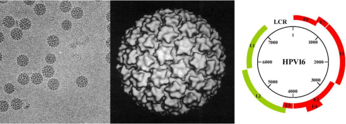
Papillomaviruses are small, non-enveloped, DNA viruses infecting squamous epithelial cells. Viral particles are approximately 55 nanometers in diameter [30] containing a single double-stranded circular DNA molecule of about 8,000 base pairs within an icosahedral capsid of 72 capsomers (Fig I.2).
Figure I.2: Papillomavirus particles and genome. Left: Electron micrograph of Bovine papillomavirus 1 (BPV1). Middle: Three dimension image reconstitution of virion particle structures (Adapted from Baker et al. [30]). Right: HPV16 genomic map. The number in the circle indicate the nucleotide position. The ORF regions are represented either in red for the early proteins or in green for the late proteins. The long control region (LCR) is represented at the top.
The genomes of numerous human and animal papillomaviruses have been fully se-quenced and it appears that the genomic organization of most PVs is similar (Fig I.2). All open reading frames are located on the same DNA strand, meaning that only one strand serves as a template for transcription. ORFs are classified either as early (E), encoding the regulatory proteins or late (L), encoding the structural proteins. A region called LCR (for Long control Region) of about 1kb devoid of ORF contains the viral origin of replication as well as important transcriptional control elements. Each of these regions are described in the following sections.
1 . LCR
The LCR of papillomaviruses contains sequence elements that are responsive to cellular factors, as well as virally encoded regulatory proteins (Fig I.3). Typically, the LCR includes a tissue-specific enhancer that plays an important role for viral gene expression in keratinocytes and may also be important for viral latency. The LCR contains numerous transcription binding sites such as AP1, SP1, Oct1, and YY1 among others [31–34]. No specific factors have been identified to be responsible for the keratinocyte-restricted activity of the enhancer. It seems that the specificity would rather be conferred by a complex interplay among these multiple ubiquitous transcription factors (see [35] for review).
Figure I.3: HPV16 Long Control Region. Schematic map of the long control region of HPV16. The end of the L1 and the beginning of the E6 ORFs are indicated. The E2 binding sites (E2BS) are numbered from 1 to 4 and colored in red.
The LCR also harbors a glucocorticoid response element that is differentially regulated among the HPVs [36–38]. In addition to the binding sites for cellular transcription factors, the LCR contains recognition elements for the virally encoded E2 protein (referred to as E2 binding sites or E2BS) as well as the origin of viral DNA replication where the E1 helicase is loaded. At least four E2BS are present in the LCR of all α-HPVs and display a conserved relative position near the initiation start of the early promoter allowing the coordinated regulation of viral DNA transcription and replication. The LCR of the β-type HPVs is smaller and the regulatory elements are positioned differently than in the α-type HPVs [39, 40]. In particular, the E2 binding sites are located farther from the early promoter, which greatly influences the regulation of viral gene expression [41]. This will be developed later in this manuscript.
2 . Early region
The early region represents about half of the total genome and encodes proteins re-quired during the early steps of the viral life cycle, before the onset of viral genome amplification. This region encodes four to seven proteins:
- E1 - E1 is the longest of all HPV proteins with 600 to 650 amino acids, but is also well conserved in sequence [42, 43]. E1 contains a DNA-binding domain as well as a helicase domain and an ATP fixation motif [44, 45]. E1 has DNA-dependent adenosine triphosphatase (ATPase) and DNA helicase activities [46, 47], which are pivotal for viral replication. E1 functions both as a DNA binding protein that binds with a weak affinity the viral origin of replication and as an helicase to unwind DNA ahead of the replication fork. The binding of E1 to the LCR is stabilized through its cooperative binding with the E2 protein [48, 49] (and [35] for review) which, in contrary to E1, associates with high affinity to its E2BS in the viral origin. Through interactions with E1 and E2, cellular replication factors such as DNA polymerase α-primase [50–52], topoisomerase I [53], and the single-stranded DNA-binding protein RPA [54] are recruited to the replication origin for assembly into an active replication complex. E1 then assembles in a hexameric complex that, in the presence of ATP, unwinds and bends the viral DNA and initiates replication [55]. In addition to being required for replication initiation, E1 is also important for DNA elongation and moves along the viral DNA template [49].
- E2 - E2 is a key protein for the viral life cycle and has a wide impact on the viral gene expression. The role of E2, being the focus of this thesis, will be detailed in a
specific section later in the introduction.
- E4 - The E4 ORF overlaps with that of E2 but is encoded in a different frame. Although E4 is classified as an early gene, it is expressed during the late stages of the viral life cycle, concomitantly with L1 and L2 [56–59]. The E4 protein has been shown to be synthesized from a spliced mRNA obtained from a donor site in E1 and an acceptor site in E2 at the beginning of the E4 ORF. It therefore results in the translation of the first five amino acids of E1 at the N-terminus of E4 and generates a protein often referred to as E1ˆE4. Recently, other transcripts were described and correspond to fusions between the N-terminal part of E2 and the complete ORF of E4 [60].
E1ˆE4 transcripts are produced throughout the HPV life cycle; however, the highest levels are found in the differentiated suprabasal layers [61]. E4 is primarily a cytoplasmic protein and colocalizes with the intermediate filament network. It has been shown to induce the reorganization and degradation of this keratin filament network to potentially favor viral particle release by weakening the upper layer of the epithelia [62, 63]. In addition, E4 was also shown to be localized at the mitochondria which leads to their detachment from the microtubules followed by a reduction of the mitochondria membrane potential and subsequent apoptosis induction. It was hypothesized that it could further facilitate the release of newly synthesized viral particles [64]. E4 is also able to arrest the cell cycle in G2, presumably to create a pseudo S-phase that optimizes conditions for viral DNA replication [65].
- E5 - The HPV E5 proteins are small hydrophobic proteins localized predomi-nantly at the membranes of the endoplasmic reticulum, and occasionally at other cellular membranes [66, 67], whose role is still nebulous [68]. The E5 ORF is absent in the genome of many HPVs, such as beta-, gamma- and mu-HPVs, indicating that this protein is not essential for the life cycle of these viruses but rather can participate in infection and transformation. HPV16 E5 self-associates and this oligomerization is mainly mediated by hydrophobic interactions [69]. The E5 protein displays some transforming activities since it was shown to induce cell growth and tumorigenic transformation in transgenic mice in association with host factors [70–72]. It has been proposed that E5 can associate directly with the EGF (Epidermal Growth Factor) receptor, which may augment downstream receptor signaling and thus promote cell proliferation. In addition, E5-mediated effects on endosomal maturation and movement have been implicated in alteration of host cell antigen presentation [73, 74], therefore potentially favoring immune evasion.
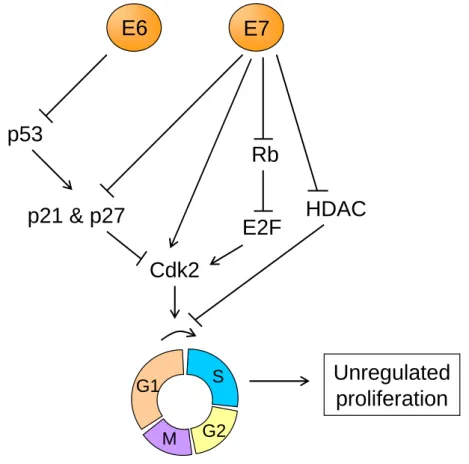
- E6 & E7 - Overall, E6 and E7 disrupt or usurp multiple cellular signaling pathways to maintain infected cells in a proliferative state necessary for viral replication. However, in the case of HR-HPVs, it can lead to an increased genomic instability, and can result in full transformation. To understand the key role of E6 and E7 in HPV infection, it is important to keep in mind that the HPV life cycle is closely linked to the differentiation process of keratinocytes. Indeed, HPVs rely on host factors and in particular, on the host replication machinery to achieve their replication. However, the cells normally exit the cell cycle upon detachment from the epithelium to enter the differentiation process. One crucial aspect of HPV infection is therefore to uncouple the cellular proliferation and differentiation capacities, and this is mainly mediated through the activities of E6 and E7. The HPV E7 protein binds to Rb (Retinoblastoma) family members and targets them
for degradation [75]. This results in the release and activation of E2F transcription factors that drive expression of S-phase genes. The interaction between E7 and Rb is conserved for different types of HPVs but a much higher affinity is observed with High-risk HPVs [76, 77]. The binding of Rb by E7 and the subsequent forced S-phase gene expression would normally lead to cell growth inhibition and apoptosis through p53-dependent pathways. However, the E6 proteins from HR-HPVs have evolved to target the tumor suppressor p53 for degradation [78], thereby preventing cell growth inhibition and other p53-mediated responses. To interfere with p53 functions, E6 recruits the cellular E3 ubiquitin ligase E6-associated protein (E6AP), which leads to the ubiquitination and proteasomal degradation of p53 [79, 80]. Low-risk HPV E6 proteins can also complex with E6AP but this does not result in p53 degradation [81, 82]. The E6 protein of high-risk HPV types also plays a role in mediating cell proliferation independently of E7 through its C-terminal PDZ-binding domain, which can mediate suprabasal cell proliferation [83, 84]. These combined activities make high-risk HPV E6 and E7 proteins the primary transforming viral oncoproteins. The action of high-risk E6 and E7 proteins in targeting cell cycle regulators to maintain S-phase competence in differentiating cells results in perturbation of many cell cycle checkpoints. In long-term HPV-infected cells, this may lead to the accumulation of cellular mutations over a long period of time, which further promotes progression toward cancer [85].
Both E6 and E7 are small proteins, approximately 18 and 13 kDa in size, respectively. Despite this small size, E6 interacts with numerous cellular partners [86, 87] by, among others, recognizing Leucine-rich motifs containing the LxxLL consensus sequence or PDZ domains [83, 88–90]. Possessing nuclear localization signals, High-risk HPV E6 proteins are localized in the nucleus and in the cytoplasm while those of Low-risk HPVs are mainly cytoplasmic [91]. E7 accumulates both in the cytoplasm and in the nucleus and the presence of both nuclear localization and export signals in its C-terminal domain suggests that E7 is able to shuttle between the nucleus and the cytoplasm [92].
To conclude, the E6 and E7 proteins do not have an intrinsic enzymatic activity but rather act on the targeting of host cellular factors and the subsequent deregulation of cellular pathways to promote viral replication. The main oncogenic activities of HR-HPVs can be attributed to E6 and E7, but other early viral proteins also contribute to the overall carcinogenic conversion, like the E5 proteins and as we will discussed later, the E2 protein.
3 . Late region
The late region of the HPV genome encodes two structural proteins: the major capsid protein L1 and the minor capsid protein L2. Both proteins are essential for the assembly of the viral capsid but also for viral entry.
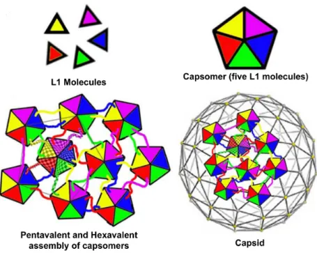
- L1 - L1 is the main factor for viral capsid oligomerization resulting in the forma-tion of capsomers, the basic structural unit of the viral capsid.
L1 is synthesized during the late stages of infection, in the upper layer of infected epithelia, and is required for virion production and assembly. The viral capsid is composed of 360 L1 molecules, and up to 72 copies of the minor capsid protein, L2. L1 spontaneously oligomerises into pentamers, termed capsomers, which are the primary constituents of the
Figure I.4: Interaction between capsomers. L1 molecules associate to form pentameric capsomers. Pentamer contacts form pentavalent or hexavalent capsomers. Adapted from Modis et al. and Pereira et al. [93, 94].
outer capsid shell. Capsomers are linked together by the carboxy-terminal domains of L1 and are stabilized by intercapsomeric disulfide bonds between highly conserved cysteine residues (Fig I.4).
L1 mediates the primary attachment of viral particles to the cell surface or extra-cellular matrix (ECM) of target cells [95, 96]. Surface binding triggers changes in the conformation of the L1 protein resulting in the exposure of the N-terminal end of the minor capsid protein L2, necessary for further internalization steps. After viral uncoat-ing, L1 segregates from the L2/viral genome complex in an endocytic compartment and is targeted to lysosomes for degradation [97].
The L1 protein contains all the intrinsic information required to form the capsid struc-ture and is therefore sufficient to produce virus like particles (VLP) that mimic the native virion structure [98]. VLPs provide an efficient system to understand papillomavirus particle assembly, structure, and the binding of virus particles to the host cell.
Two vaccines against HPV infections have passed rigorous human trials and are cur-rently commercialized: namely, Gardasil (Merck) and Cervarix (GSK). These vaccines exploit the fact that L1 expression alone leads to formation of VLPs, which have proven highly effective at producing a strong immune response that can protect against infection in humans [99, 100].
- L2 - L2 is a multifunctional structural protein naturally incorporated in the capsid. The L2 proteins are located beneath the L1 pentamers [101] meaning that there are 72 potential sites in the capsid in which L2 could be found (72 L1 capsomers in the capsid), although not all of them are believed to be occupied. L2 molecules seem to
interact with each other in an intercapsomeric-dependent manner, with the C-terminal region of one L2 molecule closely apposed to the N-terminal region of another [101]. Moreover, L2 is able to interact with L1 primarily through hydrophobic contacts [102]. L2 is critical for establishment of infection and evidence suggests an essential role for the L2 protein in many different steps of viral uptake by the infected cell [94, 103, 104]. L2 has been implicated in virion binding to the cell surface following its cleavage by furin [105], and is essential for the release of viral DNA from the endosomes [103], which seems facilitated by an L2 membrane-destabilizing motif [106] and to require L2 furin precleavage despite this step occurs at the cell surface [107]. L2 also helps in the transport and entry of the viral DNA to the nucleus [108]. However, the mechanisms involved in these processes are still evasive. Interactions of the L2 protein with microtubule motor proteins [109, 110] have been reported, suggesting that movement along microtubules might take part in the transport of HPV DNA to the nucleus. L2 proteins harbor two nuclear localization signals, suggesting that L2 could mediate nuclear import of viral genomes via nuclear pore complexes [111].
Interestingly, L2 also plays a pivotal role in virion production during viral life cycle, helping both in virion components gathering during the assembly of viral particles [112] and in the encapsidation of the viral genome through its interaction with the HPV DNA [104, 113, 114]. An interaction between L2 and E2 has been identified [115], another viral genome binding protein, which could further help the recruitment of HPV DNA to the site of virion assembly.
L2 is also necessary to generate virus-like particles. VLPs can form in the absence of L2 but L1-only VLPs have been noted to be more variable in size and shape than L1/L2 VLPs, leading to the speculation that L2 improves capsid formation. Similarly, L2 seems to increase VLP internalization when included in the particle [104, 116].
Immunological research on L2 has revealed promising cross-neutralisation potential [117, 118]. These characteristics are important in considering L2 or L2-containing structs as vaccine candidates. Currently, phase-I trials of a vaccine utilizing an L2 con-struct are planned, and results of such trials are eagerly anticipated [119–121].
d . Viral cycle
HPVs are strictly epitheliotropic viruses whose life cycle is closely linked to the dif-ferentiation process of keratinocytes. Once the viral particle reaches the basal layer of the epithelium, presumably following microwounding, it enters the cell and establish in-fection. As the infected cell undergoes differentiation and moves toward the upper layer in the epithelium, the HPV proteins hijack the replication machinery to trigger genome amplification and later, viral production. Ultimately, virions are shed from the uppermost layer of the epithelium (Fig I.5).
1 . Non-productive cycle
In the undifferentiated cells comprising the proliferating part of the epithelium, the viral genome is maintained at low copy number as a stable nuclear episome. Only a subset of viral genes, the early genes, is selectively expressed in the basal compartment,
Figure I.5: HPV life cycle. Names of the epithelium layers are indicated on the left and the different viral steps on the right.
and therefore no new viruses are produced. Consequently, the first phase of the HPV life cycle in the epithelium basal layers is commonly referred to as the non-productive cycle.
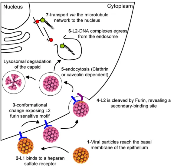
Attachment and entry
For non-enveloped viruses such as HPVs, the protein coat covers and protects the viral DNA and provides the initial interaction site of the viral particle with the host cell [94]. After receptor engagement, the viral particle is internalized and its coat is disassembled to allow the encapsidated genome to access the cellular transcription and replication machinery in the nucleus (Fig I.6).
Initial binding is believed to occur at the basal membrane which underlies the epithe-lium. The viral particle reaches these regions only after their exposure by mechanical or chemical trauma [122, 123]. The initial interaction depends primarily on L1 [124–126]. Early work investigating host cell entry of HPVs showed that this process is initiated by binding of the virus particle to cell surface receptors, which are widely expressed and evolutionary conserved. Heparan sulfate proteoglycans (HSPG) are frequently found in the extracellular matrix and on the surface of most cells and were proposed as initial attachment receptors for HPV particles [95, 127]. HSPG function as more than simple attachment factors as they promote essential conformational changes in the viral capsid. However, HSPGs are clearly not the cell surface receptors that mediate virion internaliza-tion or later events in infecinternaliza-tion [105]. Accumulating evidence suggests that a secondary receptor or co-receptor is also involved in the infectious internalization of HPV subsequent to interaction with HSPG [128]. A role for L2 in facilitating infection via interaction with a secondary receptor has been suggested [108].
Initial capsid interaction with HSPG results in a conformational change which induces the exposure of a highly conserved furin cleavage motif in the N-terminal part of the L2 protein. After cleavage, an additional conformational change is likely to expose the binding site for the secondary cell receptor [105].
Figure I.6: Model for HPV entry. Schematized representation of viral entry processes. Modification in the virion color symbolizes the conformational change. Adapted from Pereira et al. [94].
Internalization and uncoating
After binding to cell surface receptors, HPV particles are internalized into the cell to establish the infection. To date, the dynamics of HPV interaction with the cell surface during the initial stages of infection are not completely understood and both the entry mechanisms and the molecules involved are still a matter of scientific debate [129]. Pro-ductive entry of HPVs involves internalization by endocytosis, a process that for HPVs occurs slowly and asynchronously over a period of several hours [130]. Several endocytic pathways have been described and clathrin- and caveolae-mediated are two main path-ways presumably used by HPVs [131–135]. Both trafficking routes eventually converge to the endosomal pathway.
A carrier vesicle intermediate is then used to deliver virions into endosomes and lyso-somes. Uncoating is not observed until approximately 8-12 hours after cell surface binding. Viruses have evolved several mechanisms to exit the endosomal compartment in order to
access the cytoplasm. For enveloped viruses, it usually requires fusion of membranes, ei-ther at the cell surface or after internalization [136]. Non-enveloped viruses can eiei-ther lyse [137] or generate a pore in the limiting vesicular membrane [138] to allow escape of the viral genome into the cytosol. Pathogens that proceed through the endosomal pathway during trafficking in the host cell also typically take advantage of the pH acidification of the endosomal compartments [139–141]. Acidic pH acts as a trigger for many viruses to undergo conformational changes, leading to a number of events that facilitate endo-somal escape of virion proteins and genomes. Such events may include modification of the viral-receptor interaction, exposure of protease digestion motifs or partial to complete uncoating of the viral genome. Although a C-terminal region of the HPV minor capsid protein L2 has been identified to display pH-dependent membrane-destabilizing activity [106], the exact mechanism by which this structure may assist in endosomal escape re-mains unclear. L2 was shown to interact with the sortin nexin 17 (SNX17) and this was demonstrated to mediate the retention of HPV virions in late endosomes, preventing their rapid lysosomal degradation and thereby favoring L2-DNA complexes to egress from the endosomes [103]. L2 was also recently demonstrated to contain transmembrane motifs which were hypothesized to be able to oligomerize, potentially forming a pore through the endosomal membrane and therefore important for the transfer of the L2-DNA com-plexes through the membrane [142]. All data combined point to L2 as a critical factor for HPV infection and more particularly as a key protein for viral genome egress from the endosome. Meanwhile, L1 does not appear to exit from the endosomal compartment but is ultimately destructed in lysosomes, confirming that L2 is the main factor allowing viral DNA egress from the endosome.
Nuclear import
Infection by DNA viruses replicating in the nucleus requires transport of the viral genome through the cytoplasm, a complex barrier due to its viscosity and the presence of a dense network of microtubules, actin, and intermediate filaments. A common strat-egy adopted by viruses to overcome this obstacle has been to use the cellular transport machinery to move along microtubules. In particular, the microtubule disrupting drug nocodazole inhibits HPV infection [131, 132] suggesting that the microtubule network integrity is essential for the early steps of HPV infection. Cytoplasmic transport along microtubules is mediated by motor protein complexes, and L2 has been found to interact with the microtubule network via motor proteins [109, 110] suggesting that the L2-DNA complexes reach the nucleus by moving along the microtubule network. Additional ques-tions are being asked concerning the way the genome enters the nucleus but recent data suggest that it may follow nuclear membrane breakdown during mitosis [143] rather than through active transport of the L2-genome complex via karyopherins [111]. Once in the nucleus, the L2/DNA complexes predominantly localize in distinct punctuate nuclear do-mains designated ND10 bodies or promyelocytic leukaemia (PML) oncogenic dodo-mains (PODs), as determined by their co-localization with PML, the ND10-defining protein [144]. The subsequent steps of the viral life cycle will be initiated in the nucleus.
2 . Productive cycle
In a normal epithelium, basal cells proliferate and undergo cell division, but at some point, a daughter cell loses contact with the underlying basement membrane, migrates toward the upper layers of the epithelia and this serves as a signal to exit the cell cycle and initiate a terminal differentiation process. As cells move through the distinct epi-dermal layers, they acquire a more differentiated phenotype, ultimately resulting in cell cornification, cell death, and shedding into the environment. However, for a sustained viral replication and genome amplification, HPVs need the cell to maintain an active replication machinery within the suprabasal layers of the infected epithelium and have therefore evolved activities to counteract the natural cell cycle exit. Because of the com-plex interplay between the arrest of cell division and the onset of terminal differentiation, the ability of HPV proteins to reinitiate cellular replication implies an underlying capac-ity to alter the cellular differentiation program [145]. As discussed earlier, E6 and E7 are involved in the hijacking of numerous host cell pathways to maintain the prolifera-tive state and hamper the normal differentiation of keratinocytes [146], which generates hyperproliferative lesions or hyperplasia, like warts, characteristics of HPV productive infections. Also, continuing cell multiplication increases the reservoir of cells that will ultimately produce high amounts of infectious virions. On the other hand, completion of the HPV productive cycle also requires cell progression toward its natural differentiation course, since both capsid genes transcription from the late promoter and high levels of E1 and E2 production required for viral genome amplification depend on cellular fac-tors only present in these differentiated cells. Virion release also occurs via the natural shedding of the cornified cells, the last step of epithelial differentiation. Consequently, despite the function of E6 and E7 in inducing proliferation, hyperproliferative cells at one point commit to the differentiation program. This probably occurs through the combined action of viral proteins opposing E6/E7 as will be discussed in more details later for the E2 protein, and of cellular events naturally directing keratinocytes differentiation. The HPV productive cycle thus depends on a complex and timely manipulation of the balance between cell proliferation and differentiation, which constitutes a unique characteristic of HPV infection.
Replication and genome amplification
Historically, the dependence of the HPV life cycle on cellular differentiation has im-peded the study of the viral late functions. Most cell lines used to study HPVs are derived from malignancies and contain integrated viral genomes with impaired late func-tions. Continuing advances in organotypic or raft tissue culture systems have permitted the growth of differentiated keratinocytes in vitro and provided a permissive experimental system for the complete HPV life cycle [147, 148].
Together with the cellular replication machinery, replication of HPV genomes requires the viral helicase E1 and the origin-binding protein E2. Upon entry of the viral genome into the nucleus, a first step of limited viral DNA amplification establishes the genome at low copy number per cell. The viral genomes are then replicated concomitantly with the host cell for an average of one time per cell cycle and are maintained as stable episomes to ensure persistence in the basal dividing cells of the epithelium. The E2 protein has
a central role in maintaining the viral DNA in these replicating cells both by activating replication in association with E1 and by tethering the viral episome to the host chromo-some during mitosis. The precise mechanism will be discussed later in this manuscript. Upon differentiation of infected cells, a burst of viral DNA synthesis occurs in cells that express high levels of E1 and E2 corresponding to productive replication and giving rise to viral genome amplification to more than 1000 copies per cell [149].
Assembly and virion egress
Little is known about viral particle assembly and release. The assembly of viral parti-cles together with the encapsidation of viral DNA has been proposed to occur in the PML bodies [144, 150] but the exact mechanisms and precise sequence of events that lead to virion formation remain largely unknown. Viral particles can be observed in the granular layer of the epithelium but not in the lower layers. HPV is not a lytic virus, release of viral particles thereby does not occur by cell disruption but rather benefits from the natural course of keratinocyte differentiation when the cornified cells are shed from the epithelium. Indeed, the last steps of keratinocyte maturation involve nucleus loss and a type of apop-tosis, resulting in the natural release of the uppermost cell from the epithelium referred to as desquamation. Liberation of virions might also be favored by the action of E4 on the cytokeratin network and/or by the induction of apoptosis by E2 as we will discussed later.
Overall, HPV infection takes place in a continuously renewing keratinized epithelium, an environment normally tightly controlled. HPVs both depend on and benefit from the host differentiation and proliferation regulation, and require the complex hijacking of these processes to ensure proper spreading of progeny. Subsequent development of HPV-associated cancers can be envisioned as a by-product of the infection that could arise from the delicate manipulation of the regulation of host cell cycle and differentiation program. The following section describes the critical steps that can lead to the establishment of cancer.