Review
Gelsolin superfamily proteins: key regulators of cellular
functions
P. Silaccia,*, L. Mazzolaib, C. Gaucia, N. Stergiopulosa, H. L. Yincand D. Hayozb
a Laboratory of Hemodynamics and Cardiovascular Technology, Swiss Federal Institute of Technology, Lausanne
(Switzerland), Fax: +41 21 693 86 60, e-mail: paolo.silacci@epfl.ch
bService of Angiology, University Hospital, Lausanne (Switzerland)
cDepartment of Physiology, University of Texas Southwestern Medical Center, Dallas, Texas (USA)
Received 28 May 2004; received after revision 29 June 2004; accepted 8 July 2004
Abstract. Cytoskeletal rearrangement occurs in a variety
of cellular processes and involves a wide spectrum of proteins. Among these, the gelsolin superfamily proteins control actin organization by severing filaments, capping filament ends and nucleating actin assembly [1]. Gelsolin is the founding member of this family, which now con-tains at least another six members: villin, adseverin, capG, advillin, supervillin and flightless I. In addition to their respective role in actin filament remodeling, these proteins have some specific and apparently
non-overlap-DOI 10.1007/s00018-004-4225-6 © Birkhäuser Verlag, Basel, 2004
ping particular roles in several cellular processes, includ-ing cell motility, control of apoptosis and regulation of phagocytosis (summarized in table 1). Evidence suggests that proteins belonging to the gelsolin superfamily may be involved in other processes, including gene expression regulation. This review will focus on some of the known functions of the gelsolin superfamily proteins, thus pro-viding a basis for reflection on other possible and as yet incompletely understood roles for these proteins.
Key words. Gelsolin; gelsolin superfamily; cytoskeleton; apoptosis; phagocytosis; actin network.
Introduction
The gelsolin protein superfamily is a conserved family of proteins present in mammalian as well as in non-mam-malian organisms. This review will focus on the role of gelsolin and gelsolin-related proteins in mammals. Gel-solin superfamily consists of seven different proteins: gelsolin, adseverin, villin, capG, advillin, supervillin and flightless I. All contain three or six homologous repeats of a domain named gelsolin-like (G) domain (fig. 1 A). Gelsolin, a protein of 82 – 84 kDa, is the founding mem-ber of this family, exists as a cytoplasmic as well as a plasma isoform, and can bind, sever and cap actin fila-ments (reviewed in [2]). These isoforms are encoded by a single gene on chromosome 9 [3, 4]. The two messenger
*Corresponding author.
RNAs (mRNAs) encoding cytoplasmic and plasma iso-forms result from alternative splicing and use alternative transcriptional initiation sites. Gelsolin is expressed in a wide variety of cell types. Its secreted form differs from the intracellular one by a 25-amino acid signaling peptide and the presence of a disulfide bond between cysteine residues at positions 188 and 201 [3, 5]. Recent studies show that the plasma gelsolin level decreases dramati-cally as a result of major trauma, and that reinfusion of gelsolin can protect against lung damage associated with major burn injury and other types of insults [6 – 8]. Recently, a third isoform (gelsolin-3) has been described [9]. Gelsolin-3 is cytoplasmic and is characterized by 11 additional residues at the N-terminus. Gelsolin-3 is ex-pressed in oligodendrocytes and mainly in the brain, lungs and testis [9], but its specific function is still un-known.
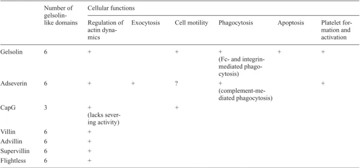
Table 1. Summary of the main proven cellular functions for gelsolin superfamily proteins. Number of Cellular functions
gelsolin-like domains Regulation of Exocytosis Cell motility Phagocytosis Apoptosis Platelet
for-actin dyna- mation and
mics activation Gelsolin 6 + + + + + (Fc- and integrin-mediated phago-cytosis) Adseverin 6 + + ? + + (complement-me-diated phagocytosis) CapG 3 + + (lacks sever-ing activity) Villin 6 + Advillin 6 + Supervillin 6 + Flightless 6 +
Absence of an indication means absence of studies addressing the specific role of a protein in the cellular process concerned.
Figure 1. Schematic representation of gelsolin structure (A). Residues are numbered as in human plasma gelsolin. Gelsolin can bind, sever and cap actin filaments (B). Actin molecules within a filament are shown as red circles. (+) and (–) indicate barbed and minus ends of actin filament, respectively. After binding to actin filament, and in response to an increase in intracellular Ca2+concentration, gelsolin undergoes
a change in conformation that allows the severing process to proceed. After severing, gelsolin remains attached to the barbed end of actin filament as a cap.
Gelsolin contains six gelsolin-like (G) domains and was first described as a protein able to bind and sever actin fil-aments, and to control polymerization of barbed ends. Furthermore, this protein also initiates formation of actin filaments by binding two monomeric actin molecules. Gelsolin activity is regulated by Ca2+, intracellular pH,
phosphoinositides and tyrosine phosphorylation. Crystal-lographic studies of gelsolin complexed with its substrate have been recently performed [10 – 14]. Results have been reviewed in [15] and will not be discussed here. The other gelsolin superfamily members; villin, advillin, supervillin and flightless, have additional domains be-yond the sixfold repeat. Villin has been demonstrated to participate in cytoskeleton remodeling in response to var-ious stimuli in the intestine [16]. Flightless is the only member of gelsolin superfamily that has been shown to be essential for mouse development [17]. Among all of the proteins forming the gelsolin superfamily, adseverin (also named scinderin) is the one that shares the highest degree of homology with gelsolin. Indeed, adseverin shares 60 % amino acid homology with gelsolin [18], and displays Ca2+-regulated actin filament severing activity.
As compared to gelsolin, adseverin has a more restricted expression. Adseverin was first discovered in platelets, megakaryocytes and chromaffin cells [19]. This protein is present in all secretory cells and is involved in actin cy-toskeleton remodeling occurring during exocytosis [19 – 22]. With flightless [23] and supervillin [24, 25], capG (also named gCap39, Mbh1 or macrophage capping protein) is the only other protein of gelsolin family shar-ing a nuclear localization [26, 27]. CapG only contains three G domains and can bind and cap actin filaments but cannot sever them [28]. CapG has been demonstrated to play an important role as a mediator of endothelial cell re-sponse to mechanical forces [29].
Actin filament remodeling
In the absence of Ca2+, gelsolin exists in a globular
con-formation. Crystal structure analysis has revealed the ex-istence of a C-terminal tail (‘latch helix’) which is in close contact with the actin binding region of G2 domain. The importance of this latch helix in Ca2+regulation of
gelsolin-actin interactions was first revealed by the demonstration that a lack of 20 residues in the C-termi-nus tail abrogates the Ca2+regulation of actin binding [30,
31]. The latch hypothesis suggests that Ca2+binding to G6
domain induces a first conformational change in the gel-solin structure, releasing tail latch inhibition of G2 bind-ing to actin. G2 bindbind-ing to actin directs the G1 domain to its actin binding site. In the absence of Ca2+, G4 and G6
domains are close together. Ca2+opens this structure, and
the G6 domain forms new contacts with the G5 domain, releasing the G4 domain, which now can form a Ca2+
binding domain coordinated by actin and G4 itself. The actin binding sites at the N- and C-terminal parts bind to two adjacent actin filaments. Slowly, gelsolin severs the two actin filaments, remaining bound to the newly formed actin + end (barbed end) of one of the two shorter filaments (fig. 1 B). Further uncapping of actin filaments requires binding of gelsolin to phosphatidylinositol lipids. This process exposes the barbed end for polymer-ization [32, 33]. Until recently, phosphatidylinositol 3,4 or 4,5-bisphosphate (PIP2) isomers were the only known intracellular agents inhibiting gelsolin severing and in-ducing dissociation of gelsolin from actin. More recently, another protein of bacterial origin was shown to inhibit gelsolin severing activity. Indeed, some bacteria have the possibility to infiltrate cells delivering effector proteins that trigger a cytoskeleton rearrangement and membrane ruffling (reviewed in [34, 35]). Salmonella invasion pro-tein a (SipA) is one of these propro-teins. SipA promotes actin polymerization, inducing an arrest of actin turnover. This arrest is partly exerted through inhibition of severing activity of gelsolin [36].
Additional evidence exists proving that tyrosine phos-phorylation can also induce conformational change in villin and gelsolin proteins, promoting their severing ac-tivities at submicromolar concentrations of Ca2+[37]. An
additional factor that has been shown to regulate actin network reorganization via gelsolin is intracellular pH. It has been demonstrated that the Ca2+requirement for both
severing and nucleating activities decreases with lowered pH. Half-maximal activities require 10 mM Ca2+at a pH
of 7.4 and decrease to 3 mM at pH 6.5. At low pH direct
activation of gelsolin severing activity can also be ob-served in complete absence of Ca2+. This feature is not
general to other gelsolin superfamily members and may be due to a conformational change of gelsolin occurring at low pH [38], resulting in increased association of gel-solin with actin. This increase is similar to that observed at pH 7.0 in the presence of Ca2+. Finally, low
pH-acti-vated gelsolin can still be inhibited by PIP2.
Cell motility
Properties of the cytoskeleton depend on different para-meters, such as filament length, flexibility, concentration and presence of cross-links. Proteins with the capacity to alter such properties are potentially important for regu-lating cellular morphology and function, as is the case for cell shape and regulation of cell motility. A first clue to evidence of the link between the expression of gelsolin or gelsolin-related proteins and cell motility was provided by transfection experiments of cultured fibroblasts [39]. Overexpression of gelsolin in these cells resulted in in-creased motility and concomitant inhibition of phospho-lipase C (PLC) -b and -g [40, 41]. Surprisingly, despite
the lack of severing activity, capG overexpression also increased fibroblast motility [42].
Gelsolin has also been shown to regulate hematopoietic stem cell motility. Lin–Sca+Kit+and Lin–Sca+Kit–, are two
hematopoietic stem cell populations which show different basal and induced motility capacity. Indeed, they exhibit differential cell motility in response to stromal derived fac-tor 1 (SDF-1), a chemokine influencing cell motility through phosphatidylinositol signaling. Lin–Sca+Kit+cells,
which are more primitive than Lin–Sca+Kit–cells, exhibit a
lower response in terms of cell motility, although the acti-vation of the phosphatidylinositol pathway was activated at the same level in the two populations. Proteomics analysis documented a lower expression of gelsolin and adseverin in Lin–Sca+Kit+than in Lin–Sca+Kit–, providing an
expla-nation for this differential response to SDF-1 [43]. CapG, which lacks severing activity, has also been shown to be involved in regulation of cellular motility in fibrob-lastic cells [42]. More recently, we were able to demon-strate that capG is also an important regulator of motility function in endothelial cells [29]. Endothelial cells are able to discriminate between different combinations of mechanical forces. In vivo, this capability results in a fo-calization of vascular areas subjected to atherosclerotic plaque development [44 – 46]. Indeed, plaques develop at bifurcations and curvatures which are exposed to a par-ticular pattern of blood flow, with a low mean shear stress value and cyclic reversal of flow direction, which is in contrast with the unidirectional pattern of flow character-izing vascular areas protected against plaque develop-ment. In response to a plaque-free hemodynamic envi-ronment, capG expression and consequently endothelial motility are increased [29], resulting in a faster wound healing process [47], as compared to endothelial cells ex-posed to plaque-prone conditions.
In vivo evidence for gelsolin involvement in cell motility was provided by gelsolin null mice (Gsn–/–), in which motility of osteoclasts was decreased [48]. In these mice, osteoclasts were unable to form cell adhesion structures (podosomes), and therefore their basal as well as their os-teopontin-induced motility was affected. Analysis of these mice identified an additional role of gelsolin in the regulation of neuronal growth, cone formation and re-traction [49]. Neuronal growth cones are highly motile structures from which lamellipodia and filopodia form and retract. Formation of these structures is a Ca2+
-con-trolled process dependent on actin cytoskeleton remodel-ing. In Gsn–/– mice, only retraction processes appeared to be delayed, as compared to wild-type mice, suggesting that retraction is solely dependent on the presence of gel-solin, whereas formation of filopodia is mainly depen-dent on adseverin activity (adseverin is indeed expressed in these cells) [49]. Finally, knockout mice revealed the essential role of capG in macrophage ruffling [50]. In-deed, macrophages rapidly change shape forming
protru-sions resulting from local actin filament assembly [51], a process that is Ca2+sensitive. In capG–/– mice, but not in
Gsn–/– mice, basal and macrophage colony stimulating factor (MCSF)-induced ruffling activities were decreased [50].
A further role in phagocytosis
Phagocytosis is a complex cellular process that necessi-tates a continuous rearrangement of actin cytoskeleton. In polymorphonuclear leukocytes three types of phagocyto-sis can be distinguished: a complement-opsonized, im-munoglobulin G (IgG)-opsonized, and integrin-mediated phagocytosis. This latter is essential for remodeling of connective tissue. The three types of phagocytosis are mediated by three separate sets of cell surface receptors: the complement receptors (CRs), the Fcg receptors and
integrin molecules [52 – 54]. Using Gsn–/– mice, it has been possible to demonstrate that gelsolin plays a pri-mordial role in Fc receptor- and integrin- but not in com-plement-mediated phagocytosis [50, 54 – 56]. Other func-tions associated with Fc receptor-mediated phagocytosis, such as the activation of NADPH-oxidase, were not af-fected in Gsn–/– mice [55]. An impairment of comple-ment-opsonized phagocytosis was observed in capG/Gsn double-null mice, providing evidence for distinctive and non-overlapping functions of capG and gelsolin in phagocytic processes [50]. Finally, binding and internal-ization of collagen beads, a process dependent on a2b1
integrin [53], was also shown to be affected in Gsn–/– fi-broblasts [56]. Despite the obvious need for actin cy-toskeleton remodeling, allowing cells to undergo phago-cytosis, details of the mechanisms relating gelsolin and capG to this process are not yet clear. Work addressing the mechanisms of regulation of actin dynamics in the context of cell migration revealed a role of gelsolin as a downstream effector of the GTP-binding protein Rac [57 – 59]. Once activated, Rac promotes dissociation of gelsolin and actin, allowing actin remodeling to proceed [57]. Rac activation is also a key step in Fc receptor- and integrin-mediated phagocytosis [56, 60 – 63], and was shown to be affected in Gsn–/– fibroblasts, providing a first possible step in the mechanisms controlling phago-cytosis process. Indeed, in Gsn–/– fibroblasts, activation of Rac after binding to collagen was impaired compared to wild-type fibroblasts [56]. This activation could be re-stored after transfection of Gsn–/– cells with gelsolin-ex-pressing constructs and with calcium ionophores [56]. Based on these results, the authors proposed a mecha-nism starting from a gelsolin-mediated remodeling of actin cytoskeleton necessary for a subsequent calcium in-flux and Rac activation. This situation is probably more complicated because of the need of Rac for gelsolin-actin dissociation [57 – 59].
Gelsolin and apoptosis
The apoptotic process is activated through a cascade of as-partate-specific cysteine protease family named caspases. Fourteen members of this family are known in mammals and are present in the cell as precursors (zymogens). They can be divided into two main groups: the first group is ac-tivated following apoptotic stimuli and their major role is to activate downstream caspases. They are aggregated by caspase adaptor molecules (FAS-associated proteins with death domain, or Apaf-1), which promote autoactivation. The second group of caspases, the executioners (caspases-3, -6 and -7), are activated by apical caspases (reviewed in [64, 65]). Once activated, executioner caspases will cleave structural proteins and proteins involved in the repair process. One of the targets of caspase-3 is gelsolin [66]. Indeed, this enzyme cleaves gelsolin protein at D352of the
DQTD352G sequence, generating two fragments of 39 and
41 kDa. Cleaved gelsolin loses Ca2+control of severing
activity and the capacity to bind monomeric actin. Mi-croinjection of the N-terminal gelsolin fragment (1 – 352), which contains the severing activity, but not of the COOH-terminal fragment (353 – 731), triggered rapid depolymer-ization of the actin cytoskeleton [66]. Moreover, HeLa cells that normally do not express gelsolin are rendered more susceptible to apoptosis by overexpressing gelsolin [66]. Caspases other than caspase-3 have been suggested to be capable of cleaving gelsolin. In MCF-7 breast carci-noma cells lacking caspase-3 activity, gelsolin was still cleaved during an apoptotic process triggered by Fas anti-body or tumor necrosis factor (TNF) [67]. Nevertheless, in more recent work, a possible cleavage of gelsolin by one of the two other executioner caspases (-6 and -7) was excluded using a cell-free system depleted of each of these caspases. Only in the absence of caspase-3 was gelsolin not cleaved [68].
Gelsolin behaves not only as a regulator but also as an in-hibitor of the apoptotic process. Initial evidences of such a dual effect of gelsolin was provided by the observation that a point mutation in mouse gelsolin confers on this protein tumor-suppressor activity against H-ras oncogene-transformed NIH 3t3 cells [69, 70]. Moreover, transfec-tion of the gelsolin gene in a human bladder cancer cell line strongly reduced colony-forming ability and tumori-genicity in vivo [71]. More direct evidence of the in-hibitory role of gelsolin was provided by Ohtsu et al., who generated Jurkat transfectants expressing up to threefold more gelsolin than wild-type cells. These transfectants ex-hibited a phenotype more resistant to apoptosis induced by several stimuli [72]. Different mechanisms have been proposed to explain such an inhibitory capacity. First, overexpression of gelsolin was demonstrated to prevent caspase-3 activation [72]. Second, using a cell-free system to study the apoptotic process, Azuma et al. were able to show that gelsolin can form a complex with PIP2 and
cas-pase-3 to inhibit cascas-pase-3 activity [73]. In the same work, the authors were able to demonstrate that gelsolin-PIP2 complex forms in vivo and that the formation of this com-plex correlates with a delay in the apoptotic process. An-other hypothesis, explaining the inhibitory action of gel-solin overexpression in the apoptotic process, involves its possible action at the level of mitochondrial membrane potential. Indeed, one of the first stages of apoptosis is an increase in permeability of the mitochondrial membrane. Loss of cytochrome c has been linked to this loss of mem-brane potential [74, 75]. When cytochrome c associates to the caspase-9/Apaf 1 complex, it triggers activation of an effector caspase [65]. Overexpression of gelsolin pre-vented this loss in mitochondrial membrane potential [76]. Gelsolin has been proposed to participate in regula-tion of apoptosis by binding to voltage-dependent anion channel (VDAC), the putative permeability transition pore [77]. Gelsolmediated protection against apoptosis is in-hibited by phosphoinositides known to bind with high affinity to gelsolin, suggesting that the effect of gelsolin is due to its conformation or to its ability to bind actin [78 – 80]. The observation that in gelsolin-null mice neu-rons exhibit enhanced apoptosis due to hypoxia after a stroke further supports the hypothetic inhibitory action of gelsolin on apoptosis. In this work a protective role of gel-solin on neuronal cells in such conditions has been sug-gested [81].
Despite all these data, the inhibitory action of gelsolin on apoptosis remains controversial. Indeed, Posey et al. re-cently failed to observe a delay in Fas-induced apoptotic processes following different stimuli in T lymphocytes overexpressing gelsolin [82]. This discrepancy could eventually be explained by different pathways leading to apoptosis. Indeed, Fas-ligand can induce apoptosis via two mechanisms, one of which involves caspase-8 activa-tion, and that is independent of changes in mitochondrial membrane potential [82]. Alternatively, differences on basal and transfected gelsolin expression in the cell pop-ulations used could explain this lack of effect.
Role of the gelsolin superfamily during development
Despite the variety of processes in which gelsolin and other related proteins have been involved, Gsn–/–, capG–/– and Gsn/capG double null mice do not exhibit developmental disorders. This is probably due to the re-dundancy in the functions of these proteins. Mice lacking the gelsolin gene reproduce and develop normally, but adult mice exhibit some alteration in cellular processes involving cell motility. In these mice, bleeding time is in-creased, suggesting an alteration in platelet function, and the inflammatory process is affected because of a de-creased migratory capacity of neutrophils and dede-creased motility of fibroblasts, resulting in slower wound healing
[83]. Interestingly, Gsn –/– also showed some develop-mental alterations, such as delay in postnatal mammary gland development [84]. Ductal outgrowth was delayed until week 9, and the mammary epithelium was unre-sponsive to hormone stimulation. Osteoclasts were de-void of podosomes, a cell-adhesion structure. Despite the observation that gelsolin null mice do not display any neuronal defects, alterations were observed in formation of the growth cone of neurites [49].
Analysis of gelsolin, adseverin and capG proteins during mouse development has revealed complementary expres-sion patterns for these proteins. Gelsolin mRNA is pre-sent in multiple tissues, with the highest levels in cardiac ventricle, diaphragm and other muscles. Adseverin ex-pression is limited to endochondral bone primordial, re-nal tubules and intestire-nal microvilli during embryogene-sis. CapG is expressed in squamous epithelia, as well as specific regions of the kidney, adrenal gland and spleen and in the developing brain cortex [85]. In adult mice, ad-severin expression is mainly observable in the kidney and in the intestine, and capG expression is strongly ex-pressed in the heart, in uterus, lung, kidney and at a lower level in other tissues. Gelsolin is strongly expressed in the heart and lung, and at a lower level in skeletal muscle, kidney and testis [85].
In zebrafish, gelsolin has been shown to be specialized for high corneal expression [86], and to be required for proper dorsalization during embryogenesis [87].
Finally, fliih, the mouse homologue of Drosophila
melanogaster flightless I, is the only protein belonging
gelsolin superfamily family essential for normal mouse development [17]. Disruption of the fliih gene in mice re-sults in a rapid degeneration of the embryo. Recently, flightless I was shown to function as a nuclear coactivator in response to estrogen stimulation in MCF-7 cells [23].
Platelet activation
Platelet formation and activation are processes that in-volve reorganization of the actin cytoskeleton. Indeed, platelets form from megakaryocytes after proliferation, differentiation, nuclear polyploidization and apoptosis [88, 89]. Mature megakaryocytes form long and thin cy-toplasmic extensions, from which platelets are released. This process is highly related to remodeling of the actin cytoskeleton [90]. Megakaryoblastic cells express gel-solin, but not adseverin and capG [91]. Forced expression of adseverin induces megakaryoblastic cell maturation, leading to platelet formation [91]. Interestingly, expres-sion of adseverin also causes downregulation of gelsolin expression, suggesting that adseverin may act as an apop-tosis regulator in megakaryoblastic cells, while gelsolin protects megakaryoblasts from apoptosis, inhibiting their final differentiation process. These data strongly support
an important role of gelsolin and related proteins in the regulation of platelet formation. Evidence exists that these proteins also play a role also in platelet activation. The first stage in this process is a change in cell shape [92], which depends on actin filament remodeling. In resting platelets actin filaments are stabilized by VASP, a cytoskeleton focal adhesion protein which shows actin binding properties. VASP protects actin filaments from the severing effects of gelsolin but does not inhibit gel-solin from binding to the filaments [93]. Moreover, rest-ing platelets contain ~ 2 mM Cap Z, a calcium-insensitive
heterodimeric capping protein associated with the barbed ends of actin filaments [94], thus protecting barbed ends from polymerization. After stimulation, rapid severing activity of existing actin filaments is triggered [95]. This process allows amplification of barbed ends. Dissocia-tion of gelsolin and CapZ from barbed ends, necessary for actin filament polymerization, necessitates phospho-inositide 3-kinase (PI3-kinase) activity [96]. Changes in platelet cell shape are dependent on a rise in intracellular Ca2+and PI3-kinase activity [97, 98]. As a consequence
of this remodeling process cells lose their resting discoid form in favor of an activated form with long thin filopods containing bundles of F-actin.
Familial amyloidosis of the Finnish type
Although the role of plasma gelsolin is still not com-pletely understood, it is now known that a mutation in cir-culating gelsolin is the basis for a genetic disease. Indeed, association of a mutation at position 187 of the gelsolin gene with familial amyloidosis of the Finnish type (FAF) has been described [99 – 103]. FAF is an autosomal, dom-inant amyloid polyneuropathy, characterized by corneal lattice dystrophy, progressive cranial and peripheral neu-ropathy as well as skin changes. Mutation of Asn residue a Tyr 187 results in destabilization of the G2 domain, pre-dicting an unfolding that will predispose gelsolin to pro-teolysis [104, 105]. This aberrant cleavage generates the FAF amyloid precursor fragment of 68 kDa. Further cleavage at position 244 is required for the generation of FAF amyloid protein, which accumulates in patient tis-sues [106 – 109]. Moreover, plasma gelsolin isolated from homozygous FAF patients lacks both actin severing and nucleating activities [104]. In contrast, cytoplasmic gel-solin is not aberrantly cleaved in these patients [110], and the normal actin modulating function of intracellular gel-solin is not affected [111].
Concluding remarks
The major, intriguing observation concerning the gelsolin superfamily proteins is that despite their key role in
con-trolling cellular processes, involving cytoskeleton remod-eling, deletion of their genes in many cases is not lethal nor does it result in a clear phenotype. Processes involv-ing cell shape changes and cell motility are slightly sup-pressed, but not abrogated. This apparent paradox may be explained by a compensatory mechanism by other pro-teins belonging to this superfamily. This observation im-plies that experiments affecting the expression of just one of these proteins need to be carefully interpreted. The ex-pression of the other gelsolin superfamily proteins has to be assessed in the targeted cells and tissues. An absence of effect does not necessarily means an absence of func-tion in vivo.
Several investigations still need to be performed in order to completely understand the function of the protein members of this family. Several aspects will need further analysis, such as the increase in nuclear capG in endothe-lial cells exposed to plaque-free flow [29]. In fact, the role of nuclear capG is not yet clear. It has been demonstrated that nuclear capG can interfere with nuclear PI3-kinase [112]. This may eventually suggest that capG is involved in phosphatidylinositol-driven chromatin remodeling [113, 114] and therefore plays a role as a general regula-tor of gene transcription.
A better understanding of the function of gelsolin super-family proteins will certainly help us in comprehending cellular physiology.
Acknowledgments. We thank Dr André Mercanzini for critical
read-ing of the manuscript. Cited works carried out by the authors of this review were supported by the Swiss National Research Foundation (grant 3200-065129.01 to P.S. and D.H.), by the National Institutes of Health (grants GM21681 and GM066110 to H.Y.) and by the Welch Foundation.
1 Yin H. L. (1987) Gelsolin: calcium- and polyphosphoinosi-tide-regulated actin-modulating protein. Bioessays 7(4): 176 – 179
2 Sun H. Q., Yamamoto M., Mejillano M. and Yin H. L. (1999) Gelsolin, a multifunctional actin regulatory protein. J. Biol. Chem. 274(47): 33179 – 33182
3 Yin H. L., Kwiatkowski D. J., Mole J. E. and Cole F. S. (1984) Structure and biosynthesis of cytoplasmic and secreted vari-ants of gelsolin. J. Biol. Chem. 259(8): 5271 – 5276
4 Kwiatkowski D. J., Mehl R. and Yin H. L. (1988) Genomic or-ganization and biosynthesis of secreted and cytoplasmic forms of gelsolin. J. Cell Biol. 106(2): 375 – 384
5 Wen D., Corina K., Chow E. P., Miller S., Janmey P. A. and Pepinsky R. B. (1996) The plasma and cytoplasmic forms of human gelsolin differ in disulfide structure. Biochemistry
35(30): 9700 – 9709
6 Rothenbach P. A., Dahl B., Schwartz J. J., O’Keefe G. E., Ya-mamoto M., Lee W. M. et al. (2004) Recombinant plasma gel-solin infusion attenuates burn-induced pulmonary microvas-cular dysfunction. J. Appl. Physiol. 96(1): 25 – 31
7 Christofidou-Solomidou M., Scherpereel A., Solomides C. C., Muzykantov V. R., Machtay M., Albelda S. M. et al. (2002) Changes in plasma gelsolin concentration during acute oxi-dant lung injury in mice. Lung 180(2): 91 – 104
8 Becker P. M., Kazi A. A., Wadgaonkar R., Pearse D. B., Kwiatkowski D. and Garcia J. G. (2003) Pulmonary vascular
permeability and ischemic injury in gelsolin-deficient mice. Am. J. Respir. Cell Mol. Biol. 28(4): 478 – 484
9 Vouyiouklis D. A. and Brophy P. J. (1997) A novel gelsolin iso-form expressed by oligodendrocytes in the central nervous system. J. Neurochem. 69(3): 995 – 1005
10 McLaughlin P. J., Gooch J. T., Mannherz H. G. and Weeds A. G. (1993) Structure of gelsolin segment 1-actin complex and the mechanism of filament severing. Nature 364(6439): 685 – 692
11 Kiselar J. G., Janmey P. A., Almo S. C. and Chance M. R. (2003) Structural analysis of gelsolin using synchrotron pro-tein footprinting. Mol. Cell. Proteomics 2(10): 1120 – 1132 12 Irobi E., Burtnick L. D., Urosev D., Narayan K. and Robinson
R. C. (2003) From the first to the second domain of gelsolin: a common path on the surface of actin? FEBS Lett. 552(2 – 3): 86 – 90
13 Narayan K., Chumnarnsilpa S., Choe H., Irobi E., Urosev D., Lindberg U. et al. (2003) Activation in isolation: exposure of the actin-binding site in the C-terminal half of gelsolin does not require actin. FEBS Lett. 552(2 – 3): 82 – 85
14 Choe H., Burtnick L. D., Mejillano M., Yin H. L., Robinson R. C. and Choe S. (2002) The calcium activation of gelsolin: in-sights from the 3A structure of the G4-G6/actin complex. J. Mol. Biol. 324(4): 691 – 702
15 McGough A. M., Staiger C. J., Min J. K. and Simonetti K. D. (2003) The gelsolin family of actin regulatory proteins: mod-ular structures, versatile functions. FEBS Lett. 552(2 – 3): 75 – 81
16 Ferrary E., Cohen-Tannoudji M., Pehau-Arnaudet G., Lapil-lonne A., Athman R., Ruiz T. et al. (1999) In vivo, villin is re-quired for Ca(2+)-dependent F-actin disruption in intestinal brush borders. J. Cell Biol. 146(4): 819 – 830
17 Campbell H. D., Fountain S., McLennan I. S., Berven L. A., Crouch M. F., Davy D. A. et al. (2002) Fliih, a gelsolin-related cytoskeletal regulator essential for early mammalian embry-onic development. Mol. Cell. Biol. 22(10): 3518 – 3526 18 Kwiatkowski D. J. (1999) Functions of gelsolin: motility,
sig-naling, apoptosis, cancer. Curr. Opin. Cell Biol. 11(1): 103 – 108
19 Rodriguez Del Castillo A., Vitale M. L., Tchakarov L. and Tri-faro J. M. (1992) Human platelets contain scinderin, a Ca(2+)-dependent actin filament-severing protein. Thromb. Haemost.
67(2): 248 – 251
20 Tchakarov L., Vitale M. L., Jeyapragasan M., Rodriguez Del Castillo A. and Trifaro J. M. (1990) Expression of scinderin, an actin filament-severing protein, in different tissues. FEBS Lett. 268(1): 209 – 212
21 Vitale M. L., Rodriguez Del Castillo A., Tchakarov L. and Tri-faro J. M. (1991) Cortical filamentous actin disassembly and scinderin redistribution during chromaffin cell stimulation precede exocytosis, a phenomenon not exhibited by gelsolin. J. Cell Biol. 113(5): 1057 – 1067
22 Marcu M. G., Zhang L., Nau-Staudt K. and Trifaro J. M. (1996) Recombinant scinderin, an F-actin severing protein, increases calcium-induced release of serotonin from perme-abilized platelets, an effect blocked by two scinderin-derived actin-binding peptides and phosphatidylinositol 4,5-bisphos-phate. Blood 87(1): 20 – 24
23 Lee Y. H., Campbell H. D. and Stallcup M. R. (2004) Devel-opmentally essential protein flightless I is a nuclear receptor coactivator with actin binding activity. Mol. Cell. Biol. 24(5): 2103 – 2117
24 Pestonjamasp K. N., Pope R. K., Wulfkuhle J. D. and Luna E. J. (1997) Supervillin (p205): a novel membrane-associated, F-actin-binding protein in the villin/gelsolin superfamily. J. Cell Biol. 139(5): 1255 – 1269
25 Wulfkuhle J. D., Donina I. E., Stark N. H., Pope R. K., Pe-stonjamasp K. N., Niswonger M. L. et al. (1999) Domain analysis of supervillin, an F-actin bundling plasma membrane
protein with functional nuclear localization signals. J. Cell Sci. 112 ( Pt 13): 2125 – 2136
26 Prendergast G. C. and Ziff E. B. (1991) Mbh 1: a novel gel-solin/severin-related protein which binds actin in vitro and ex-hibits nuclear localization in vivo. EMBO J. 10(4): 757 – 766 27 Onoda K., Yu F. X. and Yin H. L. (1993) gCap39 is a nuclear
and cytoplasmic protein. Cell Motil. Cytoskeleton 26(3): 227 – 238
28 Southwick F. S. and DiNubile M. J. (1986) Rabbit alveolar macrophages contain a Ca2+-sensitive, 41,000-dalton protein which reversibly blocks the ‘barbed’ ends of actin filaments but does not sever them. J. Biol. Chem. 261(30): 14191 – 14195
29 Pellieux C., Desgeorges A., Pigeon C. H., Chambaz C., Yin H., Hayoz D. et al. (2003) Cap G, a gelsolin family protein modulating protective effects of unidirectional shear stress. J. Biol. Chem. 278(31): 29136 – 29144
30 Kwiatkowski D. J., Janmey P. A. and Yin H. L. (1989) Identi-fication of critical functional and regulatory domains in gel-solin. J. Cell Biol. 108(5): 1717 – 1726
31 Way M., Pope B., Gooch J., Hawkins M. and Weeds A. G. (1990) Identification of a region in segment 1 of gelsolin crit-ical for actin binding. EMBO J. 9(12): 4103 – 4109
32 Yu F. X., Sun H. Q., Janmey P. A. and Yin H. L. (1992) Iden-tification of a polyphosphoinositide-binding sequence in an actin monomer-binding domain of gelsolin. J. Biol. Chem.
267(21): 14616 – 14621
33 Liepina I., Czaplewski C., Janmey P. and Liwo A. (2003) Mol-ecular dynamics study of a gelsolin-derived peptide binding to a lipid bilayer containing phosphatidylinositol 4,5-bisphos-phate. Biopolymers 71(1): 49 – 70
34 Cossart P. and Sansonetti P. J. (2004) Bacterial invasion: the paradigms of enteroinvasive pathogens. Science 304(5668): 242 – 248
35 Cossart P. (2004) Bacterial invasion: a new strategy to domi-nate cytoskeleton plasticity. Dev. Cell. 6(3): 314 – 315 36 McGhie E. J., Hayward R. D. and Koronakis V. (2004) Control
of actin turnover by a salmonella invasion protein. Mol. Cell.
13(4): 497 – 510
37 Kumar N. and Khurana S. (2004) Identification of a func-tional switch for actin severing by cytoskeletal proteins. J. Biol. Chem.
38 Lamb J. A., Allen P. G., Tuan B. Y. and Janmey P. A. (1993) Modulation of gelsolin function. Activation at low pH over-rides Ca2+ requirement. J. Biol. Chem. 268(12): 8999 – 9004 39 Cunningham C. C., Stossel T. P. and Kwiatkowski D. J. (1991)
Enhanced motility in NIH 3T3 fibroblasts that overexpress gelsolin. Science 251(4998): 1233 – 1236
40 Sun H., Lin K. and Yin H. L. (1997) Gelsolin modulates phos-pholipase C activity in vivo through phospholipid binding. J. Cell Biol. 138(4): 811 – 820
41 Banno Y., Nakashima T., Kumada T., Ebisawa K., Nonomura Y. and Nozawa Y. (1992) Effects of gelsolin on human platelet cytosolic phosphoinositide-phospholipase C isozymes. J. Biol. Chem. 267(10): 6488 – 6494
42 Sun H. Q., Kwiatkowska K., Wooten D. C. and Yin H. L. (1995) Effects of CapG overexpression on agonist-induced motility and second messenger generation. J. Cell Biol.
129(1): 147 – 156
43 Evans C. A., Tonge R., Blinco D., Pierce A., Shaw J., Lu Y. et al. (2004) Comparative proteomics of primitive hematopoietic cell populations reveals differences in expression of proteins regulating motility. Blood 103(10): 3751 – 3759
44 Ravensbergen J., Ravensbergen J. W., Krijger J. K., Hillen B. and Hoogstraten H. W. (1998) Localizing role of hemodynam-ics in atherosclerosis in several human vertebrobasilar junction geometries. Arterioscler. Thromb. Vasc. Biol. 18(5): 708–716 45 Glagov S., Zarins C., Giddens D. P. and Ku D. N. (1988)
He-modynamics and atherosclerosis. Insights and perspectives
gained from studies of human arteries. Arch. Pathol. Lab. Med. 112(10): 1018 – 1031
46 Ku D. N., Giddens D. P., Zarins C. K. and Glagov S. (1985) Pulsatile flow and atherosclerosis in the human carotid bifur-cation. Positive correlation between plaque location and low oscillating shear stress. Arteriosclerosis 5(3): 293 – 302 47 Vyalov S., Langille B. L. and Gotlieb A. I. (1996) Decreased
blood flow rate disrupts endothelial repair in vivo. Am. J. Pathol. 149(6): 2107 – 2118
48 Chellaiah M., Kizer N., Silva M., Alvarez U., Kwiatkowski D. and Hruska K. A. (2000) Gelsolin deficiency blocks po-dosome assembly and produces increased bone mass and strength. J. Cell Biol. 148(4): 665 – 678
49 Lu M., Witke W., Kwiatkowski D. J. and Kosik K. S. (1997) Delayed retraction of filopodia in gelsolin null mice. J. Cell Biol. 138(6): 1279 – 1287
50 Witke W., Li W., Kwiatkowski D. J. and Southwick F. S. (2001) Comparisons of CapG and gelsolin-null macrophages: demonstration of a unique role for CapG in receptor-mediated ruffling, phagocytosis, and vesicle rocketing. J. Cell Biol.
154(4): 775 – 784
51 Heidemann S. R., Kaech S., Buxbaum R. E. and Matus A. (1999) Direct observations of the mechanical behaviors of the cytoskeleton in living fibroblasts. J. Cell Biol. 145(1): 109 – 122
52 Vidarsson G., Sigurdardottir S. T., Gudnason T., Kjartansson S., Kristinsson K. G., Ingolfsdottir G. et al. (1998) Isotypes and opsonophagocytosis of pneumococcus type 6B antibodies elicited in infants and adults by an experimental pneumococ-cus type 6B-tetanus toxoid vaccine. Infect. Immun. 66(6): 2866 – 2870
53 Lee W., Sodek J. and McCulloch C. A. (1996) Role of inte-grins in regulation of collagen phagocytosis by human fibrob-lasts. J. Cell. Physiol. 168(3): 695 – 704
54 Arora P. D., Manolson M. F., Downey G. P., Sodek J. and Mc-Culloch C. A. (2000) A novel model system for characteriza-tion of phagosomal maturacharacteriza-tion, acidificacharacteriza-tion and intracellular collagen degradation in fibroblasts. J. Biol. Chem. 275(45): 35432 – 35441
55 Serrander L., Skarman P., Rasmussen B., Witke W., Lew D. P., Krause K. H. et al. (2000) Selective inhibition of IgG-medi-ated phagocytosis in gelsolin-deficient murine neutrophils. J. Immunol. 165(5): 2451 – 2457
56 Arora P. D., Glogauer M., Kapus A., Kwiatkowski D. J. and McCulloch C. A. (2004) Gelsolin mediates collagen phagocy-tosis through a rac-dependent step. Mol. Biol. Cell 15(2): 588 – 599
57 Arcaro A. (1998) The small GTP-binding protein Rac pro-motes the dissociation of gelsolin from actin filaments in neu-trophils. J. Biol. Chem. 273(2): 805 – 813
58 Azuma T., Witke W., Stossel T. P., Hartwig J. H. and Kwiatkowski D. J. (1998) Gelsolin is a downstream effector of rac for fibroblast motility. EMBO J. 17(5): 1362 – 1370 59 De Corte V., Bruyneel E., Boucherie C., Mareel M.,
Vande-kerckhove J. and Gettemans J. (2002) Gelsolin-induced epi-thelial cell invasion is dependent on Ras-Rac signaling. EMBO J. 21(24): 6781 – 6790
60 Crowley M. T., Costello P. S., Fitzer-Attas C. J., Turner M., Meng F., Lowell C. et al. (1997) A critical role for Syk in sig-nal transduction and phagocytosis mediated by Fcgamma re-ceptors on macrophages. J. Exp. Med. 186(7): 1027 – 1039 61 Greenberg S. (1995) Signal transduction of phagocytosis.
Trends Cell Biol. 5(3): 93 – 99
62 Caron E. and Hall A. (1998) Identification of two distinct mechanisms of phagocytosis controlled by different Rho GT-Pases. Science 282(5394): 1717 – 1721
63 Patel J. C., Hall A. and Caron E. (2002) Vav regulates activa-tion of Rac but not Cdc42 during FcgammaR-mediated phagocytosis. Mol. Biol. Cell 13(4): 1215 – 1226
fibroblast responses are blunted in mice lacking gelsolin. Cell
81(1): 41 – 51
84 Crowley M. R., Head K. L., Kwiatkowski D. J., Asch H. L. and Asch B. B. (2000) The mouse mammary gland requires the actin-binding protein gelsolin for proper ductal morphogene-sis. Dev. Biol. 225(2): 407 – 423
85 Arai M. and Kwiatkowski D. J. (1999) Differential develop-mentally regulated expression of gelsolin family members in the mouse. Dev. Dyn. 215(4): 297 – 307
86 Xu Y. S., Kantorow M., Davis J. and Piatigorsky J. (2000) Ev-idence for gelsolin as a corneal crystallin in zebrafish. J. Biol. Chem. 275(32): 24645 – 24652
87 Kanungo J., Kozmik Z., Swamynathan S. K. and Piatigorsky J. (2003) Gelsolin is a dorsalizing factor in zebrafish. Proc. Natl. Acad. Sci. USA 100(6): 3287 – 3292
88 MacPherson G. G. (1972) Origin and development of the de-marcation system in megakaryocytes of rat bone marrow. J. Ultrastruct. Res. 40(1): 167 – 177
89 Leven R. M. (1995) Differential regulation of integrin-medi-ated proplatelet formation and megakaryocyte spreading. J. Cell. Physiol. 163(3): 597 – 607
90 Tablin F., Castro M. and Leven R. M. (1990) Blood platelet formation in vitro. The role of the cytoskeleton in megakary-ocyte fragmentation. J. Cell Sci. 97 (Pt 1): 59 – 70
91 Zunino R., Li Q., Rose S. D., Romero-Benitez M. M., Lejen T., Brandan N. C. et al. (2001) Expression of scinderin in megakaryoblastic leukemia cells induces differentiation, mat-uration and apoptosis with release of plateletlike particles and inhibits proliferation and tumorigenesis. Blood 98(7): 2210 – 2219
92 Casella J. F., Flanagan M. D. and Lin S. (1981) Cytochalasin D inhibits actin polymerization and induces depolymerization of actin filaments formed during platelet shape change. Na-ture 293(5830): 302 – 305
93 Bearer E. L., Prakash J. M., Manchester R. D. and Allen P. G. (2000) VASP protects actin filaments from gelsolin: an in vitro study with implications for platelet actin reorganiza-tions. Cell Motil. Cytoskeleton 47(4): 351 – 364
94 Nachmias V. T., Golla R., Casella J. F. and Barron-Casella E. (1996) Cap Z, a calcium insensitive capping protein in resting and activated platelets. FEBS Lett. 378(3): 258 – 262 95 Hartwig J. H. (1992) Mechanisms of actin rearrangements
mediating platelet activation. J. Cell Biol. 118(6): 1421 – 1442 96 Barkalow K., Witke W., Kwiatkowski D. J. and Hartwig J. H. (1996) Coordinated regulation of platelet actin filament barbed ends by gelsolin and capping protein. J. Cell Biol.
134(2): 389 – 399
97 Falet H., Barkalow K. L., Pivniouk V. I., Barnes M. J., Geha R. S. and Hartwig J. H. (2000) Roles of SLP-76, phosphoinosi-tide 3-kinase and gelsolin in the platelet shape changes initi-ated by the collagen receptor GPVI/FcR gamma-chain com-plex. Blood 96(12): 3786 – 3792
98 Barkalow K. L., Falet H., Italiano J. E. Jr, van Vugt A., Car-penter C. L., Schreiber A. D. et al. (2003) Role for phospho-inositide 3-kinase in Fc gamma RIIA-induced platelet shape change. Am. J. Physiol. Cell Physiol. 285(4): C797 – 805 99 Maury C. P., Alli K. and Baumann M. (1990) Finnish
heredi-tary amyloidosis. Amino acid sequence homology between the amyloid fibril protein and human plasma gelsoline. FEBS Lett. 260(1): 85 – 87
100 Maury C. P. and Baumann M. (1990) Isolation and character-ization of cardiac amyloid in familial amyloid polyneuropathy type IV (Finnish): relation of the amyloid protein to variant gelsolin. Biochim. Biophys. Acta 1096(1): 84 – 86
101 Ghiso J., Haltia M., Prelli F., Novello J. and Frangione B. (1990) Gelsolin variant (Asn-187) in familial amyloidosis, Finnish type. Biochem. J. 272(3): 827 – 830
102 de la Chapelle A., Kere J., Sack G. H. Jr, Tolvanen R. and Maury C. P. (1992) Familial amyloidosis, Finnish type: G654 64 Philchenkov A. A. (2003) Caspases as regulators of apoptosis
and other cell functions. Biochemistry 68(4): 365 – 376 65 Green D. and Kroemer G. (1998) The central executioners of
apoptosis: caspases or mitochondria? Trends Cell Biol. 8(7): 267 – 271
66 Kothakota S., Azuma T., Reinhard C., Klippel A., Tang J., Chu K. et al. (1997) Caspase-3-generated fragment of gelsolin: ef-fector of morphological change in apoptosis. Science
278(5336): 294 – 298
67 Janicke R. U., Ng P., Sprengart M. L. and Porter A. G. (1998) Caspase-3 is required for alpha-fodrin cleavage but dispens-able for cleavage of other death substrates in apoptosis. J. Biol. Chem. 273(25): 15540 – 15545
68 Slee E. A., Adrain C. and Martin S. J. (2001) Executioner cas-pase-3, -6 and -7 perform distinct, non-redundant roles during the demolition phase of apoptosis. J. Biol. Chem. 276(10): 7320 – 7326
69 Mullauer L., Fujita H., Ishizaki A. and Kuzumaki N. (1993) Tumor-suppressive function of mutated gelsolin in ras-trans-formed cells. Oncogene 8(9): 2531 – 2536
70 Fujita H., Laham L. E., Janmey P. A., Kwiatkowski D. J., Stos-sel T. P., Banno Y. et al. (1995) Functions of [His321]gelsolin isolated from a flat revertant of ras-transformed cells. Eur. J. Biochem. 229(3): 615 – 620
71 Tanaka M., Mullauer L., Ogiso Y., Fujita H., Moriya S., Furu-uchi K. et al. (1995) Gelsolin: a candidate for suppressor of human bladder cancer. Cancer Res. 55(15): 3228 – 3232 72 Ohtsu M., Sakai N., Fujita H., Kashiwagi M., Gasa S.,
Shimizu S. et al. (1997) Inhibition of apoptosis by the actin-regulatory protein gelsolin. EMBO J. 16(15): 4650 – 4656 73 Azuma T., Koths K., Flanagan L. and Kwiatkowski D. (2000)
Gelsolin in complex with phosphatidylinositol 4,5-bisphos-phate inhibits caspase-3 and -9 to retard apoptotic progres-sion. J. Biol. Chem. 275(6): 3761 – 3766
74 Petit P. X., Zamzami N., Vayssiere J. L., Mignotte B., Kroemer G. and Castedo M. (1997) Implication of mitochondria in apoptosis. Mol. Cell. Biochem. 174(1-2): 185 – 188
75 Saleh A., Srinivasula S. M., Acharya S., Fishel R. and Alnemri E. S. (1999) Cytochrome c and dATP-mediated oligomeriza-tion of Apaf-1 is a prerequisite for procaspase-9 activaoligomeriza-tion. J. Biol. Chem. 274(25): 17941 – 17945
76 Koya R. C., Fujita H., Shimizu S., Ohtsu M., Takimoto M., Tsujimoto Y. et al. (2000) Gelsolin inhibits apoptosis by blocking mitochondrial membrane potential loss and cy-tochrome c release. J. Biol. Chem. 275(20): 15343 – 15349 77 Granville D. J. and Gottlieb R. A. (2003) The mitochondrial
voltage-dependent anion channel (VDAC) as a therapeutic target for initiating cell death. Curr. Med. Chem. 10(16): 1527 – 1533
78 Ahn J. S., Jang I. S., Kim D. I., Cho K. A., Park Y. H., Kim K. et al. (2003) Aging-associated increase of gelsolin for apopto-sis reapopto-sistance. Biochem. Biophys. Res. Commun. 312(4): 1335 – 1341
79 Ahn J. S., Jang I. S., Rhim J. H., Kim K., Yeo E. J. and Park S. C. (2003) Gelsolin for senescence-associated resistance to apoptosis. Ann. N. Y. Acad. Sci. 1010: 493 – 495
80 Mejillano M., Yamamoto M., Rozelle A. L., Sun H. Q., Wang X. and Yin H. L. (2001) Regulation of apoptosis by phos-phatidylinositol 4,5-bisphosphate inhibition of caspases, and caspase inactivation of phosphatidylinositol phosphate 5-ki-nases. J. Biol. Chem. 276(3): 1865 – 1872
81 Endres M., Fink K., Zhu J., Stagliano N. E., Bondada V., Ged-des J. W. et al. (1999) Neuroprotective effects of gelsolin dur-ing murine stroke. J. Clin. Invest. 103(3): 347 – 354
82 Posey S. C., Martelli M. P., Azuma T., Kwiatkowski D. J. and Bierer B. E. (2000) Failure of gelsolin overexpression to reg-ulate lymphocyte apoptosis. Blood 95(11): 3483 – 3488 83 Witke W., Sharpe A. H., Hartwig J. H., Azuma T., Stossel T. P
sion reveals an abnormal gelsolin fragment. Hum. Mol. Genet. 3(12): 2223 – 2229
109 Paunio T., Kangas H., Heinonen O., Buc-Caron M. H., Robert J. J., Kaasinen S. et al. (1998) Cells of the neuronal lineage play a major role in the generation of amyloid precursor frag-ments in gelsolin-related amyloidosis. J. Biol. Chem. 273(26): 16319 – 16324
110 Kangas H., Paunio T., Kalkkinen N., Jalanko A. and Peltonen L. (1996) In vitro expression analysis shows that the secretory form of gelsolin is the sole source of amyloid in gelsolin-re-lated amyloidosis. Hum. Mol. Genet. 5(9): 1237 – 1243 111 Kangas H., Ulmanen I., Paunio T., Kwiatkowski D. J.,
Lehtovirta M., Jalanko A. et al. (1999) Functional conse-quences of amyloidosis mutation for gelsolin polypeptide – analysis of gelsolin-actin interaction and gelsolin processing in gelsolin knock-out fibroblasts. FEBS Lett. 454(3): 233 – 239
112 Lu P. J., Hsu A. L., Wang D. S., Yan H. Y., Yin H. L. and Chen C. S. (1998) Phosphoinositide 3-kinase in rat liver nuclei. Bio-chemistry 37(16): 5738 – 5745
113 Steger D. J., Haswell E. S., Miller A. L., Wente S. R. and O’Shea E. K. (2003) Regulation of chromatin remodeling by inositol polyphosphates. Science 299(5603): 114 – 116 114 Rando O. J., Chi T. H. and Crabtree G. R. (2003) Second
mes-senger control of chromatin remodeling. Nat. Struct. Biol.
10(2): 81 – 83.
– a mutation of the gelsolin gene in Finnish families and an unrelated American family. Genomics 13(3): 898 – 901 103 de la Chapelle A., Tolvanen R., Boysen G., Santavy J.,
Bleeker-Wagemakers L., Maury C. P. et al. (1992) Gelsolin-derived familial amyloidosis caused by asparagine or tyrosine substitution for aspartic acid at residue 187. Nat. Genet. 2(2): 157 – 160
104 Weeds A. G., Gooch J., McLaughlin P. and Maury C. P. (1993) Variant plasma gelsolin responsible for familial amyloidosis (Finnish type) has defective actin severing activity. FEBS Lett.
335(1): 119 – 123
105 Isaacson R. L., Weeds A. G. and Fersht A. R. (1999) Equilib-ria and kinetics of folding of gelsolin domain 2 and mutants involved in familial amyloidosis-Finnish type. Proc. Natl. Acad. Sci. USA 96(20): 11247 – 11252
106 Maury C. P., Kere J., Tolvanen R. and de la Chapelle A. (1990) Finnish hereditary amyloidosis is caused by a single nu-cleotide substitution in the gelsolin gene. FEBS Lett.
276(1 – 2): 75 – 77
107 Haltia M., Prelli F., Ghiso J., Kiuru S., Somer H., Palo J. et al. (1990) Amyloid protein in familial amyloidosis (Finnish type) is homologous to gelsolin, an actin-binding protein. Biochem. Biophys. Res. Commun. 167(3): 927 – 932
108 Paunio T., Kangas H., Kalkkinen N., Haltia M., Palo J. and Peltonen L. (1994) Toward understanding the pathogenic mechanisms in gelsolin-related amyloidosis: in vitro