Skeletal Radiol DOI 10.1007/s00256-006-0147-8 C A S E R E P O RT M. Ziadé P. Zufferey A. K.-L. So Received: 25 January 2006 Revised: 2 March 2006 # ISS 2006
Recurrent acute low back pain secondary
to lumbar epidural calcification
Abstract Introduction: Epidural calcification is a rare cause of back pain, and spontaneous epidural calci-fication has not been reported pre-viously. Case report: We describe a patient with acute low back pain and signs of lumbar nerve root compres-sion due to epidural calcification, as demonstrated by CT-scan and MRI. Radiological signs of spondylodiscitis led to a search for an infectious cause, which was negative, and her symp-toms responded rapidly to NSAID treatment alone. Her symptoms re-curred 18 months later, and further imaging studies again revealed
epi-dural calcification, but with a changed distribution. Her symptoms were re-lieved once more by NSAID treat-ment alone. Discussion: We propose that epidural calcification secondary to aseptic spondylodiscitis is the main cause of acute back pain in this patient. A possible mechanism may be the pro-inflammatory effects of calcium pyrophosphate or hydroxy-apatite crystal deposition within the epidural space.
Keywords Epidural calcification . Low back pain . Crystal deposition disease . Spondylodiscitis
Introduction
Epidural calcification is rarely described as a cause of acute spinal pain. Calcification secondary to prior traumatic hematomas, meningioma or following intra-discal injection of triamcinalone hexacetonide has been previously re-ported. In some of these cases, calcification led to signs of nerve root or spinal cord compression. Spontaneous epidural calcification has not been previously reported.
Case report
A 71-year-old woman with stable, long-standing rheuma-toid arthritis presented with acute low back pain. The pain radiated to both thighs, and was so severe that she was unable to walk or even stand. On clinical examination, a lumbar scoliosis was noted, and the lumbar spine was tender to percussion. The femoral stretch test was positive bilaterally. Neurological examination showed abolition of the patellar reflex and weakness (M4) of the quadriceps but
no sphincter disturbances. The FBC was normal, and there was no leucocytosis. The ESR was 57 mm/h and CRP 23 mg/l. Blood and urine cultures were sterile.
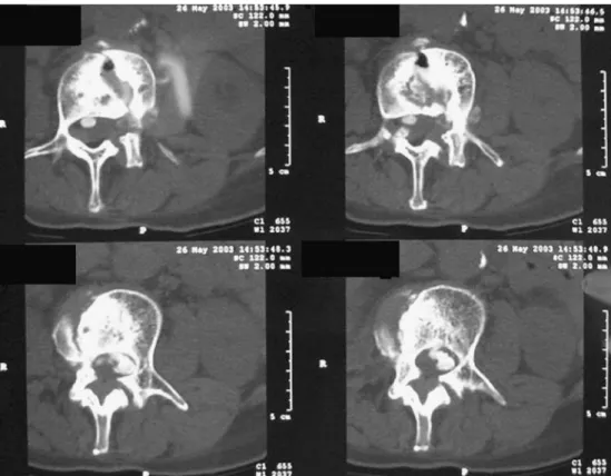
Plain radiographs showed degenerative changes in the lumbar spine and a scoliosis. The CT-Scan revealed spinal stenosis, due to a combination of degenerative change and protrusion of the L2-L3 disc. In addition, there were multiple calcifications within the spinal canal, with a dense deposit adjacent to the L2-L3 disc. These calcifications extended from the level of the L2-L3 disc distally to the L4-L5 level. (Figs.1 and3a). MRI confirmed the finding of spinal stenosis, and revealed collapse of the vertebral end-plates at L2-3 level, accompanied by diffuse perivertebral inflammatory changes (Fig.2).
In order to exclude an infectious spondylodiscitis, a lumbar puncture was performed. The CSF was inflamma-tory, with a raised protein level of 29.4 G/L (normal 0.15– 0.45 G/L), leucocytosis with 119×106/L mononuclear cells (normal<3×10⁶/L) but a normal glucose level. Bacteriological cultures, including that for mycobacteria, were sterile, and PCR to detect MTB sequences was also
M. Ziadé . P. Zufferey . A. K.-L. So (*) Service de Rhumatologie, Centre Hospitalier Vaudois, Avenue Pierre Decker,
CH 1011 Lausanne, Switzerland e-mail: [email protected] Tel.: +41-21-3141450 Fax: +41-21-3141533 Accepted: 16 March 2006 Published online: 20 May 2006
negative. The CSF did not reveal any calcium pyrophos-phate crystals by polarising light microscopy. Further radiographs of hands and knees showed no signs of chondrocalcinosis. A clinical diagnosis of aseptic spondy-lodiscitis and epidural calcification secondary to probable hydroxyapatite crystal deposition was made, and no antibiotic treatment was prescribed. The patient’s motor deficits and pain responded rapidly to indomethacin 75 mg bid and tramadol 50 mg q 4 hr, and she was discharged.
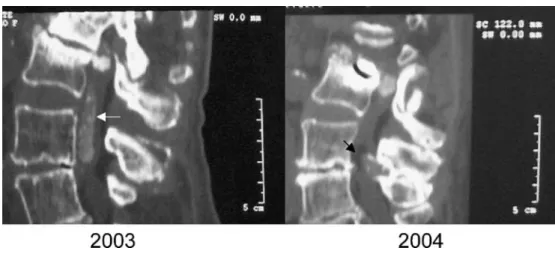
Eighteen months later, the patient developed a further exacerbation of low back pain, though this time there was no irradiation to the lower limbs. A new CT scan showed the persistence of intra-canal calcification at L2-L3 level but a disappearance of the epidural calcified trail. However, new intra-spinal calcifications appeared adjacent to the facet joint at the L4-L5 level (Fig.3). Once again, the pain decreased over a few days with NSAIDs. The clinical
Fig. 1 Transverse CT scan of the lumbar spine at the level of L2-3, showing disc calcification with a paravertebral extension as well as intra-canal calcification
Fig. 2 a Sagittal, b Transverse sections of T2 flair weighted MRI sequence of the lumbar spine, demonstrating severe ste-nosis of the lumbar canal with collapse of the L2-L3 vertebral end-plates and diffuse inflam-matory involvement of the dura mater (black arrow)
features were consistent with spinal stenosis due to degenerative disease.
Discussion
The case presents an unusual cause of recurrent acute low back pain due to epidural calcification associated with severe disc degeneration. We propose that the epidural calcification caused spinal stenosis as well as nerve-root compression and the accompanying inflammatory CSF changes. The lack of an infectious agent, despite extensive investigations and the rapid response to NSAIDs, is compatible with such a mechanism. Aseptic spondylodis-citis due to degenerative changes and microcristalline arthropathy could be the origin of the spontaneous epidural calcification. A similar process probably accounts for the transient epidural calcification observed in children with disc calcification. Although gouty tophi can give rise to epidural deposits, they are usually not as dense, and in the case of the patient described, her uric acid level was normal. As we were unable to detect other radiological signs of calcium pyrophosphate arthropathy, hydroxy-apaptite-related calcinosis is the most likely explanation of the epidural calcification in this patient.
Epidural calcification is a rare phenomenon, and its reported associations include meningioma, post-traumatic hematoma formation, and previous intra-disc injection of
long-acting corticosteroids such as triamcinalone hexace-tonide. Spontaneous hydroxyapatite deposition and calci-fication in the spine has been described most commonly in the cervical region [1], when it can present with acute disabling pain, often associated with inflammatory symp-toms and more rarely with vertebral destruction and transient neurological deficits, as in the “crowned-dens syndrome” [2]. The co-occurrence of calcification and an inflammatory syndrome in this case is best accounted for by crystal-induced inflammation, which would account for the acute presentation. In other forms of hydroxyapatite arthropathy (shoulder, hip) [3–7], an acute episode can be followed by the disappearance of the calcification. At the lumbar level, hydroxyapatite deposition disease is rare, and acute inflammatory manifestations are infrequent and may present a diagnostic challenge [8,9].
In our patient, the first episode of back pain resolved with conservative therapy with no neurological sequelae. However, the recurrence of inflammatory back pain was associated with a new site of spinal calcification which has not been previously reported. The CSF during the acute episode showed inflammatory changes with leucocytosis and raised protein content, confirming the presence of intra-thecal inflammatory involvement. The importance of distinguishing this from other causes of acute back pain is that it responds rapidly to conservative treatment and has a good prognosis.
Fig. 3 Comparison between sagittal CT scan view of the lumbar spine in 2003 and 2004, showing persistence of the intra-canal calcifications at L2-L3 level. Meanwhile, the distal epi-dural calcified trail (white arrow) had disappeared and new calci-fication observed close to the posterior articular process of L4-L5 (black arrow)
References
1. Baba H, Maezawa Y, Kawahara N, Tomita K, Furusawa N, Imura S. Calcium crystal deposition in the ligamentum flavum of the cervical spine. Spine. 1993;18:2174–2181
2. Ojemann JG, Grubb RL, Kyriakos M, Baker KB. Calcium carbonate apatite deposition in the cervical spine with associated vertebral destruction. Case report. J Neurosurg. 1997;86:1022–1026
3. Ea HK, Liote F. Calcium pyrophos-phate dihydrate and hydroxyapatite crystal induced arthropathies: update on pathogenesis, clinical features, and therapy. Curr Rheumatol Rep. 2004;6:221–227
4. Pay S, Terkeltaub R. Calcium pyro-phosphate dihydrate and hydroxyapa-tite crystal deposition in the joint: new developments relevant to the clinician. Curr Rheumatol Rep. 2003;5:235–243 5. Hayes CW, Conway WF. Calcium
hydroxyapatite deposition disease. Radiographics. 1990;10:1031–1048
6. Steinbach LS. Calcium pyrophosphate dihydrate and calcium hydroxyapatite crystal deposition diseases: imaging perspectives. Radiol Clin North Am. 2004;42:185–205
7. Li-Yu J, Clayburne GM, Sieck MS, et al. Calcium apatite crystals in syno-vial fluid rice bodies. Ann Rheum Dis. 2002;61:387–390
8. Baba H, Maezawa Y, Furusawa N, Imura S, Tomita K. The role of calcium deposition in the ligamentum flavum causing a cauda equina syndrome and lumbar radiculopathy. Paraplegia. 1995;33:219-223
9. Case Records of the Massachusetts General Hospital. New England J Med 1991;325:794–799