ORIGINAL ARTICLE
Intra-session test
–retest reliability of pelvic floor
muscle electromyography during running
H. Luginbuehl&C. Greter&D. Gruenenfelder&
J-P. Baeyens&A. Kuhn&L. Radlinger
Received: 14 June 2012 / Accepted: 20 December 2012 / Published online: 30 January 2013 # The International Urogynecological Association 2012
Abstract
Introduction and hypothesis The prevalence of female stress urinary incontinence is high, and young adults are also affected, including athletes, especially those involved in “high-impact” sports. To date there have been almost no studies testing pelvic floor muscle (PFM) activity during dynamic functional whole body movements. The aim of this study was the description and reliability test of PFM activity and time variables during running.
Methods A prospective cross-sectional study including ten healthy female subjects was designed with the focus on the intra-session test–retest reliability of PFM activity and time variables during running derived from electromyography (EMG) and accelerometry.
Results Thirteen variables were identified based on ten steps of each subject: Six EMG variables showed good reliability (ICC 0.906–0.942) and seven time variables did not show good reliability (ICC 0.113–0.731). Time variables (e.g. time difference between heel strike and maximal acceleration of vaginal accelerator) showed low reliability. However, relevant PFM EMG variables during running (e.g., pre-activation, minimal and maximal activity) could be identified and showed good reliability.
Conclusion Further adaptations regarding measurement methods should be tested to gain better control of the kinetics
and kinematics of the EMG probe and accelerometers. To our knowledge this is the first study to test the reliability of PFM activity and time variables during dynamic functional whole body movements. More knowledge of PFM activity and time variables may help to provide a deeper insight into physical strain with high force impacts and important functional reflex-ive contraction patterns of PFM to maintain or to restore continence.
Keywords Jogging . Pelvic floor . Reproducibility . Stress urinary incontinence
Introduction
Stress urinary incontinence (SUI) is a widespread condition among women of every age, meaning that young female adults are also affected [1]. Brown et al. [2] investigated the prevalence, frequency, and severity of urinary symptoms by questioning nulliparous women aged≥18 years during preg-nancy: 10.8 % of the women reported urinary incontinence according to the authors’ definition, which consisted of experiencing leakage of urine at least once per month during the period of 12 months before pregnancy with their first child.
Among the young women suffering from SUI, young female athletes are also affected, and the highest prevalence is found in those involved in“high-impact” sports [3]. Carls [4] found that 28 % of the 14- to 21-year-old college and high school female athletes responding to a survey assessing the prevalence of SUI and the need for preventative urinary incontinence education reported symptoms of SUI during sports activities. More than 16 % of the responding female athletes with incontinence reported a negative effect on their quality of life that had an impact on their social life or desire to continue participating in sports [4]. Simeone et al. [5] assessed
H. Luginbuehl (*)
:
C. Greter:
D. Gruenenfelder:
L. RadlingerBern University of Applied Sciences, Health, Murtenstrasse 10, 3008 Bern, Switzerland
e-mail: [email protected]
H. Luginbuehl
:
J.-P. BaeyensVrije Universiteit Brussel, Faculty of Physical Education and Physiotherapy, Brussel, Belgium
A. Kuhn
Urogynaecology, Women’s Hospital, Bern University Hospital
the prevalence of lower urinary tract symptoms (LUTS) and incontinence in female athletes. They found a prevalence of urinary incontinence of about 30 % (9.1 % SUI, 7.7 % mixed incontinence, 13.1 % urge incontinence) and also a more frequent association of high-impact sports with incontinence as opposed to low-impact sports with LUTS [5]. Eliasson et al. [6] investigated trampolining, which exerts extreme forces on the pelvic floor muscles (PFM) with every impact on the trampoline, and found that 80 % of the 35 trampolinists with a mean age of 15 years participating in the National Swedish Trampoline team reported urinary leakage during trampolin-ing. All 28 subjects reporting urine leakage experienced the incontinence during trampoline training, and two subjects also reported leakage during other sports activities. None reported leakage during coughing, sneezing or laughing. However, not only female athletes suffer from SUI; Salvatore et al. [7] found that SUI affects a significant proportion of young women practicing non-competitive sports activities, and that it can cause sufferers to give up the sport or limit its practice.
Women who are physically active raise their intra-abdominal pressure more frequently than sedentary women [8], and according to Bø et al. [3] it is therefore debatable whether female athletes have strong PFM as a result of their regular training, thus preventing SUI, or whether excessive loading of the PFM may stretch and weaken them and cause SUI.
To guarantee continence, the PFM must be able to con-tract strongly [9], rapidly, and reflexively [10, 11]. The ability of PFM to generate rapid and strong contractions results in the generation of an adequate squeeze pressure in the proximal urethra, which maintains a pressure higher than that in the bladder, thus preventing leakage [12]. Rapid PFM contractions are crucial for maintaining continence preceding an abrupt rise in the intra-abdominal pressure associated with coughing and sneezing [11, 12]. Several studies showed that the PFM function regarding rate of force development was impaired in incontinent women compared with continent women [10,11].
According to Hodges et al. [13], it is well known that muscles surrounding the abdominal cavity, such as the dia-phragm and abdominal muscles, are active in association with tasks that challenge spinal stability. In their investiga-tion of whether PFM contribute to the pre-programmed postural activity of the trunk muscles prior to predictable challenges to spinal stability, they found a mean onset of the PFM electromyography (EMG) activity of 28.4 ms prior to the onset of EMG activity of the deltoid during single fast arm movements [13]. Sjödahl et al. [14] found even earlier onsets of PFM activity relative to arm lifts and also showed a feed-forward response of the PFM to the start of a leg lift. To date there have been no such studies investigating PFM activity preceding movements of the whole body, as occurs, for example, during sports activities.
The highest prevalence of SUI is found in sports involv-ing high-impact activities [3]. Running is one of the most popular physical activities worldwide. Zadpoor and Nikooyan [15], in their systematic review investigating the relationship between lower-extremity stress fractures and the ground reaction force, found peak ground reaction forces between 2.40 and 3.87 times the body weight. With increas-ing runnincreas-ing speed, the ground contact gets shorter and the ground reaction force higher [16].
To date there have been no devices measuring the strength of the PFM during dynamic functional whole body move-ments. Consequently, EMG measurements are the only avail-able appropriate method and therefore this investigation focuses on PFM activity during running by means of surface EMG. EMG in general is an often used and well-established method of measuring and investigating the voluntary or invol-untary activity of skeletal muscles [17]. Electromyography during dynamic functional whole body movements allows the investigation of inter- and intramuscular coordination. Through determination of time variables of muscle activity, critical elements of muscle activity can be examined, e.g., precipitate or lagged activity [13,14]. To the authors’ knowledge there are no such studies investigating the time parameters of the PFM during dynamic functional whole body movements.
A better understanding of the PFM activity pattern during running would be relevant to the development of more spe-cific diagnostic methods and therapy. Parameters such as PFM activity peak, time to activity peak (“EMG–time curve”), pre-contraction and time of pre-pre-contraction (anticipation, feed forward), all in relation to the time of the heel-strike, could be crucial for gaining a better insight.
The aim of this study was to investigate PFM activity during running. The specific goals were to describe and test the reliability of the PFM EMG activity and time variables during running.
Materials and methods Study design
The current investigation was designed as a prospective cross-sectional study to characterize PFM activity during running by means of vaginal EMG. It focuses on the intra-session test– retest reliability of time and activity variables derived from accelerometry and EMG. The study was conducted in accor-dance with the Declaration of Helsinki, and all subjects gave written informed consent. Following an unwritten agreement with the ethics committee, approval was not required as the investigation concerned a physiotherapy-relevant reliability low-risk study involving healthy volunteer students only and the investigation was a mandatory part of the physiotherapy study program.
Subjects
Ten female subjects from the physiotherapy bachelor program at Bern University of Applied Sciences, Health Section, were included on condition that they were aged between 20 and 35 years, were nulliparae, were anamnestically healthy, had a BMI between 20 and 30 kg/m2and were physically able to cope with the requirements of treadmill running. Subjects who were pregnant, who were having their period or who had had surgery in the urogenital region as well as those with acute vaginal infection, incontinence, pelvic floor complaints, any pain during jogging, acute back or joint complaints or nickel or latex allergy were excluded. All subjects were familiar with the correct PFM contraction through their urogynecological lessons at the physiotherapy school. For the demographics of the subjects see Table1.
Instrumentation
The running treadmill system used was a Quasar med (h-p-cosmos, Traunstein, Germany). All subjects had to perform their running at a velocity of 8 km/h and 1° inclination. A vaginal surface EMG probe (Periform®; Neen UK, Oldham, Lancashire, UK) was used to measure pelvic floor muscle activity. The differential adhesive surface electrode (Nicolet; Viasys Healthcare, Warwick, UK) was fixed on the right iliac crest. EMG was measured with a Tele Myo 2400 G2 device (Noraxon USA; Scottsdale, AZ, USA).
Three accelerometers (Model 317A 3-D accelerometers; Noraxon USA Inc) were attached with double- and single-sided adhesive tape (3 M and 3 M Durapor, Rueschlikon, Switzerland) on the lateral malleolus of the right leg to identify the time point of heel strike (T0), on the sacrum and on the external part of the vaginal probe to identify the vertical impacts at certain time points of the running cycle (see Table2). The vaginal accelerometer was additionally coated with a latex finger stall (Sanor Lamprecht, Regensdorf, Switzerland).
Procedures
After emptying their bladder, receiving the test information and giving their written informed consent, the subjects were
equipped with the accelerometers and EMG reference elec-trode. The subjects were instructed in the vaginal insertion of the EMG probe using ultrasound lubrication and then performed the insertion themselves. The subjects wore a loose jogging suit and running shoes.
Electromyography was measured twice for 30 s while the subjects were at rest and totally relaxed, and twice for 5 s during maximum voluntary contraction in both a standing and supine position. Thereafter, the subjects performed a warm-up of walking (5 km/h) for 30 s, then running for 2 min 30 s in a steady state (8 km/h) to get familiar with the treadmill. After a short break of 1 min the treadmill was started again and accelerated up to 8 km/h. When the tread-mill and the subject reached the final velocity of 8 km/h and were in a steady state the data acquisition was started: EMG and accelerometry were measured continuously for 60 s and the first 10 steps of these 60 s were analyzed. The subjects were instructed to run and to breathe as normally as possible and not to activate their pelvic floor muscles voluntarily and not to talk for 60 s.
Data reduction
Electromyography and accelerometer data were amplified with a gain of 500 and 1,000 and sampled at a rate of 2 kHz using a 12-bit analog-to-digital converter (ME-2600i; SisNova Engineering, Zug, Switzerland) and the software package“Analoge und digitale Signalverarbeitung” (ADS) version 1.12 (uk-labs, Kempen, Germany).
For the sampling frequency of 2 kHz, the sampling interval (dt) was 0.5 ms. The EMG signals were first low-pass filtered with a cut-off frequency of 1 kHz to avoid aliasing and high-pass filtered with 10 Hz (2nd order zero lag Butterworth filter, 24 dB/octave filter steepness). Second, EMG data were calculated as amplitude (ųV) or as moving average (time window 200 ms) root-mean-square values and normalized to maximum voluntary contraction (MVC; (%) EMG).
One hundred percent of EMG equals the average of the maximum values during the two times 5 s of the MVC. This MVC testing while standing was chosen instead of the usual MVC testing while in a supine position [18] because it seems more functional in comparison with running.
Accelerometer signals were low-pass (2nd order zero lag Butterworth) filtered with a cut-off frequency of 30 Hz.
The different time and activity variables are shown in Table 2. All variables were analyzed using the software package“Analoge und digitale Signalverarbeitung” (ADS) version 1.12 (uk-labs, Kempen, Germany).
Electromyography data at rest and at MVC were aver-aged over the test and retest, as were EMG data during running consisting of ten steps of the right leg for each subject.
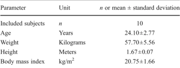
Table 1 Subject demographics
Parameter Unit n or mean ± standard deviation
Included subjects n 10
Age Years 24.10±2.77
Weight Kilograms 57.70±5.56
Height Meters 1.67±0.07
Statistical analysis
Descriptive statistics were performed for each variable (mean, standard deviation).
The reliability test followed the three steps suggestions of Weir [19]: In order to identify possible systematic errors be-tween the repeated measures, the Wilcoxon rank order test was applied at rest and the MVC tests, and the Friedman test to compare the dependent time and EMG variables over the ten steps. Intra-session test–retest reliability (consistency) was cal-culated with the intraclass correlation coefficient (ICC 3.1; i.e., relative reliability) and the standard error of measurement (SEM; i.e., absolute reliability). Additionally, the minimal de-tectable differences (MDDs) using a 95 % confidence interval and the mean absolute differences among all repeated measures were calculated.
Portney and Watkins [20] suggest as a general guideline that ICC values above 0.75 are indicative of good reliability and those below 0.75 of poor to moderate reliability.
The level for significances was set at P<0.05. All statistics were calculated using SPSS 17 for Windows (SPSS; Chicago, IL, USA) as well as Microsoft Excel 2007 (Microsoft, Redmond, WA, USA).
Results
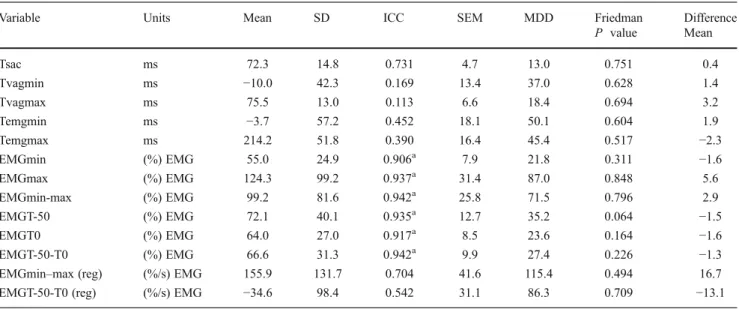
Figure1shows samples of the amplitude and time variabil-ity of EMG–time curves within the ten steps of one subject. Thirteen variables were selected based on ten steps of each subject. Descriptive statistics and reliability calcula-tions are presented in Tables 3 and4. All baseline data of EMG during rest or MVC show good reliability. The anal-ysis of systematic errors within repeated measures by Wilcoxon or Friedman tests revealed only nonsignificant values for all variables. ICCs for the time variables (ms) ranged from 0.113 to 0.731. The highest reliability indexes were found for all EMG-amplitude-derived variables
Table 2 Labels, units, and description of time and activity variables derived from accelerometry and electromyography (EMG)
Variable (unit) Description To identify
T0 Heel strike The initial time point of the impact and beginning
loading phase and strain of the PFM
Tsac (ms) Time difference between heel strike (T0)
and first acceleration of the sacrum accelerator
To identify the time difference (delay) between T0 and Tsac as a more bony part compared with the vaginal accelerator, which is attached to rather less rigid muscle structures
Tvagmin (ms) Time difference between heel strike (T0)
and minimal acceleration of the vaginal accelerator
Periodic characteristics of the acceleration of the vaginal sensor/PFM movements during cyclic running (from minimum to maximum)
Tvagmax (ms) Time difference between heel strike (T0)
and maximal acceleration of the vaginal accelerator
Periodic characteristics of the acceleration of the vaginal sensor/PFM movements during cyclic running (from maximum to minimum)
Temgmin (ms) Time difference between heel strike (T0)
and minimal EMG activity
Periodic characteristics and an“ON/OFF criterion” of the
PFM activity in comparison to rest activity during supine position or standing
Temgmax (ms) Time difference between heel strike (T0)
and maximal EMG activity
Periodic characteristics of PFM activity
EMGmin ((%) EMG) Minimal EMG activity Periodic characteristics and an“ON/OFF criterion” of the
PFM activity in comparison to rest activity during supine position or standing
EMGmax ((%) EMG) Maximal EMG activity Periodic characteristics of PFM activity
EMGmin-max ((%) EMG) Mean EMG activity between EMGmin
and EMGmax
The range of PFM activity
EMGT-50 ((%) EMG) EMG activity at heel strike (T0) minus 50 ms The pre-activity of PFM as a regulatory component
of anticipation
EMGT0 ((%) EMG) EMG activity at heel strike (T0) The EMG amplitude at the initial time point of the
impact and beginning the loading phase and strain of the PFM
EMGT-50-T0 ((%) EMG) Mean EMG activity between minus
50 ms and T0
The mean pre-activity EMGmin-max
(reg) ((%/s) EMG)
Linear regression of EMG activity between EMGmin and EMGmax
The gradient of range of amplitude EMGT-50-T0
(reg) ((%/s) EMG)
Linear regression of EMG activity
between−50 ms and T0
((%) EMG), which were superior to 0.750 and ranged be-tween 0.906 and 0.942. The combined time–amplitude var-iables ((%/s) EMG) did not reach excellent reliability, with values of 0.542 and 0.704.
Discussion
The purpose of this study was to describe and reliability test the variables that characterize PFM activity and time behavior during running. We selected a total of 13 variables (see Table2), which were tested with regard to their intra-session retest reliability based on ten steps of each subject. Generally, EMG variables showed good time variables and poor reliability. EMG variables
Among the technologies used to study PFM neural control, EMG of the PFM has gained the most attention and clinical relevance [21]. Grape et al. [18] tested the retest reliability of isolated PFM contraction exercise by means of EMG in healthy nulliparae women and showed good reliability
(ICC=0.82–0.96) of average activity (during a 10-s long squeeze), peak (maximum value), work (the area under the curve), and baseline (resting value); the ICC for speed (peak divided by time taken to reach the peak) was between 0.65 and 0.85. In contrast to our investigation, Gollhofer et al. [22] investigated the EMG reliability in leg muscles during running at constant velocity. They found high reliability coefficients for day-to-day and week-to-week comparisons and also, with regard to qualitative comparisons of EMG, high reproducibil-ity of the shape of the patterns. However, to date there have been no reliability studies concerning vaginal EMG during dynamic functional whole body movements of the subjects.
In our study 6 of the 8 identified EMG variables of PFM activity during running showed good ICC values. Moderate to almost good ICCs of EMG measurements were found for regressions.
The minimal EMG (EMGmin) activity during running shows good reliability. Mean minimal EMG activity during running shows an activity about 24 % higher than mean PFM EMG activity during rest in a standing position. These findings show a constantly increased PFM tone during run-ning, and an aerobic strain of PFM during running could be hypothesized. The increased tonicity of PFM activity was also found in the study of Hodges et al. [13] during repet-itive arm movements.
The maximal EMG (EMGmax) activity shows good re-liability. The mean activity level was at 124.3 (%) EMG. Hodges et al. [13] found, during repetitive arm movements, bursts of anal and vaginal EMG activity in association with the frequency of arm movement, but they did not provide information about the activity level. These findings suggest a higher PFM activity compared with MVC owing to re-flexive and reactive force generation during running.
The mean EMG activity between EMGmin and EMGmax variables shows good reliability and a mean level of 99.2 (%) EMG. These values are about 68 (%) EMG higher than during standing at rest. Schulte-Frei [23] examined PFM activity while subjects stood on stable and unstable ground and during one-leg stands. As her MVC normalization referred to an MVC taken individually during co-contraction while performing a set of other exercises for muscle testing, these values cannot be compared with those found in our study. Nonetheless, as she found increasing PFM mean activity from standing on stable ground compared with standing on unstable ground and an even higher activity during the one-leg stands, these findings are of interest as, during running a temporary one-leg stand takes place as well.
The variable linear regression of EMG activity between EMGmin and EMGmax shows almost good reliability.
The EMG activity at heel strike (T0) minus 50 ms shows good reliability and an activity level of 72.1 (%) EMG, which is about 40 (%) EMG higher than during rest. Hodges et al. [13] also found a mean PFM pre-activity of
Fig. 1 Exemplary EMG–time curves derived from the ten steps of
subject 1. The curves start and end at the minimum EMG (EMGmin) activity of each step cycle. The dotted line at −50 ms (EMGT-50) represents the start of the pre-activity phase which ends at T0 (dotted line at 0 ms = heel strike). Subsequently, EMG–time curves increase to their activity maximum (EMGmax)
28.4 ms prior to arm movements and Sjödhal et al. [14] a median PFM pre-activity of 206 ms prior to the start of a leg lift at a level of 20 (%) EMG. These findings confirm the assumption that the increased PFM activity 50 ms prior to the heel strike could indicate reliable PFM pre-activity dur-ing runndur-ing.
The EMG activity at heel strike (T0) shows good reli-ability. As the PFM activity shows higher values than the activity at rest in a standing position, this variable confirms the assumption of a PFM pre-activity.
The mean EMG activity between minus 50 ms and T0 shows good reliability. Again, the values are higher compared with the PFM activity during rest in a standing position, which could indicate PFM pre-activity.
The linear regression of EMG activity between−50 ms and T0 before the heel strike shows moderate reliability. This value
((%/s) EMG) cannot be used to investigate PFM pre-activity, as it cannot be compared with the PFM MVC ((%) EMG). Time variables
The time variables show large ranges and therefore low reli-ability. Four (time difference between heel strike (T0) and minimal acceleration of VagAcc; time difference between heel strike (T0) and maximal acceleration of VagAcc; time differ-ence between heel strike (T0), and minimal EMG activity; time difference between heel strike (T0) and maximal EMG activity) out of five time variables showed low ICC values.
The only variable showing almost good reliability was the time difference between heel strike (T0) and first accel-eration of SacAcc. A possible explanation could be a more precise force transmission on bony structures than on less rigid muscle structures.
Table 3 Descriptive statistics (mean ± SD), reliability indexes (intraclass correlation coefficient, standard error of measurement), minimal detectable difference for variables, and test for systematic error (Wilcoxon, difference) for activity variables
Variable Units Mean SD ICC SEM MDD Wilcoxon Difference
P value Mean
Rest supine (%) EMG 14.2 9.7 0.999a 3.0 8.3 0.398 0.1
Rest standing (%) EMG 31.3 4.0 0.993a 4.0 11.1 0.173 0.7
MVC supine ųV 64.7 27.4 0.867a 8.7 24.1 0.445 1.3
MVC standing ųV 57.6 20.1 0.914a 6.3 17.6 0.508 1.1
Mean arithmetic mean, SD standard deviation, ICC intraclass correlation coefficient, SEM standard error of measurement, MDD minimum detectable difference, Wilcoxon Wilcoxon signed-rank test; Difference arithmetic mean of test–retest differences
aIndicates ICCs>0.750
Table 4 Descriptive statistics (mean ± SD), reliability indexes (ICC, SEM), MDD for variables, and test for systematic error (Friedman, difference) for time and activity variables derived from accelerometry and electromyography
Variable Units Mean SD ICC SEM MDD Friedman Difference
P value Mean Tsac ms 72.3 14.8 0.731 4.7 13.0 0.751 0.4 Tvagmin ms −10.0 42.3 0.169 13.4 37.0 0.628 1.4 Tvagmax ms 75.5 13.0 0.113 6.6 18.4 0.694 3.2 Temgmin ms −3.7 57.2 0.452 18.1 50.1 0.604 1.9 Temgmax ms 214.2 51.8 0.390 16.4 45.4 0.517 −2.3 EMGmin (%) EMG 55.0 24.9 0.906a 7.9 21.8 0.311 −1.6 EMGmax (%) EMG 124.3 99.2 0.937a 31.4 87.0 0.848 5.6 EMGmin-max (%) EMG 99.2 81.6 0.942a 25.8 71.5 0.796 2.9 EMGT-50 (%) EMG 72.1 40.1 0.935a 12.7 35.2 0.064 −1.5 EMGT0 (%) EMG 64.0 27.0 0.917a 8.5 23.6 0.164 −1.6 EMGT-50-T0 (%) EMG 66.6 31.3 0.942a 9.9 27.4 0.226 −1.3
EMGmin–max (reg) (%/s) EMG 155.9 131.7 0.704 41.6 115.4 0.494 16.7
EMGT-50-T0 (reg) (%/s) EMG −34.6 98.4 0.542 31.1 86.3 0.709 −13.1
Mean arithmetic mean, SD standard deviation, ICC intraclass correlation coefficient, SEM standard error of measurement, MDD minimum detectable difference, Friedman Friedman test nonparametric repeated measures comparisons; difference arithmetic mean of test–retest differences a
To our knowledge, there has been to date no literature published on the time variables of PFM contraction character-istics, and therefore the results of this study can only be discussed in the context of other findings. Leitner et al. [24] were able to show good reliability regarding time variables in stair ascent in the elderly, but clearly lower reliability in stair descent, which seems to be more comparable to impacts during running at slow velocity. Masani et al. [25] investigated the variability of the ground reaction force pattern during walking in relation to different constant speeds. He found an increasing trend in variability with speeds from 3 to 8 km/h; however, the minimum variability speeds of 5.5–5.8 km/h were within the limits of usual walking speeds. This result indicates that the neuromuscular locomotor system was most stable at usual walking speed.
Methodical problems of vaginal probe measurements Because vaginal probes cannot be fixed directly onto the muscles, as they use self-adhesive electrodes, minimal shifts of vaginal probes might go unnoticed. In the present study the attachment with tape of the accelerometer to the vaginal probe seemed to be sufficient. However, greater movement of the vaginal probe (EMG and accelerometer) in contrast to the sacral time variables (Tsac) cannot be totally excluded and is supported by the lower ICCs of the vaginal time variables (Tvagmin and Tvagmax). In a future study this issue could be addressed by using a device such as a STIMPON® electrode (Innocept Biobedded Medizintechnik, Gladbeck, Germany). This electrode adapts its shape to individual vaginal cavities and therefore movement could be avoided.
Cross-talk
Electromyography is a reliable method of assessing PFM activity in healthy women [18]. However, Bo and Sherburn [26] conclude in their overview assessing PFM function and strength that the EMG signals must be interpreted with caution because the risk of cross talk from other muscles is high and because of the variability in electrode placement within the vagina. To date, cross-talk during functional dynamic whole body movements for PFM activity has not yet been examined and is therefore difficult to estimate. Treadmill
Some subjects were not familiar with treadmill running, which possibly resulted in a nonphysiological gait compared with running on natural ground at usual individual speeds. However, running on natural ground at individual speeds would be problematic with regard to speed standardization. To address the problem of a lack of familiarity with tread-mill running, all the subjects performed a warm-up of
5 km/h for 30 s and then a running-in of 8 km/h for 2.5 min, both on the treadmill, before the measurements took place.
Shoes
According to Maurer et al. [27], the characteristics of human locomotion may change for different boundary conditions such as footwear. In our study, the subjects wore their own running shoes. Wright et al. [28] found no difference be-tween soft and hard shoe impact forces. However, peak rates of loading were greater for the hard shoe than for the soft shoe. Therefore, an influence of footwear on gait and force transmission cannot be excluded. Future studies should re-quire standardized running shoes or running barefoot.
Conclusion
To the best of our knowledge, the current study is the first to test PFM activity and time variables during dynamic func-tional whole body movements with regard to their reliabil-ity. It provides a first insight into the PFM activity of healthy women during running. EMG variables of good reliability could be shown, while poor evidence was gained for the reliability of time variables. In particular, further studies should consider adaptations regarding the vaginal probe and footwear. The test application and reliability will likely have to be modified and retested in different popula-tions and given the anatomical changes seen associated with SUI and pelvic organ prolapse. More knowledge of PFM EMG variables and time variables may help to provide a deeper insight into physical strain with high force impacts and functionally important reflexive con-traction patterns of PFM to maintain or to restore continence. A further consequence is support for the development of more sophisticated diagnostic instruments and therapeu-tic approaches.
Acknowledgements The authors thank Parsenn-Produkte AG
(Küblis, Switzerland) for providing the vaginal surface EMG probes.
Conflicts of interest None
References
1. Botlero R, Davis SR, Urquhart DM, Shortreed S, Bell RJ (2009) Age-specific prevalence of, and factors associated with, different types of urinary incontinence in community-dwelling Australian women
assessed with a validated questionnaire. Maturitas 62:134–139
2. Brown SJ, Donath S, MacArthur C, McDonald EA, Krastev AH (2010) Urinary incontinence in nulliparous women before and
during pregnancy: prevalence, incidence, and associated risk
fac-tors. Int Urogynecol J 21:193–202
3. Bø K (2004) Urinary incontinence, pelvic floor dysfunction,
exer-cise and sport. Sports Med 34:451–464
4. Carls C (2007) The prevalence of stress urinary incontinence in high school and college-age female athletes in the Midwest: impli-cations for education and prevention. Urol Nurs 27:21–24 5. Simeone C, Moroni A, Pettenò A et al (2010) Occurrence rates and
predictors of lower urinary tract symptoms and incontinence in
female athletes. Urologia 77:139–146
6. Eliasson K, Larsson T, Mattsson E (2002) Prevalence of stress incontinence in nulliparous elite trampolinists. Scand J Med Sci
Sports 12:106–110
7. Salvatore S, Serati M, Laterza R, Uccella S, Torella M, Bolis PF (2009) The impact of urinary stress incontinence in young and middle-age women practising recreational sports activity: an
epi-demiological study. Br J Sports Med 43:1115–1118
8. Eliasson K, Edner A, Mattsson E (2008) Urinary incontinence in very young and mostly nulliparous women with a history of regular organised high-impact trampoline training: occurrence
and risk factors. Int Urogynecol J 19:687–696
9. Shishido K, Peng Q, Jones R, Omata S, Constantinou CE (2008) Influence of pelvic floor muscle contraction on the profile of vaginal closure pressure in continent and stress urinary incontinent women. J Urol 179:1917–1922
10. Deffieux X, Hubeaux K, Porcher R, Ismael SS, Raibaut P, Amarenco G (2008) Abnormal pelvic response to cough in women with stress urinary incontinence. Neurourol Urodyn
27:291–296
11. Morin M, Bourbonnais D, Gravel D, Dumoulin C, Lemieux MC (2004) Pelvic floor muscle function in continent and stress urinary incontinent women using dynamometric measurements. Neurourol
Urodyn 23:668–674
12. Miller J, Kasper C, Sampselle C (1994) Review of muscle phys-iology with application to pelvic muscle exercise. Urol Nurs
14:92–97
13. Hodges PW, Sapsford R, Pengel LHM (2007) Postural and respi-ratory functions of the pelvic floor muscles. Neurourol Urodyn
26:362–371
14. Sjödahl J, Kvist J, Gutke A, Öberg B (2009) The postural response of the pelvic floor muscles during limb movements: a methodo-logical electromyography study in parous women without lumbo-pelvic pain. Clin Biomech 24:183–189
15. Zadpoor AA, Nikooyan AA (2011) The relationship between lower-extremity stress fractures and the ground reaction force: a
systematic review. Clin Biomech 26:23–28
16. Keller TS, Weisberger AM, Ray JL, Hasan SS, Shiavi RG, Spengler DM (1996) Relationship between vertical ground reac-tion force and speed during walking, slow jogging, and running. Clin Biomech 11:253–259
17. Merletti R, Parker PJ (2004) Electromyography: physiology, engi-neering, and non-invasive applications. Wiley, New Jersey 18. Grape HH, Dedering Å, Jonasson AF (2009) Retest reliability of
surface electromyography on the pelvic floor muscles. Neurourol
Urodyn 28:395–399
19. Weir JP (2005) Quantifying test-retest reliability using the intra-class correlation coefficient and the SEM. J Strength Cond Res
19:231–240
20. Portney LG, Watkins MP (2009) Foundations of clinical research: applications to practice, 3rd edn. Pearson/Prentice Hall, Upper Saddle River
21. Enck P, Vodušek DB (2006) Electromyography of pelvic floor
muscles. J Electromyogr Kinesiol 16:568–577
22. Gollhofer A, Horstmann GA, Schmidtbleicher D, Schönthal D (1990) Reproducibility of electromyographic patterns in stretch-shortening type contractions. Eur J Appl Physiol 60:7– 14
23. Schulte-Frei B (2006) Sport- und Bewegungstherapie für den wei-blichen Beckenboden. Alltagsrelevanz, Analyse und Therapie unter besonderer Berücksichtigung der neuromuskulären Ansteuerung. Dissertation, Institut für Rehabilitation und Behindertensport der Deutschen Sporthochschule Köln
24. Leitner M, Schmid S, Hilfiker R, Radlinger L (2011) Test-retest reliability of vertical ground reaction forces during stair climbing
in the elderly population. Gait Posture 34:421–425
25. Masani K, Kouzaki M, Fukunaga T (2002) Variability of ground
reaction forces during treadmill walking. J Appl Physiol 92:1885–1890
26. Bø K, Sherburn M (2005) Evaluation of female pelvic-floor
mus-cle function and strength. Phys Ther 85:269–282
27. Maurer C, Federolf P, von Tscharner V, Stirling L, Nigg BM (2012) Discrimination of gender-, speed-, and shoe-dependent movement patterns in runners using full-body kinematics. Gait Posture 36:40–45
28. Wright IC, Neptune RR, van den Bogert AJ, Nigg BM (1998) Passive regulation of impact forces in hell-toe running. Clin Biomech 13:521–531