HAL Id: tel-02973648
https://tel.archives-ouvertes.fr/tel-02973648
Submitted on 21 Oct 2020
HAL is a multi-disciplinary open access
archive for the deposit and dissemination of sci-entific research documents, whether they are pub-lished or not. The documents may come from teaching and research institutions in France or abroad, or from public or private research centers.
L’archive ouverte pluridisciplinaire HAL, est destinée au dépôt et à la diffusion de documents scientifiques de niveau recherche, publiés ou non, émanant des établissements d’enseignement et de recherche français ou étrangers, des laboratoires publics ou privés.
Pathophysiological role of Pyk2 in the nervous system
Benoit De Pins
To cite this version:
Benoit De Pins. Pathophysiological role of Pyk2 in the nervous system. Neurobiology. Sorbonne Université, 2019. English. �NNT : 2019SORUS073�. �tel-02973648�
Thèse de doctorat - Sorbonne Université
Ecole doctorale Cerveau Cognition ComportementPrésentée par
Benoit de Pins
Pathophysiological role of
Pyk2 in the nervous system
Soutenue le 12 septembre 2019 Devant le jury composé de :
Dr. Jocelyne Caboche Présidente
Dr. Véronique Sgambato Rapporteur
Dr. Emmanuel Valjent Rapporteur
Pr. Jean-Christophe Corvol Examinateur
1
Acknowledgments
Ce manuscrit conclue quatre années de travail dans un environnement aussi riche qu’agréable, entouré de collègues qui sont finalement devenus mes amis. Il est normal que je le commence par un remerciement à tous ceux sans qui ce travail n’aurait pu être réalisé. A vous tout d’abord Jean-Antoine, pour m’avoir reçu dans votre bureau il y a exactement cinq ans. Je suis très fier d’avoir été votre étudiant. Vous m’avez encadré tout en me donnant toujours beaucoup de liberté. J’ai aimé nos discussions et « faire de la science » avec vous.
Denis, merci pour tous les précieux conseils que tu m’as donnés au cours de ces cinq années. Merci pour ta gentillesse et ton accessibilité.
Je voudrais remercier tout particulièrement les Dr. Jocelyne Caboche, Dr. Véronique Sgambato, Dr. Emmanuel Valjent ainsi que le Pr. Jean-Christophe Corvol d’avoir accepté d’être membre de mon jury et de relire ce manuscrit.
Au 5ème étage : Merci les amis, j’ai passé cinq années formidables autour de vous.
Vous allez tous beaucoup me manquer.
A mes amis, de Versailles, de prépa, de l’ENS, de Roscoff, de l’IFM : je me suis senti bien entouré pendant ces quelques années. Je sais à quel point je suis chanceux de vous connaitre.
Papa, Maman, Ronan, Solène, Amicie, Peyo, Antoine, Pierre, Olivier, Gilles, Chantal, Xavier, Christine et toute la famille, merci pour votre présence et votre soutien quotidiens. Je vous aime très fort.
A toi Albert, bien évidemment, un énorme merci. Sans toi, tout cela n’est rien. Merci de m’avoir fait rentrer dans ton monde. Ens tornarem a trobar !
3
Contents
Acknowledgments ... 1
Contents ... 3
List of abbreviations ... 7
Context and objectives ... 9
INTRODUCTION ... 11
Tyrosine kinase signaling and the FAK family ... 13
1. The origin of TKs and FAK family ... 13
2. FAK family ... 16
Identification of FAK family kinases ... 16
Structure of FAK family kinases ... 16
2.2.1. FERM domain ... 17
2.2.2. Linker 1 ... 18
2.2.3. Kinase domain ... 18
2.2.4. Linker 2 ... 19
2.2.5. FAT domain ... 19
Expression of FAK and Pyk2 ... 20
Cellular localization of FAK family kinases ... 21
Isoforms of FAK family kinases ... 21
2.5.1. FAK isoforms ... 22
2.5.2. Pyk2 isoforms ... 22
Biological functions of FAK ... 23
2.6.1. Cellular functions of FAK ... 23
2.6.2. Physiological functions of FAK ... 25
Pyk2: a nRTK of FAK family ... 25
1. Regulation of Pyk2 activity in non-neuronal cells ... 25
Activation and phosphorylation of Pyk2 ... 25
1.1.1. Canonical activation of Pyk2 ... 25
1.1.2. Regulation of Pyk2 activation by Ca2+-activated kinases ... 26
1.1.2.1. PKC ... 26
1.1.2.2. CaMKII ... 27
Dephosphorylation of Pyk2... 27
1.2.1. Tyrosine phosphatases ... 27
4 1.2.1.2. SHP-2 ... 27 1.2.1.3. PTP-PEST ... 27 1.2.1.4. STEP ... 28 1.2.2. Ser/Thr phosphatases ... 28 SUMOylation of Pyk2 ... 28 S-nitrosylation of Pyk2 ... 28
2. Pyk2 functions in non-neuronal cells ... 29
Pyk2 cellular functions ... 29
2.1.1. Cell adhesion... 29
2.1.2. Cell migration ... 29
2.1.3. Cell division ... 30
2.1.4. Cell survival ... 30
2.1.5. Cell differentiation ... 31
Physiological role of Pyk2 ... 31
2.2.1. Bone physiology ... 31
2.2.2. Vascular system integrity ... 32
2.2.3. Immune system function ... 32
2.2.4. Kidney function ... 32
2.2.5. Sperm capacitation ... 33
2.2.6. Generation of Pyk2 knockout mice ... 33
3. Pathological role of Pyk2 ... 34
Pyk2 and inflammatory diseases... 34
Pyk2 and cancers ... 34
Pharmacological inhibitors of Pyk2 ... 35
Roles of Pyk2 in the CNS ... 37
1. Specific regulation of Pyk2 in the CNS ... 37
Activation of Pyk2 in neurons ... 37
Regulation of Pyk2 localization ... 38
2. Pyk2 biological functions in the CNS ... 39
Ionic channels regulation ... 39
2.1.1. Kv1.2 ... 39
2.1.2. BK channels ... 39
2.1.3. NMDA receptor (NMDAR) ... 39
Development ... 40
5
Neuronal survival ... 41
Pyk2 in glial cells ... 42
3. Pyk2 in CNS diseases ... 42
Alzheimer·s disease ... 42
Parkinson·s disease ... 43
Huntington·s disease ... 43
Neuroinflammation ... 43
Glioma and neuroblastoma ... 43
Cerebral ischemia ... 44
Psychiatric disorders ... 44
RESULTS ... 47
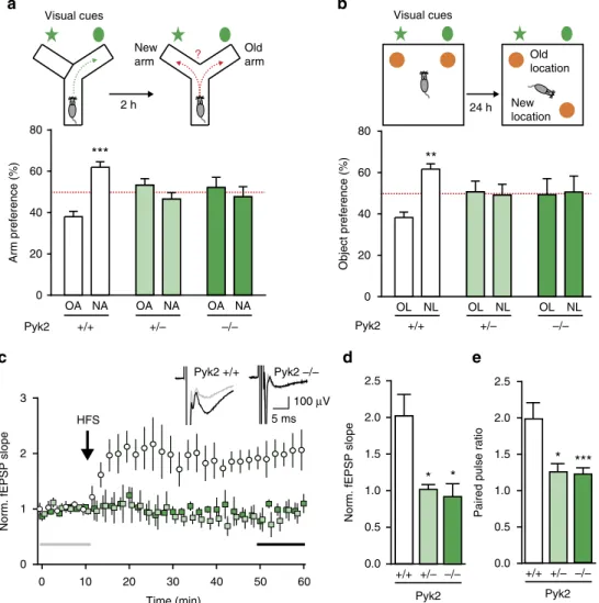
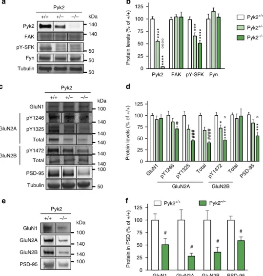
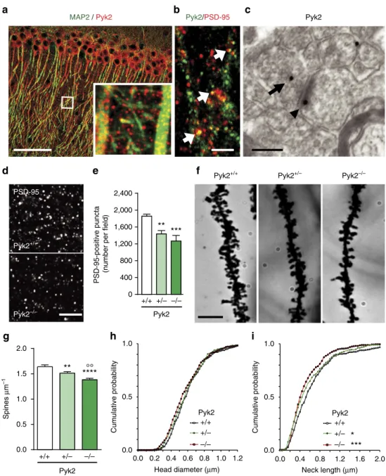
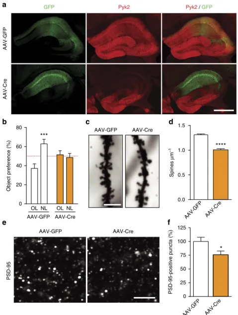
Pyk2 modulates hippocampal excitatory synapses and contributes to cognitive deficits in a Huntington's disease model... 51
1. Context and objectives ... 51
2. Contribution to the work... 51
3. Article ... 53
4. Summary of the findings and conclusions ... 55
Pyk2 in the amygdala modulates chronic stress sequelae via PSD-95-related micro-structural changes... 57
1. Context and objectives ... 57
2. Contribution to the work... 57
3. Article ... 59
4. Summary of the findings and conclusions ... 61
PTK2B/Pyk2 overexpression improves a mouse model of Alzheimer's disease ... 63
1. Context and objectives ... 63
2. Contribution to the work... 63
3. Article ... 65
4. Summary of the findings and conclusions ... 67
Conditional BDNF Delivery from Astrocytes Rescues Memory Deficits, Spine Density, and Synaptic Properties in the 5xFAD Mouse Model of Alzheimer Disease. ... 69
1. Context and objectives ... 69
2. Contribution to the work... 69
3. Article ... 71
4. Summary of the findings and conclusions ... 73
Pyk2 in nucleus accumbens D1 receptor-expressing neurons is selectively involved in the acute locomotor response to cocaine ... 75
6
1. Context and objectives ... 75
2. Contribution to the work... 75
3. Article ... 77
4. Summary of the findings and conclusions ... 80
Supplementary data: spine density and morphology in the NAc of Pyk2-/- mice .. 82
1. Materials and methods ... 82
2. Results ... 82
DISCUSSION ... 84
Role of Pyk2 in memory ... 86
Kinase-dependent and independent functions of Pyk2 ... 87
Antagonistic effect of Pyk2 on spine density and morphology ... 88
BDNF and Pyk2 merging functions ... 89
Pyk2 and AD: risk or rescue factor? ... 89
Contrasted function of Pyk2 in the striatum ... 92
7
List of abbreviations
AAV-Cre Cre-expressing adeno-associated virus
AD Alzheimer's disease
APP amyloid precursor protein
ArgBP2 Arg kinase binding protein 2
ASAP2 ArfGAP with SH3 domain, ankyrin repeat and PH domain 2
ATP adenosine triphosphate
AƢ amyloid Ƣ peptide
AƢo AƢ oligomers
BDNF brain-derived neurotrophic factor
BK ´big potassiumµ large conductance voltage-gated K+ channel
BY-kinase bacterial tyrosine kinase
CADTK calcium dependent-protein tyrosine kinase
CAKƢ cell adhesion kinase-Ƣ
cAMP 3·,5·-cyclic adenosine monophosphate
CSPG chondroitin sulphate proteoglycan
CUMS chronic unpredictable mild stress
DHPG (S)-3,5-dihydroxy-phenylglycine
DNA deoxyribonucleic acid
EGFR epidermal growth factor receptor
eNOS endothelial nitric oxide synthase
EOAD early onset AD
ePK eukaryotic protein kinase
ERK extracellular signal-regulated kinase
FAK focal adhesion kinase
FAT focal adhesion targeting
FERM four-point-one, ezrin, radixin, moesin
FIP200 FAK family kinase-interacting protein of 200 kDa
FNRK FAK-related non-kinase
GluN2A NMDAR subunit 2A
GWAS genome-wide association studies
HD Huntington·s disease
HTT huntingtin (human gene)
Htt huntingtin (protein)
JNK c-Jun NH2-terminal kinase
KIR kinase inhibitory region
LOAD late onset AD
LTP long-term potentiation
LTP-IE long-term potentiation of intrinsic excitability
MAPK mitogen activated protein kinase
MAP4K4 mitogen-activated protein kinase kinase kinase kinase 4
MECP2 methyl-CpG-binding protein 2
MKK3 mitogen-activated protein kinase kinase 3
mGluR5 metabotropic glutamate receptor 5
mRNA messenger RNA
8
NAPOR neuroblastoma apoptosis-related RNA-binding protein
NES nuclear export signal
NLS nuclear localization sequence
NMDAR NMDA receptor
nRTK non-receptor tyrosine kinases
NTS nuclear targeting sequence
PARP poly(adenosine diphosphate ribose)polymerase
PD Parkinson's disease
PDGFR platelet-derived growth factor receptor
PITPNMs membrane-associated phosphatidylinositol transfer proteins
PKC protein kinase C
PR proline-rich
PRAP proline-rich acidic protein
PRNK Pyk2-related non-kinase
PrPc prion protein cellular precursor
Prx2 peroxiredoxin 2
PSTPs protein serine-threonine phosphatases
PTP protein tyrosine phosphatase
pTyr phosphotyrosine
Pyk2 proline-rich tyrosine kinase 2
RAFTK related adhesion focal tyrosine kinase
RB1CC1 retinoblastoma 1-inducible coiled-coil 1
RNA ribonucleic acid
ROS reactive oxygen species
RTK receptors having a tyrosine kinase activity
SAP102 synapse-associated protein 102
SFKs Src-family kinases
SH2 Src homology 2
SH3 Src homology 3
SNP single nucleotide polymorphism
SPN striatal projection neuron
STEP striatal-enriched protein-tyrosine phosphatase
STK serine/threonine kinase
TK tyrosine kinases
9
Context and objectives
Protein function regulation by phosphorylation is implicated in the great majority of cellular processes in eukaryotes, and to a lesser extent in prokaryotes. Regulatory phosphorylation of proteins is a post-translational modification in which a phosphoryl group is transferred from a donor, usually adenosine triphosphate (ATP), to the substrate, a residue of an amino acid: either serine, threonine or tyrosine in metazoans. Protein phosphorylation is a reaction catalyzed by protein kinases, usually reversible due to the action of protein phosphatases which catalyze dephosphorylation. Protein kinases and phosphatases contribute to the cellular adaptation to the environment in response to extra and intra-cellular signals and often act as integrators.
Genes coding for protein kinases represent about 2% of eukaryotes protein coding genes and embody one of the major classes. Protein kinases and protein phosphatases are both comprised of different families. In the human, on the ~540 genes coding for protein
kinases, 90 code for ´realµ tyrosine kinases (TK) of which 32 are non-receptor tyrosine
kinases (nRTK) and 58 are transmembrane receptors having a tyrosine kinase activity (RTK) (Manning et al., 2002b; Wilson et al., 2018). Our laboratory is particularly interested in the nRTK group named focal adhesion kinase (FAK) family containing only two members: FAK and the proline-rich tyrosine kinase 2 (Pyk2).
FAK and Pyk2 share roughly 45% amino acid sequence identity and 65% similarity (Avraham et al., 1995, 2000; Sasaki et al., 1995) but display very distinct expression patterns. Two products of alternative splicing characterized by the presence or absence of an alternative exon were described for Pyk2. The variant lacking this exon is highly expressed in hematopoietic cells whereas the complete form is enriched in the brain. Pyk2 has a key role in numerous cellular physiological and pathological processes such as cell migration, proliferation, survival and death. In the nervous system, it could participate in learning and memory. The major step initiating Pyk2 activation is its autophosphorylation on Tyr-402. This phosphorylation allows the recruitment and the activation of Src family members, the phosphorylation of other residues of Pyk2 and the recruitment of other partners leading to the activation of several pathways.
Works of many laboratories, including ours, have shown that Pyk2 is involved in NMDA receptor regulation and synaptic plasticity in the hippocampus, suggesting maybe a participation of Pyk2 in neurological diseases. However, Pyk2 function remains unclear and the mechanism of action of Pyk2 is still poorly understood. The objective of my thesis was to clarify the role of Pyk2 in the central nervous system and its implication in several neurological pathologies.
We studied the involvement of Pyk2 in cognitive deficits associated to Huntington's disease (Article 1). We also investigated a putative role of Pyk2 in the resilience of mice to a model of depression (Article 2) and in a mouse model of Alzheimer's disease (AD) (Article 3). In parallel with this last study, we used the same mouse model of AD to investigate the potential benefit of astrocyte-targeted delivery of BDNF in this disease (Article 4). We finally characterized the expression of Pyk2 in the striatum and its role in cocaine response (Article 5, submitted).
In the bibliographic introduction, I will address several topics associated to my subject: the presentation of FAK family kinases, the characterization of Pyk2, and the specific role of Pyk2 in the central nervous system.
11
13
Tyrosine kinase signaling and the FAK family
In animals, phosphotyrosine (pTyr) signaling is an essential system that regulates hormone, growth factor, immune, and adhesion-based signaling, therefore allowing cell-cell communication (Hunter and Cooper, 1981; Shattil and Brugge, 1991; Myers et al., 1994; Weiss and Littman, 1994).
This signaling relies on a simple mechanism: a TK phosphorylates certain tyrosine residues on the substrate protein (or itself). Then, an effector protein recognize the pTyr by its Src homology 2 (SH2) domain or other pTyr-binding domains, and activates downstream signaling. The pathway can be stopped by a protein tyrosine phosphatase (PTP) which dephosphorylates the pTyr (Figure 1).
Figure 1. Mechanism of TK signaling. Tyrosines are phosphorylated by TKs and
dephosphorylated by PTPs. The phosphorylated tyrosine is then recognized by an SH2-containing effector.
1. The origin of TKs and FAK family
The eukaryotic protein kinase (ePK) superfamily is divided into two kinase families according to their substrate specificity: the TKs and the serine/threonine kinases (STKs) families. TKs originally evolved from STKs. Although some STKs can phosphorylate tyrosine residues and are referred to as dual specificity protein kinases (Lindberg et al., 1992), TKs are discriminated from STKs by their overall sequence similarity and also notably by their characteristic catalytic loop motif (Hanks and Hunter, 1995; Manning et al., 2008).
The evolutionary history of TK family has been specifically studied and discussed for many years (Miller, 2012; Tong et al., 2017). It was initially believed that tyrosine kinases were specific to metazoans. In contrast with the high diversity of TKs in many metazoan phyla, they were initially not found in plants, fungi and other analyzed eukaryotes (Hanks and Hunter, 1995; Manning et al., 2002a). Although some bacteria evolved unique tyrosine kinases known as bacterial tyrosine kinases (BY-kinases), metazoan-like TKs were not found in bacteria (Grangeasse et al., 2012). Accordingly, TK were hypothesized to be a metazoan-specific evolutionary innovation that permitted cell-cell communication and thus contributed to the origin of metazoan multicellularity.
However, in 2001, King and Carroll discovered the first TK (a receptor tyrosine kinase designated MBRTK1) outside of the metazoan taxon (King and Carroll, 2001). This TK was found in Monosiga brevicollis, a unicellular member of choanoflagellates that are the closest known living relatives of metazoans (Richter and King, 2013). Successive papers
14
then confirmed the existence of multiple active tyrosine kinases in this organism (King et al., 2003) and in several other choanoflagellate species (Segawa et al., 2006; Suga et al., 2008).
Genomic analyses then revealed that choanoflagellates contain a rich and complex repertoire of TKs that is comparable to those observed in the most complex multicellular animals (King et al., 2008; Manning et al., 2008; Pincus et al., 2008). Many choanoflagellates can form colonies, suggesting that their common ancestor with the metazoan could be a transitional form between unicellular and multicellular organisms and that these unicellular tyrosine kinases may have facilitated the evolution of multicellular animals (King, 2004).
Afterward, new genome/transcriptome sequencing has uncovered TKs in other eukaryotes thus forcing to reconsider every time the apparition date of TKs during evolution. In this way, in addition to the metazoans and the choanoflagellates, tyrosine kinases were discovered in three other sister lineages: the filastereans, the ichthyosporeans and the corallochytreans (forming, with the metazoans and the choanoflagellates, the holozoans) (Shalchian-Tabrizi et al., 2008; Suga et al., 2012, 2014; Fairclough et al., 2013; Sebé-Pedrós et al., 2016). TKs were also found in two amoebozoans (Clarke et al., 2013; Schaap et al., 2015) and in the apusozoan Thecamonas trahens (Suga et al., 2012). Moreover, TKs were discovered in some bikonts such as the green alga Chlamydomonas reinhardtii or the higher plants Arabidopsis thaliana and Oryza sativa (Shiu and Li, 2004; Miranda-Saavedra and Barton, 2007; Kerk et al., 2008; Wheeler et al., 2008) but also the oomycete Phytophthora
infestans (Shiu and Li, 2004; Judelson and Ah-Fong, 2010). Nevertheless and so far, TKs
were not found in the non-holozoan opistokonts (the fungi (Shiu and Li, 2004; Miranda-Saavedra and Barton, 2007; Suga et al., 2012, 2014), and the fungi relatives cristidiscoideans (Suga et al., 2014)), and in many other species (Shiu and Li, 2004; Miranda-Saavedra and Barton, 2007; Suga et al., 2008; Liu et al., 2011; Clarke et al., 2013) (Figure 2).
Figure 2. Distribution of tyrosine kinases and FAK family within eukaryotic evolution. The
branches in the phylogenetic tree are not proportional to the divergence time. Presence or absence of tyrosine kinases or FAK orthologs in a clade is marked by a green tick or a red cross respectively. One can thus hypothesize that the appearance of TKs dates back up to the beginning of eukaryotic evolution and that some species then lost these enzymes (Shiu and Li, 2004;
15
Miranda-Saavedra and Barton, 2007; Kerk et al., 2008; Wheeler et al., 2008; Schaap et al., 2015). However, one cannot exclude absolutely the possibility of multiple independent appearance of TKs or eventually some horizontal gene transfers. In any case, the prime hypothesis of a phosphotyrosine signaling at the origin of metazoan multicellularity is invalidated by these recent findings. Nevertheless, it is worth noting that the number of TKs underwent a great expansion in the holozoans (Manning et al., 2008; Suga et al., 2012, 2014; Fairclough et al., 2013; Sebé-Pedrós et al., 2016) suggesting a more prominent role of phosphotyrosine signaling in this monophyletic group. This expansion is the result of several gene duplications and domain shuffling (Shiu and Li, 2004; Suga et al., 2008, 2012, 2014; Liu et al., 2011; Jin and Pawson, 2012; Liu and Nash, 2012) and, as reviewed in Tong et al., it was certainly allowed by the fact that holozoan pTyr signaling had little cross-interference with pre-existing signaling systems such as pSer/Thr signaling and had therefore liberty to evolve novel functions (Tong et al., 2017).
Within holozoan lineage, a central set of TK families rapidly arose and remained conserved throughout the evolution. Among them, orthologs of the focal adhesion kinase (FAK) family have been discovered in filastereans and choanoflagellates but not in more evolutionary-distant clades (Sebe-Pedros et al., 2010; Fairclough et al., 2013; Suga et al., 2014) (Figure 2). This suggests an urholozoan origin of the FAK family.
However, the appearance of paralogs within FAK family seems to have occurred early in the vertebrate lineage. FAK family is composed of FAK and the proline rich kinase 2 (Pyk2) sharing together around 45% amino acid identity. Sequence alignments from different species suggests that gene duplication leading to the appearance of FAK and Pyk2 occurred after the urochordate branch and is consequently specific to vertebrates (Corsi et al., 2006) (Figure 3). Besides, the aforementioned alignments showed that FAK genes are more closely related to the common unique ancestor than Pyk2 genes. This gives rise to the idea that Pyk2 probably underwent less evolutionary pressures and had thus more possibilities to evolve.
Figure 3. Evolution of FAK family within eumetazoan clade. The branches in the phylogenetic
tree are not proportional to the divergence time. The dendrogram summarizes the results obtained in Corsi et al., 2006.
16
2. FAK family
Identification of FAK family kinases
FAK was first identified in the beginning of the 90·s as a 120-kDa protein that is phosphorylated on tyrosine in cells attached to fibronectin-coated surfaces (Guan et al., 1991). It was then cloned independently by two laboratories from chicken embryo cells infected with v-Src (Schaller et al., 1992) and by sequence homology in mouse (Hanks et al., 1992). As its name implies, FAK is located to focal adhesions which are interaction sites mediated by integrins, between extracellular matrix and actin cytoskeleton through the plasma membrane (Carragher and Frame, 2004). FAK was then cloned in multiples vertebrate and invertebrate species including human (Whitney et al., 1993), xenope (Hens and DeSimone, 1995; Zhang et al., 1995), zebrafisch (Henry et al., 2001; Crawford et al., 2003), sea urchin (GarcÖ٨a et al., 2004) and drosophila (Fox et al., 1999; Fujimoto et al., 1999; Palmer et al., 1999).
Pyk2 (Lev et al., 1995), also known as cell adhesion kinase-Ƣ (CAKƢ) (Sasaki et al., 1995), calcium dependent-protein tyrosine kinase (CADTK) (Yu et al., 1996) or related adhesion focal tyrosine kinase (RAFTK) (Avraham et al., 2000) was initially described in PC12 cells as an nRTK activated by cytosolic calcium increase and stimulation of protein kinase C (PKC) (Lev et al., 1995). These two signaling pathways can be, in some case, independently activated by the same stimulus but can have an additive effect on Pyk2 activation (Brinson et al., 1998). As mentioned above, Pyk2 is a vertebrates-specific protein.
Structure of FAK family kinases
FAK and Pyk2 are composed of three conserved domains (Figure 4): an amino-terminal four-point-one, ezrin, radixin, moesin (FERM) domain, a central kinase domain and carboxy-terminal focal adhesion targeting (FAT) domain (Girault et al., 1999b; Lipinski and Loftus, 2010; Hall et al., 2011; Walkiewicz et al., 2015). These three domains are connected by two linker regions which contain proline-rich (PR) motifs. Unlike many nRTKs, neither FAK nor Pyk2 encompass Src-homology domains 2 and 3 (SH2 and SH3). FERM and FAT domains both contribute to the regulation of the enzymatic activity of FAK and Pyk2 and allow their interaction with many proteins playing a key role in signal transduction.
Figure 4. Comparison of human FAK and Pyk2 structures. FERM, kinase, FAT and PR
domains, as well the main phosphorylated tyrosines are indicated. Percentages of identity (aminoacids) of each domain are specified.
17
2.2.1. FERM domain
FERM domains are roughly 300 amino-acids domains commonly found in proteins that bind cytoplasmic regions of transmembrane proteins and often act as linker between the cytoskeleton and plasma membrane (Chishti et al., 1998; Girault et al., 1999b; Riggs et al., 2011). Besides, FERM domains can also mediate intramolecular interactions. For instance, the functional activity of the prototypical FERM domain proteins ezrin, radixin, and moesin is regulated by FERM domain mediated intramolecular associations (Pearson et al., 2000; Edwards and Keep, 2001).
The FERM domain has three lobes, namely: F1, F2 and F3, together forming a cloverleaf-shaped structure that mediates both protein-membrane targeting as well as protein-protein interactions (Figure 5). It is worth noting FAK FERM domain shares only 12²15% identity with the sequences of other FERM domains but they adopt a quite similar tertiary structure as shown by hydrophobic cluster analysis (Girault et al., 1999b).
FERM proteins are targeted to the membrane due to the interaction between basic residues in a cleft between subdomains F1 and F3 and PIP2 (Hirao et al., 1996; Hamada et al., 2000). This interaction further induces conformational changes of FERM proteins that would stimulate their interaction with the cytoplasmic tails of transmembrane proteins (Hamada et al., 2003).
The FERM domain also appears be a domain of interaction with various cytosolic and nuclear proteins. Among them, the three membrane-associated phosphatidylinositol transfer proteins (PITPNMs), a family of protein associated with metastasized cancers, interact with the FERM domain of Pyk2 but not FAK (Lev et al., 1999). In contrast, the transcription factor p53 was shown to interact with the FERM domain of both FAK (Golubovskaya and Cance, 2011) and Pyk2 (Lim et al., 2010). The mitogen-activated protein kinase kinase kinase kinase 4 (MAP4K4) was shown to interact with the FERM domain of Pyk2 but not of FAK (Loftus et al., 2013).
Another function of the FERM domain is a potential autoregulatory role due to interactions with the kinase domain. This autoinhibitory interaction has been described in FAK (Lietha et al., 2007), and also, more recently, in Pyk2 (Loving and Underbakke, 2019). Moreover, in both FAK and Pyk2, the FERM domain is supposed to be mandatory for the activation-induced homodimerization (Kohno et al., 2008; Riggs et al., 2011; Brami-Cherrier et al., 2014).
Finally, a nuclear localization sequence (NLS) and a nuclear export signal (NES) were found in the F2 and F1 subdomain respectively of both FAK and Pyk2 showing a particular standing of this region for FAK family kinases nucleocytoplasmic shuttling (Lim et al., 2008a; Ossovskaya et al., 2008).
18
Figure 5. Cartoon structure of FAK (left) and Pyk2 (right) FERM domain. FAK FERM
structure was taken from Ceccarelli et al., 2006. Pyk2 FERM structure was taken from PDB ID:
4EKU Savarimuthu et al.
2.2.2. Linker 1
The FERM domain of FAK family kinases is followed by a linker of approximately 70 aminoacids which precedes the kinase domain. This sequence is poorly conserved with the exception of PR1 and the Tyr-397/Tyr-402 (FAK /Pyk2) whose phosphorylation plays a key role in the activation of FAK and Pyk2 and in the recruitment of Src-family kinases (SFKs) (Dikic et al., 1996; Hall et al., 2011). Indeed, this recruitment is based on a two-point interaction between: 1. the SH2 domain of the SFK and the phospho-tyrosine (pTyr-397 of FAK/pTyr-402 of Pyk2) 2. The SH3 domain of the SFK and the PR1 of FAK/Pyk2 (Schaller et al., 1994; Thomas et al., 1998; Arold et al., 2001).
In FAK specifically, this linker also contains the Tyr-407, which phosphorylation was shown to negatively regulate FAK activity (Lim et al., 2007).
2.2.3. Kinase domain
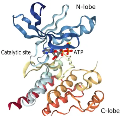
The central region is the kinase domain which is highly conserved between FAK and Pyk2 (Sasaki et al., 1995). This domain adopts a typical bi-lobal structure, very similar to that of other kinase domains, surrounding the catalytic site and the ATP binding pocket (Han et al., 2009) (Figure 6). The N-lobe contains a five-stranded beta-sheet and one alpha-helix whereas the C-lobe is mainly composed of alpha-helixes and the activation loop. Phosphorylation of Tyr-579 and Tyr-580 (Tyr-576 and Tyr-577 for FAK) within this activation loop is crucial to maximize Pyk2 kinase activity.
This kinase domain was also shown to interact directly with the retinoblastoma 1-inducible coiled-coil 1 (RB1CC1) which is supposed to be an inhibitor of Pyk2 kinase activity (Ueda et al., 2000).
Moreover, a NES was found in the C-lobe of both FAK and Pyk2 suggesting an unexpected role of the kinase domain in nucleocytoplasmic shuttling of FAK family kinases (Ossovskaya et al., 2008).
19
Figure 6. Cartoon structure of Pyk2 kinase domain co-crystalized with ATPgS. (From
Han et al., 2009a).
2.2.4. Linker 2
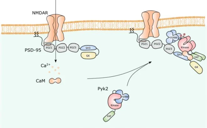
The linker 2 is located between the kinase domain and the FAT domain. This sequence contains two PR sequences, named PR2 (residues 713 to 720 in Pyk2) and PR3 (residues 855 to 860, ibid.), that mediate the interaction of Pyk2 with a number of SH3 domain-containing proteins that also interact with FAK such as p130Cas (Astier et al., 1997; Xiong et al., 1998; Lakkakorpi et al., 1999), SAPAP3, and Graf (Ohba et al., 1998; Xiong et al., 1998). More specifically in Pyk2, these PR sequences were also shown to interact with PSD-95 (Seabold et al., 2003), synapse-associated protein 102 (SAP102) (Seabold et al., 2003), ArfGAP with SH3 domain, ankyrin repeat and PH domain 2 (ASAP2) (Andreev et al., 1999), Arg kinase binding protein 2 (ArgBP2) (Haglund et al., 2004), nephrocystin (Benzing et al., 2001) and proline-rich acidic protein (PRAP) (Takahashi et al., 2003).
Additionally, a NES and a nuclear targeting sequence (NTS) were found in the linker 2 of Pyk2, playing an important role in subcellular trafficking of Pyk2 (Faure et al., 2013).
2.2.5. FAT domain
The C-terminal FAT domain of FAK family kinases is highly conserved between Pyk2 and FAK. It exhibits an anti-parallel four-helix bundle structure in both FAK (Hayashi et al., 2002; Liu et al., 2002) and Pyk2 (Lulo et al., 2009) (Figure 7). In FAK, this domain is consubstantially linked to focal adhesion targeting (Hildebrand et al., 1993; Shen and Schaller, 1999). It was reported to interact with talin (Chen et al., 1995) and paxillin (Tachibana et al., 1995; Brown et al., 1996), two proteins highly enriched in focal adhesions. Conversely, Pyk2 FAT domain was shown to interact with paxillin (Lulo et al., 2009) but not talin (Zheng et al., 1998). Moreover, it was demonstrated that the binding mechanism between Pyk2 and FAK for paxillin was different and that paxillin thus formed a much
20
more stable complex with the FAT domain of FAK than with the FAT domain of Pyk2 (Vanarotti et al., 2014). Instead of paxillin, Pyk2 FAT domain showed a preferential interaction with some proteins from the same paxillin superfamily such as leupaxin (Vanarotti et al., 2016) or the molecular scaffold and transcription co-regulator Hic-5 (Matsuya et al., 1998). These differences between FAK and Pyk2 perhaps explain why only a small proportion of Pyk2 is localized in focal contacts in most cell types.
Moreover, the FAT domain of FAK and Pyk2 associates with gelsolin, an actin binding protein, showing a common regulatory role of FAK and Pyk2 in actin cytoskeleton organization (Wang et al., 2003; Chan et al., 2009). Finally, within the FAT domain, Tyr-925 in FAK and Tyr-881 in Pyk2, when phosphorylated by Src, constitute a binding site for the adaptor Grb2 leading to the initiation of the MAP kinase signaling pathway (Schlaepfer and Hunter, 1996; Blaukat et al., 1999). In Pyk2, this phosphorylated tyrosine was also reported to be an anchoring site for the oncogenic TK c-Abl (Zrihan-Licht et al., 2004).
Figure 7. Cartoon structure of Pyk2 FAT domain. (From Lulo et al. 2009).
The structural differences between FAK and Pyk2 highlighted in this part constitute a basis for the explanation of the many differences between these two proteins.
Expression of FAK and Pyk2
FAK and Pyk2 are encoded by PTK2 and PTK2B genes, respectively, in humans (Ptk2 and
Ptk2b in mice). These genes are localized, in human, on chromosome 8 and, in mouse, on
chromosomes 15 and 14 respectively (Fiedorek and Kay, 1995; Herzog et al., 1996). Their regulation mechanisms are not well understood yet. However, FAK promotor characterization allowed to highlight regulations by several transcription factors. FAK transcription is positively regulated by NF-ƪB and N-Myc, and negatively regulated by p53 (Golubovskaya et al., 2004; Beierle et al., 2007). FAK is ubiquitously expressed during development and in the adult (Hanks et al., 1992; André and Becker-André, 1993; Furuta et al., 1995; Ilic et al., 1995; Kanazawa et al., 1995; Zachary, 1997). The highest expression of FAK is observed in the brain, lung, testis and osteoclasts (Hanks et al., 1992; André and Becker-André, 1993; Barry and Critchley, 1994; Burgaya et al., 1995; Grant et al., 1995).
21
In fetus, Pyk2 is mostly, although modestly, expressed in the brain. In adult, it is expressed in the brain but also in the lung, kidney, spleen and thymus (Avraham et al., 1995; Sasaki et al., 1995; Menegon et al., 1999). In adult brain, Pyk2 is mainly concentrated in forebrain neurons but it is also present in astrocytes (Cazaubon et al., 1997) and microglial cells (Combs et al., 1999; Tian et al., 2000).
Cellular localization of FAK family kinases
In adhering non-neuronal cells, FAK is located in focal adhesions which are sub-cellular complexes forming mechanical links between intracellular actin bundles and the extracellular matrix (Schaller et al., 1992). Pyk2 is essentially located in the perinuclear region (Sieg et al., 1998; Avraham et al., 2000) but can be found, to a lesser extent than FAK, in focal adhesions (Du et al., 2001).
In neurons, FAK is enriched is cellular body, growth cones and dendrites (Renaudin et al., 1999). Moreover, FAK can be enriched along perinuclear microtubules associated to centrosomes (Xie et al., 2003). In neurons, Pyk2 is enriched in cell body and dendritic shafts (Menegon et al., 1999; Corvol et al., 2005).
However, FAK and Pyk2 can also shuttle to the nucleus. Indeed, FAK was observed in the nucleus of cardiomyocytes under some pathological conditions (Lobo and Zachary, 2000; Yi et al., 2003). Pyk2 was also observed in the nucleus of various cell types including keratinocytes (Schindler et al., 2007), chondrocytes (Arcucci et al., 2006), and in depolarized neurons or PC12 cells (Faure et al., 2007). The dynamic of nucleocytoplasmic shuttling of FAK and Pyk2 results from the presence of multiple regulatory sequences detailed above. For instance, in the case of Pyk2, it was shown that the NES located on linker 2 was regulated by phosphorylation at residue Ser-778, a substrate of 3·,5·-cyclic adenosine
monophosphate (cAMP)-dependent protein kinase and calcineurin, a Ca2+
/calmodulin-activated protein phosphatase. The detailed mechanism of this regulation will be discussed in III.1.2.
Isoforms of FAK family kinases
Both FAK and Pyk2 present various isoforms resulting from alternative splicing of their messenger ribonucleic acid (mRNA) or from alternative transcription initiation sites (Figure 8).
22
Figure 8. FAK and Pyk2 isoforms.
2.5.1. FAK isoforms
To date, FAK possesses four additional peptides (called boxes) which can be included or not to the sequence and are named according to the number of aminoacids contained in the box: box 28, box 6, box 7 and box 3 (or PWR, based on the single letter amino acid code for three residues of this box, Pro, Trp, and Arg; FAK isoform containing this exon
is termed FAK+).
These inserts provide biological differences between isoform. Besides, every isoform present a specific expression pattern. For instance the isoform containing the boxes 3, 6
and 7 (called FAK+6,7) is predominant in neurons (Burgaya et al., 1997).
Moreover, FAK gene possesses a second promotor located in the intron following the last exon coding for the catalytic domain. Expression of the C-ter domain from this promotor produces the so-called FAK-related non-kinase (FNRK). This variant is an in vitro inhibitor of FAK as it competes with FAK for the localization to focal adhesions by its intact FAT domain (Schaller, 2010). FRNK was shown to have physiological functions. Increased expression of FRNK prevented metastatic adhesion of hepatocellular carcinoma cells (von Sengbusch et al., 2005). Moreover, FRNK negatively regulates IL-4-mediated inflammation by attenuating eosinophil recruitment in a way that seems independent of FAK pathway (Sharma et al., 2015).
2.5.2. Pyk2 isoforms
Pyk2 has an isoform lacking 42 amino acids in the linker 2 (Dikic et al., 1998; Xiong et al., 1998; Keogh et al., 2002; Kacena et al., 2012). This isoform, called Pyk2-H (hematopoietic) or Pyk2-S (short), is abundantly expressed in hematopoietic cells where it is implied in cell response to some chemokines.
23
As for FAK, another isoform of Pyk2 results from the expression of the C-terminal domain only and is termed Pyk2-related non-kinase (PRNK) (Xiong et al., 1998). This variant also inhibits Pyk2 function and was tested for this reason in many experiments. Thus, exogenous expression of PRNK could prevent myocardial fibrosis (You et al., 2015), impair CD11b/CD18-mediated phagocytosis in macrophages (Paone et al., 2016), and could reduce squamous carcinoma cells viability, migration, invasiveness and adhesion ability (Yue et al., 2015). However, PRNK may act as an inhibitor of Pyk2 only in some cell types as it does not interact with the Pyk2 partner p130Cas or Graf (Xiong et al., 1998).
Finally, Arcucci et al discovered a nuclear fragment of Pyk2 of 68 kDa in chick embryo epiphyseal chondrocytes which is not recognized by an antibody raised against the N-terminal region of Pyk2 suggesting that this fragment lacks the N-terminal region (Arcucci et al., 2006). However, neither the mechanism of formation of this fragment nor its biological function is known.
Biological functions of FAK 2.6.1. Cellular functions of FAK
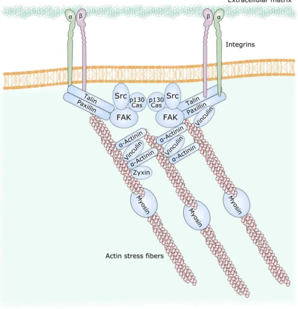
Among the adhesion sites mediated by integrins, the focal adhesions are long flat structures often located in the periphery of cells (Sastry and Burridge, 2000). They are mainly composed of integrin, paxillin, vinculin, talin and FAK.
Upon engagement of integrins by extracellular matrix, FAK is recruited to the focal adhesions and rapidly phosphorylated on tyrosine (Hanks et al., 1992; Kirchner et al., 2003; Zaidel-Bar et al., 2003). Activated FAK can interact with Src which phosphorylate many partners such as ơ-actinin which then recruits vinculin and regroups the actin bundles to the focal adhesions (Mitra et al., 2005) (Figure 9). FAK-/- fibroblasts have reduced and
immature focal adhesions with a default of binding to the actin bundles showing that FAK is essential to the function of these structures (Iliý et al., 1995; Katoh, 2017). Particularly, loss of FAK results in the persistence of old focal adhesions and in the inability to form new ones showing the importance of FAK in focal adhesion turnover (Ren et al., 2000).
24
Figure 9. Structure of focal adhesions. Adapted from Mitra et al., 2005.
FAK-/- fibroblasts also show decreased motility (Iliý et al., 1995) whereas FAK
overexpression in many cell types stimulates migration (Schlaepfer and Mitra, 2004). Cell migration can be defined as the ability of a cell to move in response to a stimulus. It participates in many physiological processes such as embryogenesis, wound healing, angiogenesis, inflammation, or in some pathological processes such as tumorigenesis and metastasis formation. Several studies demonstrated the importance of FAK in many steps of cell migration such as stimuli sensing (Katsumi et al., 2004; Schlaepfer and Mitra, 2004), lamellipodia (Tilghman et al., 2005) and filipodia (Wu et al., 2004) formation and microtubules stabilization (Siegrist and Doe, 2007). FAK thus functions as a biosensor or integrator to control cell motility by influencing the cytoskeleton and the renewing of focal adhesion and membrane protrusions (Mitra et al., 2005).
FAK was also shown to contribute to cell proliferation. Inhibition of FAK results in cell proliferation inhibition (Parsons, 2003; Owen et al., 2011; Mierke et al., 2017) whereas FAK overexpression in fibroblasts speeds it up (Zhao et al., 1998). FAK would particularly promote cyclin D expression and cyclin-dependent kinases activation during G1/S transition allowing a faster transition (Zhao et al., 2003; Bond et al., 2004; Owen et al., 2011).
25
Finally, FAK is involved in cell survival and was shown to be an anti-apoptotic kinase. In response to some pro-apoptotic stimuli such as oxidative stress, FAK can activate the PI3K/Akt pathway (Chen et al., 1996) thereby inhibiting many actors of the apoptotic machinery such as BAD or caspase-9 (Hanks et al., 2003). FAK would also be an inhibitor of the pro-apoptotic p53 (Golubovskaya and Cance, 2013). Furthermore, FAK was shown to undergo cleavage during apoptosis thereby stopping its anti-apoptotic function (Manzur et al., 2018). This function is particularly engaged for the inhibition of anoikis, a form of programmed cell death that occurs in anchorage-dependent cells when they detach from the surrounding extracellular matrix (Duxbury et al., 2004). Integrin interaction with the extracellular matrix activates FAK thereby preventing cell from anoikis (Beauséjour et al., 2012).
2.6.2. Physiological functions of FAK
FAK knockout mice is embryonic lethal around E8.5 showing a major role in development (Furuta et al., 1995). This phenotype principally results of a migration defect as detailed in the previous part (Iliý et al., 1995). Interestingly the function of FAK during early development involves more than its autophosphorylation mechanism. For example mice homozygous for a deletion of the exon that codes for Tyr-397 have a normal development up to E12.5 and die later (Corsi et al., 2009).
FAK is also important in the nervous system in vivo as shown by several studies. It is highly expressed during brain development whereas its levels decrease in the adult (Burgaya et al., 1995). Conditional deletion of FAK in the cortex demonstrated its major role in basal lamina formation (Beggs et al., 2003) and in dendritic shaft formation (Gorski et al., 2002). Conforming to its role in focal adhesion turnover, FAK was shown to be a negative regulator of axonal branching and synapse formation (Rico et al., 2004). FAK is also critical for neuronal migration and axon guidance during development (Xie et al., 2003; Li et al., 2004b; Bechara et al., 2008; Chacón et al., 2012; Kerstein et al., 2017) as well as for the control of cortical dendrite arborization (Garrett et al., 2012). Additionally, conditional deletion of FAK in Schwann cells proved the requirement of FAK for axonal myelination (Grove and Brophy, 2014).
According to these various physiological functions, FAK is involved in many diseases such as cancer (Naser et al., 2018), ischemia (Bikis et al., 2015; Gao et al., 2018), beta peptide accumulation (Zhang et al., 1996; Williamson et al., 2002) and cardiac hypertrophy (Mohanty and Bhatnagar, 2018) and is thus studied as a potential target for many molecular therapies.
Pyk2: a nRTK of FAK family
1. Regulation of Pyk2 activity in non-neuronal cells
Activation and phosphorylation of Pyk2 1.1.1. Canonical activation of Pyk2
Canonically, Pyk2 is activated in response to increases in intracellular calcium levels (Lev et al., 1995; Girault et al., 1999a; Alier and Morris, 2005; Cao et al., 2007; Schaller, 2008). Calmodulin appears to play an important role in this activation. However the mechanism
26
proposed for this activation is still not well understood. Kohno et al. proposed that
Ca2+/CaM binds to the FERM domain of Pyk2 allowing dimerization and
trans-autophosphorylation of Pyk2 (Kohno et al., 2008). Conversely, Xie et al. suggested that the
interaction site of Pyk2 with Ca2+/CaM was located in the kinase domain of Pyk2 (Hens
and DeSimone, 1995). This binding would enhance Pyk2 homodimerization. In 2011, Riggs et al. demonstrated that EGTA could not dissociate Pyk2 homodimers leading to the
hypothesis that Ca2+/CaM is required for Pyk2 homodimer formation but that this dimer
undergoes a conformational change in another state which does not include Ca2+/CaM
(Riggs et al., 2011). Following this dimerization, Pyk2 is phosphorylated on Tyr-402. Several enzyme can catalyze this reaction. In 2004, Park et al. transfected Pyk2 in SYF cells which are deprived of the Src, Yes and Fyn kinases and they observed phosphorylation of Pyk2 on Tyr-402 supporting the hypothesis of an autophosphorylation independent of Src. They also provided evidence for an autophosphorylation acting in a trans (and not cis) manner by transfecting various mutant forms of Pyk2 and observing that a kinase dead mutant form of Pyk2 was phosphorylated by Pyk2 with an active kinase domain (Park et al., 2004). However in 2016, Zhao et al. reassessed this question by proving that Src was necessary to adhesion-induced phosphorylation of Pyk2 in vitro (Zhao et al., 2016). They proposed that the phosphorylation of Pyk2 observed by Park et al. in SYF cells was due to the overexpression of Pyk2 and that, in physiological conditions, Src would be needed for the initial phosphorylation of Tyr-402 of Pyk2 and that this pre-activated form of Pyk2 could then auto-trans-phosphorylate the other Pyk2 proteins thus bringing back together the two hypothesis.
This latter mechanism may nonetheless be limited to the activation of Pyk2 upon integrin ligation. Additionally, in some cell types, Pyk2 activation seems to be independent of CaM. In cytotoxic T lymphocytes, Pyk2 phosphorylation was not inhibited by the
calmodulin inhibitor W7 (Lysechko 2010). In these cells, increased intracellular Ca2+
induces the production of reactive oxygen species, which activate the mitogen activated protein kinase (MAPK)/extracellular signal-regulated kinase (ERK) pathway, leading to Src-family kinase-dependent Pyk2 phosphorylation (Lysechko 2010). In human sperm, calmodulin inhibition even enhances Pyk2 phosphorylation by an unknown mechanism (Battistone 2014).
The mechanisms leading to the phosphorylation of Pyk2 on Tyr-402 are thus likely to be different according to the stimulus activating Pyk2. The specific mechanism of activation of Pyk2 in neurons will be discussed in III.1.1.
The phosphorylated tyrosine then constitutes a binding site for the SH2 domain of the SFKs which phosphorylate Tyr-579 and Tyr-580 within the activation loop of Pyk2 kinase domain triggering full activation (Li et al., 1999; Schlaepfer et al., 1999). SFKs also phosphorylate Tyr-881 which recruits the Grb2/Sos complex thus leading to the ERK pathway activation (Blaukat et al., 1999; Lev et al., 1999).
Other residues of Pyk2 can be phosphorylated but the biological function of these phosphorylation sites still remains unclear (Oppermann et al., 2009).
1.1.2. Regulation of Pyk2 activation by Ca2+-activated kinases
1.1.2.1. PKC
The role of PKC in Pyk2 activation is still not clear. In 1995, Lev et al. demonstrated that PKC activation was able to activate Pyk2 (Lev et al., 1995). Many studies then confirmed the involvement of PKC in Pyk2 activation. Pharmacological inhibition of PKC by
27
staurosporine and Ro31-8220 in platelet cells inhibited Pyk2 phosphorylation in response to thrombin (Ohmori et al., 1999). In accordance with this, inhibition of PKC in neurons also blocked depolarization-induced Pyk2 phosphorylation (Siciliano et al., 1996; Corvol et al., 2005). Although, the precise mechanism of PKC function in Pyk2 activation is unknown, residue(s) phosphorylated by PKC on Pyk2 have not been identified so far, and
one can hypothesize that it acts indirectly by favoring Ca2+ elevation in cells.
1.1.2.2. CaMKII
Another enzyme involved in Pyk2 regulation is CaMKII. Pharmacological inhibition by KN-62 reduced Pyk2 phosphorylation induced by depolarization but not by bradykinin (Zwick et al., 1999). Ribonucleic acid (RNA) interference of CaMKII in PC12 cells blocked KCl-induced phosphorylation of Pyk2 (Banno et al., 2008). Treatment of vascular smooth
muscle cells with the inhibitor KN-93 also inhibited H202 induced Pyk2 phosphorylation
(Bouallegue et al., 2009). Specific knockdown of CaM kinase IIƤ2 in cultured hypothalamic neurons and CaM kinase IIƢ'e in cultured gonadotroph cells both inhibited the GnRH-induced activation of Pyk2 (Okitsu-Sakurayama et al., 2019). Although the specific mechanism of Pyk2 activation by CaM kinases remains unknown, these results clearly show
a direct or indirect role for Ca2+-dependent kinases in Pyk2 regulation.
Dephosphorylation of Pyk2
Pyk2 signaling can also be regulated by dephosphorylation. The protein phosphatases involved are divided in two groups according to their substrates: the PTPs and the protein serine-threonine phosphatases (PSTPs).
1.2.1. Tyrosine phosphatases 1.2.1.1. SHP-1
Pyk2 is a substrate of SHP-1 (Kumar et al., 1999; Ganju et al., 2000). SHP-1 associates with Pyk2 (Ganju et al., 2000) and overexpression of SHP-1 inhibits Pyk2 phosphorylation (Kumar et al., 1999).
1.2.1.2. SHP-2
Pyk2 is also a substrate of SHP-2 (Chauhan et al., 2000; Tang et al., 2000). An interaction between SHP-2 and Pyk2 was shown in some cell types. This interaction was constitutive (Tang et al., 2000), induced by interleukins (Chauhan et al., 2000) or by platelets engagement (Wiiger and Prydz, 2004). The domain involved in this interaction is unknown but it does not involved SH2 (Chauhan et al., 2000; Tang et al., 2000).
1.2.1.3. PTP-PEST
The Tyr-402 and Tyr-579/580 of Pyk2 were shown to be substrates of PTPN12, also known as PTP-PEST (Davidson and Veillette, 2001; Lyons et al., 2001). PTP-PEST interacts with Pyk2 by its N-terminal PR domain (Gupta et al., 2003; Chellaiah et al., 2007). PTP-PEST deficiency results in Pyk2 hyperphosphorylation and increased cell motility in endothelial (Souza et al., 2012), cancer (Sahu et al., 2007; Li et al., 2015) and dendritic cells (Rhee et al., 2014). A PTP-PEST/Pyk2 pathway is also involved in the response of T cells (Davidson et al., 2010), macrophages fusion (Rhee et al., 2013) and osteoclasts function (Eleniste et al., 2012).
28
1.2.1.4. STEP
PTPN5, also known as striatal-enriched protein-tyrosine phosphatase (STEP) binds to and dephosphorylates Pyk2 at Tyr-402 (Xu et al., 2012). The role of this brain-specific PTP in Pyk2 regulation will be discussed further in III.
1.2.2. Ser/Thr phosphatases
In 2002, Lin et al. described the implication of Pyk2 and calcineurin, also known as PP2B, in the carbachol-induced signaling pathway (Lin et al., 2002). Treatment of cells with cyclosporine A, a calcineurin inhibitor, was shown to decrease Pyk2 phosphorylation suggesting a role of calcineurin in Pyk2 activation (Xia et al., 2006). Calcineurin dephosphorylates Pyk2 at Ser-778, a residue phosphorylated by PKA and involved in the regulation of the nuclear export motif of Pyk2 (Faure et al., 2007, 2013). This dephosphorylation leads to Pyk2 nuclear accumulation but mutation of Ser-778 do not.
SUMOylation of Pyk2
Recently, Uzoma et al. discovered that four lysines of Pyk2 could be SUMOylated thus promoting autophosphorylation of Pyk2 at Tyr-402 (Uzoma et al., 2018). This result suggests a crosstalk between phosphorylation and SUMOylation and enriches the spectrum of Pyk2 regulatory mechanisms.
S-nitrosylation of Pyk2
In 2015, Yan et al. demonstrated that oxidative stress induced S-nitrosylation of Cys-534 of Pyk2 (Yan et al., 2015). The Cys-534 mutant Pyk2 overexpression in cells reduced Pyk2 S-nitrosylation and decreased the oxygen glucose deprivation-induced Pyk2 phosphorylation suggesting a role for this post-translational modification in the regulation of Pyk2 phosphorylation.
The principal post-translational modifications of Pyk2 are compiled in Figure 10.
Figure 10. Pyk2 post-translational modifications. SUMOylation and S-nitrosylation of Pyk2
promote phosphorylation. Tyr-402 phosphorylation recruits SFKs which phosphorylate Tyr-579 and Tyr-580 leading to full activation of Pyk2, and Tyr-881 inducing the recruitment of the Grb2/Sos complex. Ser-778 phosphorylation/dephosphorylation regulate Pyk2 cytonuclear shuttling.
29
2. Pyk2 functions in non-neuronal cells
Pyk2 cellular functions 2.1.1. Cell adhesion
As FAK, Pyk2 possesses a FAT domain which targets it to the membrane and the focal adhesions. In fibroblasts, Pyk2 localizes at focal adhesions and microinjection of Pyk2 in these cells leads to reorganization of focal adhesions and cell rounding (Du et al., 2001). In macrophages as well, Pyk2 localizes within F-actin-rich membrane ruffles (Williams and Ridley, 2000). Pyk2 deletion in macrophages resulted in a delay in the formation of the leading edge lamellipodia in response to chemokine stimulation and the incapacity to detach from the substrate at the trailing edge thus reducing cell migration (Okigaki et al., 2003). Latter, Hashido et al. showed that Pyk2 was involved in cell repulsion by promoting focal adhesion disassembly (Hashido et al., 2006). In T-cells, Pyk2 activation leads to the separation of the cell with dendritic cells (Raab et al., 2017). It is thus likely that Pyk2 promotes the turnover of focal adhesions in cells. This may involve the activation of Rho, a GTPase controlling cytoskeletal morphology (Lim et al., 2008b).
2.1.2. Cell migration
In concordance with its role in focal adhesions turnover, Pyk2 also regulates migration in different cell types including vascular smooth muscle cells (Soe et al., 2009), PC-12 cells (Park et al., 2007), B-2 cells and marginal zone B cells (Tse et al., 2012), macrophages (Okigaki et al., 2003), cytotoxic T lymphocytes (Cheung and Ostergaard, 2016a), eosinophils(Zhu et al., 2008), monocytes (Watson et al., 2001) and neutrophils (Allingham et al., 2007).
Rho family of GTPases are crucial in this molecular process with Rho acting in cytoskeletal reorganization in stress fibers and Rac stimulating formation of lamellipodia (Lawson and Ridley, 2018). Pyk2 was shown to interact with these two signaling proteins. PLC-gamma1 activates Pyk2 which then activates Rac thus upregulating the formation of lamellipodia and cell migration (Choi et al., 2007). Pyk2-Rac pathway is also involved in macrophages migration (Miao et al., 2016). Rho is activated by Pyk2 in vascular smooth muscles cells (Ohtsu et al., 2005) and Pyk2 deficient macrophages were shown to have decreased Rho activation and lamellipodia contraction (Okigaki et al., 2003). By stimulating both Rac and Rho, Pyk2 thus embodies a pro-migratory role.
However, in osteoblasts, Pyk2 was shown to have an anti-migratory role as Pyk2 deletion increases osteoblast migration (Eleniste et al., 2016). Such contradictory results need further study to be understood precisely.
One should note that FAK loss is accompanied by a compensatory increase of Pyk2 expression and function (Sieg et al., 1998; Lim et al., 2008b; Weis et al., 2008) suggesting that FAK can be partly replaced by Pyk2 for the regulation of cellular migration. On the contrary, FAK does not compensate for Pyk2 in Pyk2-deficient B cells and macrophages suggesting that Pyk2 developed particular features during evolution (Guinamard et al., 2000).
30
2.1.3. Cell division
Pyk2 is also involved in the control of the cell cycle and cell proliferation. Expression of inactive Pyk2 decreases lymphocytes (Miyazaki et al., 1998) and prostatic cells (Picascia et al., 2002) proliferation.
The Wnt/Ƣ-catenin pathway is known for promoting cellular proliferation by initiating the transcription of cyclin D1 and cMyc (Klaus and Birchmeier, 2008). Pyk2
increases nuclear accumulation of Ƣ-catenin by phosphorylating Ƣ-catenin on tyrosine
residues (Otero et al., 2009), associating with the Wnt5a/Frizzled/LRP5 complex which
also activates Ƣ-catenin (Despeaux et al., 2012), and protecting Ƣ-catenin from
GSK3Ƣ-induced degradation (Zhang et al., 2014).
Pyk2 thus promotes cell proliferation and is the object of many studies focusing of its oncogenic function (Shen and Guo, 2018).
2.1.4. Cell survival
Pyk2 plays a dual role in cell survival as it can be either pro or anti-apoptotic depending on the cell type and the conditions.
In mammary epithelial cells, Pyk2 activates an anti-apoptotic pathway involving epidermal growth factor receptor (EGFR), Akt and the inhibition of p53 in response to benzo[a]pyrene exposure (Burdick et al., 2006). In thymocytes, Pyk2 is activated by interleukine-7 in an anti-apoptotic pathway involving Jak activation (Benbernou et al., 2000). In pathological conditions, Pyk2 is also associated with pathways preventing cell death. In breast cancer cells for instance, Pyk2 activates an EGFR/Akt signaling inhibiting apoptosis (Burdick et al., 2006). Inhibition of the Pyk2/Ƣ-catenin pathway in multiple myeloma cells leads to increased cell death, showing an anti-apoptotic role of Pyk2-activated pathway in this situation (Kamihara et al., 2016). After ischemia/reperfusion, Pyk2 is involved in the activation of the prosurvival signaling molecules ERK and Akt, thereby preventing excessive increases in reactive oxygen species and providing protection from injury (Miller et al., 2019).
Pyk2 also has a pro-apoptotic role. In 1997, Xiong and Parsons induced apoptosis in fibroblasts by inducing the expression of Pyk2 (Xiong and Parsons, 1997). Latter, Ueda et al. reported this effect in vitro and prevented apoptosis by the expression of the FAK family kinase-interacting protein of 200 kD (FIP200), which is a natural inhibitor of Pyk2 (Ueda et al., 2000). Inhibition of Pyk2 by the expression of its C-terminal domain also improved survival of ventricular myocytes in vivo following myocardial infarction suggesting a role of Pyk2 in cell death (Hart et al., 2008). In osteoblasts as well, kinase-deficient or phosphorylation-defective Pyk2 mutations were shown to inhibit the glucocorticoid-induced apoptosis (Plotkin et al., 2007). In the bone marrow, Pyk2 deletion increases the number of monocytes suggesting a pro-apoptotic role of Pyk2 in these cells thus regulating the turnover of these cells (Llewellyn et al., 2017). Pharmacological blockade of Pyk2 or
inhibition of Pyk2 activation by Na2S reduced myocardial infarct size in mice (Bibli et al.,
2017).
The molecular mechanisms involved may be different depending on the cell type and the environmental condition. In fibroblasts, Pyk2 may function as an inhibitor of the anti-apoptotic PI3K/Akt pathway (Xiong and Parsons, 1997). Pyk2 was also shown to phosphorylates and inhibits endothelial NO synthase (eNOS) which produces protective amounts of NO (Bibli et al., 2017). The effect of Pyk2 on cell survival is also often related to the activation of MAPKs. As mentioned in II.1.1.1, Pyk2 can recruit the Grb2/Sos
31
complex which activates the ERK pathway (Blaukat et al., 1999; Lev et al., 1999). Pyk2 also
act as an upstream regulator of the c-Jun NH2-terminal kinase (JNK) pathway through the
phosphorylation, by Src, of p130Cas and subsequent recruitment of Crk, C3G and DOCK180, leading to JNK activation (Blaukat et al., 1999). Furthermore, p38 was shown to be activated by Pyk2 through a pathway involving mitogen-activated protein kinase kinase 3 (MKK3) (Pandey et al., 1999; Sorokin et al., 2001). These signaling pathways regulate a variety of cellular activities including cell survival and death. In osteoblasts, the apoptotic effect of Pyk2 in appears to be due to the activation of JNK (Plotkin et al., 2007). In cardiomyocytes, Pyk2 activates p38 leading to the cleavage of PARP, the activation of caspase-3 and deoxyribonucleic acid (DNA) degradation (Melendez et al., 2004). Conversely, in these same cells but following ischemia/reperfusion, Pyk2 has a protective role through the activation of the ERK pathway notably (Miller et al., 2019).
2.1.5. Cell differentiation
Pyk2 is involved in keratinocytes differentiation by increasing the expression of transcription factors promoting the expression of differentiation markers (Schindler et al., 2007).
However, Pyk2 is more often associated with de-differentiation of cells. Pyk2 inhibits the differentiation of myeloid progenitors (Dylla et al., 2004) and of osteoprogenitors (Buckbinder et al., 2007). Recently, Pyk2 was confirmed to inhibit osteoblast differentiation (Posritong et al., 2018). Besides, Pyk2 inhibition promotes differentiation of vascular smooth muscle cells into a contractile phenotype thus promoting the integrity of the vascular wall (Grossi et al., 2017). This aspect of Pyk2 also contributes to its involvement in many cancers.
Physiological role of Pyk2 2.2.1. Bone physiology
Bone mass regulation results from the equilibrium between the actions of osteoblastic cells which produce bone matrix and osteoclasts which induce matrix resorption.
Pyk2-/- mice have increased bone-mass due to increased osteoblast proliferation,
migration and activity and decreased osteoclast function (Buckbinder et al., 2007; Gil-Henn et al., 2007; Eleniste et al., 2016).
In osteoclasts, Pyk2 is involved in the assembly and disassembly of podosomes which are conical, actin-rich membrane protrusions which serve as both sites of attachment and degradation along the extracellular matrix. Pyk2 interacts with structural proteins
surrounding the actin ring of the podosome such as vinculin, talin, ơ-actinin and gelsolin
(Shyu et al., 2007). Pyk2 is activated by the attachment of osteoclasts on different substrates (Duong et al., 1998). One should note that the role of Pyk2 appears to be dependent on its autophosphorylation site (Tyr-402) but not on its kinase activity. Pyk2 appears to recruit Src which then activates Cbl and PI3K allowing the development of osteoclasts polarity and function (Lakkakorpi et al., 2003; Horne et al., 2005; Buckbinder et al., 2007).
In osteoblasts, mechanical stimulations were shown to activate and re-localize Pyk2 (Guignandon et al., 2006). Pyk2 has an inhibitory function on osteoblast activity (Posritong et al., 2018). Accordingly, a research team developed a PEGDA-gelatin hydrogel containing an inhibitor of Pyk2 which promoted, in vitro, increased osteoblast activity and mineral
32
deposition possibly constituting a suitable treatment of skeletal defect (Posritong et al., 2019).
2.2.2. Vascular system integrity
In endothelial cells, Pyk2 is activated upon stimulation by different signals from integrin binding to growth factors or mechanical stimulations (Orr and Murphy-Ullrich, 2004). Neovessel formation after hind-limb ischemia is impaired in Pyk2-/- mice. Pyk2 is involved in Src/PLCgamma1 and Src/PI3-kinase/Akt pathways, leading to endothelial NO synthase phosphorylation, and modulation of vasoactive function and angiogenic response (Matsui et al., 2007). Pyk2 would actually have an inhibitory action on eNOS thus blocking its vasodilatation activity (Fisslthaler et al., 2008).
Pyk2 is an important determinant of thrombin-induced endothelial inflammation. Thrombin stimulation of endothelial cells was shown to activate IKK, through Pyk2, and to promote the release and the transcriptional capacity of RelA/p65 (Soni et al., 2017).
Pyk2 may also have a role in endothelial integrity by modulating cell adhesion. VE-cadherin loss of function was shown to induce tyrosine phosphorylation of Ƣ-catenin by Pyk2 (van Buul et al., 2005). Pyk2 phosphorylation of VE-PTP constitutes a docking site for Src which phosphorylates VE-cadherin leading to endothelial barrier dysfunction (Soni et al., 2017).
2.2.3. Immune system function
Pyk2 is expressed in cells of hematopoietic origin where it controls the molecular mechanisms of migration and activation.
In macrophages, Pyk2 is activated by FcƣR stimulation during phagocytosis (Kedzierska et al., 2001). Both FAK and Pyk2 are involved in phagocytosis (Bruce-Staskal et al., 2002; Hudson et al., 2005). However, the two proteins do not display the same function. Pyk2 activation is sufficient for phagocytosis of YadA expressing bacteria whereas FAK is sufficient for phagocytosis of invasin expressing bacteria (Owen et al., 2007).
Pyk2 is also involved in cytokines secretion by macrophages such as 1Ƣ and IL-18 (Välimäki et al., 2013). Pyk2 phosphorylates the inflammasome adaptor protein ASC which participates in speck formation that leads to caspase-1 activation and secretion of IL-1Ƣ and IL-18 (Chung et al., 2016). Pyk2 might thus be involved in HIV-associated dementia characterized by the infiltration of macrophages in perivascular sites of the central nervous system and the production of IL-1Ƣ in the brain (Cheung et al., 2008).
As detailed above, Pyk2 is implicated in cell migration. Particularly, Pyk2 plays a crucial role in the migration of macrophages (Owen et al., 2007), T cells (Cheung and Ostergaard, 2016b), marginal zone B cells (Tse et al., 2009), platelets (Cipolla et al., 2013), eosinophils (Zhu et al., 2008), and neutrophils (Kamen et al., 2011). The role of Pyk2, probably involving the RhoA-ROCK signaling pathway, is underlined by the inability of Pyk2 deficient cells to detach form the rear (Okigaki et al., 2003; Cheung and Ostergaard, 2016a).
The production of reactive oxygen species (ROS) by macrophages (Lin et al., 2008) and neutrophils (Zhao and Bokoch, 2005) also involves Pyk2 activity and is necessary to inflammatory response of these cells.
2.2.4. Kidney function
Pyk2 plays an important role in acidobasic homeostasis. Acidosis activates Pyk2 in kidney cells, promoting MAPK- and JNK-dependent transcription and activation of the NHE3
33
transporter, thus increasing acid secretion by these cells (Gluck, 2004; Li et al., 2004a; Preisig, 2007).
Pyk2 is also involved in the acid-stimulated citrate reuptake by NaDC-1 cotransporter in kidney cells (Zacchia et al., 2018).
Diabetes was also shown to induce a Pyk2-dependent inactivation of GSK3Ƣ thus causing elevated protein synthesis and kidney hypertrophy (Mariappan et al., 2014).
2.2.5. Sperm capacitation
Northern blot and immunoprecipitation analysis demonstrate that, among germinal cells, Pyk2 is more abundant in spermatocytes and spermatids (Chieffi et al., 2003). Inhibition of Pyk2 results in decreased sperm motility, acrosome reaction and ability to penetrate eggs
(Battistone et al., 2014). Pyk2 may phosphorylate CatSper, a Ca2+ channel, thus promoting
the Ca2+ wave necessary to sperm capacitation (Brukman et al., 2019).
2.2.6. Generation of Pyk2 knockout mice
Genetic knockout is a useful approach to better study the physiological function of a protein in an organism.
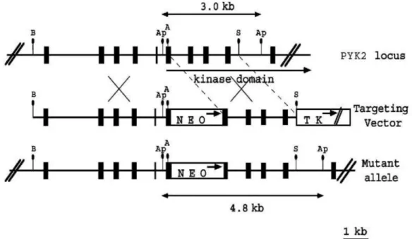
The Schlessinger group was the first to develop Pyk2 knockout mice by inserting a Neo expression cassette into the 5Ȩ portion of Pyk2 sequence encoding for the protein tyrosine kinase domain as shown in Figure 11 (Okigaki et al., 2003). In contrast to the embryonic lethality in FAK-deficient mice, Pyk2 knockout mice are viable and fertile (Guinamard et al., 2000; Okigaki et al., 2003). They display a mild immunological phenotype with reported defects in immune cells activation and mobility as described in II.2.2.3 (Guinamard et al., 2000; Okigaki et al., 2003; Cipolla et al., 2013).
Figure 11. Generation of Pyk2 knockout by Okigaki et al., 2003. Schematic diagrams and
partial restriction maps of Pyk2 locus, the Pyk2 targeting vector, and the predicted structure of the Pyk2 locus after homologous recombination with targeting vector. The filled box represents Pyk2 coding sequence, the open box indicates the Neo expression cassette and thymidine kinase (TK) expression cassette. The arrows in open box indicate the direction of transcription. A, Ap, B, and S represent AccI, ApaI, BamHI, and SacI, respectively. From Okigaki et al., 2003.