The cell-cell junctions of mammalian testes: I. The
adhering junctions of the seminiferous epithelium
represent special differentiation structures
The MIT Faculty has made this article openly available. Please share
how this access benefits you. Your story matters.
Citation
Domke, Lisa M., Steffen Rickelt, Yvette Dörflinger, Caecilia Kuhn,
Stefanie Winter-Simanowski, Ralf Zimbelmann, Rina
Rosin-Arbesfeld, Hans Heid, and Werner W. Franke. “The Cell–cell
Junctions of Mammalian Testes: I. The Adhering Junctions of
the Seminiferous Epithelium Represent Special Differentiation
Structures.” Cell and Tissue Research 357, no. 3 (June 8, 2014):
645–665.
As Published
http://dx.doi.org/10.1007/s00441-014-1906-9
Publisher
Elsevier B.V.
Version
Final published version
Citable link
http://hdl.handle.net/1721.1/96531
Terms of Use
Creative Commons Attribution
REGULAR ARTICLE
The cell
–cell junctions of mammalian testes: I. The adhering
junctions of the seminiferous epithelium represent special
differentiation structures
Lisa M. Domke
&Steffen Rickelt
&Yvette Dörflinger
&Caecilia Kuhn
&Stefanie Winter-Simanowski
&Ralf Zimbelmann
&Rina Rosin-Arbesfeld
&Hans Heid
&Werner W. Franke
Received: 14 March 2014 / Accepted: 28 April 2014 / Published online: 8 June 2014 # The Author(s) 2014. This article is published with open access at Springerlink.com
Abstract The seminiferous tubules and the excurrent
ducts of the mammalian testis are physiologically
separat-ed from the mesenchymal tissues and the blood and
lymph system by a special structural barrier to paracellular
translocations of molecules and particles: the
“blood–testis
barrier”, formed by junctions connecting Sertoli cells with
each other and with spermatogonial cells. In combined
biochemical as well as light and electron microscopical
studies we systematically determine the molecules located
in the adhering junctions of adult mammalian (human,
bovine, porcine, murine, i.e., rat and mouse) testis. We
show that the seminiferous epithelium does not contain
desmosomes, or
“desmosome-like” junctions, nor any of
the desmosome-specific marker molecules and that the
adhering junctions of tubules and ductules are
fundamen-tally different. While the ductules contain classical
epithe-lial cell layers with E-cadherin-based adherens junctions
(AJs) and typical desmosomes, the Sertoli cells of the
tubules lack desmosomes and
“desmosome-like” junctions
but are connected by morphologically different forms of
AJs. These junctions are based on N-cadherin anchored in
cytoplasmic plaques, which in some subforms appear thick
and dense but in other subforms contain only scarce and
loosely arranged plaque structures formed by
α- and
β-catenin, proteins p120, p0071 and plakoglobin, together
with a member of the striatin family and also, in rodents,
the proteins ZO-1 and myozap. These N-cadherin-based
AJs also include two novel types of junctions: the
“areae
adhaerentes
”, i.e., variously-sized, often very large cell-cell
contacts and small sieve-plate-like AJs perforated by
cytoplasm-to-cytoplasm channels of 5–7 nm internal diameter
(“cribelliform junctions”). We emphasize the unique character
of this epithelium that totally lacks major epithelial marker
molecules and structures such as keratin filaments and
des-mosomal elements as well as EpCAM- and PERP-containing
junctions. We also discuss the nature, development and
pos-sible functions of these junctions.
Keywords Adherens junction . Desmosomes . Sertoli cells .
Seminiferous tubules . Areae adhaerentes . Cribelliform
junctions
Electronic supplementary material The online version of this article (doi:10.1007/s00441-014-1906-9) contains supplementary material, which is available to authorized users.
L. M. Domke
:
S. Rickelt:
Y. Dörflinger:
C. Kuhn:
S. Winter-Simanowski
:
R. Zimbelmann:
H. Heid:
W. W. Franke (*)
Helmholtz Group for Cell Biology, German Cancer Research Center (DKFZ), Im Neuenheimer Feld 280, 69120 Heidelberg, Germany e-mail: w.franke@dkfz.de
L. M. Domke
Brandenburg University of Technology, Senftenberg, Germany
C. Kuhn
:
W. W. FrankeProgen Biotechnik GmbH, Heidelberg, Germany R. Rosin-Arbesfeld
Department of Anatomy, Sackler School of Medicine, Tel-Aviv University, Tel-Aviv, Israel
Present Address: L. M. Domke
Whitehead Institute for Biomedical Research, Massachusetts Institute of Technology (MIT), Cambridge, MA, USA Present Address:
S. Rickelt
David H. Koch Institute for Integrative Cancer Research,
Massachusetts Institute of Technology (MIT), Cambridge, MA, USA Cell Tissue Res (2014) 357:645–665
Introduction
A unique type of epithelium-like tissue element, in which
somatic cell proliferation and differentiations occur side by
side with meiotic divisions and male germ cell differentiation
(spermatogenesis), is the tubulus seminiferus of the
mamma-lian testis. Here, basal lamina-founded somatic cells, the
“Sertoli cells”, are laterally connected to each other and to
spermatogenic cells with multiple cell-to-cell attachment
structures (Dym and Fawcett
1970
; Dym
1977
; Russell and
Peterson
1985
; Pelletier
2001
). Moreover, the Sertoli and
the germ cells form an obviously tight-fitting barrier for
paracellular translocations of molecules and particles, the
tight junction-based blood–testis barrier (BTB) and support
the development of the germ cells, at least up to the point
of spermatid differentiation, in specific Sertoli cell
inden-tations (“pockets”) harboring the spermatid heads (e.g.,
Dym
1977
; Vogl et al.
1991
,
2008
,
2013
; Southwood and
Gow
2001
; Wong and Cheng
2005
). Although prima facie the
mature Sertoli cell layer looks like a typical epithelium, these
cells are profoundly different from all other epithelial cells
with respect to their biochemical and morphological
compo-nents as well as their general architecture. This holds in
particular for the absence of intermediate-sized filaments
(IFs) of the keratin type, for the presence of vimentin IFs
(Franke et al.
1979
; see also Spruill et al.
1983
; Paranko and
Virtanen
1986
; Franke et al.
1989
; Stosiek et al.
1990
; Steger
and Wrobel
1994
; Steger et al.
1994
), for the additional
occurrence of neurofilaments in human Sertoli cells (see,
e.g., Davidoff et al.
1999
) and for the presence of various
types of specific adherens junctions (AJs) between the
Sertoli cells (homotypic) and between Sertoli cells and
spermatogonial cells in the basal part of the Sertoli cells
(heterotypic-basolateral junctions) and between the adluminal
pockets of the Sertoli cells and the spermatid heads
(heterotypic-apical junctions).
Originally, in the early years of transmission electron
microscopy, certain AJs connecting Sertoli cells with each
other or with spermatogonial cells had been seen as typical
desmosomes or as desmosome-related and thus classified as
“desmosomes”, “rudimentary desmosomes”, or
“desmosome-like junctions” (e.g., Nicander
1967
; Altorfer et al.
1974
;
Russell
1977a
,
b
,
c
; Connell
1978
; Nagano and Suzuki
1978
; Osman
1978
; Osman and Plöen
1978
). Although our
laboratory has repeatedly reported the total absence of both
specific desmosomal structures and desmosomal marker
mol-ecules from Sertoli cells of the mature mammalian testis for
more than three decades (e.g., Franke et al.
1979
,
1981
,
1982
,
1983
,
1986
;
1989
; Mueller and Franke
1983
; Moll et al.
1986
;
Schmelz et al.
1986
; Theis et al.
1993
; see also Pelletier and
Byers
1992
; Schäfer et al.
1994
; Nuber et al.
1995
; Mertens
et al.
1996
), other authors have claimed, again and again, the
occurrence of desmosomes or
“desmosome-like” junctions in
Sertoli cells of mature mammals active in spermatogenesis
(Vogl et al.
2008
; Li et al.
2009
; Lie et al.
2010
,
2011
; Cheng
et al.
2011
; Mruk and Cheng
2011
; see Table
1
and Electronic
Supplementary Material, Table
S1
). Because of this long and
still ongoing controversy, the potential diagnostic value of
molecular markers in histology and pathology and also in
view of the worldwide interest in the development of male
contraceptive agents based on the interference with cell–cell
interactions in the testis (e.g., O’Donnell et al.
2000
; Cheng
and Mruk
2002
,
2011
,
2012
; Lee and Cheng
2004
; Mruk and
Cheng
2004a
,
b
; Wong et al.
2005
; Xia et al.
2005
; Lee et al.
2009
; Mok et al.
2012
,
2013a
,
b
), we decided to study the cell
biology of the tubuli seminiferi of diverse mammalian species.
We were particularly interested in the connections and
inter-actions of Sertoli cells with each other and with the
spermato-gonial cells. Therefore, we analyzed these interactions in
ultrastructural and molecular biological detail using the
Table 1 Reports claiming that desmosomes or desmosome-like junc-tions or desmosome-specific molecules occur in the tubuli seminiferi of mammalian testes (only references since 1983 are considered here, as identifications using molecule-specific antibodies against desmosomal components have been generally available since that year); for completereferences, see Electronic Supplementary Material, TableS1
Alves et al. (2013)
Bergmann et al. (1984)
Chapin et al. (2001)
Cheng and Mruk (2002,2011,2012)
Cheng et al. (2011, 2013)
Goossens and van Roy (2005) Johnson and Boekelheide (2002a,b)
Kopera et al. (2010)
Lee and Cheng (2004)
Lee et al. (2009)
Li JCH et al. (2001)
Li MWM et al. (2009, 2010)
Lie et al. (2010,2011)
Mok et al. (2013a)
Morrow et al. (2010)
Mruk and Cheng (2004a,b,2011)
Mruk et al. (2013) Mullholland et al. (2001)
Russell and Peterson (1985)
Su et al. (2013)
Vogl et al. (2000,2008, 2013a,b)
Wine and Chapin (1999)
Wong and Cheng (2005)
Wong et al. (2004,2005)
Xia et al. (2005)
Yan et al. (2007)
Yan and Cheng (2005) Zhang et al. (2005a,b)
epithelium of the excurrent duct system as parallel controls.
The results concerning the adhering junctions of the tubuli
seminiferi are presented in this report. Results on gap and
tight junctions, other cell–cell adhesion structures, the
junctions in the peritubular and interstitial cells of the
testis, junctional and cytoskeletal structures identified in
early developmental stages, in aged and pathological
forms, and in cultured Sertoli cells will be dealt with in
further publications.
Materials and methods
Tissues and cells
Bovine testicular tissue samples were obtained in the regional
slaughterhouse (Mannheim, Germany). Murine (rat and
mouse) testes from sexually mature animals were obtained
from the Central Animal Laboratory of the German Cancer
Research Center (Heidelberg, Germany; for details, see
Franke et al.
2006
,
2013
). In addition, testis tissue specimens
from 3-year-old boars of German landrace pigs were provided
by the Institute of Farm Animal Genetics
(Friedrich-Loeffler-Institute, Mariensee, Germany; cf. Rickelt et al.
2011
).
Cryopreserved and aldehyde-fixed human testis samples were
obtained from surgical material taken, examined for
diagnos-tic pathology and processed in compliance with the
regula-tions of the Ethics Committees of the Universities of
Heidelberg and Marburg (Germany; see also Langbein et al.
2003
).
In general, the tissue samples were fixed at ca. 4 °C or at
room temperature in most cases with 4 % formaldehyde,
freshly prepared from paraformaldehyde, or in
phosphate-buffered saline (PBS), with or without millimolar additions
of divalent cations and embedded in paraffin. Alternatively,
the tissues was snap-frozen in isopentane that had been
precooled in liquid nitrogen and then stored at
−80 °C
until use (see references cited above). Suitable frozen tissue
samples were used for preparations of
“semi-thin” cryostat
sections (ca. 5–15 μm thick), or thin sections for
immunoflu-orescence microscopy (see below) and analyses by
SDS-polyacrylamide gel electrophoresis (SDS-PAGE) of
polypep-tides and immunoblotting (see below).
Antibodies
The antibodies (Abs) used in immunofluorescence and
immunoelectron microscopy or in immunoblotting analyses
of gel-electrophoretically-separated polypeptides are listed in
Supplementary Table
S2
. Antigen-bound primary Abs were
visualized with goat anti-rabbit, goat anti-guinea pig, or goat
anti-mouse IgG (H+L) secondary Abs coupled to Cy3 (Dianova,
Hamburg, Germany) or Alexa 488 (MoBiTec, Göttingen,
Germany). For immunoblot analysis,
horseradish-peroxidase-conjugated secondary Abs were used (Dianova).
Gel electrophoresis and immunoblotting
Proteins of tissue samples, including microdissected ones,
were analyzed by SDS-PAGE, followed by immunoblotting,
as previously described (Rickelt et al.
2010
,
2011
; Straub et al.
2011
; Pieperhoff et al.
2012
). Usually 100–200 cryostat
sec-tions of ca. 5–15 μm thickness were collected and proteins
of tissue lysates were solubilized in the same sample
buffer (cf. Pieperhoff et al.
2012
; Franke et al.
2013
).
After homogenization, the tissue lysates were heated to—
and kept at—ca. 95 °C for 4–5 min, then centrifuged at
15,000g for 10 min. Both the supernatant and the pelleted
proteins and glycoproteins were subjected to SDS-PAGE,
followed by transfer to polyvinylidene difluoride membranes
(ImmobilonP; Millipore, Bedford, MA, USA). For immunoblot
analyses, horseradish-peroxidase-conjugated secondary Abs
were applied in combination with an enhanced
chemilumines-cence system (ECL; Fisher Scientific, Schwerte, Germany).
Immunofluorescence microscopy
The methods used for immunofluorescence microscopy have
been described (Langbein et al.
2003
; Franke et al.
2006
,
2013
,
2014
; Rickelt et al.
2008
,
2011
; Pieperhoff et al.
2012
;
Rickelt
2012
). The frozen tissue sections were mounted on
coverslips, air-dried and fixed for 5 or 10 min in
−20 °C
acetone. The specimens were rehydrated in PBS and
pre-incubated in PBS containing 0.2 or 0.3 % Triton X-100 for
5 min before application of the primary Abs. In parallel, the
tissue samples were fixed for 5–7 min in PBS (pH 7.4)
con-taining 2 % formaldehyde, freshly prepared from
paraformal-dehyde and the cells were permeabilized with PBS containing
0.1 or 0.2 % saponin (same pH value; 5 min), followed by
exposure to the specific primary Abs in PBS for 1 h.
Following several washes in PBS for 5–10 min each, the
samples were exposed to the specific secondary Abs for
30 min. After two or three washes for 5–10 min in PBS, the
cell preparations or cryosections were rinsed in distilled water,
fixed for 1 or 5 min in ethanol and mounted in Fluoromount G
(Southern Biotech; obtained through Biozol Diagnostica,
Eching, Germany). Finally, immunofluorescence microscopic
images were recorded with an Axiophot II photomicroscope
(Carl Zeiss, Jena, Germany), equipped with an AxioCam HR
(Carl Zeiss). For confocal laser scanning microscopy, a Zeiss
LSM 510 Meta instrument was used.
Electron and immunoelectron microscopy
The electron and immunoelectron microscopy protocols were
essentially as described (see Langbein et al.
2003
; Franke et al.
2006
,
2013
; Rickelt et al.
2008
,
2011
). For immunoelectron
microscopy of cryostat sections, the tissue samples were fixed
in PBS containing 2 or 3 % formaldehyde for 5
–7 min and
permeabilized with PBS containing 0.1 % saponin (3–5 min),
followed by incubation with primary Abs for at least 2 h. After
three washing steps, the samples were incubated with
second-ary Abs conjugated with 1.4-nm gold particles (Nanogold;
Biotrend, Cologne, Germany) for 4 h, followed by silver
enhancement for various periods of time (5, 7, or 9 min).
Electron micrographs were taken at 80 kV in an EM 900 or
EM 10 (Carl Zeiss) instrument.
Results
For comparison and control: the epithelia of the excurrent
ducts
In all species examined, we used tissue samples from various
portions of the excurrent ducts, which are known to contain
typical simple or columnar epithelia, as controls of the
methods applied and to determine the specificity of the
antibodies used in immunocytochemistry of the seminiferous
tubules (for references, see, e.g., Dym
1974
,
1976
,
1977
;
Kasper and Stosiek
1989
; Cyr et al.
1995
,
2007
; Piomboni
1997
; Pelletier
2001
; DeBellefeuille et al.
2003
). Frequently,
it was possible for us to study testicular tissue containing
seminiferous tubules in parallel with preparations of excurrent
duct tissues, including the epididymis, from the same animal.
Biochemical results
When we compared the adhering junction proteins of specific
intercepts or portions of the excurrent system with those of the
seminiferous tubule system by gel electrophoresis and
immu-noblot identification, we observed striking differences
(Fig.
1
). Certain cytoskeletal control proteins such as actin
and vimentin (Fig.
1a, j
), as well as constituents known to
occur in all kinds of AJs such as the armadillo-type plaque
proteins
β-catenin, protein p120 and plakoglobin, which is
known also to occur in desmosomes (Cowin et al.
1986
), were
detected in all samples (e.g., Fig.
1f, g
). Other molecules
showed marked differences between the AJs of the excurrent
duct and the seminiferous tubule epithelia. In all species
examined, E-cadherin was found as a major component in
all excurrent duct regions but was not detected in the
semini[-]ferousrous epithelium (e.g., Fig.
1d, e
; for a special
minor non-epithelial cell type, see below). Vice versa,
N-cadherin was present as a major component in the
seminifer-ous tubule epithelia but was absent in the excurrent duct
epithelia (e.g., Fig.
1b, c
). No other AJ cadherins examined
were found in either kind of epithelia, while the AJ plaque
proteins,
α- and β-catenin, p120, p0071, myozap and a
mem-ber of the striatin family, were abundantly present in both
excurrent duct and seminiferous tubule epithelia (see Table
2
for specific comments).
The results for desmosomal proteins of both kinds were
very impressive and clear: As a representative example of
the desmosomal cadherins, the absence of desmoglein
Dsg-2 in the seminiferous tubules is shown in Fig.
1i
and the absence of the plaque protein desmoplakin in
Fig.
1h
. Negative results in the seminiferous tubules were
also obtained for desmocollin Dsc-2 and plakophilin Pkp-2
as well as molecules Dsg-1 and Dsg-3, Dsc-1 and Dsc-3
and Pkp-1 and Pkp-3 (not shown). In contrast, certain
desmosomal molecules (Dsc-2, Dsg-2, Pkp-2 and
desmo-plakin) were abundantly present in all the epithelia of the
excurrent duct system (Table
2
).
The transmembrane proteins EpCAM and PERP, which
were not detected in the seminiferous epithelia, were regular
constituents of the subapical region close to the zonula
adhaerens AJs of the excurrent ducts (these molecules will
be specifically dealt with in one of the next publications in this
series).
Immunofluorescence microscopy results
Excurrent duct epithelia
The results revealed in all the epithelia of the excurrent duct
system a keratin IF cytoskeleton and an abundance of
desmo-somes that could generally be demonstrated by
co-immunolocalization of the corresponding transmembrane
and the cytoplasmic plaque molecules (Table
2
; an example
for desmoglein Dsg-2 and desmoplakin is shown in Electronic
Supplementary Material, Fig.
S1
). By contrast, differential
localization of E-cadherin and N-cadherin is seen in the tissues
in which N-cadherin was selectively absent in the epithelia
and only seen as a very minor element in some of the
inter-stitial cells (Fig.
2
). As to the cytoplasmic plaque proteins of
the epithelial AJs, we noted that some occurred in all junction
structures, i.e., zonula adhaerens plus fasciae adhaerentes and
puncta adhaerentia, such as the catenins and protein p120
(Fig.
3a–a
''), whereas others such as proteins myozap, p0071
and a member of the striatin family appeared to be largely
restricted to the subapical zonula adhaerens (see, e.g.,
Fig.
3b
). Protein PERP was a particularly prominent
compo-nent associated with the zonula adhaerens structure (not
shown; for comparison with other epithelia, see Franke et al.
2013
) and specific zonula immunolocalizations were also
noted with some of the antibodies directed against protein
LUMA (cf. Franke et al.
2014
). The massive aggregates of
spermatozoa frequently seen in the lumina of these ductules
were totally negative for the various junctional proteins
ex-amined (e.g., Figs.
2
,
3
).
Seminiferous tubule epithelium
In correspondence with the biochemical results (Fig.
1
), the
tubule cells of the layer lining the adluminal side of the basal
lamina were positive for both vimentin IFs and N-cadherin AJs
(Electronic Supplementary Material, Figs.
S2
,
S3
), the latter in
confirmation of Cyr et al. (
1992
,
1993
), Newton et al. (
1993
) and
Byers et al. (
1994
) but negative for keratins (for references see
“
Introduction
”), E-cadherin and all the other cadherins examined
(Figs.
4
,
5
,
6
, and
7
), including P-cadherin, VE-cadherin and
cadherin-11 (Table
2
; see also Cyr et al.
1992
). In addition, a
surprising positive E-cadherin reaction was detected in a thin
non-epithelial cell layer of the interstitium of the bovine testis,
a so-called
“myoid” cell layer surrounding the tubules
(red-stained cell-cell junctions in Fig.
4a–a
'''), a layer also
positive for keratins 8 and 18 (cf. Electronic Supplementary
Material, Fig.
S2
).
A very clear result of these immunolocalization
experi-ments was that no significant reactions were seen for any of
the cadherin family glycoproteins of either the desmoglein and
the desmocollin group, in particular not for Dsg-2 and Dsc-2
(see, e.g., Electronic Supplementary Material, Fig.
S4a
). Also
negative were the reactions for the desmosomal plaque
pro-teins desmoplakin (Fig.
5
) and plakophilin Pkp-2 (e.g.,
Electronic Supplementary Material, Fig.
S4b
), or any other
member of the plakophilin group (Table
2
). These negative
results were obtained for all desmosomal marker molecules
using antibodies that abundantly demonstrated positivity in all
the species studied.
The N-cadherin-positive, punctate or elongated junction
structures were positive for several of the cytoplasmic plaque
proteins of the armadillo family such as
β-catenin (Fig.
5
),
plakoglobin (not shown; see Table
2
and Byers et al.
1994
),
proteins p0071 (Figs.
6
and
7
) and p120. They were also
immunostained with antibodies to the actin-interacting protein
α-catenin (for co-localization with β-catenin see Electronic
Supplementary Material, Fig.
S5
; see also Table
2
) as well as
for the proteins myozap and striatin (Figs.
7
and
8
; Table
2
).
On the other hand, the Sertoli cell membranes were totally
negative for the transmembrane, junction-associated proteins
Fig. 1 Identification of proteins and glycoproteins of cell-cell adheringjunctions in dissected tissue parts of mammalian testis, enriched in seminiferous tubules, or excurrent duct tissues, demonstrated after SDS-PAGE of total protein lysates by immunoblotting with specific antibodies
a Antibodies againstβ-actin identify a single polypeptide band at 43 kDa
in cultured human HaCaT keratinocytes (lane 1 in all blots shown, i.e. a–
j), bovine testis (lane 2) and epididymis (lane 3), boar testis (lane 4) and boar excurrent duct-containing tissue (lane 5), rat testis (lane 6) and rat excurrent duct tissue (lane 7). b Antibodies against N-cadherin recognize this glycoprotein in testicular tissue of bull (lane 2), boar (lane 4) and rat (lane 6) but not to a significant extent in excurrent duct tissues of bovine (lane 3), porcine (lane 5) and murine (rat, lane 7) origin. c As in (b), N-cadherin is detected to comparable intensities in near-equal amounts of testicular tissue of bovine (lane 2), rat (lane 3), mouse (lane 4) and human (lane 5) origin. d In a mutually exclusive way, E-cadherin is recognized in the excurrent duct tissues of bovine (lane 3), porcine (lane 5) and murine (rat, lane 7) origin but not in the corresponding testicular tissues enriched
in seminiferous tubules (material in lanes 2, 4 and 6 of bovine, porcine, and murine origin). e E-cadherin is not detected in tissue material from microdissected testicular tissues enriched in seminiferous tubules of bovine (lane 2), rat (lane 3), mouse (lane 4) and human (lane 5)
origin. f The major plaque protein of AJs, β-catenin, is detected in
all these tissues (lanes 2–7 as in a, b and d), although in different
intensities (note the weak reaction in lanes 5 and 6). g Similarly, the AJ plaque protein p120 is found in all samples, although at a rather low intensity in boar excurrent duct tissues (lane 5; note, however, a major proteolytic degradation product of about 62 kDa in lanes 3, 5 and 7). h In comparison with human HaCaT keratinocytes (lane 1), desmo-plakin is not recognized in bovine (lane 2), rat (lane 3), mouse (lane 4,) and human (lane 5) seminiferous tubules of testis. i Correspondingly, desmoglein Dsg-2 (HaCaT cells, lane 1) is not detected in bovine (lane 2), rat (lane 3), mouse (lane 4) and human (lane 5) testicular tissue contain-ing seminiferous tubules. j Control showcontain-ing the presence of vimentin in
all tissues shown in (h–j)
PERP and EpCAM (Table
2
; for comparison with positively
stained cells, see, e.g., Rickelt et al.
2011
; Pieperhoff et al.
2012
; Franke et al.
2013
).
Electron microscopy
As some of the AJ-related structures in the seminiferous
tubules are rather small, others very complex or extremely
large and some of them also appear to be organized in
cell-type-specific ways, they were examined in detail by
transmis-sion electron microscopy and immunoelectron microscopy.
Only the AJ-type structures connecting Sertoli cells with each
other or with spermatogenic cells will be dealt with in the
present report.
It is a striking observation that in well-fixed, optimally
preserved tubuli seminiferi, the cells are intimately associated
with each other by junction-like plasma membrane structures
for very large proportions of the cell surface, often exceeding
50 % (e.g., Fig.
9a–a
''', b). This rather consistently close and
parallel contact with a membrane-to-membrane distance of 8–
20 nm often appears to be accompanied by small and sparse
submembranous densities, which are only loosely associated
with the specific membrane regions (see, e.g., Fig.
9b–d
).
Such extended, close and parallel junctions are seen in both
homotypic Sertoli cell associations as well as in Sertoli cell
associations with spermatogenic cells.
In some regions of such plaque-based cell–cell associations,
local differences of the intermembranous space are notable
(Figs.
9d
,
10a–c
) and, in some of these junction-like structures,
a linear punctate midline array of granular-looking
“dots” 2–
5 nm in diameter is resolved (e.g., Fig.
10b, b
', c; see also the
junction denoted by an arrowhead in Fig.
10d
'''). Closely
spaced, small (diameters up to 500 nm) AJ-like structures
with rather irregularly contoured, electron-dense plaques
appear as almost regular arrays, interrupted only by some
direct membrane-membrane
“touch sites” of an as yet
un-identified molecular nature (Fig.
10d
, bottom: the series of
AJ contacts between cell 1 and cell 2; for details, see also
Fig.
10d
'', d'''). Immunolocalization electron microscopy
ex-periments published by Byers et al. (
1991
) and Pelletier and
Byers (
1992
) have indicated the presence of protein ZO-1 at
these sites that we can confirm. On the other hand, ZO-1–3
proteins cannot be considered unequivocal markers of tight
junctions (TJs) as they have been identified in both TJ and
AJ structures (for references. see, e.g., Franke
2010
).
Some of these close membrane-to-membrane associations
(Fig.
10a–d
show many such cell–cell contact regions
be-tween several Sertoli cell processes, numbered 1–5) are
ac-companied in a conspicuous way by so-called
“ectoplasmic
specializations” (ES; for morphological and
immunocyto-chemical references, see, e.g., Dym and Fawcett
1970
;
Russell
1977c
; Franke et al.
1978
; Russell and Peterson
1985
; Vogl
1989
; Vogl et al.
1991
,
2000
,
2008
; Mruk and
Cheng
2004a
; Wong et al.
2005
; Yan et al.
2007
; Kopera et al.
2010
; Cheng and Mruk
2012
; Qian et al.
2013
). These are
paracrystalline actin microfilament bundles that, in some
re-gions, are directly connected to the inner side of the plasma
membrane by lateral, rather closely spaced, up to 4-nm-thick
cross-bridge structures (see, e.g., the inset in Fig.
10d
'),
whereas other portions of such bundles may protrude into
the cytoplasm (see, e.g., the cells labelled nos. 3 and 4 in
Fig.
10d
). Microfilament bundles of such ES structures,
Table 2 Homotypic adhering junctions: results of immunolocalizationexperiments (summary for all species examined)
Molecule Sertoli–Sertoli and Sertoli–
Spermatogonia cell junctions
Excurrent duct epithelia Transmembrane glycoproteins (Cadherins)
E-cadherin − + VE-cadherin − − N-cadherin + − P-cadherin − − Cadherin-11 − − Desmoglein 1 (Dsg-1) − − Desmoglein 2 (Dsg-2) − + Desmoglein 3 (Dsg-3) − − Desmocollin 1 (Dsc-1) − − Desmocollin 2 (Dsc-2) − + Desmocollin 3 (Dsc-3) − −
Other transmembrane molecules
EpCAM − +
Protein PERP − +
Cytoplasmic plaque proteins
Desmoplakin I+II − + Plakophilin 1 − − Plakophilin 2 − + Plakophilin 3 − + β-Catenin + + Protein p120 + + Protein p0071 + + Plakoglobin + + Neurojungin − − α-Catenin + + Protein ZO-1 (+/−)a + Myozap (+/−)a + Striatinb + + Protein LUMA – + a
For unexplained reasons, we have consistently found reactions of the plaque proteins myozap and ZO-1 in adherens junctions of Sertoli cells in the rodent testes but not in the other species examined in which only the adherens junctions of the endothelial and some interstitial mesenchymal cells showed myozap- and/or ZO-1-positive junctions
b
Whether it is striatin or a closely related member of the striatin family of proteins is not yet clear
associated with three or even more cells, were also
repeat-edly noted (e.g., Fig.
11d
). Careful studies of the
distribu-tions of such ES structures have not allowed the
identifi-cation of terminal associations (“anchorages”) at AJ plaque
structures.
While we have not yet been able to elucidate the molecular
composition of the
“close contact” intercepts between the
plaque-bearing, ES-associated AJ structures (cf., e.g., arrows
and arrowheads in Figs.
10d
'', d''',
11a
), we often noted a fuzzy
coating of electron-densely stained material at these
struc-tures (Figs.
10d, d
'', d''',
11h–j
). Obviously, these rather
loose but regular associations with the
“close” junctions
have also impressed some previous authors who have
even included them in model drawings of such structures
(see, e.g., Pelletier and Byers
1992
). A most surprising
substructure in these cell–cell contact regions were the
relatively small, sieve-plate-like cribelliform junctions (areae
cribelliformes) characterized by regularly sized,
membrane-bounded cell–cell “channels” or “pores”, often associated with
short filamentous bundles (Fig.
11d–f
; for similar short
“bushes”
of filaments, see also Fig.
3
of Connell
1978
).
What was also conspicuous in these Sertoli cells was
the abundance of bundles of vimentin IFs that did not
insert at the AJs but were more or less parallel with
respect to the plasma membrane as well as with the
nuclear envelope (a typical overview is presented in
Electronic Supplementary Material, Fig.
S6
; see also
fig. 1 of Franke et al.
1979
). Not infrequently, such
vimentin IF bundles assumed almost paracrystalline order
in which the IFs were separated by mean center-to-center
distances of 10–25 nm (see, e.g., Electronic Supplementary
Material, the bottom part of Fig.
S6
).
Immunoelectron microscopy
Our immunoelectron microscopic results confirmed the
nega-tive immunofluorescence reactions of all desmosome-specific
proteins and glycoproteins (desmoplakin, plakophilins,
desmogleins, and desmocollins) in the adult tubuli seminiferi
of all the species examined (not shown). By contrast, we saw
more or less continuous immunogold labeling along the Sertoli
cell associations with other Sertoli cells or with spermatogonial
Fig. 2 Double-label immunofluorescence microscopy of anear-longitu-dinal cryostat section through frozen rat testis tissue containing excurrent ducts (L lumen; M mesenchymal space with interstitial cells) after reactions with antibodies to E-cadherin (a, red) or N-cadherin (a', green), demon-strating the mutually exclusive localization of both cadherins (a''; a''' with
phase contrast background). Note the extensive and intensive E-cadherin reaction along the plasma membranes of the ductal epithelium (a, a'') as well as the weak reaction of N-cadherin in some of the interstitial cells of the mesenchyme (green in a'). Note that the ductal lumen is filled with
masses of aggregated spermatozoa (S in a'''). Bar 20μm
cells. Examples for the armadillo plaque protein
β-catenin are
shown in Fig.
12a–d
(the continuous-appearing labeled region
in Fig.
12a
, for example, is more than 6
μm long; the regions
shown in Fig.
12b–d
are also very long). Similar results were
obtained for N-cadherin (Fig.
12e
) and the member of the
striatin family present in the testis (Fig.
12f
).
Discussion
The Sertoli cells of the mature mammalian testis have to be
classified as
“epithelial” cells as they are based on a
remark-ably thick basal lamina, show a polar—basolateral-apical—
architecture with lateral cell–cell junctions and border on a
Fig. 3 This double-label immunofluorescence microscopy shows thespecific immunostaining of the AJs connecting excurrent duct epithelial cells of rat testis after reactions with the armadillo plaque proteins p120
(a, red, mouse mAb) andβ-catenin (a', green, rabbit antibodies),
resulting in colocalization indicated by the yellow merger staining (a'', on a phase contrast background) in the subapical zonula adhaerens as well as in the numerous AJs along the lateral
membrane-membrane contacts. By contrast in (b), colocalization
ofβ-catenin with the non-armadillo plaque protein, myozap (red,
mouse mAb), indicates that in this case protein myozap also occurs in the subapical zonula but is not detectable in significant amounts in most of the lateral membrane junctions. L lumen; M
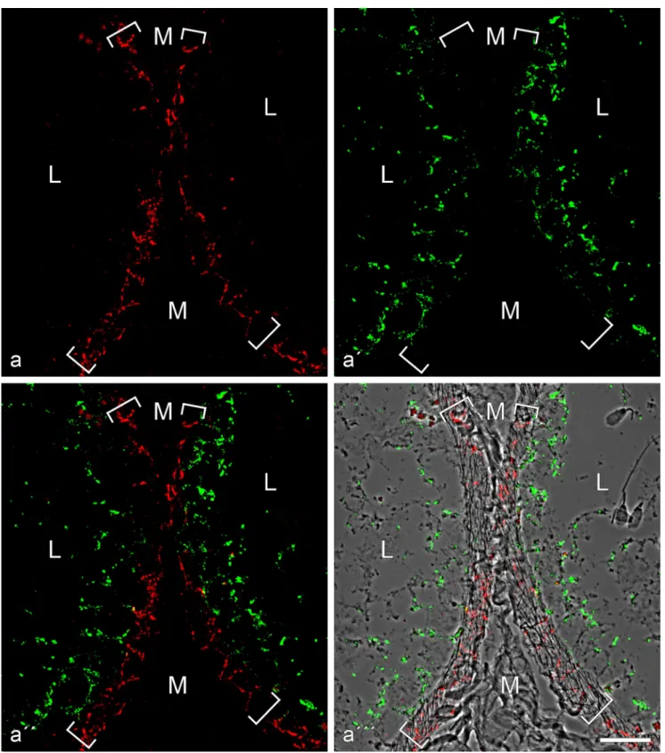
Fig. 4 Double-label immunofluorescence microscopy of cryostat cross-sections through tubuli seminiferi of frozen bull testis after reactions with antibodies against E-cadherin (red; a, a'', a''') and N-cadherin (green; a', a'', a'''). In a''', the reactions are shown on a phase contrast background. Note the mutually exclusive immunostaining of cell-cell junctions of the adherens type, N-cadherin-based ones (green) in the Sertoli cells and
spermatogonia of the tubuli seminiferi and E-cadherin-containing junc-tions exclusively in a special layer of myoid cells surrounding the tubuli (demarcated by the parentheses). M mesenchymal region with interstitial cells; L lumen of the seminiferous tubules, with individual spermatids
(e.g., on the right-hand side of a'''). Bar 20μm
luminal space. On the other hand, during development and
maturation, they have changed their molecular character as
they have lost all keratin IFs and possess abundant bundles of
vimentin IFs. They have also lost the typical epithelial
junc-tion architecture and instead are connected to each other and to
the interspersed spermatogenic cells by a wealth of special and
rather extended forms of adherens junctions.
Absence of desmosomes and desmosome-specific molecules
One of the clearest and most important results of our study is
the demonstration that the mature tubuli seminiferi of all five
mammalian species examined do not contain any desmosome
structures (maculae adhaerentes in the morphological
definition of Farquhar and Palade
1963
). Neither do they
possess any of the desmosome-specific cadherins, i.e.,
desmogleins and desmocollins, nor any of the
desmosome-specific cytoplasmic plaque proteins, i.e., desmoplakin or one
of the plakophilins Pkp1-3. In contrast, plakoglobin as a protein
component of plaques of both desmosomes and adherens
junc-tions (AJs; cf., e.g., Cowin et al.
1986
; Franke et al.
1987
,
2009
)
is also found in AJs of Sertoli cells. So, in Sertoli and
sper-matogonial cells, there are no desmosome-like structures or
related junctions with desmosome-typical molecules.
The significance of the absence of desmosomes and even
of any desmosome-specific molecules in junctions connecting
Sertoli cells with each other or with spermatogenic cells is also
based on direct experimental comparisons with their abundant
presence in all portions of the excurrent duct epithelia (see
also, e.g., Table
2
). So we hope that, from now on, words such
as
“desmosomes”, “desmosome-like structures” or
“desmosomal proteins” will no longer appear in the
liter-ature on mliter-ature Sertoli cells (the need for this sentence is
evident from Table
1
and Electronic Supplementary
Material, Table
S1
), although the similar conclusion of
Pelletier and Byers in
1992
(
“Therefore, the term
desmo-some-like is seemingly inappropriate to designate these
junctions…”) has already been widely ignored by
re-searchers in this field.
Of course, we are aware of the fundamental controversy of
our present report and the recent articles of several other
authors (see Table
1
and Electronic Supplementary Material,
Table
S1
), in particular the publication of Lie et al. (
2010
).
These authors have specifically claimed, based on
experi-ments at the mRNA (RT-PCR) and protein or glycoprotein
(immunoblots and immunolocalization) level, that Sertoli
cells of rats contain desmogleins Dsg-1, Dsg-2 and Dsg-4,
desmocollins Dsc-1 and Dsc-3 and plakophilins Pkp-1, Pkp-2,
and Pkp-4, as well as desmoplakin. While the mentioning of
“plakophilin-4” as a desmosomal component is obsolete since
it has been demonstrated that this plaque protein does not
occur in any kind of desmosome but, as
“protein p0071”, is
restricted to AJs (Hofmann et al.
2008
,
2009
). In addition, the
reported discovery of desmoglein Dsg-4 is highly disturbing
as this glycoprotein has so far been identified only in the
uppermost layers of the epidermis and in certain hair follicle
layers (for references, see, e.g., Godsel et al.
2004
; Schmidt
and Koch
2008
). However, as many of the claims reported by
Lie et al. (
2010
) specifically address Sertoli cell cultures, a
detailed critical discussion will have to be postponed to a
future article in our series, which will deal with cell cultures
and tumors assumed to be derived from Sertoli cells.
Fig. 5 Double-label immunofluorescence microscopy showing thereac-tions of antibodies to N-cadherin (a, red, mouse mAb) and desmoplakin (b, green, guinea pig antibodies) on a cross-section through tubuli seminiferi of bull testis (L, tubular lumen; M, mesenchymal space). While the N-cadherin reaction identifies the AJs of the Sertoli and
spermatogonial cell layer (a and a'' show the reaction in the three neighbouring tubular structures) there is no desmoplakin reaction (a';
for a visualization“control” this picture has been selected as, accidentally,
a very small artifical green particle is seen here, denoted by a white
On the other hand, in this context, we do not forget to
mention the specific occurrences of individual desmosomal
molecules in other ensembles such as desmoplakin in the
complexus adhaerentes of parts of lymphatic endothelia
(Schmelz and Franke
1993
; Hämmerling et al.
2006
; Moll
et al.
2009
), plakophilin Pkp-2 in AJs of certain very
prolifer-ative stages of mesenchymal cells (e.g., Barth et al.
2009
,
2012
;
Rickelt et al.
2010
; Rickelt
2012
) and the
“free” Dsg-2
glyco-proteins dispersed on the surfaces of certain types of melanoma
cells (Schmitt et al.
2007
; Rickelt et al.
2008
). Particularly
complex
“hybrid junctions” are the composite junctions (areae
compositae) connecting mammalian cardiomyocytes (Franke
et al.
2006
) and the
“meningioma junctions” in which E- or
N-cadherin can be associated not only with
α- and β-catenin,
plakoglobin and protein p120 but also with the
“desmosomal
protein
”, plakophilin Pkp-2 (Akat et al.
2008
).
The absence of desmosomes and any desmosome-specific
molecules in mature Sertoli cells is also remarkable as these
structures and molecules are present in stages of
embryogene-sis, fetal as well as early postnatal development, upon advanced
aging and in various pathological situations and also in
Sertoli-type cells of a wide variety of non-mammalian vertebrates (for
references, see Baccetti et al.
1983
; Bergmann et al.
1984
;
Pfeiffer and Vogl
2002
).
Like keratin IF bundles, vimentin IF bundles can also be
anchored at desmosomal plaques (e.g., Kartenbeck et al.
1984
;
Schwechheimer et al.
1984
; Moll et al.
1986
; for references,
see Franke et al.
2009
). As desmosomes are absent in mature
Sertoli cells, it is thus noteworthy that the abundantly present
vimentin IF bundles are not anchored at—nor in any other way
firmly attached to—the plaques of the diverse subtypes of AJs
aforementioned (see, e.g., Electronic Supplementary Material,
Fig. 6 Double-label immunofluorescence microscopy of cross-sectionsthrough seminiferous tubules of frozen bull testis, showing the
near-complete colocalization ofβ-catenin (a, red, mouse mAb) and
N-cadherin (a', green, rabbit antibodies) in the AJs of the Sertoli cell layer
of the tubules, demonstrated by the yellow merged colour (a'', a'''; the latter is presented on a phase contrast background and with nuclei stained
blue with DAPI). Note alsoβ-catenin-positive structures in several types
of interstitial cells. L, lumen. Bar 50μm
Fig.
S6
). The same seems to be true for the neurofilament
bundles (see also Davidoff et al.
1999
).
The various forms of AJs in the mammalian tubuli seminiferi,
including the areae adhaerentes
Since the classic report by Farquhar and Palade (
1963
), the
non-desmosomal adhering junctions, the adherens junctions
(AJs), of epithelia have been found to occur in one of three
morphological forms, the zonula adhaerens, the fascia
adhaerens and the punctum adhaerens. Clearly, structures of
the two latter types also exist in both the tubuli seminiferi and
the excurrent duct epithelia, the excurrent ducts also having
typical subapical zonulae adhaerentes (see also Figs.
1
,
2
,
3
).
In addition to these three forms, we now define a fourth major
form, the areae adhaerentes, i.e., extended, often very large
surface regions (e.g., including areas larger than 30
μm
2as determined from serial sections), which obviously
pro-vide very important structures of monolayer organization
in the seminiferous tubules, both in homotypic AJs and in
heterotypic connections with spermatogenic cells. As this
study shows, they generally contain N-cadherin and the
typical AJ plaque ensemble of proteins (Table
2
). Whether
such extended regions of the AJ type also occur in
N-Fig. 7 Double-label immunofluorescence microscopy of cross-sectionsthrough seminiferous tubules of frozen bull testis, showing the near-complete colocalization of N-cadherin (a, red, mouse mAb) and the plaque protein p0071 (a', green, guinea pig antibodies) in the AJs of the Sertoli cells, demonstrated by the yellow merger colour (a'', a'''; the
latter is shown on a phase contrast background and with DAPI-staining of nuclei). Note also the intensive green immunostaining of the p0071 reaction in the junctions connecting endothelial cells in blood and lymph vessels (here indicated, e.g., by the arrow in the lower right-hand corner of a'–a'''). L, lumen. Bar 50 μm
cadherin-based cell–cell contacts of other cells cannot yet
be stated.
Morphologically, one can distinguish three major subforms
of AJ structures in the seminiferous tubules (for diverse
morphological AJ subtypes; see also fig. 28.1 of Pelletier
2001
): One subform (type I) with a relatively thick and dense
cytoplasmic plaque (e.g., Fig.
10d
–d
'''), a second subform
(type II) with less and only loosely associated filamentous
plaque substructures (e.g., Figs.
9d
,
10a–c
) and a third
subform (type III) with very little and very thin cytoplasmic
plaque filaments (MPM-AJs), which even in electron
micro-graphs are often not distinctly resolved (e.g., Figs.
9
,
10a–d
,
11i–e
). In certain areas of cell–cell contacts, diverse AJ
sub-types may occur side by, side, often alternating or group-wise
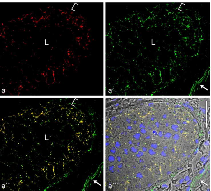
Fig. 8 Double-label immunofluorescence microscopy of a cross-sectionthrough a seminiferous tubule of frozen mouse testis tissues, showing the very frequent colocalization of the plaque protein of the striatin family (a,
red, mouse mAb) andβ-catenin (a', green, rabbit antibodies) in the basal
parts of Sertoli cells and in spermatogonial cells, demonstrated by the
yellow merger colour (a'', a'''; the latter is shown on a phase contrast background and with staining of the nuclei). Note also the DAPI-stained elongated spermatid heads typical for rodents. L, lumen of the
seminiferous tubules. Bar 50μm
Fig. 9 Electron micrographs of ultrathin sections through seminiferous
tubules of boar (a–a''') and bull (b–d) testis, showing a survey
picture including large parts of a nucleus (N) and details of the very extended, rather regularly narrow-spaced (membrane-to-membrane
in-terspace 8–18 nm) area adhaerens junctions; B, basal lamina; M,
mitochondrion (a'–a''' present details at higher magnification; C,
cytoplasm). Such extended, narrow-spaced plasma membrane
connections of the “minimal plaque material” AJ type are also seen
in bovine Sertoli cells (b–d) and only occasionally rather thin,
loosely and irregularly arranged plaque-like structures are detected (see, e.g., d, parentheses). Note that these MPM-AJ associations are also maintained at sites where the plasma membranes of three cells
Fig. 10 Electron micrographs of ultrathin sections through testicular tissue of a bull, showing various subtypes and aspects of AJs connecting Sertoli cells in the tubuli seminiferi. a Junctions characterized by a rather
narrow distance between the membrane profiles (8–18 nm) loosely
asso-ciated with some cytoplasmic plaque material that is highly variable in size and configuration. b, b' Overview (b) and partial magnification (b') of a region containing an AJ with a strictly planar arrangement of 6- to
7-nm-thick membranes, an intermembrane space of 8–18 nm, with serially
arranged“punctate midline” granules of 2–5 nm diameter (b') and a
general but loose cytoplasmic plaque coverage. c This small and rather narrow junction (ca. 5 nm intermembrane distance) is covered asymmet-rically with irregularly shaped cytoplasmic plaque material. d Survey micrograph showing five tight-packed Sertoli cell processes and extended cell–cell contact regions (areae adhaerentes) between 5 pairs of cells (numbered 1–5). Note in these Sertoli cell processes, the dense package of, e.g., mitochondria (M) and the so-called“ectoplasmic specializations”,
i.e., cortical paracrystalline actin microfilament bundles that in some
regions are parallel to—and rather closely associated with—cell–cell
junction plasma membrane regions, often revealing lateral up to 4-nm-thick cross-bridges between the filament paracrystals and the plasma membrane (see, e.g., the bundle in the upper right of cell process num-bered 2 and the parenthesis in the insert labeled d'). Note also the
extended region with cell–cell junctions of the MPM-AJ type connecting
cells numbered 1 and 2 in (d) (with higher partial magnifications in d'' and d'''), showing numerous, closely spaced, dense arrangements of typical AJs with cytoplasmic plaque material separated by tight-adpressed
special junctions of 10–30 nm diameter (arrows in d'', d'''). All three
major junction types are seen side-by-side in (d'''): a punctum adhaerens (arrowhead), a tightly adpressed membrane junction (arrow) and a region
of the MPM-AJ type (parenthesis; cf. Fig.10). Bars (a, b, d, d'', d''')
200 nm, (b', c, d') 100 nm
interspersed (e.g., Fig.
10d
'''). These different AJ subforms
also occur in the seminiferous tubules of many other
mam-malian species (see in particular Pelletier
1988
,
2001
; Pelletier
and Byers
1992
).
From a series of observations in the neuronal system, it may
also be hypothesized that, in the seminiferous tubules,
N-cadherin has in addition a developmental biological role in
topogenesis and specific cell
–cell interactions, including the local
stabilization of other cell–cell connection structures, as it has first
been proposed for certain synapses (e.g., Ushida et al.
1996
;
Arikkath
2010
; Mendez et al.
2010
; Tan et al.
2010
; Gärtner et al.
2012a
,
b
). Whether such roles and interactions can also be
ascribed to some of the various subtypes of the N-cadherin–
based junctional structures of the testis remains to be examined.
Cribelliform junctions
With great surprise, we noticed, amidst the AJs of Sertoli cells,
a further distinct and totally novel kind of cell–cell junction
structure that is characterized by clusters of channel-like, ca.
6- to 7-nm-wide cytoplasmic continuity between adjacent
Sertoli cells, i.e., pore structures formed by the fused plasma
membranes of both cells. As we have shown, these
sieve-plate-like, close-spaced assemblies of channels, areae
cribelliformes, occur as small, distinct groups of cell–cell
continuities that would allow the passage of relatively
large molecules or particles. At present, we cannot answer
questions as to their frequencies and functions or whether
they contain any specific molecules, questions that we are
currently trying to answer in quantitative and
morphomet-ric studies. Moreover, we will have to demonstrate the
cell-to-cell passages of fluorescent or electron microscopic
markers between Sertoli cells and determine the nature
and sizes of molecules and particles that can take that
route.
Of course, the question arises why these cribelliform
junctions with their characteristic cytoplasmic sieve pore
structures between Sertoli cells have not been described
before. Here, however, careful examination of the
pub-lished electron micrographs has revealed a few illustrations
in works on guinea pig and dog seminiferous epithelia
that show structures suggestive of such groups of
cribelliform connections (see, e.g., figs. 3 and 4 of
Connell
1978
, or figs. 7 and 17 in Pelletier and Friend
1983
). Thus, we expect that such junctions will be
detect-able in the Sertoli cells of diverse mammalian species.
Concluding remarks
Our findings confirm and extend the view that the Sertoli cells
of mature tubuli seminiferi and their cell
–cell junction system
represent a special and complex epithelial system, profoundly
different from those of all other epithelial cells: Keratins are
lost, desmosomal structures and desmosome-typical
mole-cules are lost, E-cadherin is lost, EpCAM-containing
junc-tions are lost. Instead, various subtypes of other adhaerens
Fig. 11 Electron micrographs of ultrathin sections through bovinetesticular tissue, showing details of adherens regions (areae adhaerentes) and cribelliform junctions connecting Sertoli cells of a specific subtype. Sertoli cells of this subtype are characterized by a high packing density of endoplasmic reticulum cisternae in a cytoplasm of marked electron density and with extended regions of cell–cell junctions of the adherens type as well as some rather small cribelliform junctions and frequent junction-associated, paracrystalline actin microfilament bundles (“ectoplasmic
specializations”). a Interdigitating processes of Sertoli cells (SC1–SC4)
are connected by extended plasma membrane regions of“normal”
intermembrane distance AJs, interspersed with small tightly adpressed membrane junctions some of which even suggest direct molecular interaction (a', b, d). Distinct narrow channels between the cytoplasms of two Sertoli cells are indeed resolved in some very thin sections and appear as sieve-plate junctions (some positions are denoted by arrows in d and some of them are shown at higher magnification in e and f): cribelliform
junctions (areae cribelliformes; e, f). The channel-like cell–cell continuities
of these cribelliform junctions (e, f) have an inner“pore” diameter of 6–
7 nm and a total length of 6–9 nm. Note that these cell–cell channels are
often also characterized by electron-dense, plaque-like structures on one or on both sides of the channel (arrowheads or brackets in d, f, h, j). All in all, a major part of the plasma membrane indicates a junction-like association with adjacent actin filament bundles, which are often cross-bridged to the plasma membrane by short structures (c, g, i, j; see also arrowheads in g). Not infrequently, these parallel and close-spaced membrane-membrane junction-like structures are coated with loose and irregularly shaped
cytoplasmic dense materials (j–l). Bars (a) 1 μm, (a') 500 nm, (b–d, g–l)
200 nm, (e, f) 50 nm
Fig. 12 Immunoelectron microscopy of bull testicular tissue using
anti-bodies againstβ-catenin (a–d), N-cadherin (e) and striatin (f). The
silver-enhanced immunogold grains show specific binding ofβ-catenin in the
extended regions of these Sertoli cell contacts with neighboring cells,
including very long (4–6 μm) stretches with almost continuous β-catenin
labelling (a–d). The reaction antibodies used in the preparation (d) have
been enlarged by an especially intensive silver enhancement. All the
diverse morphological subforms of Sertoli–Sertoli cell junctions also
appear positive for N-cadherin (e.g., e) as well as for the other major
adherens plaque proteins such as striatin (f) Bars (c) 1μm, (d, f) 500 nm,
(a, b, e) 200 nm
structures have formed that are based on N-cadherin. While
such a transition from E- to N-cadherin is known from other
cell-type changes in embryonal and fetal development as well
as from certain pathological transformations as in the invasion
and metastasis of diverse kinds of tumor cells (for review, see,
e.g., Kalluri and Weinberg
2009
), another form of advent of
N-cadherin in epithelial cells, i.e., the formation of AJs based
on E–N heterodimer clusters, as described for mammalian
hepatocytes and liver tumor cells in situ and in culture
(Straub et al.
2011
), seems to be excluded from the mature
and active Sertoli cells.
Acknowledgments We wish to thank Dr. Michael Rogers (German
Cancer Research Center) for reading and correcting the English text and Eva Gundel for careful assembly of the manuscript text.
This work was supported by a grant from the German-Israeli Founda-tion for Scientific Research and Development (GIF grant I-1098-43.11/ 2010). A major part of this work has been presented as a Bachelor thesis of the first author (Brandenburg University of Technology, Senftenberg, 2013).
Open Access This article is distributed under the terms of the Creative Commons Attribution License, which permits any use, distribution and reproduction in any medium, provided the original author(s) and the source are credited.
References
Akat K, Bleck CKE, Lee Y-MA, Haselmann-Weiss U, Kartenbeck J (2008) Characterization of a novel type of adherens junction in meningiomas and the derived cell line HBL-52. Cell Tissue Res
331:401–412
Altorfer J, Fukuda T, Hedinger C (1974) Desmosomes in human semi-niferous epithelium. An electron microscopic study. Virchows Arch
B 16:181–194
Arikkath J (2010) N-cadherin: stabilizing synapses. J Cell Biol 189:
397–398
Baccetti B, Bigliardi E, Talluri MV, Burrini AG (1983) The Sertoli cell in
lizards. J Ultrastruct Res 85:11–23
Barth M, Schumacher H, Kuhn C, Akhyari P, Lichtenberg A, Franke WW (2009) Cordial connections: molecular ensembles and structures of adhering junctions connecting interstitial cells of cardiac valves in
situ and in cell culture. Cell Tissue Res 337:63–77
Barth M, Rickelt S, Noffz E, Winter-Simanowski S, Niemann H, Akhyari P, Lichtenberg A, Franke WW (2012) The adhering junctions of valvular interstitial cells: molecular composition in fetal and adult hearts and the comings and goings of plakophilin-2 in situ, in cell culture and upon re-association with scaffolds. Cell Tissue Res 348:
295–307
Bergmann M, Schindelmeiser J, Greven H (1984) The blood-testis barrier in vertebrates having different testicular organization. Cell Tissue
Res 238:145–150
Byers S, Graham R, Dai HN, Hoxter B (1991) Development of Sertoli cell junctional specializations and the distribution of the tight-junction-associated protein ZO-1 in the mouse testis. Am J Anat
191:35–47
Byers SW, Sujarit S, Jegou B, Butz S, Hoschutzky H, Herrenknecht K, MacCalman C, Blaschuk OW (1994) Cadherins and cadherin-associated molecules in the developing and maturing rat testis.
Endocrinology 134:630–639
Cheng CY, Mruk DD (2002) Cell junction dynamcis in the testis: Sertoli-germ cell interactions and male contraceptive development. Physiol
Rev 82:825–874
Cheng CY, Mruk DD (2011) Regulation of spermiogenesis, spermiation and blood-testis barrier dynamics: novel insights from studies on
Eps8 and Arp3. Biochem J 435:553–562
Cheng CY, Mruk DD (2012) The blood-testis barrier and its implications
for male contraception. Pharmacol Rev 64:16–64
Cheng CY, Wong EWP, Lie PPY, Li MWM, Mruk DD, Yan HHN, Mok D-W, Mannu J, Mathur PP, W-y L, Bonanomi M, Silvestrini B (2011) Regulation of blood-testis barrier dynamics by desmosome, gap junction, hemidesmosome and polarity proteins. An unexpected
turn of events. Spermatogenesis 1:105–115
Connell CJ (1978) A freeze-fracture and lanthanum tracer study of the complex junction between Sertoli cells of the canine testis. J Cell Biol 76:57–75
Cowin P, Kapprell H-P, Franke WW, Tamkun J, Hynes RO (1986) Plakoglobin: a protein common to different kinds of intercellular adhering junctions. Cell 46:1063–1073
Cyr DG, Blaschuk OW, Robaire B (1992) Identification and develop-mental regulation of cadherin messenger ribonucleic acids in the rat testis. Endocrinology 131:139–145
Cyr DG, Hermo L, Robaire B (1993) Developmental changes in epithelial cadherin messenger ribonucleic acid and immuno-cytochemical localization of epithelial cadherin during post-natal epididymal development in the rat. Endocrinology 132: 1115–1124
Cyr DG, Robaire B, Hermo L (1995) Structure and turnover of junctional complexes between principal cells of the rat epididymis. Microsc Res Tech 30:54–66
Cyr DG, Gregory M, Dubé E, Dufresne J, Chan PTK, Hermo L (2007) Orchestration of occludins, claudins, catenins and cadherins as players involved in maintenance of the blood-epididymal barrier in animals and humans. Asian J Androl 9:463–475
Davidoff MS, Middendorff R, Pusch W, Müller D, Wichers S, Holstein AF (1999) Sertoli and Leydig cells of the human testis express neurofilament triplet proteins. Histochem Cell Biol 111:173–87
DeBellefeuille S, Hermo L, Gregory M, Dufresne J, Cyr DG (2003) Catenins in the rat epididymis: their expression and regulation in adulthood and during postnatal development. Endocrinology 144:
5040–5049
Dym M (1974) The fine structure of monkey Sertoli cells in the transi-tional zone at the junction of the seminiferous tubules with the tubuli
recti. Am J Anat 140:1–25
Dym M (1976) The mammalian rete testis– a morphological
examina-tion. Anat Rec 186:493–523
Dym M (1977) The male reproductive system. In: Weiss L, Greep RO
(eds) Histology. McGraw-Hill, New York, pp 979–1038
Dym M, Fawcett DW (1970) The blood-testis barrier in the rat and the physiological compartmentation of the seminiferous epithelium.
Biol Reprod 3:308–326
Farquhar MG, Palade GE (1963) Junctional complexes in various
epi-thelia. J Cell Biol 17:375–412
Franke WW (2010) Discovering the molecular components of intercel-lular junctions-a historical view. In: Nelson J, Fuchs E (eds) Cold
spring harbor perspectives in biology. Cell–Cell junctions. Vol 1.
Cold Spring Harbor Laboratory, New York, pp 1–34
Franke WW, Grund C, Fink A, Weber K, Jockusch BM, Zentgraf H, Osborn M (1978) Location of actin in the microfilament bundles associated with the junctional specializations between Sertoli cells