Brainstem processing of vestibular sensory exafference:
implications for motion sickness etiology
Charles M. Oman
1and Kathleen E. Cullen
21
Man Vehicle Laboratory, Massachusetts Institute of Technology, Cambridge, MA
02139 USA
2
Aerospace Medical Research Unit, Department of Physiology, McGill University,
Montreal, Quebec, Canada
For the special issue: “Neurobiology of Nausea and Vomiting” (Yates and Horn, Eds.)
Address Correspondence to:
Charles M. Oman, PhD
Man Vehicle Laboratory
Room 37-219
MIT 77 Massachusetts Avenue
Cambridge, MA 02139 USA
[email protected]
617 273-7508
Submitted Jan 30, 2014
Revised March 17, 2014, accepted April 20, 2014, published online May 18, 2014
DOI 10.1007/s00221-014-3973-2
Abstract
(244 words)
The origin of the internal “sensory conflict” stimulus causing motion sickness has been debated for more than four decades. Recent studies show a subclass of neurons in the vestibular nuclei and deep
cerebellar nuclei that respond preferentially to passive head movements. During active movement, the semicircular canal and otolith input (“reafference”) to these neurons is cancelled by a mechanism comparing the expected consequences of self-generated movement (estimated with an internal model- presumably located in the cerebellum) with the actual sensory feedback. The un-cancelled component (“exafference”) resulting from passive movement normally helps compensate for unexpected postural disturbances. Notably, the existence of such vestibular “sensory conflict” neurons had been postulated as early as 1982, but their existence and putative role in posture control, motion sickness has been long debated. Here we review the development of “sensory conflict” theories in relation to recent evidence for brainstem and cerebellar reafference cancellation, and identify some open research questions. We propose that conditions producing persistent activity of these neurons, or their targets, stimulates nearby brainstem emetic centers – via an as yet unidentified mechanism. We discuss how such a mechanism is consistent with the notable difference in motion sickness susceptibility of drivers as opposed to passengers, human immunity to normal self-generated movement, and why head restraint or lying horizontal confers relative immunity. Finally, we propose that fuller characterization of these mechanisms, and their potential role in motion sickness could lead to more effective, scientifically based prevention and treatment for motion sickness.
Keywords
Introduction
Most researchers and clinicians concerned with nausea and vomiting in the context of cancer
chemotherapy, cyclic vomiting or GI syndromes are aware that vestibular stimulation can also provide a strong emetic stimulus. However, it is also generally appreciated that the physiology of the vestibular-emetic linkage appears different. For instance, drugs notably effective against motion sickness (e.g. scopolamine) are relatively ineffective against nausea produced by other stimuli, and conversely (e.g. 5HT3 antagonists) (Yates et al. 1998).
When compared to our present understanding of the chemo- and gastric syndromes, the physiology and pharmacology underlying motion sickness largely remains a puzzle. Seasickness, carsickness and airsickness are ubiquitous phenomena for which nausea and vomiting often occur. Since similar symptoms are also commonly experienced with acute vestibular disease, motion sickness is frequently attributed simply to “vestibular overstimulation”. Indeed, clinical and experimental evidence reviewed by (Money 1970) indicates that humans and animals who lack functional vestibular organs are entirely immune to motion sickness.
Over half a century ago, Wang and Chinn (1956) induced motion sickness in dogs using swing exposure. Because animals did not display vomiting after bilateral labyrinthectomy or lesions of the nodulus and uvula of the vestibular cerebellum, they argued that “motion stimulates the labyrinthine receptors, and the vestibular impulses traverse the nodulus and uvula of the cerebellum, to the chemoreceptive emetic trigger zone (CTZ) , and finally reach the medullary vomiting center “. However, this proposal was not supported by subsequent experiments indicating that the CTZ was not essential in motion sickness (Borison and Borison 1986), that the “vomiting center” was not discretely localizable in the medulla (Miller and Wilson 1983b), and that even an intact cerebellum was not essential (Miller and Wilson 1983a).
Vestibular physiologists and psychologists (e.g. Reason and Brand (1975)) further proposed that vestibular overstimulation could not explain other established motion sickness characteristics. For instance: Why is it that jumping and other athletic activities that create significant vestibular stimulation never produce sickness? Why do sailors that are well adapted to ship motion or astronauts who fly long missions experience disorientation and nausea upon return to a normal environment? Why is it that some people experience nausea in wide screen movie theaters, where the head is not moving at all? Why are the drivers of real or virtual cars or the pilots of aircraft notably less susceptible than their passengers (Reason and Brand 1975; Reason 1978; Rolnick and Lubow 1991; Dong et al. 2011) yet it is the experienced pilots and drivers who are more susceptible than trainees in simulators (Kennedy et al. 1990)? When standing subjects view a moving visual surround, why does the magnitude of postural disturbance correlate with the intensity of subsequent symptoms (Owen et al. 1998; Smart et al. 2002) ? Why does providing head support or resting gravitationally supine or prone (Manning and Stewart 1949; Tyler and Bard 1949; Johnson and Mayne 1953) reduce motion sickness susceptibility?
Sensory conflict hypotheses
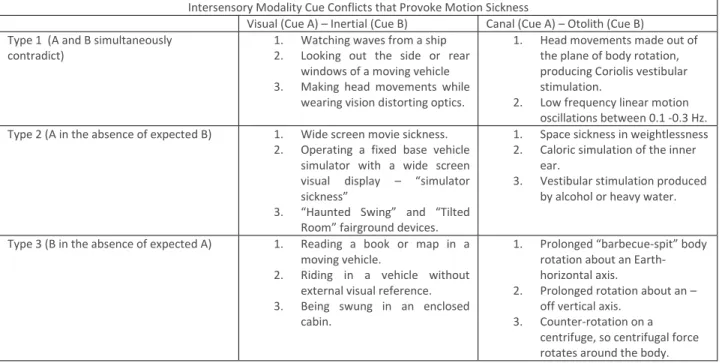
Claremont (1931) originally suggested that sea sickness was due to “unaccustomed conflict between sensations normally combined in other ways” originating in the vestibular, visual and proprioceptive senses. This intuitive “inter-sensory cue conflict” hypothesis was later scientifically elaborated by Guedry (1968), Steele (1963),and Reason (1969), leading to four decades of scientific debate aimed at establishing the essential internal stimulus for motion sickness. The detailed but notional taxonomy for inter-sensory cue conflict proposed by Reason and Brand (1975) - shown in Table 1 – is illustrative and self-explanatory.
Intersensory Modality Cue Conflicts that Provoke Motion Sickness
Visual (Cue A) – Inertial (Cue B) Canal (Cue A) – Otolith (Cue B) Type 1 (A and B simultaneously
contradict) 1. Watching waves from a ship 2. Looking out the side or rear windows of a moving vehicle 3. Making head movements while
wearing vision distorting optics.
1. Head movements made out of the plane of body rotation, producing Coriolis vestibular stimulation.
2. Low frequency linear motion oscillations between 0.1 -0.3 Hz. Type 2 (A in the absence of expected B) 1. Wide screen movie sickness.
2. Operating a fixed base vehicle simulator with a wide screen visual display – “simulator sickness”
3. “Haunted Swing” and “Tilted Room” fairground devices.
1. Space sickness in weightlessness 2. Caloric simulation of the inner
ear.
3. Vestibular stimulation produced by alcohol or heavy water. Type 3 (B in the absence of expected A) 1. Reading a book or map in a
moving vehicle.
2. Riding in a vehicle without external visual reference. 3. Being swung in an enclosed
cabin.
1. Prolonged “barbecue-spit” body rotation about an Earth-horizontal axis.
2. Prolonged rotation about an – off vertical axis.
3. Counter-rotation on a centrifuge, so centrifugal force rotates around the body.
Table 1 - After: Reason and Brand (1975), Table 6.
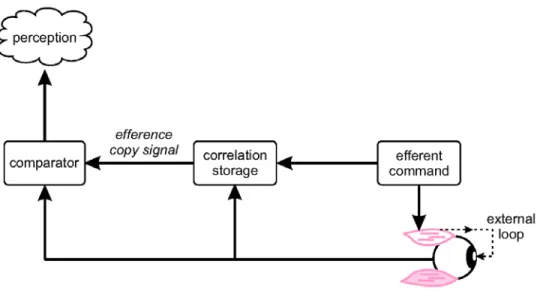
However, in a landmark paper that followed, Reason (1978) rejected his own inter-sensory modality conflict definition, arguing that the signals from various sense organs have different dynamic response and coding, and what is “normal” depends on prior sensory-motor experience. Reason suggested that instead the essential conflict stimulus causing motion sickness was related to the difference between actual and anticipated sensory inputs. This idea followed the theoretical and behavioral work of Von Holst (1954) that had addressed the question of how does the CNS distinguishes changes in visual input resulting from active body movements (“re-afference”) from those associated with passive movement of the entire visual surround (“ex-afference”). Von Holst had suggested that the brain compares an ‘image’ or “efference-copy” of the motor command (“efference”) to the re-afference caused by the movement in a manner similar to comparing “the negative of a photograph compares to the print”. Once this comparison is made, only the “exafferent” component remains, such that the anticipated component of incoming sensory information is cancelled. Held (1961) soon after completed a series of conceptually related perceptual adaptation studies. Specifically, humans wore prism glasses that systematically changed the relationship between head movements and sensory return. Held termed these conditions “sensory rearrangements”, and proposed a hypothetical model (Figure 1) to explain
the adaptation. While the model was similar to that proposed by von Holst, it included an additional hypothetical element , a neural network (“correlation storage”) that accounted for the normal
relationship between motor outflow and sensory return. Each time the correlation storage receives an efferent signal, it generates an efference copy signal that – based on prior experience - most likely would cancel the incoming sensory information. A difference between the efference-copy and sensory return generated an expectancy conflict signal of sensory dimension, which Held proposed triggered the updating of the correlation storage dictionary, and to adapt perception and motor performance
appropriately for the specific sensory rearrangement.
Fig 2 Hypothesized mechanism for reafference cancelation via cerebellar internal model. Adapted from
Cullen 2012
Accordingly building on von Holst’s “reafference cancellation principle” and Held’s “correlation storage” explanation for sensory adaptation, Reason (1978) posited that motion sickness was caused by sensory rearrangements. He extended Held’s conflict modeling scheme by including semicircular canal and otolith cues in addition to vision. Notably, Reason proposed that the drive for motion sickness depended on the magnitude of cue conflict in each sensory modality, increased with the number of discrepant modalities, and varied inversely with the prior exposure to the discordance. He speculated but did not demonstrate that the correlation storage element (renamed the “neural store”) might be located in the cerebellum, and did not formally consider posture control or the circuit underlying the emetic linkage.
Further progress towards understanding the etiology of motion sickness was next made by applying engineering control and estimation theory to develop a general bio-mathematical model of sensory-motor integration by Oman (1982); (1990; 1991). He argued that simple reflexes would be inadequate to estimate body movement to stabilize head and body posture based on the available incomplete set of
noisy but partially redundant sensory inflow (i.e., vestibular, visual and somatosensory/proprioceptive signals). Instead he proposed that the CNS employed an “internal model” referenced scheme to estimate posture. The internal model employed functioned as an association network, analogous to Held’s “correlation storage” and Reason’s “neural store”. It received motor efference and sensory afference as inputs, and produced as outputs continuous head and body posture estimates, and
concurrent “efference copy” estimates of expected sensory inputs for each modality, including effects of gravitational stimulation. An important feature of the internal model estimator was that actual sensory afference was continuously compared with the “efference copy” estimate. During normal active movement, the difference between the two signal sets - “sensory conflict”- was small, resulting in almost complete reafference cancellation. However if the body was moved passively, or if the normal relationship between body movement and sensory afference somehow systematically changed, the resulting “sensory conflict” signals - weighted based on sensory noise and dynamic characteristics - continuously corrected motor outflow and triggered sensory motor learning. This “Observer” head orientation estimation scheme is arguably optimal in a Bayesian/Kalman sense (Selva and Oman 2012). Observer models for head and eye movement have since been employed to interpret a variety of human and animal vestibular experimental data (Merfeld et al. 1993; Haslwanter et al. 2000; Merfeld and Zupan 2002; Vingerhoets et al. 2007) as well as to model human reaching movements (Wolpert et al. (1995); (1998)). Notably, Wolpert and colleagues argued that the cerebellum contained internal “forward” models used to predict the sensory consequences of motor commands and thereby
compensate for time delays (see also Ito (1970). Wolpert also proposed that the cerebellum contained internal “inverse” models used to create the motor command required to achieve a desired arm movement, and that both types may also contribute to cognition, including perception of the external world.
Interestingly, there was a common thread linking the work of Held, Reason and Oman with important implications for understanding of motion sickness. Each posited that any conditions creating sustained sensory conflict would lead to internal model relearning/updating via gradual interactions with
prevailing sensory environment. For example, Oman noted that prolonged periods of conflict occurred not only when head movements were made repeatedly during conditions of “sensory rearrangement” - as defined by Held and Reason - but whenever posture was disturbed by external forces or
accelerations, as when riding as a passenger in an aircraft or on shipboard. To account for motion sickness, Oman more specifically posited that vestibular conflict signals somehow coupled to CNS emetic centers, via an “emetic linkage” mechanism. After a passive motion stimulus is applied, there is typically a latency of several minutes before nausea appears. Nausea then rises exponentially, with vomiting the usually inevitable result. If the stimulus is removed before vomiting occurs, nausea gradually decays, suggesting that the emetic linkage has the dynamic characteristics of a nonlinear “leaky integrator”. In order to account for latency and why symptoms do not result merely from sensory noise or the
occasional postural disturbances of daily life, the integration mechanism must have an output rather than input threshold. Experimental data (e.g. Bock and Oman (1982); Golding and Stott (1997))
supports this view. Since Oman’s model accounted for motion sickness due to passive motion as well as sensory rearrangements, with conflict signals playing an indirect but essential role in postural
Do vestibular sensory conflict neurons exist?
Oman (1990) posited that conflict signals were computed at the first stage of CNS sensory processing, but cautioned that the theory should be regarded as a “black-box” or “as-if” model, since the
physiological locus of the internal model had not been determined and the existence of vestibular neurons that responding to passive but not active movement had not been shown. Lacking
experimental evidence, debate about internal models and conflict signals continued for several decades. Some proposed that the essential conflict causing motion sickness might not originate at the first stage of vestibular processing, but might result at subsequent levels of processing as a result of competing internal estimates of orientation derived from different senses (Treisman 1977; Zupan et al. 2002) or alternatively from deviations in the perceived direction of the gravitational vertical (Bos and Bles 1998). The evolutionary significance of motion sickness was also debated (reviewed by Oman (2012)).
Ecological psychologists Stoffregen and Riccio (1991) argued that all conflict notions – sensory or otherwise –remained unproven reifications, and noted that motion sickness symptoms appear causally related to postural sway in standing subjects, e.g. (Smart et al. 2002).
Meanwhile neuroscientists continued to seek evidence for reafference cancellation in the cerebellum. For instance, (Blakemore et al. 1999) showed large differences in cerebellar fMRI activation to passive vs. self-produced tactile hand stimulation. Additional progress was made in the study of the mormyrid fish. As shown by Bell and colleagues, single unit recording revealed that electroreceptor reafference resulting from the fish’s own electric organ pulses was cancelled in its cerebellum-like structures (reviewed in Bell et al. (2008)). Since the output neurons of the cerebellar cortex (i.e., Purkinjie cells) project to neurons in the deep cerebellar nuclei and vestibular nucleus, it made sense to look for evidence of reafference cancellation in these areas as well.
Rhesus brainstem and cerebellar neurons that respond primarily to
passive motion.
The first evidence for reafference cancellation in the vestibular system was obtained from a distinct class of neurons in the vestibular nucleus of the primate brainstem. Vestibular nucleus units receiving direct input from the sensory afferents of inner ear comprise three major classes: Two (Position Vestibular Pause Neurons and Floccular Target Neurons) play important roles in vestibulo-ocular reflex stabilization and calibration, but their responses are eye position dependent, and they show no
evidence of reafference cancellation. (Nor does making eye movements alone trigger motion sickness). In contrast, the third class of central neurons, termed “vestibular only”(VO) neurons, characteristically respond to semicircular canal and/or otolith afferent input but not to eye position and exhibit
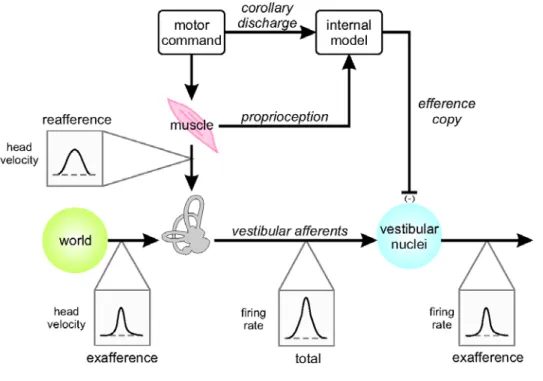
reafference cancellation. Notably, VO neurons show reafference cancellation in response to semicircular canal (McCrea et al. 1999; Roy and Cullen 2002; Roy and Cullen 2004; Cullen et al. 2009; Sadeghi et al. 2009) and/or otolith (Carriot et al. 2013) stimulation. These neurons contribute to vestibulo-collic and vestibulo-spinal reflexes via direct and indirect projections to the spinal cord (reviewed in Cullen (2012)). The mechanism proposed by Cullen and colleagues for the suppression of vestibular reafference is shown schematically in Fig 2 (Roy and Cullen 2002; Carriot et al. 2013). In this model, the motor
command to neck muscles creates neck proprioceptor and vestibular reafference. An internal model – most likely in the cerebellar cortex/deep cerebellar nuclei – uses an efference copy of the motor command to predict the expected proprioceptive input. Vestibular reafference is then canceled if and only if there is a close match between the actual and expected proprioceptive signals. However, cancellation does not occur for movements where the difference between actual and expected proprioceptive signals (also termed ‘sensory prediction error’) is significant. As a result, VO neurons primarily respond to externally applied motion (i.e., vestibular exafference), which normally then contributes to vestibulo-collic/-spinal motor outflow, to help stabilize head and body posture and gait in response to unexpected/unintended self-motion.
Fig 2 Hypothesized mechanism for reafference cancelation via cerebellar internal model. Adapted from
Cullen 2012
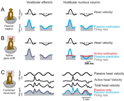
Cullen and colleagues have further shown that in contrast to central neurons, Rhesus vestibular
afferents similarly (and robustly) encode vestibular reafference and exafference (Cullen and Minor 2002; Sadeghi et al. 2007; Jamali et al. 2009) Thus, in primates the role of the vestibular efferent system does not appear to modulate the sensitivity and/or resting discharge of the end-organ response to active movement as had been previously suggested (Goldberg 2000). Figure 3 compares the response of a typical semicircular canal primary vestibular afferent input to the brainstem (left columns) along with the response of a central VO neuron (right columns) during passive, active and combined head rotations (Roy and Cullen 2001; Cullen et al. 2009). Firing rate data is shown in gray, and head velocity in black. As shown in the top row of Figure 3, if the seated animal’s head and body are passively rotated together on a turntable, the semicircular canal afferent and VO neuron response are almost identical,
demonstrating that VO cells respond vigorously to exafferent stimulation. Responses to a similar active head movement are shown in the middle row of Figure 3. The active vestibular afferent response is identical to the passive response. However the response of VO neurons – often only one synapse more central – is greatly attenuated. The blue line in the figure estimates what the neuron response would be for the identical passive stimulation. The bottom row of Figure 3 shows responses when the animal was allowed to make volitional head movement during passive rotation. Vestibular afferents respond to total head velocity as expected. However the active component of the VO neuron response is absent. The VO neuron only responds to the passive component of total head velocity. This example
demonstrates that the cancellation mechanism does not simply gate-out the semicircular canal signal, but is instead selectively cancels the active component.
Fig 3 Rhesus semicircular canal primary afferent (left columns) and brainstem vestibular nucleus VO
neuron (right columns) firing rate data (gray), during passive (top rows), active (middle rows) and combined active and passive (lower rows) angular stimulation (black). See text for details. After: Cullen et al 2009
Cullen and colleagues have recently reported similar results from vestibular afferent and VO neurons in the vestibular nuclei during passive, active and combined head translations (Jamali et al. 2009; Carriot et al. 2013). Figure 4 shows the responses of two example Rhesus otolith driven VO neurons (gray) to active translation movements in naso-occipital (A) and inter-aural (B) directions (Carriot et al. 2013).
The first, a purely otolith driven VO cell is shown in the upper row. The second responded to both rotation and translation and is shown in the bottom row. Neural activity predictions based on each neuron’s sensitivity to passive translation as previously recorded using a linear sled stimulus are superimposed in blue, and demonstrate the dramatic reduction in sensitivity of both types of units to active translations compared to the corresponding passive movement. Panel C compares the directions of maximal sensitivity (white arrows) and the spatial sensitivity tuning curves for active (red area) and passive (blue area) linear accelerations.
Fig 4 Activity of an otolith only VO neuron (upper row) and canal-otolith convergent VO neuron (lower
row) activity (gray) during active (self-generated) naso-occipital (A) and interaural (B) head translations. (C) Comparison of the tuning curves computed during self-generated head motion (red area) and those computed during passive head motion (blue area) From Carriot et al 2013
On average, during active motion, the responses of VO neurons are attenuated by 70 and 61% for rotations and translations, respectively. Cullen and colleagues have carried out a systematic series of experiments (not shown) demonstrating that in order for otolith as well as semicircular canal
reafference cancellation to occur, proprioceptive afference must closely match reafference (Roy and Cullen 2004; Carriot et al. 2013; Brooks and Cullen, 2014). Notably, these data are consistent with the
model in Figure 2, showing that proprioceptive mismatches influence vestibular reafference cancellation, as one might expect.
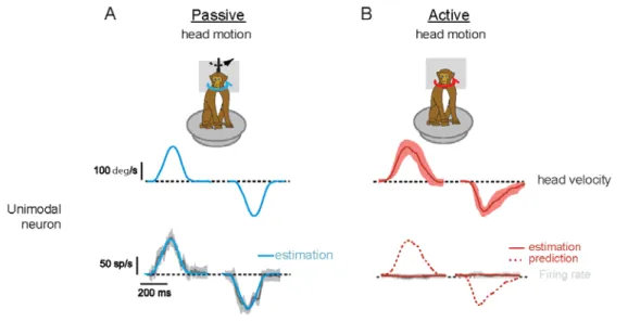
To understand the mechanism responsible for the suppression of vestibular reafference in the vestibular nuclei, Brooks and Cullen (2013) next recorded from neurons in the cerebellum. In particular, recordings were made from the rostral fastigial nucleus (rFN), which is the most medial of the deep cerebellar nuclei, and projects strongly to the vestibular nuclei, as well as to the reticular formation and spinal cord. One class of rFN neurons - responding only to passive vestibular stimulation and called
“unimodal” (u-rFN) - encode passive head movement even during concomitant active movement in a manner analogous to brainstem VO neuron. (Note, a second class of rFN neurons – responding to both vestibular and proprioceptive input and called “bimodal” - encode the position of the body in space, rather than passive head movement (Brooks and Cullen 2009).) Figure 5 shows an example of Rhesus u-rFN activity during passive (blue) and active (red) rotations. The top row illustrates average head velocities for ten movements, plus or minus one standard deviation (shading) for the trained active movements. The bottom row shows average firing rate (dark line), plus or minus one standard deviation (shading). The blue and dashed red line overlays show an estimate of the neurons previously recorded sensitivity to passive rotation on a turntable. Consistent with their hypothesis that the cerebellum plays a key role in the suppression of vestibular reafference, this neuron’s response to self-generated activity was minimal.
Fig 5 Unimodal rostral Fastigial Nucleus neuron activity in Rhesus cerebellum during passive (A blue)
and active (B red) head rotations. Top row: average head velocities for ten movements, and shaded area shows +/- one SD Bottom row: average firing rates (dark line) and one SD (shading). Details in text. Adapted from Brooks, et al 2013, Figure 1
Because these neurons are the output neurons of the cerebellum, this result provided the first evidence that computations in the cerebellum (cortex and deep nuclei) provide a precise estimate of the detailed
time course of exafference - even when experienced concurrently with active motion. In addition, both VO and u-rFN neurons participate in postural reflex stabilization of the head and body. Accordingly, the fact that they exhibit reafference cancellation means that they can do so without the associated reflexes impeding active movement. These neurons are also likely to project directly or indirectly to thalamus, and on to sensory regions of cerebral cortex where they may contribute to orientation and motion perception, perhaps indirectly by influencing internal model predictions as suggested by the models of Oman (1990) and Wolpert et al. (1998). Certainly there are other populations of vestibularly driven neurons in brainstem and deep cerebellar nuclei that do not exhibit reafference cancellation, for instance the responses of neurons in the vestibular nuclei that that mediate the vestibulo-ocular reflex, subserving other functions that are not attenuated during active motion.
Conclusions and Open Questions
An improved understanding the neural substrate mediating motion sickness is required for more effective, scientifically based methods for prevention and treatment. Below we discuss a number of open questions and suggest future research to provide further insight into the underlying neural mechanisms.
Do the brainstem and/or the cerebellar neurons that exhibit reafference cancellation project to emetic and nausea centers? If they do, this may offer a solution to the motion sickness puzzle, since it would account for the immunity of humans and animals to self -generated movement, the therapeutic effectiveness of head restraint and horizontal postures, the relative immunity of drivers and pilots, and the role of sensorimotor learning in motion sickness adaptation. It would also be parsimonious with Wang and Chinn’s trans-cerebellar theory for motion sickness, Reason and Oman’s sensory conflict hypotheses, and even Stoffregen’s evidence of relationships between posture control and motion sickness susceptibility. Because there is a persistent conflict (i.e., mismatch) between expected and actual sensory motion during active movements following vestibular sensory loss, or prolonged exposure to passive motion or conditions of sensory rearrangement, including weightlessness, we speculate that VO and u-rFN neurons display robust activity in such conditions. Yates and coworkers (Suzuki et al. 2012) suggest that pathways from vestibular nucleus cells project to the parabrachial nucleus (PBN), and then on to limbic cerebral forebrain areas responsible nausea and affective changes, whereas projections from vestibular nucleus to the nucleus tractus solitaries (NTS) and PBN and on to the lateral tegmental field (LTF) in the dorsolateral reticular formation initiate vomiting. They also found that gastrointestinal afferents project to some regions of vestibular nucleus. However their experiments were conducted on decerebrate, cerebellectomized, paralyzed animals. Hence it is not yet known whether brainstem VO neurons exhibiting reafference cancellation are the same neurons that project to these putative nausea and vomiting pathways. Demonstrating that neurons exhibiting reafference cancellation anatomically project to emetic centers is an important first step. However this is a challenge since it requires the identification of VO and u-rFN neurons in alert, behaving animals. Is the cerebellum essential for motion sickness susceptibility, as Wang and Chinn (1956) asserted ? On the one hand, based on present knowledge cerebellectomy should disrupt reafference cancellation and cerebellar sensory-motor learning. While the brainstem vestibular-emetic pathway would remain
intact, the source of the internal representation of expected sensory inflow would be eliminated. On the other hand, both Miller and Wilson (1983a) and (Uno et al. 2000) concluded that cerebellectomy (i.e., posterior vermis lesions) did not always not eliminate susceptibility to motion sickness. Future experiments examining the short and long term consequences of cerebellar ablation will be needed to further address this question.
Does sustained sensory conflict (e.g. encoded by neurons in the cerebellum and/or brainstem
exafference) initiate sensorimotor learning as proposed by Held, Reason and Oman? Ito’s (1970, 2000) theory for cerebellar motor learning endorsed the concept of an internal model but suggested that the adaptive drive was a motororic error signal descending from cerebral cortex via the inferior olive and then transmitted via climbing fibers to the cerebellar cortex. Wolpert et al. (1998) noted that although climbing fibers may appear to respond, at least in part, to motor errors during reaching and eye movements, in other systems, reafference cancellation pathways may drive climbing fibers, e.g. (Gellman et al. 1985). While the computation of a reafference cancellation signal is evident at the level of the vestibular nucleus and deep cerebellar nuclei, it remains uncertain what information is inherited from the Purkinje cells output versus which component of the computation is subsequently done within each nuclei and/or via the reciprocal connections between them.
Finally, several other important questions remain: Do other areas of cerebellum exhibit reafference cancellation ? Do brainstem VO neurons also respond differentially to active and passive roll and pitch rotations ? To tilt as well as translation ? Other than passive and active rotations and translations, what other sensory stimuli activate vestibular neurons exhibiting reafference cancellation in brainstem and cerebellum ? Oman (1990) proposed that widescreen movie sickness might result because the visual scenes are so compelling they create a vestibular efference copy outflow signaling tilt, translation or rotation, and that this was the effective stimulus for sickness as well as motor outflow. Similarly, standing subjects, who are relatively more visually dependent, are more likely to experience motion sickness symptoms while viewing an oscillating visual scene. Perhaps this occurs because the expected vestibular input does not match the actual afference produced by the nearly motionless body, and in turn this mismatch leads to a disturbance of posture and eventually motion sickness Stoffregen and Smart (1998) and Owen et al. (1998) If so, this may explain why postural sway is a marker for motion sickness susceptibility in this situation, as noted by Stoffregen. So far our attempts to activate semicircular canal VO neurons in Rhesus using moving stripe optokinetic stimuli have not been successful, and linear stimulation corresponding to that used by Stoffregen et al has not yet been attempted. Interestingly, recent research shows that VO neuronal responses to active motion are suppressed is across a wide range of species - spanning from mice (Medrea and Cullen 2013) to monkeys (reviewed in Cullen (2014). This suggests that the suppression of vestibular reafference at the earliest stages of sensory processing is a common evolutionary strategy.
In conclusion, we suggest that a fuller characterization of VO neuron and cerebellar reafferent cancellation and adaptation mechanisms and physiology/pharmacology of the VO neuron to NTS/RF emetic linkage should be a research priority. A complete physiologic definition of motion sickness – particularly the physiology of the vestibular-emetic linkage and cerebellar adaptation could eventually lead to more effective and scientifically based behavioral and pharmacologic countermeasures.
Acknowledgments
Dr. Oman was supported in part by the National Space Biomedical Research Institute through
NASA NCC 9-58. Dr. Cullen’s research was supported by the Canadian Institutes of Health
Research (CIHR), the National Institutes of Health (DC002390), and the Fonds Que´be´cois de la
Recherche sur la Nature et les Technologies (FQNRT), and US National Institute of Health Grant
R01 DC2390..
References
Bell CC, Han V, Sawtell NB (2008) Cerebellum-like structures and their implications for cerebellar function. Annual Review of Neuroscience 31:1-24 doi:
10.1146/annurev.neuro.30.051606.094225
Blakemore SJ, Wolpert DM, Frith CD (1999) The Cerebellum Contributes to Somatosensory Cortical Activity during Self-Produced Tactile Stimulation. Neuroimage 10:448-459
Bock OL, Oman CM (1982) Dynamics of subjective discomfort in motion sickness as measured with a magnitude estimation method. Aviation, Space, and Environmental Medicine 53:733-737 Borison HL, Borison R (1986) Motion sickness reflexarc bypasses the area postrema in cats. Exp Neurol
92:723-737
Bos JE, Bles W (1998) Modeling motion sickness and subjective vertical mismatch detailed for vertical motions. Brain Res. Bull 47:537-542
Brooks JX, Cullen KE (2009) Multimodal integration in rostral fastigial nucleus provides an estimate of body movement. J Neurosci 29:10499-10511 doi: 10.1523/JNEUROSCI.1937-09.2009
Brooks JX, Cullen KE (2013) The primate cerebellum selectively encodes unexpected self-motion. Current Biology 23:947-955 doi: 10.1016/j.cub.2013.04.029
Brooks JX, Cullen KE (2014) Early vestibular processing does not distinguish active from passive self motion if there is a discrepancy between predicted and actual proprioceptive feedback. J. Neurophysiol doi: 10.1152/jn.00600.2013
Carriot J, Brooks JX, Cullen KE (2013) Multimodal Integration of Self-Motion Cues in the Vestibular System: Active versus Passive Translations. Journal of Neuroscience 33:19555-19566 doi: 10.1523/JNEUROSCI.3051-13.2013
Claremont CA (1931) The psychology of sea-sickness. Psyche 11:86-90
Cullen KE (2012) The vestibular system: multimodal integration and encoding of self-motion for motor control. Trends in Neurosciences 35:185-196 doi: 10.1016/j.tins.2011.12.001
Cullen KE (2014) The neural encoding of self-generated and externally applied movement: implications for the perception of self-motion and spatial memory. Front Integr Neurosci 7:108 doi:
10.3389/fnint.2013.00108
Cullen KE, Brooks JX, Sadeghi SG (2009) How actions alter sensory processing: reafference in the
vestibular system. Annals of the New York Academy of Sciences 1164:29-36 doi: 10.1111/j.1749-6632.2009.03866.x
Cullen KE, Minor LB (2002) Semicircular canal afferents similarly encode active and passive head-on-body rotations:implications for vestibular efference. J. Neuroscience 22:RCC226(221-227) Dong X, Yoshida K, Stoffregen TA (2011) Control of a virtual vehicle influences postural activity and
Gellman R, Gibson AR, Houck JC (1985) Inferior Olivary Neurons in the Awake Cat: Detection of Contact and Passive Body Displacement. J. Neurophysiol 54:40-60
Goldberg JM (2000) Afferent diversity and the organization of central vestibular pathways. Experimental Brain Research 130:277-297
Golding JF, Stott JRR (1997) Objective and subjective time courses of recovery from motion sickness assessed by repeated motion challenges. Journal of Vestibular Research 7:421-428
Guedry FE (1968) Conflicting sensory orientation cues as a factor in motion sickness. In: Graybiel A (ed) Fourth Symposium on the Role of the Vestibular Organs in Space Exploration, vol NASA SP-187. US Government Printing Office, Pensacola, FL, pp 45-51
Haslwanter T, Jaeger R, Mayr S, Fetter M (2000) Three-dimensional eye-movement responses to off-vertical axis rotations in humans. Exp Brain Res 134:96-106
Held RM (1961) Exposure history as a factor in maintaining stability of perception and coordination. J. Nerv. Ment. Dis. 132:26-32
Ito M (1970) Neurophsyiological aspects of the cerebellar motor control system. Int. J. Neurol. 7:162-176 Ito, M. (2000). "Mechanisms of motor learning in the cerebellum." Brain Research 886: 237-245.
Jamali M, Sadeghi SG, Cullen KE (2009) Response of vestibular nerve afferents innervating utricle and saccule during passive and active translations. Journal of Neurophysiology 101:141-149 doi: 10.1152/jn.91066.2008
Johnson WH, Mayne R (1953) Stimulus required to produce motion sickness: restriction of head movment as a preventive of airsickness - field studies of airbourne troops. Journal of Aviation Medicine:400-411, 452
Kennedy AR, Hettinger LJ, Lilllienthal MG (1990) Simulator sickness. In: Crampton GH (ed) Motion and Space Sickness. CRC Press, Boca Raton, FL, pp 317-341
Manning GW, Stewart WG (1949) Effect of body position on the incidence of motion sickness. Journal of Applied Physiology 1:619-628
McCrea RA, Gdowski GT, Boyle R, Belton T (1999) Firing Behavior of Vestibular Neurons During Active and Passive Head Movements: Vestibulo-Spinal and Other Non-Eye-Movement Related Neurons. J. Neurophysiol 82:416-428
Medrea I, Cullen KE (2013) Multisensory integration in early vestibular processing in mice: the encoding of passive vs. active motion. J. Neurophysiol 110:2704-2717 doi: 10.1152/jn.01037.2012.-The Merfeld DM, Young LR, Oman CM, Shelhamer MJ (1993) A multidimensional model of the effect of
gravity on the spatial orientation of the monkey. J Vestib Res 3:141-161
Merfeld DM, Zupan LH (2002) Neural processing of gravitoinertial cues in humans. III. Modelling tilt and translation responses. J. Neurophysiol. 87:819-833
Miller AD, Wilson VJ (1983a) Vestibular-induced vomiting after vestibulocerebellar lesions. Brain, Behavior and Evolution 23:26-31
Miller AD, Wilson VJ (1983b) 'Vomiting center' reanalyzed: an electrical stimuration study. Brain Research 270:154-158
Money KE (1970) Motion sickness. Physiol. Rev. 50:1
Oman CM (1982) A heuristic mathematical model for the dynamics of sensory conflict and motion sickness. Acta Otolaryngologica (Stockholm) 94(S392):4-44
Oman CM (1990) Motion sickness: a synthesis and evaluation of the sensory conflict theory. Can. J. Physiol. Pharmacol. 68:294-303
Oman CM (1991) Sensory conflict in motion sickness: an Observer Theory approach. In: Ellis S (ed) Pictorial communication in real and virtual environments. Taylor and Francis, London, pp 362-367
Oman CM (2012) Are evolutionary hypotheses for motion sickness "Just-So" stories ? Journal of Vestibular Research 22:117-127
Owen N, Leadbetter AG, Yardley L (1998) Relationship between postural control and motion sickness in healthy subjects. Brain Res Bull 47:471-474
Reason JT (1969) Motion sickness - some theoretical considerations. Int. J. Man Machine Studies 1:21-38 Reason JT (1978) Motion sickness adaptation: a neural mismatch model. J. R.. Soc. Med. 71:819-829 Reason JT, Brand JJ (1975) Motion Sickness. Academic Press, London
Rolnick A, Lubow RE (1991) Why is the driver rarely motion sick? The role of controllability in motion sickness. Ergonomics 34:867-879 doi: 10.1080/00140139108964831
Roy EA, Cullen KE (2001) Selective processing of vestibular reafference during self-generated head motion. J. Neuroscience 21:2131-2142
Roy EA, Cullen KE (2002) Vestibuloocular Reflex Signal Modulation During Voluntary and Passive Head Movements. J. Neurophysiol 87:2237-2357
Roy JE, Cullen KE (2004) Dissociating self-generated from passively applied head motion: neural mechanisms in the vestibular nuclei. J. Neuroscience 24:2102-2111
Sadeghi SG, Minor LB, Cullen KE (2007) Response of vestibular-nerve afferents to active and passive rotations under normal conditions and after unilateral labyrinthectomy. Journal of
Neurophysiology 97:1503-1514 doi: 10.1152/jn.00829.2006
Sadeghi SG, Mitchell DE, Cullen KE (2009) Different neural strategies for multimodal integration: comparison of two macaque monkey species. Exp Brain Res 195:45-57 doi: 10.1007/s00221-009-1751-3
Selva P, Oman CM (2012) Relationships between Observer and Kalman filter models for human dynamic spatial orientation. Journal of Vestibular Research 22:69-80
Smart LJ, Stoffregen TA, Bardy BG (2002) Visually Induced Motion Sickness Predicted by Postural Instability. Human Factors 44:451-465
Steele JE (1963) Motion sickness and spatial perception:a theoretical study. In, vol AMRL-TDR-63-25. U.S. Army Aeromedical Research Laboratory, Wright Patterson AFB, Ohio
Stoffregen TA, Riccio GE (1991) An ecological critique of the sensory conflict theory of motion sickness. Ecological Psychology 3:159-194
Suzuki T, Sugiyama Y, Yates BJ (2012) Integrative responses of neurons in parabrachial nuclei to a nauseogenic gastrointestinal stimulus and vestibular stimulation in vertical planes. Am J Physiol Regul Integr Comp Physiol 302:R965-975 doi: 10.1152/ajpregu.00680.2011
Treisman M (1977) Motion Sickness: an evolutionary hypothesis. Science 197 (493-496) Tyler DB, Bard P (1949) Motion Sickness. Physiological Reviews 29:311-369
Uno A, Takeda N, Kitahara T, Sakata Y, Yamatodani A, Kubo T (2000) Effects of Vestibular Cerebellum Lesion on Motion Sickness in Rats. Acta Otolaryng. (Stockh.) 120:386-389
Vingerhoets RAA, Van Gisbergen JAM, Medendorp WP (2007) Verticality perception during off-vertical axis rotation. J. Neurophysiol 97:3256-3268
Von Holst E (1954) Relations between the central nervous system and the peripheral organs. British Journal of Animal Behavior 2:89-94
Wang SC, Chinn HI (1956) Experimental motion sickness in dogs: Importance of labyrinth and vestibular cerebellum. Am. J. Physiol. 185
Wolpert DM, Ghahramani Z, Jordan MI (1995) An internal model for sensorimotor integration. Science 269:1880
Wolpert DM, Miall RC, Kawato M (1998) Internal models in the cerebellum. Trends in Cognitive Sciences 2:338-347
Yates BJ, Miller AD, Lucot JB (1998) Physiological basis and pharmacology of motion sickness: an update. Brain Res Bull 47:395-406
Zupan LH, Merfeld DM, Darlot C (2002) Using sensory weighting to model the influence of canal, otolith and visual cues on spatial orientation and eye movements. Biol. Cybern. 86:209-230