ABSTRACTS
Abstracts of the 6th Cachexia Conference, Milan, Italy,
December 8
–10, 2011
1-01
Leucine modulates the effects of the Walker tumour’s proteolysis-inducing factor on gene expression and cells activity in C2C12 myotubes
Estela M. Gonçalves, Emilianne M. Salomão, Maria Cristina C. Gomes-Marcondes (Nutrition and Cancer Laboratory, Department of Physiology and Biophysics, IB, UNICAMP, Campinas, SP, Brazil)
Cancer cachexia involves a severe weight lost, particularly from waste of skeletal muscle, which can arise from the increase on protein catabolism and/or the decrease on protein synthesis. The protein degradation in skeletal muscle of cancer patients is mediated by the cytokine proteolysis-inducing factor (PIF), produced by tumour, which increases the expression of the ubiquitin–proteasome pathway, and the skeletal muscle protein synthesis in these patients can be affected by several agents, including nutrient signalling. Some nutrients, such as branched-chain amino acid leucine, can decrease the expression of the ubiquitin–proteasome pathway and improve the protein content in skeletal muscle of cachectic animals. In this study, we investigated the effects of leucine on cell viability, morphology, functional proteasome activity, enzymatic activities, and protein synthesis and degradation in C2C12skeletal muscle cells exposed to the PIF-like protein, purified from the Walker tumour-bearing rat. The Walker factor (WF), immunologically identical to PIF and with the same molecular weight, presented no cytotoxic effect in myotube cells, which had no alterations in their morphological characteristics in the presence of WF and/or leucine. However, increased phosphatase alkaline activity was observed, especially at low WF concentrations. At higher WF concen-trations, chymotrypsin-like activity and also the 20S proteasome gene expression were increased, as well as the cathepsin B activity trended to increase. Adding leucine previously to WF-treated myotubes cells, proteasome activity decreased and phosphatase alkaline activity increased. Total protein synthesis was decreased in WF-treated cells in parallel to increase in protein degradation. These changes were minimized or reverted after leucine exposure. Taken together, these results suggested an important modulatory effect of leucine under the WF actions in C2C12myotube cells. 1-02
Habitual skeletal muscle protein fractional synthetic rate in healthy individuals as determined by a novel oral tracer technique
Alisdair J. MacDonald1, Carolyn A. Greig2, Holger Husi1, Nathan A. Stephens1, Jim Ross1, Alexandra C. Small3, Kenneth C.H. Fearon1, Tom Preston3 (1Tissue Injury and Repair Group, University of Edinburgh,
Edinburgh, UK;2Department of Clinical and Surgical Sciences, University of Edinburgh, Edinburgh, UK;3Stable Isotope Biochemistry Laboratory, Scottish Universities Environmental Research Centre, East Kilbride, UK) Background/aims: Using current methodology skeletal muscle fractional synthetic rate (FSR) is measured over short time periods in response to specific stimuli (e.g. feeding/exercise) resulting in wide variation in FSRs. In clinical studies, interventions occur over weeks or months and measures over longer periods may be more representative. We aimed to develop a novel method to determine skeletal muscle protein FSR to estimate habitual FSR in healthy individuals, over timescales comparable with clinical interventions, avoiding intravenous amino acid tracer.
Method: Four healthy males, median (range) age: 37 years (32–52), height 179 cm (177–185), weight 80 kg (71–87) were given 100 g water enriched to 70 atom% with 2H2O as a single oral bolus. Vastus lateralis biopsies were performed using a Bergstrom needle at intervals 4–12 days post-dose. Serum was collected at baseline and three time-points between 3 and 14 days, measuring body water enrichment and analysis of plasma alanine enrichment (GC–pyrolysis–IRMS). Myofibrillar protein was isolated, acid hydrolysed and 2H-alanine enrichment measured. Skeletal muscle protein FSR was calculated (% day−1) both using free 2H-alanine and body water to predict precursor enrichment.
Results: Body water 2H increased to 1520 ppm excess (1,435–1,582). Elimination half time was 8 days (7–10). The r2 for the natural log of 2H enrichment against time was >0.999 in each individual. Plasma alanine was labelled in a predictable manner (in theory, four atoms become labelled; 3.1–4.2 were measured). Skeletal muscle FSR was calculated from six biopsies in four individuals as 1.18% day−1 (0.94–1.59). For the two individuals with two biopsies each at different times, the differences in estimates of FSR were 14.3% and 20%.
Conclusions: This is the first study to describe skeletal muscle protein FSR in free-living healthy individuals over 4–12 days. Using a single oral 2H2O bolus, endogenous labelling of alanine occurs in a predictable manner giving estimates of FSR comparable with published values. 1-03
Environmental conditions in muscle tissue culture alter responses to atrophy signals
Elodie Archer-Lahlou1, Cathy Lan1, Robert T. Jagoe2 (1Segal Cancer Centre, Lady Davis Institute for Medical Research, Jewish General Hospital, Montreal, Quebec, Canada,2McGill University Department of Oncology, Segal Cancer Centre, Lady Davis Institute for Medical Research, Jewish General Hospital, Montreal, Quebec, Canada) Published online: 8 November 2011
Muscle atrophy is a powerful determinant of poor prognosis, impaired physical function, and debility in all chronic diseases, including cancer. Laboratory models of muscle wasting have suggested a number of different mechanisms which can contribute to cancer-related muscle atrophy. However, paradoxically, very little progress has been made in developing therapies which are clinically effective in treating muscle wasting in cancer patients. Muscle tissue culture still has huge potential to help develop new treatments but the factors which have impeded the effective translation of basic research into clinical treatments need to be identified and addressed. For this reason, we studied the effects of using tissue culture conditions that reproduce the in vivo physiological environment of muscles. We first established two highly defined and reproducible muscle tissue culture systems using C2C12 myotubes under standard and modified conditions. These modified conditions have been chosen to attempt to mimic certain key aspects of the in vivo environment experienced by muscle in cancer patients, on the assumption that this may promote more representative adaptive responses. We then assessed whether such modified conditions alter the responses of mature myotubes to distinct atrophy-inducing treatments. After 3 days of treatment, we observed a 20–30% reduction in protein and myotube diameter but a 46% increase in myotube number in modified conditions compared to standard culture conditions. Only small differences in most atrophy-related gene expression or protein levels for markers of muscle growth and phenotype were observed but modified conditions did induce a 70% reduction in IGF-1 mRNA levels. However, surprisingly, the modified culture conditions offered partial or complete protection against further atrophy due to added atrophy-inducing treat-ments. This protection was observed despite similar changes in the transcript levels of selected atrophy-related genes. These studies confirm the potential for environmental conditions to modulate muscle responses to atrophy-inducing conditions in vivo.
1-04
During early recovery the worsening of immobilization-induced rat tibialis anterior muscle atrophy is associated with sustained activation of proteolytic and apoptotic pathways
Lamia Slimani1,2, Anabelle Dubost2, Bruno Meunier2, Dominique Dardevet1, Anne Listrat2, Lydie Combaret1, Didier Attaix1 (1Clermont Université, Université d’Auvergne, Unité de Nutrition Humaine, BP 10448, F-63000 Clermont-Ferrand, INRA, UMR 1019, UNH, CRNH Auvergne, F-63000 Clermont-Ferrand; 2INRA, UMR 1213, URH, F-63122 Saint Genès Champanelle)
Background and aims: Muscle atrophy induced by immobilization stabilized in the shortened gastrocnemius muscle (GA), but worsened in the lengthened tibialis anterior muscle (TA) after 10 days of recovery. Thus, important remodeling within the TA following immobilization could condition the initial steps of recovery and may depend on the position of immobilization. The ubiquitin–proteasome system (UPS) and the mitochondria-associated apoptosis being involved in GA atrophy their role in muscle remodeling were studied during the very early stages of recovery in both the TA and GA.
Methods: Adult rats were subjected to unilateral hindlimb immobiliza-tion and the contralateral non-immobilized leg served as the control. After 8 days of immobilization (I8), casts were removed and animals were allowed to recover for 1 (R1) to 10 (R10) days. UPS-dependent proteolysis and apoptosis were evaluated in both the TA and GA. Results: Muscle atrophy rapidly worsened after cast removal as soon as R1 and up to R6 in the TA previously immobilized, but stabilized in the GA from R1. In addition, a more pronounced and sustained activation of proteasome and apoptosome activities prevailed in the immobilized TA from I8 until R10, but was normalized at R6 in the previously immobilized GA.
Conclusions: Altogether, the data suggest that UPS-dependent proteolysis and mitochondria-associated apoptosis are involved in the muscle remodeling during recovery and may be responsible for the worsening of muscle atrophy observed in the TA. Alterations of connective tissue being differentially altered upon stretching during immobilization (Mattiello-Sverzut et al. Histol Histopathol 2006; 21:957–964), we hypothesized that this may impact muscle proteolytic signaling pathways.
1-05
Msy-3/Csda coordinatesrepression of myogenic genes in muscle wasting
Alessandra Feraco1, Georgi Marinov3, Luciana De Angelis2, Elisabetta Ferraro1, Gilberto Hernandez3, Barbara J. Wold3, Libera Berghella1 (1IRCCS San RaffaelePisana Institute, Roma, Italy; 2University La Sapienza, Dept. of Hystology and Medical Embryology, Roma, Italy; 3
California Institute of Technology, Pasadena, USA)
Background and aims: The Y-box protein MSY-3/Csda regulates postnatal repression of the myogenic transcription factor myogenin. In postnatal muscle, myogenin plays a key role in regulating pathways involved in muscle maturation, degeneration and eventually cachexia. MSY-3 binds a highly conserved DNA cis-acting element located upstream of the myogenin promoter. We wanted to find other possible targets of MSY-3 regulated in concert during muscle maturation and atrophy.
Methods: ByChIP assay, we analyzed MSY3/Csda binding in vivo in adult muscle and in C2C12. We tested the MSY-3/MyoD conserved site transcriptional activity by luciferase assay and in vivo electro-poration. By high-throughput technology, we genome-wide analyzed MSY-3 DNA binding in adult muscle and expression profile of wt and MSY-3 knockout mice and denervated muscle.
Results: We found a conserved regulative cis-element in the MyoD locus, that matches with the myogenincis-motif and by ChIP assays we verified that both MyoD and MSY-3/Csda bind to it in vivo. Luciferase assays show that the MYOD site, is sufficient for high levels of expression in C2C12 and MSY-3 acts as a repressor. In vivo muscle electroporation demonstrates that the MYOD site is required for MyoD postnatal repression. Furthermore, in combined genome-wide analysis of MSY-3 DNA binding and global RNA expression, we found other candidate genes possibly repressed by MSY-3 during adult muscle maturation, as well as myogenin, MYOD, AChRs and HDAC4.
Conclusions: This study suggests that both MyoD and myogenin are controlled by the same repressor complex (MSY-3/Csda), recognizable by a similar cis-motif. This motif is responsible for MYOD auto activation/ maintenance during muscle differentiation and its repression MSY-3-mediated in postnatal muscle, suggesting a developmental stage depending competition for the two transcription factors at the same site. This strongly suggests that MSY-3 regulates in concert MYOD and myogenin expression. Moreover, the genome-wide analysis highlights a group of genes possibly modulated by the same regulatory complex during muscle maturation and degeneration. These findings indicate MSY3/Csda as a potential target for muscle wasting caused by neurogenic atrophy, sarcopenia, and cachexia.
1-06
Resveratrol blocks glucocorticoid-induced muscle wasting in L6 cultured myotubes through a SIRT1-dependent mechanism
Nima Alamdari, Zaira Aversa, Estibaliz Castillero, Aniket Gurav, Sreven Tizio, Per-Olof Hasselgren (Department of Surgery, Beth Israel Deaconess Medical Center, Harvard Medical School, Boston, MA, USA)
Background and aims: Muscle proteolysis during sepsis and other catabolic conditions is, at least in part, regulated by glucocorticoids. Dexamethasone-treated myotubes are a commonly used in vitro model of muscle wasting. We reported recently that decreased expression and activity of histone deacetylases (HDACs) including sirtuin (SIRT) 1 are involved in the development of muscle wasting. Here, we tested the hypothesis that treatment with the SIRT1 activator resveratrol would prevent glucocorticoid-induced protein degrada-tion and muscle atrophy, and that glucocorticoid-induced expression of atrogin-1 and MuRF1 is, at least in part, regulated by SIRT1.
Methods: L6 myotubes were treated for 24 h with 1μM dexamethasone in the absence or presence of resveratrol (100μM). After treatment for 24 h, myotubes were harvested for determination of atrogin-1 and MuRF1 mRNA and protein levels, protein degradation, and myotube diameter. In additional experiments, the effects of resveratrol treatment were tested in myotubes transfected with non-targeting or SIRT1 siRNA.
Results: Treatment with dexamethasone increased atrogin-1 and MuRF1 expression and protein degradation and reduced myotube diameter. Resveratrol treatment inhibited the dexamethasone-induced increase in atrogin-1 and MuRF1 expression and protein degradation as well as the reduction in myotube size. SIRT1 siRNA abolished the effect of resveratrol on atrogin-1 and MuRF1 expression.
Conclusions: Results suggest that resveratrol treatment may prevent glucocorticoid-induced muscle wasting through a SIRT1-dependant mech-anism. SIRT1 activation may be a novel therapeutic approach in combating muscle wasting conditions. Supported by R01 DK37908 from the NIH. 1-07
Decreased NADPH oxidase expression and antioxidant activity in cachectic skeletal muscle
Melanie J. Sullivan-Gunn, Paul A. Lewandowski (Deakin University, Medical School, Geelong, Australia)
Background: Cancer cachexia is the progressive loss of skeletal muscle protein that contributes significantly to cancer morbidity and mortality. Evidence of antioxidant attenuation and the presence of oxidised proteins in patients with cancer cachexia indicate a role for oxidative stress. This study aimed to investigate the superoxide generating NADPH oxidase (NOX) enzyme and antioxidant enzyme systems in murine adenocarcinoma tumour-bearing cachectic mice. Method: Superoxide levels, mRNA levels of NOX enzyme subunits, and the antioxidant enzymes were measured in the skeletal muscle of mice with cancer and cancer cachexia. Protein expression levels of NOX enzyme subunits and antioxidant enzyme activity were also measured in these samples.
Results: Superoxide levels increased 1.4-fold in the muscle of mice with cancer cachexia, and this was associated with a decrease in mRNA of NOX enzyme subunits, NOX2, p40phox, and p67phox along with the antioxidant enzymes SOD1, SOD2, and GPx. Cancer cachexia was also associated with a 1.3-fold decrease in SOD1 and 2.0-fold decrease in GPx enzyme activity. Conclusion: Despite increased superoxide levels in cachectic skeletal muscle, NOX enzyme subunits, NOX2, p40phox, and p67phox, were downregulated along with the expression and activity of the antioxidant enzymes. Therefore, the increased superoxide levels in cachectic skeletal muscle may be attributed to the reduction in the activity of endogenous antioxidant enzymes. 1-08
Differential adaptation of glycolytic and oxidative muscle to hypoxia Chiel C. de Theije1, Ramon C.J. Langen2, Eleonore S. Kohler1, Wouter H. Lamers1, Annemie M. W. J. Schols2 (1Department of Anatomy & Embryology, NUTRIM, Maastricht University, Maastricht, The Netherlands; 2Department of Respiratory Medicine, NUTRIM, Maastricht University, Maastricht, The Netherlands)
Background: Hypoxia may be a trigger of skeletal muscle atrophy in acute and chronic respiratory disease. In chronic obstructive pulmo-nary disease patients, muscle fiber atrophy is most pronounced in glycolytic compared to oxidative muscle fibers. Therefore, we hypothesized that hypoxia preferentially affects glycolytic muscles. Methods: Mice were maintained under normoxia or hypoxia. During 48 h, oxygen levels were reduced stepwise to 8%, which was maintained for 21 days. Food intake was monitored daily and mice were sacrificed at days 4 and 21.
Results: Pair-feeding experiments revealed that the reduction in oxidative (Soleus) muscle weight was solely attributable to hypoxia-induced reduced food intake (hypophagia), whereas hypoxia caused a greater decrease of the glycolytic muscle weight, which was not accounted for by hypophagia. Furthermore, cross-sectional area (CSA) of the individual fibers revealed reduced CSA in glycolytic muscle, whereas no reduction was seen in the oxidative muscle. Expression levels of genes involved in proteasomal protein degradation (Atrogin-1), lysosomal protein degradation (Lc3B, Atg5), endoplasmic reticu-lum (ER) stress signaling (Atf4 and Gadd34), and glucocorticoid receptor (GR) signaling (Glul and GR) were increased by hypoxia in both muscles. Interestingly, only in glycolytic muscle, the upregulation involved a hypoxia-specific component and was not merely explained by hypophagia. Furthermore, capillary contacts per muscle fiber CSA, increased in both muscles in response to hypoxia. In the oxidative muscle, this was the result of a reduction in interstitial space, whereas this adaptation was accounted for by decreased fiber CSA in the glycolytic muscle.
Conclusions: Muscle mass, proteolysis, ER stress, and GR signaling in the glycolytic muscle are more affected than in the oxidative muscle by hypoxia. In addition, in oxidative, but not in the glycolytic muscle, these alterations are accounted for by hypophagia, indicative of an intrinsic sensitivity of glycolytic muscle to hypoxia, which may involve a differential adaptive capacity of the vascular bed.
1-09
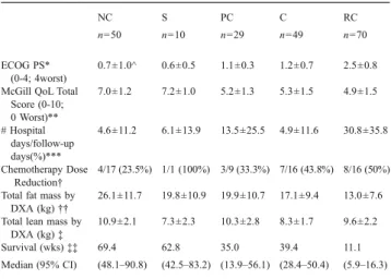
The incidence of sarcopenia and its effects on body composition, physiological function, nutrition, and fatigue in patients before allogeneic hematopoietic stem cell transplantation
Shinichiro Morishita1, Katsuji Kaida2, Shinya Yamauchi1, Tatsushi Wakasugi1, Kazuhiro Ikegame2, Norihiko Kodama3, Hiroyasu Ogawa2, Kazuhisa Domen3 (1Department of Rehabilitation, Hyogo College of Medicine Hospital, Nishinomiya, Hyōgo, Japan;2Division of Hematology, Department of Internal Medicine, Hyogo College of Medicine, Nishinomiya, Hyōgo, Japan;3Department of Rehabilitation Medicine, Hyogo College of Medicine, Nishinomiya, Hyōgo, Japan)
Background and aims: Cachexia in patients with haematological malignancies is often related to sarcopenia. We believe that allogeneic hematopoietic stem cell transplant (allo-hematopoietic stem cell transplantation (HSCT)) patients are often stricken by sarcopenia prior to transplantation. The aim of this study is to investigate the incidence of sarcopenia and the relationship of sarcopenia with body composi-tion and physiological funccomposi-tion, nutricomposi-tion, and fatigue in patients before allo-HSCT.
Methods: Study participants included patients who had undergone allo-HSCT from May 2007 to July 2011. One hundred and sixty-four patients were included in this study (median 50 years). Body composition was evaluated using bioelectrical impedance analysis. Evidence of sarcopenia was calculated using the male (<8.87 kg of skeletal muscle mass/m2) and female (<6.42 kg of skeletal muscle mass/ m2). Physiological functions were evaluated by handgrip, knee extensor strength, and 6-min walk test (6MWT).
Results: Eighty-three patients (50.6%) enrolled in our study had sarcopenia prior to allo-HSCT. Lean body mass (LBM) strongly correlated with handgrip and knee extensor strength (r=0.72∼0.75, p<0.01) and weakly correlated with 6MWT (r=0.34, p<0.01). Body weight and fat body mass correlated with fatigue (r=−0.15 and r=−0.2, respectively, p<0.05). Patients with sarcopenia experienced decreased muscular strength and increased fatigue compared with patients without sarcopenia (handgrip=−12%, knee extensor strength=−12%, fatigue=+22%, p<0.05).
Conclusion: Sarcopenia has been reported to occur at a rate of 5% (2 SD) in the elderly. In this study, roughly half of the patients had sarcopenia before allo-HSCT. Therefore, patients may have decreased muscle mass prior to allo-HSCT. In the evaluation of body composition, we discovered that muscle strength and LBM showed a high correlation. Patients with sarcopenia also have decreased muscle strength due to muscle atrophy and we believe that this causes increased fatigue in these patients.
1-10
Oleic acid accelerates myogenic differentiation through modulation of Notch and beta-catenin pathways in muscle stem cells Carlos Hermano J. Pinheiro, Kaio F. Vitzel, Renato T. Nachbar, Hilton K. Takahashi, Marco Aurélio R. Vinolo, Haroldo Fujiwara, Rui Curi (Department of Physiology and Biophysics, Institute of Biomedical Sciences, University of São Paulo, São Paulo, Brazil)
Background and aims: Marked changes in fatty acid composition occur during myogenic differentiation of muscle stem cells. In the present study, we demonstrated an increase in content of unsaturated fatty acids (as oleic acid) and delta-9-desaturase activity suggesting synthesis of oleic acid during myogenesis. The effects of oleic acid in muscle stem cell proliferation and myogenesis were investigated.
Methods: The effect of oleic acid in muscle stem cell proliferation was evaluated by 14C-thymidine incorporation. The content of markers of notch and beta-catenin pathways in undifferentiated muscle stem cells and expression of myogenic regulator factors (MyoD, myogenin, and myf-6) in differentiating myotubes were evaluated by western blotting.
Results: The addition of oleic acid to myogenic differentiation medium led to an increase in myoblast fusion, myotube diameter, and length and in expression of MyoD, myogenin, and myf-6. At 8 days of culture in myogenic differentiation medium, increased diameter of skeletal muscle cells and content of myosin heavy chain were also observed due to oleic acid addition. The treatment of undifferentiated muscle stem cells with oleic acid decreased expression of HES-1 and cell proliferation and raised p38-MAPkinase phosphorylation, NF-kappaB activity, and the content of phospho(Ser9)-GSK3beta and beta-catenin.
Conclusion: These results support the proposition that oleic acid accelerates myogenesis by mechanism associated to increased p38-MAPkinase/NF-kappaB pathway, beta-catenin stabilization, and decrease in Notch signaling. 1-11
Oleic acid accelerates myogenic differentiation through modulation of Notch and alpha-catenin pathways in muscle stem cells Carlos Hermano J. Pinheiro, Kaio F. Vitzel, Renato T. Nachbar, Hilton K. Takahashi, Marco Aurélio R. Vinolo, Haroldo Fujiwara, Rui Curi (Department of Physiology and Biophysics, Institute of Biomedical Sciences, University of São Paulo, São Paulo, Brazil)
Marked changes in fatty acid composition occur during myogenic differentiation of muscle stem cells. Increased content of unsaturated fatty acids (as oleic acid) and delta-9-desaturase activity were observed suggesting synthesis of oleic acid during myogenesis. The addition of
oleic acid to myogenic differentiation medium led to an increase in myoblast fusion, myotube diameter, and length and expression of myogenic regulator factors (MyoD, myogenin and myf-6). At 8 days of culture in myogenic differentiation medium, increased diameter of skeletal muscle cells and content of myosin heavy chain were also observed due to oleic acid addition. The treatment of undifferentiated muscle stem cells with oleic acid decreased expression of HES-1 and cell proliferation and raised p38-MAPkinase phosphorylation, NF-kappaB activity and the content of phospho(Ser9)-GSK3beta and alpha-catenin. These results support the proposition that oleic acid accelerates myogenesis by mechanism associated to increased p38-MAPkinase/NF-kappaB pathway, alpha-catenin stabilization and decrease in Notch signaling.
1-12
Dexamethasone-induced increase in FOXO1 activity and expression of atrogin-1 and MuRF1 in cultured muscle cells is PPARβ/δ-dependent Estibaliz Castillero, Zaira Aversa, Nima Alamdari, Aniket Gurav, Per-Olof Hasselgren (Department of Surgery, Beth Israel Deaconess Medical Center, Harvard Medical School, Boston, MA, USA)
Background and aims: Muscle wasting during various catabolic conditions is at least in part mediated by glucocorticoids. Corticosteroids upregulate FOXO1 activity, expression of atrogin-1 and MuRF1, and muscle proteolysis. Recent studies suggest that activation of the transcrip-tion factor PPARβ/δ upregulates the expression and activity of FOXO1 and the expression of atrogin-1 and MuRF1. The role of PPARβ/δ in glucocorticoid-regulated muscle wasting, however, is not known. We hypothesized that activation of PPARβ/δ results in increased protein degradation and muscle atrophy and that glucocorticoid-induced FOXO1 activation, expression of atrogin-1 and MuRF1, and protein degradation are at least in part regulated by PPARβ/δ.
Methods: Cultured L6 myotubes (a rat skeletal muscle cell line) were treated for 24 h with the PPARβ/δ agonist GW0742 (0.1 μM) or dexamethasone (1μM) in the absence or presence of the PPARβ/δ antagonist GSK0660 (0.1μM) followed by measurement of FOXO1 DNA-binding activity, mRNA and protein levels for atrogin-1 and MuRF1, protein degradation, and myotube diameter. In additional experiments, the effects of dexamethasone were tested in myotubes transfected with non-targeting or PPARβ/δ siRNA.
Results: Treatment of the myotubes with GW0742 increased FOXO1 DNA-binding activity, atrogin-1 and MuRF1 mRNA and protein expression, and protein degradation, and reduced myotube diameter. Dexamethasone stimulated PPARβ/δ and FOXO1 activity, atrogin-1 and MuRF1 expression, and protein degradation, and reduced myotube diameter. The effects of dexamethasone were blocked by GSK0660 or PPARβ/δ siRNA.
Conclusions: Results suggest that PPARβ/δ activates the atrophy program in skeletal muscle and that glucocortiocid-induced muscle wasting is at least in part regulated by PPARβ/δ. The transcription factor PPARβ/δ may be a target for the treatment and prevention of muscle wasting.
This work was supported by NIH R01 DK37908 (POH) and by a postdoctoral fellowship from Gobierno Vasco BFI2010-240 (EC). 1-13
Bed rest-induced muscle wasting is associated with modulation of myogenic markers
Fabrizio Pin1, Andrea Camperi1, M. Sturma2, S. Mazzucco2, Fabio Penna1, Maurizio Muscaritoli3, Filippo Rossi Fanelli3, G. Biolo2, Paola Costelli1 (1Dipartimento di Medicina e Oncologia Sperimentale, Università di Torino, Turin, Italy;2Dipartimento Clinico di Scienze Mediche, Chirurgiche e Della Salute, Università di Trieste, Trieste, Italy; 3Dipartimento di Clinica Medica,‘Sapienza’ Università di Roma, Rome, Italy)
Background/aims: Bed rest is associated with loss of skeletal muscle mass and strength, mainly due to protein hypercatabolism. In addition, inactivity has been shown to be associated with inflammation and oxidative stress that may lead to modulation of transcriptional factors that regulate myogenesis. In the present study, we investigated the relationship between prolonged muscle immobilization and gene expression of factors involved in myogenesis, such as MyoG, MyoD, Myf5 and Pax7.
Methods: Twenty healthy young male volunteers were recruited for a period of strict bed rest (33 days). All daily activities were performed in horizontal clinostatic conditions. For each volunteer, three biopsies were performed in the vastus lateralis muscle; the first, 1 day before bed rest (control), the second and the third after 7 and 33 days from the beginning of immobilization, respectively. Muscle biopsies were used to assess gene expression of MyoG, MyoD, Myf5 and Pax7 by real-time PCR.
Results: Pax7 and Myf5 expression is reduced of about 20–25% at both days 7 and 33 of immobilization. As for Myf5, at day 33, there is a tendency to restore control values. MyoD gene expression increases of about 50% after 7 days of bed rest, and returns to basal levels at day 33. Finally, MyoG expression is increased, though not significantly, due to sample variability, after both 7 and 33 days of immobilization.
Conclusions: Our results show that bed rest-induced muscle wasting is associated with downregulation of Pax7 and Myf5 mRNA levels. This could indicate a reduced satellite cell population, resulting either from enhanced differentiation, as suggested by increased MyoG expression, or by satellite cell death, due to inflammation and oxidative stress associated with inactivity. Further studies are in progress to clarify this point.
1-14
Muscle actin is polyubiquitinated in C2C12 myotubes and human muscle biopsies and targeted for breakdown by the E3 ligase MuRF1 Cécile Polge1, Anne-Elisabeth Heng1, Daniel Béchet1, Lydie Combaret1, Bernard Monsarrat2, Daniel Taillandier1, Didier Attaix1 (1INRA, UMR 1019, Unité de Nutrition Humaine, CRNH Auvergne, F-63000 Clermont-Ferrand, France; 2CNRS, Institut de Pharmacologie et de Biologie Structurale, 205 route de Narbonne, F-31077, Toulouse, France) Background and aims: Elaborating new strategies to prevent muscle wasting requires information on the precise mechanisms of the breakdown of contractile proteins. Experiments were performed (1) to demonstrate that actin is polyubiquitinated in muscle cells and human biopsies and thus degraded by the 26S proteasome and (2) to identify the ubiquitin (Ub) protein ligase E3 responsible for actin recognition and polyubiquitination. Methods: Chimeric flag-actin was stably transfected in C2C12 myotubes treated or not with 1μM dexamethasone (Dex). Poly-Ub conjugates in C2C12 cells and human muscle biopsies were purified using an affinity matrix and deubiquitination as described (Ventadour et al. J. Biol. Chem. 2007, 282:5302–9). We used glutathione S-transferase pulldown and ubiquitination assays to detect actin interaction and MuRF1-dependent actin polyubiquitination. mRNA levels of E3 ligases were measured by qRT-PCR analysis in C2C12 myotubes treated or not with Dex. C2C12 myotubes were transfected with siRNAs to induce MuRF1 knockout.
Results: Chimeric flag-actin was destabilized and polyubiquitinated in stably transfected and Dex-treated C2C12 myotubes and only proteasome inhibitors blocked its breakdown. Poly-Ub actin was also detected in human muscle biopsies from control and cancer patients. An increase in MAFbx/atrogin1, Trim32 and Ozz mRNAs was observed in Dex-treated C2C12 cells, while Nedd4, E4b, MuRF1 and MuRF3 mRNAs did not change significantly. However, MuRF1 protein content increased in Dex and Dex+MG132 treated C2C12 cells and in muscles from early-stage
cancer patients. Actin was polyubiquitinated by MuRF1 in an in vitro ubiquitination assay. Accordingly, MuRF1 siRNA stabilized the break-down of flag-actin.
Conclusions: These data demonstrate that actin is polyubiquitinated in muscle cells and human biopsies, and that MuRF1 is implicated in this process. This further supports the need for new strategies blocking specifically the activation of the UPS and/or MuRF1 in wasting diseases.
Work supported by the Association Française contre les Myopathies. 1-15
Role of ubiquitin E3 ligases in sarcopenia: towards an integrative understanding of their roles in muscle wasting
Julius Bogomolovas, Dittmar Labeit, Alexander Gasch, Siegfried Labeit (Dept. for Integrative Pathophysiology, Medical Faculty Mannheim, Theordor-Kutzer-Ufer 1–3, Mannheim, Germany)
Background and aims: Progressive loss of sarcomeres, leading ultimately to a state referred to as sarcopenia, is induced by a variety of atrophy-inducing signals, including physical inactivity, cytokines and inflammation, and metabolic disorders. It has been postulated that the induction of E3-type ubiquitin ligases represents an initial and rate limiting step in the degradation of muscle proteins, thereby providing attractive targets for atrophy attenuation.
Methods: Here, we have studied mice that were either deficient for the E3 ligase MuRF1 (MuR1-KO), or that overexpressed MuRF1 under the control of a CK promoter (MuRF1-CK-TG).
Results: We characterized their responses to a variety of atrophy inducing stimuli, including denervation, hindlimb suspension, and injection of TNF-alpha. Our results indicate that inactivation of MuRF1 leads to potent protection from muscle atrophy, and that protection occurred preferentially in fast fiber types. Intriguingly, we also noted a protection of force-bearing bones from demineralization in MuRF1-KO mice and thus in a context were MuRF1 is not known to be expressed. Consistent with systemic regulatory effects of MuRF1 during atrophy, we noted metabolic effects on lipid and glucose oxidation, and circulating amino acid levels.
Conclusions: Future studies are warranted if additional humoral pathways regulated by MuRF1 may contribute to sarcopenia protection in addition to the known intra-muscular pathways involved in the degradation of structural muscle proteins.
1-16
Mice lacking the USP19 deubiquitinating enzyme show less muscle wasting in response to several catabolic conditions due to suppression of ubiquitin ligases and autophagic genes and enhanced rates of protein synthesis
Nathalie Bedard1, Samer Jammoul1, Linda Wykes2, Patricia L. Hallauer3, Kenneth E. Hastings3, Simon S. Wing1 (1Medicine, McGill University Health Centre Research Institute, Montreal, Quebec, Canada;2School of Dietetics and Human Nutrition, McGill University, Montreal, Quebec, Canada;3Montreal Neurological Institute, Montreal, Quebec, Canada) Background and aims: Although many enzymes involved in ubiquiti-nation are activated in atrophying muscle, little is known about the role of deubiquitinating enzymes (DUBs). We previously showed that the USP19 DUB is induced in various conditions of muscle atrophy including cancer and that silencing of USP19 in muscle cells increases expression of myofibrillar proteins. To test the relevance of this in vivo, we inactivated USP19 in mice and characterized the wasting response induced by denervation, fasting, and cancer.
Methods: USP19 KO or WT mice were subjected to the above catabolic conditions. Muscle mass, myofiber area, protein synthesis (flooding dose method), mRNAs encoding ubiquitin ligases MuRF1, atrogin-1, and autophagy genes Bnip3, Atg4, Gabarap and LC3 were measured.
Results: USP19 KO mice showed 26–39% less muscle wasting than WT mice. Myofiber area measurements in the denervation studies confirmed that this was due to less myofiber atrophy in the KO mice. In the fed state, fractional rates of muscle protein synthesis were similar in WT, KO. However, in fasting, fractional synthesis rates were 260% and 135% higher in the KO in the sarcoplasmic and myofibrillar fractions respectively. In all conditions, WT, KO muscle showed levels of expression of MuRF1, Atrogin1 and autophagy genes that were similar in basal state. However, in the catabolic states, MuRF1 and Atrogin-1 and some autophagy genes in the KO were <50% that in WT. This regulation by USP19 appeared to be muscle intrinsic as silencing of USP19 in muscle cells also abrogated the increase in MuRF1 induced by dexamethasone. Conclusions: USP19 is essential for the full catabolic response to diverse wasting conditions including cancer cachexia. USP19 sup-presses protein synthesis and enhances protein degradation (ubiquitin proteasome system and autophagy) suggesting that it acts on an upstream signalling pathway(s). These results identify USP19 as a potential drug target in cachexia.
1-17
IL-6 receptor signaling antagonism maintains muscle oxidative capacity during the progression of cachexia in the ApcMin/mouse: a role for exercise
James P. White, Suichi Sato, Melissa Puppa, James A. Carson (Applied Physiology Division, Exercise Science, University of South Carolina, Columbia, South Carolina, USA)
Background and aims: Cancer cachexia is associated with the loss of skeletal muscle. Muscle mitochondria function and content have been suggested to be as key regulators of muscle protein turnover, and we have demonstrated that muscle oxidative capacity is reduced in severely cachectic ApcMin/mice. Exercise and anti-cytokine therapies have been shown to be effective in preventing the progress of cachexia tumor-bearing mice. However, limited studies have explored the effect of these therapies on alterations in muscle oxidative capacity. Therefore, the purpose of this study is to determine regulation of muscle oxidative capacity during the progression of cachexia and whether IL-6 inhibition or exercise can preserve muscle mass through the maintenance of muscle oxidative capacity.
Methods: Three experiments were conducted using ApcMin/mice. Experiment 1 stratified mice by body weight loss. Experiment 2 administered IL-6 receptor antibody for 2 weeks after the onset of cachexia. Experiment 3 examined if moderate intensity treadmill training could attenuate changes induced by systemic IL-6 over-expression.
Results: Muscle mitochondrial content was not reduced during the onset of cachexia, while protein expression related to biogenesis and fusion was repressed. As cachexia progressed, mitochondrial content decreased and biogenesis- and fusion-related proteins were further repressed, while fission protein expression increased. IL-6 receptor antibody administration attenuated mitochondrial content loss, rescued the repression of biogenesis and fusion proteins, and prevented the induction on fission proteins. Exercise training prevented the onset of cachexia due to IL-6 over-expression, and was associated with an increase in mitochondrial biogenesis and fusion protein expression, while repressing fission protein expression.
Conclusions: The loss of muscle oxidative capacity occurs during late stages of cachexia, while repressed expression of proteins regulating mitochondrial biogenesis and dynamics occur early in the development of cachexia. Attenuation of Il-6 receptor signing or exercise training can reverse these changes.
1-18
Optimization of macrophage-secreted myogenic factors and promotion of muscle regrowth
Nicolas Dumont, Jérôme Frenette (Université Laval, CHUL research center, Quebec, QC, Canada)
Background and aims: Muscle wasting is a common side effect of many pathologies and conditions such as AIDS, cancers, chronic heart diseases, aging, prolonged bed rest, space flight, etc. Using an animal model of hypogravity, we previously showed that regrowth of atrophied muscles is associated with an inflammatory reaction and that the presence of macrophages is necessary for an optimal recovery. A growing body of evidence indicates that macrophages possess important myogenic capacities raising the question of whether the activity of macrophages can be optimized to promote muscle repair and regrowth. The objective of this study was thus to determine the effect of modulating the concentration and activation of macrophages on muscle regrowth from atrophy.
Methods: Mice were hindlimb unloaded for a period of 10 days, which induces a 50% decrease in soleus muscle force and mass, followed by 3, 7, or 14 days of reloading. At the day of reloading, mice were injected with macrophage-colony stimulating factor (M-CSF, 10 μg/ml) or phosphate-buffered saline (PBS) directly into the right and left soleus muscles, respectively. In addition, we developed an in vitro co-culture system in which myotubes atrophied by dexametha-sone (1 μg/ml) were exposed or not to bone marrow-derived macro-phages (BMDM, 25,000 cells/ml) and/or M-CSF (100 ng/ml). Results: Compared to PBS injection, M-CSF induces a twofold increase in macrophage concentration in soleus muscles, which is associated with a 20% increase in muscle force and a 10% increase in muscle fiber diameter at day 7 of reloading (p< 0.05). In vitro, our data indicate that contrary to BMDM or M-CSF alone, the combination of BMDM and M-CSF increased myotube diameters by 20% and decreased the expression of the catabolic protein MuRF-1 by 25% (p< 0.05) relative to control.
Conclusion: Together, these results suggest that the myogenic capacities of macrophages can be optimized through M-CSF to promote muscle regrowth. 1-19
SOCS3 overexpression counteracts the IL-6/STAT3 signaling pathway and improves muscle wasting in experimental cachexia
Andrea Bonetto1, Tufan Aydogdu2, Felipe E. Pedroso1, Paul B. Spaulding3, Leopold Puzis3, Leonidas G. Koniaris4, Teresa A. Zimmers5 (1Dept. of Cancer Biology, Kimmel Cancer Center, Thomas Jefferson University, Philadelphia, PA, USA;2Dept. of Cell Biology and Anatomy, University of Miami Miller School of Medicine, Miami, FL, USA; 3Dept. of Surgery, University of Miami Miller School of Medicine, Miami, FL, USA; 4Dept. of Surgery, Thomas Jefferson University, Philadelphia, PA, USA; 5Dept. of Cancer Biology, Kimmel Cancer Center, Thomas Jefferson University, Philadelphia, PA, USA)
Background and aims: Elevated serum interleukin (IL)-6 correlates with muscle wasting and mortality in both cancer and severe burn injury. Systemic administration of IL-6 induces wasting, while its
inhibition can partially rescue muscle mass. The signaling pathways responsible for muscle wasting downstream of IL-6 are not completely known. IL-6 binds to its receptor, thus inducing the recruitment of gp130 and, in turn, the activation of STAT3. We recently reported that STAT3 is associated with tumor-induced muscle wasting. Since SOCS3 is one of the inhibitors of the IL-6/STAT3 signaling, here we aim at investigating whether SOCS3 can antagonize the IL-6/ STAT3 pathway, thus improving muscle wasting in cancer- and burn-associated cachexia.
Methods: C2C12 myotubes were used to investigate the IL-6/STAT3 pathway in vitro. MLC-SOCS3 C57BL/6 transgenic mice served to isolate the effects of SOCS3 on the skeletal muscle. CD2F1 mice were injected with the Colon-26 (C26) adenocarcinoma to study cancer-associated cachexia, while C57BL/6 wild-type were burned to evaluate burn-induced cachexia. SOCS3 was overexpressed by means of in vitro adenoviral infection or in vivo electroporation.
Results: C2C12 overexpressing Ad-GFP-SOCS3 proved resistant to IL-6 induced wasting, while SOCS3 alone induced hypertrophy in control myotubes. Muscle weight was increased in female MLC-SOCS3 transgenics only, thus suggesting a gender-specific function of SOCS3. STAT3 nuclear translocation was reduced in gastrocnemius muscle of MLC-SOCS3 mice, consistent with decreased p-STAT3 levels. Both C26 tumor-bearers and burned mice exhibited reduced muscle CSA and wasting, associated with increased levels of p-STAT3. Localized overexpression of SOCS3 in the tibialis muscle inhibited STAT3 activation and prevented muscle wasting in C26 and burn cachexia.
Conclusions: These data support the use of SOCS3 manipulation as a therapeutic approach in cachexia of high IL-6, enhancing both muscle mass and fiber size. Among the likely targets of SOCS3 activity is STAT3.
1-20
Serum creatinine is a biomarker of skeletal muscle mass in chronic hemodialysis patients
Sapna S. Patel1, Miklos Z Molnar1, John Tayek1,3, Deborah Benner5, Joel D. Kopple1,3,4, Csaba P. Kovesdy2, Kamyar Kalantar-Zadeh1,3,4 (1Harold Simmons Center for Kidney Disease Research and Epidemiology, Los Angeles Biomedical Research Institute at Harbor-UCLA, Torrance, CA, USA;2Salem Veteran Affairs Medical Center, Salem, VA, USA;3David Geffen School of Medicine at UCLA, Los Angeles, CA, USA;4UCLA School of Public Health, Los Angeles, CA, USA;5DaVita Nutrition, Irvine, CA, USA)
Background: Skeletal muscle mass is a major component of lean body mass (LBM) and an important nutritional measure in people with chronic kidney disease including those who undergo maintenance hemodialysis (MHD) treatment. We hypothesized that serum creatinine concentration in stable MHD patients with adequate dialysis represents skeletal muscle mass. Methods: In the“Nutritional and Inflammatory Evaluation in Dialysis” Study cohort, we examined 725 randomly selected patients receiving MHD at eight DaVita dialysis clinics in the Los Angeles South Bay area. Near-infrared interactance (NIR) in all patients and dual energy X-ray absorptiometry (DEXA) in a subset of randomly selected 118 patients were used as the reference standards for LBM measurements over a 5-year period. The index test was 3-month averaged serum concentrations of creatinine (SCr). The reference test was LBM measured via the difference between body mass and body fat assessed by NIR in the main cohort and by DEXA in the subcohort. Results: Both DEXA and NIR measured LBM correlated significantly with SCr (r=0.65 to 0.71, p<0.001). SCr in women had even stronger correlation with LBM. Across increments of 10 kg from <40 to >70 kg LBM, an incremental increase in SCr was observed which can be expressed by a polynomial equation (r=0.99):
Conclusions: Three-month averaged SCr is a good correlate of LBM, as determined from DEXA or NIR measured LBM, in MHD patients. 1-21
Total-body creatine pool size and skeletal muscle mass determination by D3-creatine dilution in rats
Stephen A. Stimpson1, Scott M. Turner2, Lisa G. Clifton1, James C. Poole1, Todd W. Shearer1, Greg M. Waitt3, Laura L. Hagerty1, Katja S. Remlinger1, Marc K. Hellerstein2, William J. Evans1(1Muscle Metabolism DPU/Metabolic Pathways and Cardiovascular TA Unit, GlaxoSmithKline, Research Triangle Park, NC, USA;2KineMed, Inc., Emeryville, CA, USA;3Platform Sciences and Technology, GlaxoSmithKline, Research Triangle Park, NC, USA)
Background and aims: There is currently no direct, facile, and inexpensive method to determine total-body skeletal muscle mass for the routine diagnosis and treatment of skeletal muscle wasting conditions such as sarcopenia, cachexia, and disuse. We sought to validate in rats the hypothesis that the enrichment of creatinine-(methyl-d3) (D3-creatinine) in a spot urine sample after a single defined oral tracer dose of D3-creatine can be used to determine total-body creatine pool size and skeletal muscle mass.
Methods: We determined (a) an oral tracer dose of D3-creatine that was completely bioavailable with minimal urinary spillage and sufficient enrichment in the body creatine pool for detection of D3-creatine in muscle and D3-creatinine in urine, and (b) the time to isotopic steady state. We then used cross-sectional studies in growing (9–22 weeks of age) rats to compare creatine pool size determined by the D3-creatine dilution method to lean body mass determined by quantitative magnetic resonance.
Results: D3-creatine (1 mg/kg) was 103±14% bioavailable, and the specific dose used in these studies (0.475 mg/rat) averaged <0.5% urinary spillage. Isotopic steady state was achieved 24–48 h after giving creatine. Creatine pool size, calculated from urinary enrichment of D3-creatinine 72 h after D3-creatine administration, significantly increased with muscle accrual during rat growth, significantly decreased in response to dexamethasone-induced skeletal muscle atrophy and was highly correlated with lean body mass (r=0.9517; p<0.0001). Enrichment of D3-creatine in muscle was greater in muscle with predominantly oxidative
Lean body mass (kg) <40 (n=97) 40 to <50 (n=226) 50 to <60 (n=234) 60 to <70 (n=115) ≥70 (n=75) p Value
versus glycolytic fibers. Creatine pool size calculated from muscle D3-creatine enrichment and converted to skeletal muscle mass based on muscle creatine content yielded expected skeletal muscle composition. Conclusions: A novel, facile, direct, noninvasive D3-creatine dilution method has been validated in rats for the determination of total-body creatine pool size and skeletal muscle mass, and holds promise for routine clinical application.
1-22
An investigation of potential skeletal muscle protein biomarkers of cancer cachexia
Nathan A. Stephens, Richard J.E. Skipworth, Carolyn A. Greig, James A. Ross, Kenneth C.H. Fearon (Department of Clinical and Surgical Sciences, University of Edinburgh, Edinburgh, EH16 4SB, UK)
Background and aims: There remains an unmet clinical need for diagnostic biomarkers/therapeutic targets in cancer cachexia. This study evaluated several skeletal muscle biomarkers (selected from previous animal/human studies) in a cohort of cachectic upper gastrointestinal cancer (UGIC) patients.
Methods: One hundred twenty patients (18 controls, 102 UGIC patients) were recruited. Mean (SD) weight loss of UGIC patients was 7.7 (9.2)%. Cachexia was defined as weight loss ≥5%. Immunoblotting of protein homogenates of rectus abdominis muscle biopsies obtained at surgery was performed probing for Akt (n=52), phosphorylated-Akt (n=52), FOXO1 (n=59), FOXO3a (n=59), LC3 (n=32), beta-dystroglycan (BDG, n=52), beta-sarcoglycan (BSG, n=52), calmodulin-kinase II (CAMKII, n=59), phosphorylated-CAMKII (n=59), and myosin heavy chain (n=47). ImageJ was used to calculate densitometry and results analysed using SPSS v15.0. Follow-up of UGIC patients was for a median of 663 days (range, 450– 1,955 days). Candidate biomarkers were assessed for: (1) differences between controls and UGIC patients, (2) diagnostic biomarkers of cancer cachexia, and (3) prognostic biomarkers of survival (lower vs. upper third). Results: Compared with controls, UGIC patients had lower Akt levels (0.49 (0.31) vs. 0.89 (0.17), p=0.001), lower total/phosphorylated-Akt ratio (1.73 (1.77) vs. 4.38 (2.62), p=0.002) and a trend towards higher CAMKII levels (0.77 (0.25) vs. 0.56 (0.30), p=0.053). Compared with noncachectic patients, cachectic patients had higher BDG levels (1.01 (0.16) vs. 0.87 (0.20), p=0.007) and a trend towards higher BSG levels (0.63 (0.28) vs. 0.55 (0.55), p=0.052). Survival was reduced in UGIC patients with lower vs. higher ratio of total/phosphorylated-Akt (median survival 483 vs. 640 days, p=0.020).
Conclusions: We suggest that Akt is suppressed and CAMKII is increased in skeletal muscle of UGIC patients. Few other potential diagnostic/prognostic biomarkers were revealed. This may reflect lack of sensitivity using immunoblotting, broad diagnostic criteria, or a heterogeneous patient group including individuals at different timepoints during the cachexia journey. 1-23
Altered skeletal muscle ultrastructure and myocyte enhancer factor (MEF) 2 C gene expression in the colon 26 carcinoma model of cachexia: a potential link?
Angie M. Y. Shum1,2,3, Ryland J. Taylor3, Arran B. Painter3, Melissa M. Moore3, Maria Tsoli3, Stephen J. Clarke3, Graham R. Robertson3 and Patsie Polly1,2,3(1Inflammation and Infection Research Centre, School of Medical Sciences, Faculty of Medicine, University of New South Wales, NSW, 2052, Australia; 2Department of Pathology, School of Medical Sciences, Faculty of Medicine, University of New South Wales, NSW, 2052, Australia;3Cancer Pharmacology Unit, ANZAC Research Institute, University of Sydney at Concord Repatriation and General Hospital, NSW, 2139, Australia.)
Background and aims: Cachexia is a highly debilitating paraneoplastic disease observed in more than 50% patients with advanced cancers and directly contributes to 20–30% of cancer deaths. Loss of skeletal muscle is a defining and often fatal characteristic of patients with cancer cachexia. Pathological mechanisms of cachexia are complex and multifactorial, which limits the development of an effective means of predicting, preventing, or treating it. This study aims to identify transcriptional regulators underlying cancer-induced skeletal muscle wasting.
Methods: The colon 26 (C26) carcinoma mouse model of cachexia was established and skeletal muscle was isolated for gene and protein expression studies by real-time qPCR and Western blotting; ultrastructural studies were performed using transmission electron microscopy. Results: The present study has identified a potential novel role for MEF2C as a regulator of skeletal muscle wasting in C26-bearing mice. MEF2C, an important transcription factor in controlling skeletal muscle maintenance and regeneration, was found to be downregulated at both transcript and protein levels during cancer-induced skeletal muscle wasting. Myogenic genes including myoglobin and myomesin, also displayed reduced expression due to cachexia; suggesting a potential link to MEF2C. These altered molecular effects imply compromised oxygen transport capacity in addition to distorted structural integrity. Changes in mitochondrial integrity were also evident in electron micrographs showing ultrastructural alterations.
Conclusion: Together, these effects may limit sarcomeric contractile ability and lead to skeletal muscle weakness and fatigue in cancer patients. Moreover, these may also predispose to structural instability in skeletal muscle and thus promote degradation along the course of disease progression. We believe that the identification of MEF2C is important as it appears to be a potential regulator of skeletal muscle loss in cancer cachexia. Targeting MEF2C could better direct intervention strategies at early stage disease and thus minimise or delay skeletal muscle wasting in cancer patients.
1-24
Effect of plasminogen activator inhibitor 1 (PAI-1) in rodent cancer cachexia and sarcopenia
Carsten Jacobi1, Shinji Hatakeyama1, Brian Latario3, Aline Mueller1, Joel Grosjean1, Angelika Meyer1, Daisy Rohner1, Anne-Ulrike Trendelenburg2, David Glass2(1Novartis Institutes for Biomedical Research, Muscle FiP, Basel, Switzerland;2Novartis Institutes for Biomedical Research, Muscle FiP, Cambridge, USA;3Developmental and Molecular Pathways Platform, Cambrige, USA3)
The plasminogen activator (PA) system has been shown to play an important role in various diseases including muscle wasting condi-tions. In particular, plasminogen activator 1 (PAI-1), a serine protease inhibitor (aka serpin E1) that targets urokinase-type (uPA) and tissue-type plasminogen activator (tPA), was shown to play an important role in muscle regeneration and hypertrophy. For example, PAI-1 knockout mice show improved muscle regeneration in cardiotoxin-induced injury model (Koh et al. 2005) and PAI-1 is upregulated during acute resistance exercise (Chen et al. 2002) as well as cancer, cardiovascular diseases, and diabetes. Moreover, PAI-1 expression is modulated by a variety of pathways known to be involved in muscle growth and regeneration such as pro-inflammatory cytokines, corticosteroids, IGF-I, and TGF-β proteins. We have further studied PAI-1’s role and mode of action in a human skeletal muscle assay system (HuSKmC) as well as in rodent cancer cachexia and sarcopenia models. We show here that HuSKmC cells increase during differentiation PAI-1 mRNA expression and secrete active PAI-1 measured by enzyme-linked immunosorbent assay. Treatment with TGF-β proteins further increase PAI-1 expression and secretion. Inhibition of PAI-1 by pharmacologic
and genetic tools increased HuSKmC differentiation indicating a tonic effect of secreted PAI-1. On differentiated myotubes, both PAI-1 targets uPA and tPA concentration-dependently induce increase in myotube diameter. Increased PAI-1 levels were detected in circulation of the mouse cancer cachexia model; in addition, cultured cancer cell lines, which are known to induce cachexia in vivo, actively secrete PAI-1. Moreover, expression of PAI-1 was increased in muscles of the cancer cachexia xenograft mouse model and sarcopenic rats. These data revealed further insights into the role and mode of action of PAI-1 in muscle cell differentiation and wasting conditions.
1-25
Autonomous CaMKII activity and SRF phosphorylation are decreased in skeletal muscle during experimental cancer cachexia
Zaira Aversa1, Nima Alamdari1, Estibaliz Castillero1, Aniket Gurav1, Maurizio Muscaritoli2, Filippo Rossi Fanelli2, Per-Olof Hasselgren1 (1Department of Surgery, Beth Israel Deaconess Medical Center, Harvard Medical School, Boston, MA, USA;2Department of Clinical Medicine, Sapienza, University of Rome, Rome, Italy)
Background and aims: Muscle wasting is an essential component of cancer cachexia. Studies suggest that calcium/calmodulin-dependent protein kinase II (CaMKII) is involved in the regulation of muscle mass. A unique feature of CaMKII is its autophosphorylation after calcium/calmodulin activation, resulting in calcium-independent autonomous activity. The transcription factor serum response factor (SRF) is an important substrate of autonomous CaMKII activity. Recent studies suggest that increased autonomous CaMKII activity and phosphoryla-tion of SRF(Ser103) result in muscle hypertrophy. Based on those observations, we hypothesized that cancer-induced muscle atrophy is associated with reduced autonomous CaMKII activity and SRF (Ser103) phosphorylation.
Methods: Cancer cachexia was induced in male Wistar rats by i.p. inoculation of AH-130 cells. Control rats were injected with corresponding volume of saline. After 7 days, gastrocnemius muscles were harvested for determination of phosphorylated CaMKII(Thr286/287) and SRF(Ser103) by Western blotting and measurement of autonomous CaMKII activity using a commercially available CaMKII kinase activity kit. In addition, muscle weight was measured and mRNA levels for atrogin-1 and MuRF1 were determined by real-time PCR.
Results: Muscle weight was reduced by 25% and atrogin-1 and MuRF1 mRNA levels were increased three- and fourfold, respectively, in tumor-bearing rats. Muscle levels of p-CaMKII(Thr236/237) and autonomous CaMKII activity were reduced by 30% in tumor-bearing rats. These changes were accompanied by a 25% decrease in nuclear levels of p-SRF (Ser103).
Conclusions: Muscle wasting in cancer cachexia is associated with reduced autonomous CaMKII activity and SRF(Ser103) phosphorylation. Stimulation of autonomous CaMKII activity and SRF phosphorylation may prevent cancer-induced muscle wasting.
Supported in part by NIH R01DK37908 to POH. ZA was supported by the Department of Clinical Medicine, Sapienza, University of Rome, Rome, Italy 1-26
Sex differences of cytokine profiles and muscle loss in pancreatic cancer patients
Bruno Gagnon1, Chrissi Galanakis2, Prosanto Chaudhury3, Alexa Gilbert2, Neil MacDonald3 (1Department of Medicine and Oncology, McGill University, Montreal, Quebec, Canada;2Division of Clinical Epidemiology, McGill University Health Center, Montreal, Quebec, Canada;3Department of Oncology, McGill University, Montreal, Quebec, Canada
Background: Cancer cachexia is a devastating wasting syndrome characterized by fat and muscle loss that affects the majority of advanced cancer patients. This syndrome, however, is poorly understood.
Objective: The objective of the study was to examine the extent to which muscle area changes over time in pancreatic cancer patients.
Methods: Forty newly diagnosed or recurring cancer patients of all stages (16 women (40%); age, 58 (SD: 10)years old) from McGill University Health Center and the Jewish General Hospital, with an Eastern Cooperative Oncology Group score of 0–3 and a life expectancy greater than 3 months were included in the study. Muscle area was assessed from computed tomography images, performed for clinical indications, of the third lumbar vertebrae with Slice-O-Matic. Cytokines were measured from plasma samples with the Bio-PlexR system and data output being analyzed with the Bio-Plex ManagerTM software. Group-based trajectory modeling was used to identify trajectories of muscle area over time.
Results: Twenty (50%) patients in the study were losing muscle over time, 13 (81%) women compared to seven (29%) men. Four trajectories of muscle area were identified: one losing muscle area over time and three with stable muscle area over time. Therefore, the losing weight group included 13 women and 7 men, while the other three groups included a total of 3 women and 24 men (z value 3.23, p value=0.001). Women had differing cytokine profiles compared to men at baseline.
Conclusion: Muscle loss occurs in half of the pancreatic cancer patients in this study, the majority of which are women. Future studies on cancer cachexia should explore the effects of sex differences among pancreatic cancer patients, especially in regard of the influence of cytokine profiles. 1-27
microRNA-451: a human specific regulator of adaptation to exercise training
Bethan Phillips1, Maria Psatha2, John Williams1, Kenneth Smith1, Valentina Gburcik2, Dominic Wells2, Paul Greenhaff3, Philip Atherton1, Michael Rennie1, James Timmons2(1University of Nottingham, School of Graduate Entry Medicine and Health, Derby, UK;2Royal Vetinary College, London, UK;3University of Nottingham, School of Biomedical Sciences, Nottingham, UK)
Background: Human skeletal muscle demonstrates remarkable hetero-geneity in capacity to adapt to external loading, such that gains in muscle strength or mass during resistance exercise training (RET) vary greatly between individuals. Our objective was to explore relationships between adaptive heterogeneity and abundance of miRNAs—the key regulators of translation of protein-coding mRNA.
Methods: Muscle biopsies were obtained from young (18–28 years old, n= 14), middle-aged (45–55 years old, n=20) and older (65–75 years old, n= 17) men and women before and after 20 weeks fully supervised whole-body RET. Using miRNA arrays that detect ∼700 human miRNAs, we hypothesised that miRNA abundance would differ between the highest (n= 5) and lowest (n = 5) responders (in terms of muscle strength) to RET. Results were confirmed using the entire cohort (n = 51), with TaqMan-based miRNA detection technology.
Results: Baseline physiology did not differ between high and low responders. We provide independent validation of a previous observation that mir-451 is specifically upregulated in young male low-responders to RET, and extended this observation to middle-aged and older male subjects. No such association was found in female subjects. Intriguingly, mir-451 is not expressed in murine C2C12 cells and its expression is absent or 10–50-fold lower in mouse/rat than in human skeletal muscle extracts. Targets of mir-451 are limited in number (∼20) and represent conserved protein coding genes, including Hamartin (TSC1), F-box protein 33 (FBXO33) and calcium-binding protein 39 (CAB39) which along with additional evidence that mir-451 can inhibit
Serine/threonine-protein kinase 11 (LKB1), places it in a central position to interact with numerous anabolic and catabolic related pathways.
Conclusion: We have validated a novel regulator of male human in vivo muscle growth. The fact that miR-451 demonstrates a human-specific expression pattern supports the notion that current preclinical murine or rodent models of muscle growth may be unable to fully mimic the human scenario.
2-01
Transcriptomic and proteomic profiling of livers from cachectic mice reveals dysfunctional metabolism
Dominic Burg1, Ryland Taylor1, Lucy Jankova1, Arran Painter1, Maria Tsoli1, Shiba Dolai2, Stephen Clarke1, Mark Molloy2, Graham Robertson1 (1Cancer Pharmacology Unit, ANZAC Research Institute, Concord RG Hospital, NSW 2139, Australia;2Australian Proteome Analysis Facility, Dept of Chemistry & Biomolecular Sciences, Macquarie University, NSW Australia)
Background: Cancer cachexia is a catabolic condition characterized by progressive weight reduction and energy imbalance associated with systemic inflammation, elevated serum C-reactive protein and cytokines. While muscle and fat loss are obvious manifestations of cachexia, it is likely that the pivotal role of the liver in nutrient uptake, metabolism, and redistribution contributes to dysregulated metabolism of cachexia.
Methods: Utilising a multiplatform approach including microarray and iTRAQ analysis, as well as novel ATP-binding protein enrichment technology coupled with label-free quantitation, we have profiled gene and protein expression patterns of livers from C26 tumor-bearing mice displaying cachexia.
Results: The transcriptomic and proteomic datasets revealed high correlation between the three approaches, with very few instances of incongruity. Pathway analysis utilizing several software packages indicated that central metabolic processes including lipid handling, glycolysis/gluconeogenesis, amino acid metabolism, tricarboxylic acid cycle and mitochondrial electron transport chain are reduced in cachectic mice. Linking these metabolic pathways to upstream regulatory events, transcriptional activation is reduced within the RXR canonical pathway (e.g., LXR, FXR, TR, PPARα/δ/γ), associ-ated with cytokine signaling through activassoci-ated JAK/STAT pathway, SOCS3, and IL-1/LPS-BP signaling. Repressed expression of genes and proteins in key energy generation pathways is counter-intuitive to the expected role of the liver in settings of food restriction/weight-loss —i.e., to adaptively utilize amino acids, carbohydrates and fatty acids, and activate ketone body production and gluconeogenesis. As a counterpoint to this dramatic disruption in metabolic pathways, we see significant acute phase protein production and a concomitant increase in protein translation, potentially mediated through phosphorylated 4E-BP downstream of mTOR.
Conclusion: Chronic stimulation of cytokine-signaling in the liver by distal tumours disrupts metabolic pathways responsible for maintaining energy homeostasis. The net outcome of impaired processing and supply of nutrients to muscle, fat, and other organs would contribute to the devastating effects of cachexia.
2-02
Sarcopenia in cirrhotic patients with and without hepatocellular carcinoma
Judith Meza-Junco1, Aldo Montano-Loza2, Carla Prado1, Jessica Lieffers1, Vicie Baracos1, Vincent Bain2, Michael Sawyer1(1Cross Cancer Institute, Edmonton, AB, Canada;2University of Alberta, Edmonton, AB, Canada)
Background/aims: Prognostic assessment of cirrhotic patients remains a challenge. Sarcopenia is defined as low levels of muscle mass; it may be present in chronic/malignant diseases. It is not well-studied/understood in cirrhotic and HCC patients. We aimed to establish sarcopenia frequency and if it predicts mortality in a cohort of cirrhotic patients with and without hepatocellular carcinoma (HCC).
Patients and methods: One hundred sixty-three patients with cirrhosis were consecutively evaluated for liver transplant and had computed tomography (CT) scan at the third lumbar vertebrae were selected. Skeletal muscle cross-sectional area was measured by CT to determine the third lumbar vertebrae (L3) skeletal muscle index (L3 SMI) defined as total lumbar muscle cross-sectional area adjusted for stature; sarcopenia was defined using previously published gender-specific cutoffs.
Results: One hundred eighteen patients were males (72%), mean age of 55 ±1 years, 51 patients (31%) had HCC at the time of CT. Median time of follow-up was 19±1 months. Sarcopenia was present in 61 patients (37%), and there was no difference in the frequency of sarcopenia among patients with and without HCC (31% vs. 40%, P=0.3). By univariate Cox analysis the bilirubin, creatinine, albumin, Model for End-Stage Liver Disease (MELD), Child–Pugh, sodium, L3 SMI, and sarcopenia were associated with mortality. The presence of HCC was not associated with increased mortality (HR 1.12, P=0.6). By multivariate Cox regression analysis, only MELD score (HR 1.08, P=0.001), and sarcopenia (HR 2.18, P=0.001) were independently associated with early mortality. Median survival for sarcopenic patients was 19±5 months, compared to 30±2 months in nonsarcopenic patients (P= 0.001). There was a poor correlation between sarcopenia and MELD score (r =−0.13, P=0.1) and sarcopenia and Child–Pugh score (r=0.04, P=0.6).
Conclusions: Sarcopenia is present in 37% of patients with cirrhosis and is not associated with the presence of HCC. Sarcopenia constitutes a strong and independent predictor of mortality in cirrhotic patients and does not correlate with degree of liver dysfunction evaluated with conventional scoring systems. Further studies including sarcopenia with conventional scores may allow better mortality prediction among cirrhotic patients with and without HCC.
2-03
Metabolic derangements in the gastrocnemius and the effect of selective NF-κB inhibition in a murine model of cancer cachexia Hirak Der-Torossian1, Ashley Wysong2, Scott Shadfar1, Monte S. Willis3, Jonathan McDunn4, Marion E. Couch5 (1Department of Otolaryngology-Head and Neck Surgery, University of Alabama at Birmingham, Birmingham, AL, USA;2Department of Dermatology, Stanford University, Stanford, CA, USA;3McAllister Heart Institute, University of North Carolina at Chapel Hill, Chapel Hill, NC, USA; 4Metabolon, Inc., Durham, NC, USA; 5
Department of Surgery, University of Vermont, Burlington, VT, USA) Cancer cachexia is a severe wasting syndrome characterized by the progressive loss of lean body mass and systemic inflammation. Inhibiting NF-κB signaling largely prevents such cancer-induced muscle wasting in murine models. We have previously shown the utility of compound A, a highly selective novel NF-κB inhibitor that targets the IkB kinase complex, to provide clinical benefit in cancer-induced skeletal muscle and cardiac atrophy. Using a metabolomics approach, we describe the changes found between cachectic and noncachectic gastrocnemius muscles, before and after compound A treatment. Of the 234 metabolites in the gastrocnemius, cachexia-induced changes in gastrocnemius metabolism reset the steady-state abundances of 42 metabolites (p<0.05). These changes, not evenly distributed across biochemical categories, are concentrated in amino acids, peptides, carbohydrates and energetics intermediates, and lipids. The gastrocnemius glycolytic pathway is markedly altered—changes consistent with tumor Warburg physiology. This is the first account of a Warburg