HAL Id: tel-01399499
https://tel.archives-ouvertes.fr/tel-01399499
Submitted on 19 Nov 2016HAL is a multi-disciplinary open access archive for the deposit and dissemination of sci-entific research documents, whether they are pub-lished or not. The documents may come from teaching and research institutions in France or abroad, or from public or private research centers.
L’archive ouverte pluridisciplinaire HAL, est destinée au dépôt et à la diffusion de documents scientifiques de niveau recherche, publiés ou non, émanant des établissements d’enseignement et de recherche français ou étrangers, des laboratoires publics ou privés.
Characterisation de l’ubiquitine Ligase PDZRN3 en tant
que nouvel acteur des voies Wnt dans la morphogenese
et l’integrite vasculaire
Raj Nayan Sewduth
To cite this version:
Raj Nayan Sewduth. Characterisation de l’ubiquitine Ligase PDZRN3 en tant que nouvel acteur des voies Wnt dans la morphogenese et l’integrite vasculaire. Cellular Biology. Université de Bordeaux, 2014. English. �NNT : 2014BORD0226�. �tel-01399499�
THÈSE PRÉSENTÉE POUR OBTENIR LE GRADE DE
DOCTEUR DE
L’UNIVERSITÉ DE BORDEAUX
ÉCOLE DOCTORALE SCIENCES DE LA VIE ET DE LA SANTE SPÉCIALITÉ BIOLOGIE CELLULAIRE ET PHYSIOPATHOLOGIE
Par Raj Nayan SEWDUTH
CARACTERISATION DE L’UBIQUITINE LIGASE PDZRN3 EN TANT QUE NOUVEL
ACTEUR DES VOIES WNT DANS LA MORPHOGENESE ET L’INTEGRITE VASCULAIRE
CHARACTERIZATION OF THE UBIQUITIN LIGASE PDZRN3 AS A NOVEL ACTOR
OF WNT PATHWAYS IN VASCULAR MORPHOGENESIS AND INTEGRITY
Sous la direction de : Dr Cécile DUPLAA (Directeur de Recherche) Soutenue le 18 Novembre 2014
Membres du jury : Pr Elisabeth Tournier
Laserve Professeur Des Universités/ Practicien Hosptialier Hopital Lariboisière/Fernand Vidal – Inserm Rapporteur Dr Stéphane Germain Directeur De Recherche– Collège De France/ Inserm Rapporteur Dr Nathalie Macrez Chargée De Recherche– Institut Des Maladies Neurodégénératives/ Cnrs Examinateur Dr Nathalie Sans Chargée De Recherche– Neurocentre Magendie/ Inserm Invité
Characterisation De L’ubiquitine Ligase PDZRN3 En Tant Que Nouvel Acteur Des Voies Wnt Dans La Morphogenese Et L’integrite Vasculaire
Résumé : Parmi les récepteurs Frizzled, Frizzled 4 est le seul à avoir un phénotype vasculaire fort. Par criblage, nous avons identifié l’ubiquitine ligase PDZRN3 en tant que nouveau partenaire de la protéine adaptatrice Dvl3 qui agit en aval de Fzd4. En utilisant des modèles murins inductibles, nous montrons que la délétion de PDZRN3 induit une létalité embryonnaire suite à des défauts de vascularisation du sac amniotique ; et que PDZRN3 est requis pour une vascularisation normale de la rétine. De par son activité d’ubiquitine ligase, PDZRN3 induit la prise en charge du complexe Fzd4/ Dvl3 par les vésicules d’endocytose ce qui permet la transduction du signal après fixation du ligand Wnt5a sur le récepteur Fzd4. PDZRN3 régule également le maintien des jonctions des cellules endothéliales et l’intégrité de la barrière hémato-encéphalique. La délétion de PDZRN3 stabilise les microvaisseaux après ischémie cérébrale. PDZRN3 induit la disruption des jonctions serrées et la rupture de la barrière hématoencéphalique en ubiquitinant la protéine d’échafaudage MUPP1.
Mots-clés: PDZRN3, Wnt/ Frizzled, endocytose, ubiquitinylation, angiogenèse, permeabilité, developpement, barrière hémato-encéphalique.
Characterization Of The Ubiquitin Ligase PDZRN3 As A Novel Actor Of Wnt Pathways In Vascular Morphogenesis And Integrity
Abstract : Fzd4 is the only Frizzled receptor that is essential for angiogenesis. By using a yeast two hybrid screening, we have identified the ubiquitin ligase PDZRN3 as a potential partner of the adaptor protein Dvl3 that acts downstream of Fzd4. By using inducible mouse models, we have shown that loss of PDZRN3 leads to early embryo lethality due to vascular defects in the yolk sac when deleted in utero, and is then required during post natal retinal vascularization. PDZRN3 would target the Fzd4/ Dvl3 complex to endosome, leading to signal transduction upon binding of Wnt5a to Fzd4. PDZRN3 also regulates integrity of the blood brain barrier by acting on tight junctions stability. Loss of PDZRN3 stabilizes microvessels after cerebral ischemia. PDZRN3 would induce tight junction disruption and blood brain barrier leakage by ubiquitinylating the scaffolding protein MUPP1.
Key words: PDZRN3, Wnt/ Frizzled, endocytosis, ubiquitinylation, angiogenesis, permeability, development, blood brain barrier.
Inserm U1034 Adaptation Cardiovasculaire à l’Ischémie
REMERCIEMENTS
A tous les membres du jury, je vous remercie d’avoir accepté de juger ce travail
Je remercie tout le laboratoire pour leur soutien ; Catherine, Béatrice, Myriam, Annabel et Jérome pour leur aide technique ; Christelle pour son aide au niveau administratif ; Aude, Claire, Nathalie, Marie-Lise et Laura pour leur bonne humeur ; Dr Marie-Ange Renault, Pr Pascale Dufourcq et Pr Thierry Couffinhal pour leur conseils et Dr Cécile Duplàa pour son encadrement au cours de ce travail.
Un merci aux étudiants en Master qui ont contribué au projet, Béatrice Desmoures, Nicolas Fritsch, Vincent Jecko et Robin Tellier.
Je tiens finalement à remercier mes parents et mes amis pour leur support moral. Je remercie tout spécialement Delphine pour son soutien tout le long de ce parcours.
TABLE OF CONTENTS
REMERCIEMENTS ... 1 TABLE OF CONTENTS ... 2 LIST OF FIGURES ... 5 ABBREVIATIONS ... 10 AVANT-PROPOS ... 12 INTRODUCTION ... 14CHAPTER I. WNT SIGNALING AND ANGIOGENESIS ... 15
1) The Wnt pathways ... 15
a) The Wnt pathways ... 16
b) Wnt (wingless-type MMTV integration site family) Factors ... 19
c) Fzd receptors ... 24
d) Coreceptors of Fzd proteins ... 28
e) Downstream effectors of Wnt signaling ... 32
2) Wnt signaling as a regulator of vascular development ... 37
a) Wnt signaling is implicated in angiogenesis during development and in pathology ... 37
b) Molecular mechanisms of regulation of angiogenesis/cell migration by Wnt signaling 46 CHAPTER II. REGULATION OF WNT SIGNALING BY ENDOCYTOSIS AND UBIQUITINYLATION... 51
1) Endocytosis as a regulator of Wnt signaling ... 51
a) Role of the early endosome in signal transduction ... 51
b) Role of the early endosome in Wnt signaling ... 52
c) Caveolin and Clathrin mediated endocytosis would regulate balance between Wnt pathways ... 53
d) Role of the endocytosis in Wnt signaling during development ... 55
2) Ubiquitinylation as a regulator of Wnt signaling ... 56
a) Role of ubiquitylation in protein stability or function ... 56
b) Ubiquitinylation of Fzd proteins ... 57
c) Ubiquitinylation of Dvl proteins ... 58
d) Ubiquitinylation of catenin ... 60
3) PDZRN3 E3 ubiquitin ligase ... 61
a) Identification of PDZRN3 ... 61
b) Structure and Function of PDZRN3 E3 ubiquitin ligase ... 62
c) PDZRN3 is a member of the LNX family of protein ... 64
CHAPTER III.WNT SIGNALING AND VASCULAR INTEGRITY ... 65
1) Wnt signaling regulates endothelial permeability in the Blood brain barrier ... 65
a) Wnt signaling as a regulator of brain vessels permeability ... 65
b) Wnt signaling as a regulator of brain vessels function in pathologies ... 69
2) Molecular mechanisms of regulation of vascular permeability by Wnt signaling ... 71
a) Structure of the tight junctions ... 71
b) Interplay between catenin and ZO1 ... 73
c) Interplay between c-Jun and ZO2 ... 75
a) MUPP1 is important for tight junctions stability ... 77
b) MUPP1 acts as a scaffold in the Crumbs complex ... 78
c) MUPP1 as a bridge between Tight Junctions and Crumbs complex ... 80
METHODS ... 81
A) DETAILED PROTOCOLS ... 82
1) Yeast Two Hybrid experiment ... 82
2) Expression constructs ... 82
3) Analysis of mRNA expression by quantitative RT-PCR ... 83
4) Cell culture ... 84
a) Wnt conditioned media production ... 84
b) Cell transfection / transduction ... 84
c) Chemical treatments ... 85
d) Proliferation (MTT) assay ... 85
e) Luciferase reporter assay ... 85
5) Protein analysis ... 85 a) Western Blot ... 85 b) Immunoprecipitation assay ... 86 c) Cell fractionation... 86 d) Ubiquitinylation assay... 87 6) Immunocytochemistry ... 87 a) Immunocytochemistry ... 87 b) Internalization assay ... 87 7) Immunohistochemistry ... 88 a) Paraffin sections ... 88 b) Cryosections ... 88 c) Vibratome sections ... 88
d) In toto embryo staining ... 88
e) Dab staining ... 88
f) Immunofluorescence ... 89
8) Image analysis ... 89
9) In vitro assays on endothelial cells ... 89
a) 2D tubulogenesis assay ... 89
b) Wound assay ... 90
c) 3D tubulogenesis (cytodex) assay ... 90
d) Chemotaxis assay ... 91
e) Permeability assay ( Transwell assay ) ... 91
f) Junctions disruption by calcium chelation ... 91
10) Experimental animals ... 91
a) Cre – Lox system ... 91
b) Tetracyclin activated system ... 93
c) Injections ... 93
11) Surgery ... 94
a) Retinal vascularization analysis ... 94
b) Middle cerebral artery occlusion ... 95
d) Evaluation of vascular leakage in embryo and pups ... 97
e) Micro computer tomography ... 97
f) Isolation of primary endothelial cells from mouse kidney ... 98
g) Vessel extraction ... 98
12) Statistical analysis ... 99
B) REAGENTS ... 100
1) List of used antibodies ... 100
2) Chemical reagents ... 101
3) Equipments ... 102
RESULTS ... 103
ARTICLE1.FRIZZLED 4 REGULATES ARTERIAL NETWORK ORGANIZATION THROUGH NONCANONICAL WNT/PLANAR CELL POLARITY SIGNALING ... 104
a) State of art ... 105
b) Hypothesis ... 105
c) Summary ... 106
ARTICLE2.THE UBIQUITIN LIGASE PDZRN3 IS REQUIRED FOR VASCULAR MORPHOGENESIS THROUGH WNT/PLANAR CELL POLARITY SIGNALING ... 123
a) State of art ... 124
b) Hypothesis ... 125
c) Strategy ... 125
d) Preliminary Results ... 126
e) Summary ... 128
ARTICLE3.THE UBIQUITIN LIGASE PDZRN3 REGULATES BLOOD BRAIN BARRIER INTEGRITY BY CONTROLLING TIGHT JUNCTIONS STABILITY VIA A WNT DEPENDANT MECHANISM ... 145
a) State of art ... 146
b) Hypothesis ... 146
c) Preliminary Results ... 146
d) Methods ... 147
e) Summary ... 150
CONCLUSION AND PROSPECTS ... 171
1) PROPOSED MODEL ... 173
2) PROSPECTS ... 177
a) Expression of PDZRN3 in pathologies ... 177
b) Interaction of PDZRN3 with MUPP1 ... 179
c) Phosphorylation status of PDZRN3 ... 179
d) Chemical inhibition of PDZRN3 ... 180
e) PDZRN3 in other tissues ... 180
LIST OF FIGURES
Figure 1: The Wnt canonical and non canonical pathways (adapted from Saadeddin et al 2009) ... 15
Figure 2: Transcriptional targets of the Wnt/ canonical pathway (Wnt homepage, Stanford) ... 16
Figure 3: PCP induces asymmetrical organization of Fzd/ Dvl and Vangl2/ Pk (Schulte 2010) ... 17
Figure 4: Distribution of Wnt Genes in Metazoans. X indicate that no member of the Wnt subfamily found in the genome of the organism; the color is different for each Wnt; two blocks indicate presence of paralogue in the specie (adapted from Prud'homme et al. 2002) ... 19
Figure 5: Phylogenetic tree of the Wnt family (adapted from Prud'homme et al. 2002) ... 20
Figure 6: List of Wnt genes (Wnt homepage, Stanford)... 22
Figure 7: Schematic presentation of Wnt/ Fzd signaling kinetics (Schulte and Shenoy 2011) ... 23
Figure 8: Phylogenetic tree of the human Frizzled receptors FZD (Schulte and Shenoy 2011) ... 24
Figure 9: Schematic representation of Frizzled protein (binding sites for Dvl/ PDZ and G proteins indicated) (Schulte and Shenoy 2011) ... 24
Figure 10: Conservation of the PDZ binding domain (TXV-COOH) in the Fzd family; functional and non functional PDZ binding domain is noted as (+) or (-) respectively (Schulte and Shenoy 2011) ... 25
Figure 11: List of Frizzled receptors (Wnt homepage, Stanford) ... 26
Figure 12: Diagram of Fzd-4 showing mutations associated to the disease (Robitaille et al. 2002, Robitaille et al. 2011) ... 27
Figure 13: Competition between Wnt ligands would regulate activation of Wnt pathways by modulating phosphorylation of LRP5/6 and Ror2 (adapted from Grumolato et al 2010) ... 28
Figure 14: Structure of LRP5 (He et al 2005) ... 29
Figure 15: Structure of the Ror2 protein (adapted from Scopus) ... 30
Figure 16: Structure of the Ryk protein ... 31
Figure 17: Structure of Dvl (SCOPUS) ... 32
Figure 18: List of Dvl proteins (adapted from Wnt homepage, Stanford)... 33
Figure 19: Positive (left) and negative (right) regulators of Dvl activity (Gao et al 2010) ... 33
Figure 20: The -catenin destruction complex (Kimelman and Xu 2006) ... 34
Figure 22: a- Expression of various components of Wnt signaling in placenta and yolk sac; b- Placenta and Yolk Sac of Fzd5 -/- embryos are not vascularized correctly; Vascular pattern is normal in Fzd5-/- embryo but
vasculature remains superficial in placenta of Fzd5-/- embryos (Tomo et al 2001). ... 37
Figure 23: a- The yolk sac of the junB KO embryo at E9.5 is translucid and shows no vessels; b- Morphological phenotypes of JunB deficient embryos (Schorpp-Kistner 1999). ... 38
Figure 24: a- Wnts and Fzd4 are mediators of Brain angiogenesis (right) and differentiation of the Blood brain barrier (left) (Paolinelli et al 2011); b- Lrp5/6 and -catenin are also essential for vessel formation in the hindbrain as KD of both proteins lead to vascular leakage (in white) (Zhou et al. 2014) ... 39
Figure 25: Schematic of retina during its vascularization (adapted from Franco et al 2009) ... 40
Figure 26: a- Neutralizing FZD4 alters development of the intermediate and deep plexus; b- leading to leakage as shown by fluorescein extravasation and PLVAP expression (Ye et al 2010) ... 41
Figure 27: a - Fzd4 deletion leads to expression of PLVAP; the cells expressing PLVAP are progressively lost, indicating that endothelial cells deleted for Fzd4 are eliminated; b- Proliferation is increased in Fzd4-/- cells as indicated by Edu staining. ... 42
Figure 28: Retinal myeloid cells (in red) interact with descending vertical sprouts (in green) ... 43
Figure 29: Retinal myeloid cells flow sorted from the deep plexus are subjected to PCR for Wnt pathway components (Stefater, Lewkowich et al. 2011) ... 43
Figure 30: a-c micro-CT images of control and stem cells injected hindlimb after ischemia; d - quantification of vessel density and connectivity; e- expression of Wnt factors as detected in ischemic muscle after injection of normal/ hypoxic stem cell (adapted from Leroux et al.) ... 44
Figure 31: Events that occur after cerebral ischemia (Sandoval 2009) ... 45
Figure 32: Wnt5a induces migration by relocalizing c-Jun to the leading edge (Nomachi 2008) ... 46
Figure 33: Cell migration and adhesion regulated by the Wnt5a pathway (Kikuchi et al. 2012) ... 46
Figure 34: Regulation of migration via the actin skeleton by Dvl and Daam (Chen et al 2003)... 47
Figure 35: Regulation of migration via microtubules by Dvl and Daam (Chen et al 2003) ... 48
Figure 36: Sprouting endothelial cells are hierarchically organized into tips cells and stalk cells during retinal angiogenesis. Tip cell formation is induced by VEGF signaling, while stalk cell are determined by Notch signaling (Herbert et al. 2011) ... 49
Figure 37: Different effects of Wnt/-catenin and Notch signaling during sprouting angiogenesis (Gerhardt et al. 2009) ... 50
Figure 38: Endosome regulates signaling by different manner (Dobrowolski et al 2011) ... 51
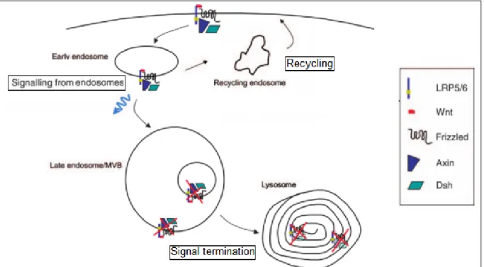
Figure 39: Endocytosis of Wnt receptors can lead to signal transduction / recycling or signal termination depending on context (Gagliardi et al 2008) ... 52
Figure 41: The dorsal determinants of the Xenopus egg may be Wnt endosomal components (adapted from
Drobowolski et al 2011) ... 55
Figure 42: a - different types of ubiquitin chains, b- 3D structure of Lysine 63 linked diubiquitin, c- 3D structure of Lysine 48 linked diubiquitin (Ikeda, Dikic 2008) ... 56
Figure 43: a- position of the different lysines residues on intracellular domains of Fzd4; ubiquitinylation of Fzd4 (smear) is mediated by Wnt3a and UBPY (Mukai et al 2010) ... 57
Figure 44: UBPY deubiquitin ligase controls availability of FZD4 for Wnts (Mukai et al 2010) ... 57
Figure 45: Different regulators of Dvl ubiquitinylation (adapted from Tauriello 2010) ... 58
Figure 46: a- Cyld mediated K63 dependant ubiquitinylation of Dvl (detected as a smear); b- model of regulation of Dvl activity by CYLD (Tauriello et al. 2010) ... 58
Figure 47: Inversin regulation of Dvl (adapted from Simons 2006) ... 59
Figure 48: Ubiquitin mediated regulation of core Wnt proteins such as catenin (adapted from Tauriello et al. 2010) ... 60
Figure 49: Orthologues of PDZRN3 (Gene Ontology) ... 61
Figure 50: Structure of the protein (Scopus, EMBL) ... 62
Figure 51: Process of ubiquitinylation (Dikic et al. 2012) ... 62
Figure 52: ClustalX alignment of the last amino acids of murine PDZRN3 (mm LNX3), PDZRN3 4 (mm LNX4) as well as orthologues from invertebrate species(Flynn et al. 2011). ... 63
Figure 53: Molecular evolution of the LNX family (Flynn et al 2011) ... 64
Fgure 54: Brain capillaries under normal and pathological conditions (Paolinelli et al 2011) ... 65
Figure 55: VE-cadherin traps FoxO1 and β-catenin at the membrane. In the nucleus, both would block claudin-5 transcription. In acute permeability condition, VE-cadherin adhesion is destabilized and FoxO1 and β-catenin are phosphorylated and translocate to the nucleus where they block claudin-5 transcription (Gavard et al 2008) ... 66
Figure 56: a- Transcription of claudin 3 is activated bycatenin (Liebner et al 2008); b- Conditional loss of -catenin leads to vascular leakage during development (Catellino et al 2003) ... 67
Figure 57: a/b- Injection of Evans Blue of Fz4 deficient in EC mice shows leakage in the cerebellum and retina; c- Leakage arises from the fact that vessels are fenestrated as indicated by Biotin / Mouse igG extravazation(adapted from Wang et al 2013/ Zhou et al 2014 ) ... 68
Figure 58: Response to stroke (adapted from Kahle et al. 2014) ... 69
Figure 59: Structure of the tight junctions (adapted from González-Mariscal et al 2000) ... 71
Figure 60: Structure of MAGUK Proteins (adapted from González-Mariscal et al 2000) ... 72
Figure 62: Differential expression and localization of -catenin and ZO-1 in migratory versus stationary
MCF10A human breast cells (adapted from Polette et al 2007) ... 73
Figure 63: Interaction of ZO1/catenin during EMT transition (Polette et al 2007) ... 74
Figure 64: In subconfluent cultures, ZO-2 and Jun are found at the nuclei and cell borders, while in the confluent condition, they are only detected at the cell membrane (Betanzos et al 2007) ... 75
Figure 65: In subconfluent cultures, ZO-2 and Jun are found at the nuclear fraction, while in confluent cultures, they are only detected in the membrane fraction as indicated by cell fractionation (Betanzos et al 2007) ... 75
Figure 66: ZO-2 immunoprecipitation realized from membrane (a) and nuclear extracts (b) from sparse and confluent cell cultures (Betanzos et al. 2004) ... 76
Figure 67: a -Interaction of claudins with scaffolding proteins ZOs and MUPP1; b- Western blot showing expression of MUPP1 in brain tissues (adapted from Soini et al 2011 and Sitek et al 2003) ... 77
Figure 68: a- Interactions of MUPP1/ Patj (first 10 PDZ domains are identical in MUPP1 and Patj); b- Relocalization RhoA to Leading edge by the Crumbs complex (Ernkvist et al. 2009) ... 78
Figure 69: a- Syx /RhoA /Amot /MUPP1 and Rab13 colocalize in response to VEGF.A , as detected 2mins after treatment in vesicles and 30mins after treatment at the leading edge of migrating cells; b - Recycling vesicles are essential for polarized migration as depletion of Rab13 dirupts polarity of migrating cells (Wu et al. 2011) ... 79
Figure 70: a -Depletion of MUPP1 and Syx disrupts junctional integrity as shown by ZO1 stainin and Transmission electron micrographs of sections of ventricle myocardium; b- Model of regulation of Junctional stability via MUPP1 (Ngok et al. 2012) ... 80
Figure 71: Dvl3 sequence used for cloning in each corresponding plasmids in red ... 82
Figure 72: PDZRN3 sequence used for cloning in each corresponding plasmids in red... 83
Figure 73: Formation of tube-like structures after cytodex plating (control condition) ... 90
Figure 74: Floxed PDZRN3 mice construct ... 91
Figure 75: Mating of PDGF-icre Pdzrn3fl/fl males with Pdzrn3 fl/fl females ... 92
Figure 76: Validation of the PDGF icre recombinase system using the mT/mG reporter mice ... 93
Figure 77: Quantification of Vascular area/ Regression and Proliferation ... 94
Figure 78: Region of retina imaged for quantification ; Representative view of the superficial plexus; a plane of the intermediate plexus and the deep plexus ... 95
Figure 79: View of the module of Tracking (Image J) and data analysis (Chemotaxis and migration tool Ibidi) .. 95
Figure 80: Scheme of the cerebral artery occlusion procedure (Rousselet et al 2012) ... 95
Figure 81: Evans Blue diffusion after MCAO: a- whole brain; b- 100µm vibratome slice observed under white light; c- observed at 545nm ... 96
Figure 83: Vessel extracts from adult - PDGF cre+ mT/mG mice injected with tamoxifen (vessels fluoresce in
green; Hoechst staining of nuclei in blue) ... 98
Figure 84: Phenotype of Fzd4-/- mice ... 105
Figure 85: Effet of deletion of Fzd4 on vessel density ... 106
Figure 86: Interaction of Fzd4 and Dvl favor Wnt non canonical pathway to favor angiogenesis ... 107
Figure 87: Alignment of the Dvl1/ 2 and 3 sequences ... 124
Figure 88: Yeast two Hybrid Strategy and Structure of the PDZRN3 protein ... 125
Figure 89 A few of the proteins found to be interacting with Dvl3 Dix using the screening ... 126
Figure 90 PDZRN3 (V5) colocalizes with clathrin, Rab5 and Rab11; wth ubiquitin ... 127
Figure 91 Depletion of Pdzrn3 using two siRNAs reduces migration of endothelial cells ... 127
Figure 92 Loss of PDZRN3 modifies the ability of endothelial cells to form tubes ... 128
Figure 93: Role of PDZRN3 for transduction of Wnt signaling and vascular morphogenesis ... 129
Figure 94: EDTA treatment induces disruption of cell to cell junctions in control Huvecs; a delay is observed in Pdzrn3 depleted cells ... 146
Figure 95: Huvec cells are orientated in a flow of 10, 20 or 30dyn/cm2; Pdzrn3 depleted cells are unable to align to the flow and reorganize their junctions accordingly... 147
Figure 96: a - validation of the PDGF Cre activation using a mt/mg reporter; b- vessel extracts were purified from pdgf cre mt/mg mice and subjected to confocal microscopy ... 148
Figure 97: Validation of the enrichment of the brain vessel fraction by q RT PCR ... 148
Figure 98: Expression of PDZRN3 in vessel / wash fraction ... 149
Figure 99: Proposed Model ... 150
Figure 100: Expression of the Pdzrn3 in Carcinoma (Oncomine) ... 177
Figure 101: Correlation of expression of PDZRN3 with other genes in Carcinoma – inset represent the scaling colour (Oncomine) ... 178
Figure 102: Phosphorylation of PSD-95 on Y397 makes access to the 3rd PDZ domain of PSD 95 possible (Zhang et al. 2011)... 179
Figure 103: Interaction of compound 3289-8625 (blue) and Dpr peptide(brown) with the PDZ domain of Dvl (Grandy et al. 2009) ... 180
ABBREVIATIONS
ABC: Active cateninAPC : Adenomatous Polyposis Coli AJ: adherens junctions
BBB: blood brain barrier BrdU : BromoDésoxyUridine BSA : Bovine Serum Albumin CD31 : Cluster of Differentiation 31 CM: conditoned medium
CNS: Central nervous system CRD : Cystein rich Domain Ctl: control
DAAM: Dishevelled-associated activator of morphogenesis DAB : Diaminobenzidine
DAG : Diacylglycérol
DEP: Dishevelled, EGL-10, Pleckstrin Dgo: Diego
DIX: Dishevelled/Axin Dkk : Dickkopf
(D)MEM: (Dulbecco's) Modified Eagle's Medium DMSO: Dimethyl sulfoxide
DNA : Deoxyribonucleic acid dNTPs : Dinucleotidtriphosphate dpc : days post-coitum
Dsh /Dvl : Dishevelled DTT : 1,4-dithio-threitol EC : Endothelial Cells
EDTA :ethylenediaminetetraacetic acid E: Embryonic day
ERK : Extracellular signal-Regulated Kinase FEVR: Familial exudative vitreoretinopathy FGF : Fibroblast Growth Factor
FITC : Fluorescein IsoThioCyanate Fmi: Flamingo
Fzd: Frizzled
GSK3β : Glycogen Synthase Kinase 3 beta I(sch): Ischemic
IB: immunoblot
i(EC)KO: inducible (endothelial cell specific) knock-out IP: intermediate plexus OR immunoprecipitate
LiCl:Lithium Chloride KO: Knock-Out
P: post natal day OR probability
PAGE : polyacrylamid gel electrophoresis PBS : phosphate buffer saline
PCR : Polymerase chain reaction PDZ: PSD95, Dlg1, ZO-1
PFA : Paraformaldehyde
PMA: Phorbol Myristate Acetate
MCAO: middle cerebral artery occlusion NI: non ischemic
NVU: neurovascular unit RNA : Ribonucleic acid
RT-PCR : Reverse-transcriptase PCR SDS :sodium dodecyl sulfate
TCF : T-cell Factor
TGF-β : transforming growth Factor β TJ: tight junctions
TNFα: Tumor Necrosis Factor alpha TTA: tetracyclin transactivator Ub: Ubiquitin
UBC: Ubiquitin C Vang: Van Gogh
VE-cadhérine : Vascular Endothelial-cadhérine VEGF : Vascular Endothelial Growth Factor W/O: without
WT: wild - type ZO: Zona occludens
AVANT-PROPOS
Les maladies cardiovasculaires sont la première cause de mortalité en France ; environ 170 000 décès sont recensés chaque année. Parmi les plus fréquentes, les cardiopathies ischémiques sont responsables de 27% des décès, les accidents vasculaires cérébraux de 25 % et les insuffisances cardiaques de 23 %. Les mécanismes d'adaptation à l'ischémie requièrent une adaptation vasculaire au niveau du tissu ischémique, avec le développement de nouveaux vaisseaux sanguins afin de corriger le défaut de perfusion tissulaire. Mieux comprendre les mécanismes favorisant la formation et le maintien de néo-vaisseaux fonctionnels dans un tissu ischémique est un pré-requis pour améliorer le traitement des ces pathologies. Ma recherche s’inscrit dans l’étude des mécanismes de formation et de maintien des vaisseaux en condition physiologique et pathologique au cours de l’embryogenèse et chez l’adulte. Les protéines Wnt et leurs récepteurs Frizzled régulent la formation des vaisseaux sanguins via une signalisation intracellulaire complexe. Comme les acteurs en aval des Frizzled restent méconnus, le laboratoire a mis en place une stratégie de criblage qui a permis d’identifier de nouveaux acteurs des voies Wnt/ Frizzled. Mes travaux ont porté sur l’étude d’un d’entre eux, l’ubiquitine ligase PDZRN3 et de son rôle dans la formation des vaisseaux sanguins.
L’introduction de ce manuscrit comporte trois chapitres. J’ai tout d’abord décrit le rôle de la voie Wnt/Frizzled dans la régulation de l’angiogenèse, en décrivant les différentes voies et en présentant les acteurs du système. J’ai détaillé les données concernant le rôle du ligand Wnt5a, du récepteur Frizzled, de la protéine adaptatrice Dishevelled 3 et de l’effecteur c-Jun, des protéines retrouvées en filigrane au cours du travail, avant de détailler les différents modèles de régulation proposés dans la littérature. La 2ème partie de l’introduction décrit comment l’endocytose permet de réguler les voies Wnt, avec différents modèles proposés qui pourraient notamment expliquer comment la balance entre les différentes voies Wnt se fait. J’ai ensuite abordé comment l’ubiquitination permet de réguler l’endocytose pour favoriser la transduction du signal dans les voies de signalisation Wnt. Dans une 3ème
les liens existant entre les voies Wnt et les jonctions serrées qui sont essentielles au maintien de l’intégrité vasculaire au niveau cérébral.
Dans le cadre de l’étude du système Wnt/Frizzled dans la formation des vaisseaux sanguins, j’ai participé à un premier travail décrivant l’implication de Frizzled 4 dans le développement vasculaire au cours du développement et dans un modèle d’ischémie du membre inférieur. Ce travail a mis en avant le rôle de Frizzled4 en tant qu’activateur des voies Wnt, requis pour la migration des cellules endothéliales.
A la suite de ce travail, une approche de criblage par double hybride a été menée permettant d’identifier un nouveau partenaire de la voie, l’ubiquitine ligase PDZRN3. Un premier travail a porté sur le rôle de la protéine dans l’angiogenèse. Des souris délétées pour la protéine de façon ubiquitaire ou spécifiquement dans les cellules endothéliales ont été générées. J’ai étudié le rôle de PDZRN3 dans le développement vasculaire au cours de l’embryogenèse et du développement de la rétine. J’ai ensuite utilisé des approches biochimiques et de culture cellulaire pour comprendre comment PDZRN3 jouait sur les voies Wnt pour favoriser le développement vasculaire.
Dans un dernier travail, le rôle physiologique de la voie Wnt via PDZRN3 dans le maintien des jonctions serrées des cellules endothéliales du cerveau et le contrôle de la barrière hémato-encéphalique a été analysé. J’ai notamment étudié l’impact de la surexpression de PDZRN3 dans les cellules endothéliales au cours de l’embryogenèse et qualifié les mutants délétés pour PDZRN3 dans les cellules endothéliales dans un modèle d’ischémie cérébral. Nous montrons par des approches de biochimie que PDZRN3 fait le lien entre voies Wnt et stabilité des jonctions en ubiquitinant la protéine associée aux jonctions MUPP1 et en favorisant la rupture des jonctions sérrées.
Chapter I. Wnt signaling and Angiogenesis
1) The Wnt pathways
Wnt signaling plays a key role in many biological processes as it regulates cell proliferation, migration and polarity establishment. Wnts are essential during development as they guide body axis specification (Barolo 2006). Ectopic placement of Wnts in Xenopus eggs during early gastrulation leads to formation of a secondary body axis and head, while inhibition of Wnt signaling results in a lack of dorsal structures in the frog embryo. Wnts are produced from specific sites during development, such as the edge of the developing fly wing or the dorsal region of the neural tube in vertebrates. Failure of closure of the neural tube is characteristic of knock-out mice of proteins of the Wnt pathway (Wang et al. 2006). Dysregulation of Wnt signaling is associated with cancer development (Saadi-Kheddouci et al. 2001) as alteration of proteins implicated in the pathway such as Axin or Ror2 are linked to carcinogenesis (Saadeddin et al. 2009, Ford et al. 2013).
……..
a) The Wnt pathways
i) Canonical pathway
The canonical Wnt signaling pathway requires β –catenin (Fig 1). Level of β -catenin is regulated by the GSK-3 containing β –catenin destruction complex. Upon binding of Wnt ligands to the Frizzled receptor (Fzd) and to its coreceptor - low density lipoprotein receptor-related protein 5/6 (LRP5/6), Fzd recruits the intracellular protein dishevelled (Dvl). Dvl then recruits the β –catenin destruction complex to the membrane, leading to inhibition of destruction of cytoplasmic -catenin. β -catenin then accumulates in the cytoplasm and translocates to the nucleus where it functions as a coactivator for the lymphoid-enhancing factor/T-cell factor (LEF/TCF) family of transcription factors.
Gene Organism/system Direct/Indirect up/down
c-myc human colon cancer Yes Up
n-myc mesenchyme limbs Up
Cyclin D human colon cancer Yes Up
Tcf-1 human colon cancer Yes Up
LEF1 human colon cancer Yes Up
PPAR human colon cancer Yes Up
matrix metalloproteinase MMP-7
human colon cancer Yes Up
Axin-2 human colon cancer Yes Up
EphB/ephrin-B human colon cancer up/down
claudin-1 human colon cancer Yes Up
VEGF human colon cancer Yes Up
endothelin-1 human colon cancer Up
Frizzled 7 EC cells Yes Up
Wnt3a EC cells
E-cadherin ES/EB Down
The canonical pathway is implicated in proliferation as transcriptional activation of Cyclin D1 is a hallmark of the activation of the pathway upon β -catenin stabilization. C-myc / axin2 / VEGF/ endothelin and claudin 5 are also transcriptional targets of the pathway (Zeng et al. 2008).
ii) Non canonical / Planar cell polarity (PCP) pathway
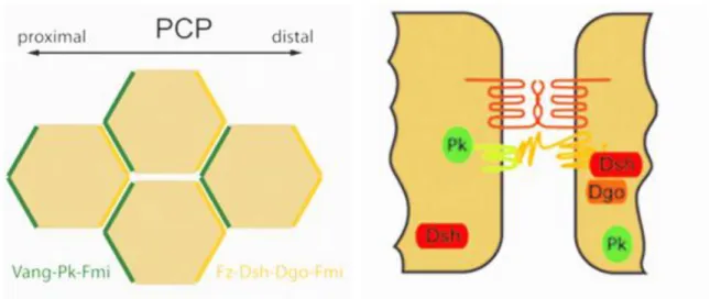
The non-canonical pathways do not require β-Catenin or LRP5/6. Their activation usually leads to inhibition of the canonical pathway (Grumolato et al. 2010). One of the non canonical pathways is the planar cell polarity signaling. PCP is distinct from apical/ basal polarity and can be defined as the alignment of cells within a cell sheet (Fig 2). PCP signaling creates an orientation of the cell by producing asymmetrical organization of Wnt signaling components: Fzd/Dvl are present at the distal side of the cell and Vangl2/ Prickle at the proximal side (Veeman et al 2003)(Montcouquiol et al. 2006). Both complex exclude each other: Prickle antagonizes Fzd/ Dvl by inducing removal of Fzd/ Dvl from the cell membrane by endocytosis while Dvl recruits E3 ubiquitin ligase Smurf to Prickle, leading to its loss (Narimatsu et al 2007).
Figure 3: PCP induces asymmetrical organization of Fzd/ Dvl and Vangl2/ Pk (Schulte 2010)
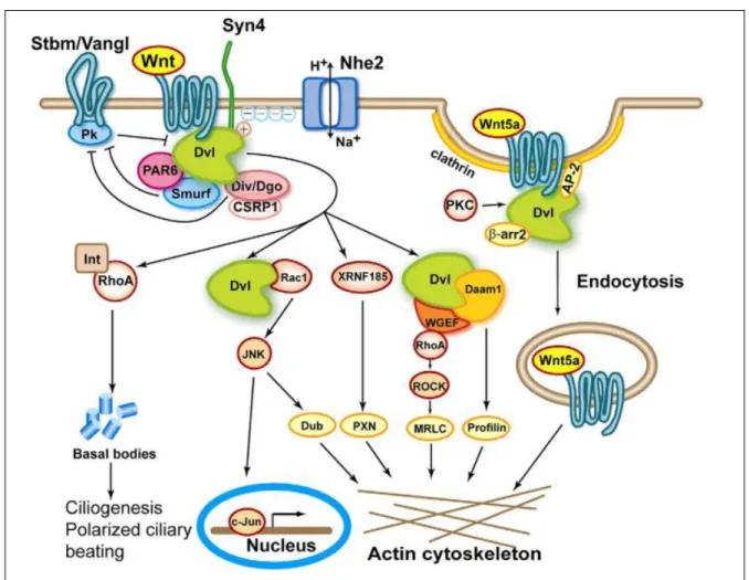
The current consensus is that for activation of the PCP pathways, Wnt ligand would bind to their receptor (Fzd with Ror2 or Ryk) and recruit Dishevelled, which then forms a complex with Disheveled-associated activator of morphogenesis 1, Daam1. Activation of Dvl leads to phosphorylation/ relocalization to the nucleus of c-JUN, where it acts as a transcription factor with c-FOS and to
activation of the small G-protein Rac, leading to activation of JNK (Gan et al. 2008). In parallel, Daam1 has been identified as a regulator of cell migration as it activates the small G-protein Rho, leading to activation of ROCK (Rho-associated kinase), which is one of the major regulators of the remodeling of the actin cytoskeleton (Ju et al. 2010, Tsuji et al. 2010). Most of the proteins involved in planar cell polarity in drosophila wing formation are conserved and used in vertebrates during regulation of cell movements during events such as gastrulation. Both processes imply control of actin filaments by the small G-proteins Rho and/ or Rac (van Amerongen and Nusse 2009). Other mechanisms are activated by PCP such as inflammation; phosphorylation of mitogen activated protein kinase p-38 would be induced by the non canonical pathway but not the canonical pathway, leading to inflammation (Verkaar et al. 2011). Interestingly, the PDZ protein GIPC is also a regulator of the localization of Vangl2 and of PCP in general (Wang et al. 1999, Giese et al. 2012).
iii) Other non canonical pathways
In the Wnt/Calcium pathway, Wnt5a and Frizzled regulate intracellular calcium levels. Wnt ligand binding to Frizzled receptor causes coupled G-protein to activate mechanisms that lead to an increase of intracellular calcium concentration. This increase activates Cdc42 (cell division control protein 42) through PKC (phosphokinase C), and calcineurin through CamKII (calcium/calmodulin-dependent kinase II). Calcineurin then induces activation of the transcription factor NFAT, which regulates ventral patterning. CamKII can also activate TAK1 and NLK kinase, which inhibit the canonical pathway(Kelly et al. 2011).
Finally, in the Wnt/GSK3 pathway, Wnt inhibition of GSK-3 activates mTOR without involvement of -Catenin. It’s why the mTOR inhibitor, Rapamycin inhibits Wnt-induced cell growth and cancer formation independently of -Catenin. The Wnt/ m-Tor pathway regulates cell cycle and is important in carcinogenesis (van Amerongen and Nusse 2009).
b) Wnt (wingless-type MMTV integration site family) Factors
i) Overview of the Wnt family of proteins
Mouse mammary tumor virus (MMTV) is a retrovirus that utilizes an insertional mutagenesis strategy to infect mammalian cells, leading to malignant transformation only in mammary epithelium, having a potent role in breast cancer formation. MMTV activates aberrant expression of number of developmental genes, via proviral insertion, leading to oncogenetic transformation. These normally silent genes activated by the MMTV have been named as ints. The association of MMTV with the Wingless family of proteins (drosophila) began with int-1 that shares 54% identity with Drosophila Wng, with a conservation of 20 cysteine residues. Thus, an amalgam was created out of the wingless-int-1 similarity (Wnt) that served for naming of the family. The Wnt family includes 19 members in mammals. Ligands such as Wnt1, Wnt3a, and Wnt8 interact with Fzd and LRP5/6 to activate the canonical pathway, while Wnt4, Wnt5a, Wnt7a and Wnt11 activate non canonical pathways.
Figure 4: Distribution of Wnt Genes in Metazoans. X indicate that no member of the Wnt subfamily found in the genome of the organism; the color is different for each Wnt; two blocks indicate presence of paralogue in the specie (adapted from Prud'homme et al. 2002)
Wnt5 and 10 are the only Wnt factors expressed in all species, indicating that their function are vital and conserved during evolution (Fig 4). Wnt3, 5 and 7 are not part of a subfamily suggesting that their function is not redundant with other Wnt factors (Fig 5).
Species name abbreviations: Av, Alopias vulpinus (shark);
Bf, Branchiostoma floridae (amphioxus);
Ce, Caenorhabditis elegans; Cs, Cupiennius salei (spider);
Dm, Drosophila melanogaster; Es, Eptatretus stoutii (hagfish);
Et, Evasterias troschelii (echinoderm);
Hs, Homo sapiens; Hv, Hydra vulgaris; Mc, Mysidium columbiae (crustacean);
Pdu, Platynereis dumerilii (annelid);
Pvu, Patella vulgata (mollusc); Sp, Strongylocentrotus purpuratus (echinoderm); Tl, Triops longicaudatus (crustacean); Tr, Terebratulina retusa (brachiopod).
ii) Phenotypes of Knockout Mice for Wnt factors
Loss of Wnt factors leads to severe phenotypes where various territories are affected. For example, loss of Wnt3 leads to gastrulation and neurogenesis defects. Loss of Wnt5 is associated to defects of morphogenesis and of formation of the reproductive apparatus. Loss of Wnt7 induces defects of the Central nervous system vascularization (Fig 6).
Gene Phenotype of Knockouts Mouse or other functions Associated diseases in Human
Wnt1 Loss of midbrain and cerebellum
With Wnt3a deletion: reduction of number of neural precursors in the neural tube
With Wnt4 deletion: reduction of number of thymocytes
Wnt2 Placental defects
With Wnt2b : defective lung development
Wnt2b/13 Retinal cell differentiation defects
With Wnt2: defective lung development
Wnt3 Early gastrulation defect
Hair growth defects
Apical ectodermal ridge defects
Medial-lateral retinotectal topography
Hippocampal neurogenesis defects
Tetra-Amelia
Wnt3a Somites, tailbud defects
With Wnt1: reduction in number of neural precursors in the neural tube together
Loss of hippocampus
Segmentation oscillation clock defects
Left right asymmetry
Hematopoetic stem cell self-renewal defects
Wnt4 Kidney defects
Defects in female development due to absence Mullerian Duct, leading to ectopic testosterone synthesis
Side-branching in mammary gland
With Wnt1: decrease in the number of thymocytes
Repression of the migration of steroidogenic adrenal precursors into the gonad
Mullerian-duct regression and virilization
SERKAL syndrome
Wnt5a Truncated limbs and anteroposterior axis
Distal lung morphogenesis defects
Associated with Susceptibility to type
Longitudinal skeletal outgrowth
Female reproductive tract defects
Shortened and widened cochlea
Mammary gland defects
Prostate gland defects
Intestinal elongation defects
Required for endothelial differentiation of ES cells
2 diabetes
Wnt5b Wnt6
Wnt7a Limb polarity defects
Infertility due to lack of Mullerian duct regression
Delayed maturation of synapses in Cerebellum
Promotes neuronal differentiation
With Wnt7b: required for central nervous system vasculaturization and maturation
Fuhrmann syndrome
Wnt7b Placental development defects
Respiratory failure due to defects in mesenchymal proliferation, required for lung development
With Wnt7a: required for central nervous system vasculaturization and maturation
Determines cortico-medullary axis in the kidney
Wnt8a
Wnt8b Changes in gene expression profile in neurons
Wnt9a Joint integrity defects
Wnt9b Regulates of mesenchymal/ epithelial transitions
Regulates renal vesicle induction
Planar cell polarity defects of the kidney
Wnt10a
Odonto-onycho-dermal dysplasia
Wnt10b Decreased trabecular bone (Bennett 2005) and bone mass loss
Required for taste papilla development
Loss activates adipogenesis
Overexpression inhibits adipogenesis
Mutations in Obesity patients
Split-Hand/Foot Malformation
Wnt11 Ureteric branching defects
Cardiogenesis defects
Wnt16
iii) Wnt3a (wingless-type MMTV integration site family, member 3A)
Wnt3a can bind Fzd1, Fzd2, Fzd4 and LRP6 directly. WIF1 and SFRP1 bind to Wnt3a and inhibit its activity (Dufourcq et al. 2008). Wnt3a is able to induce the canonical pathway by promoting caveolin-dependent internalization of LRP6/ Fzd after binding of LRP6 (Yamamoto et al. 2008).
iv) Wnt5a (wingless-type MMTV integration site family, member 5A)
Wnt5a can interact with Fzd1, Fzd2, Fzd4, Fzd7 or Ror2. Wnt5a can activate or inhibit canonical Wnt signaling, depending on receptor context. It can activate – catenin signaling by inducing caveolin-mediated endocytosis of Fzd receptors or inhibit the canonical Wnt pathway by promoting clathrin-mediated endocytosis of Fzd / Ror2(Grumolato et al. 2010). Wnt5a is required during embryogenesis for extension of the primary anterior-posterior axis and for gastrulation.
Figure 7: Schematic presentation of Wnt/ Fzd signaling kinetics (Schulte and Shenoy 2011)
Activation of -catenin (active -catenin - ABC, Fig 7, in black) or LRP6 (in blue) and interation between GSK3 and Axin are induced by Wnt3a. Phosphorylation of Dvl or Rac1 (in red/ orange) is induced by Wnt3a or Wnt5a. Phosphorylation of mitogen activated protein kinase p-38 is induced by Wnt5a only, indicating that both proteins induce signal transduction via similar protein, leading to activation of different targets (Schulte et al. 2010, Schulte and Shenoy 2011).
c) Fzd receptors
i) Overview of the Frizzled protein family
‘Frizzled’ refers to the phenotype of the frizzled mutant in Drosophila that has irregular patterned and curled hairs on the thorax, wings, and feet (Bridges and Brehme, 1944). Frizzled receptors family is a family of G protein- that act as receptors in the Wnt signaling pathway. These proteins are formed of seven-transmembrane domains, an extracellular N terminus, and an intracellular C terminus domain. Their basic architecture is reminiscent of conventional G protein-coupled receptor. Phylogenetic analysis reveals that Fzd form 4 subfamilies indicating potential redundancy of function between Fzd1,2 and 7; Fzd 3 and 6; Fzd4,9 and 10 and between Fzd5 and 8 repsectively (Fig 8).
Figure 8: Phylogenetic tree of the human Frizzled receptors FZD (Schulte and Shenoy 2011)
Figure 9: Schematic representation of Frizzled protein (binding sites for Dvl/ PDZ and G proteins indicated) (Schulte and Shenoy 2011)
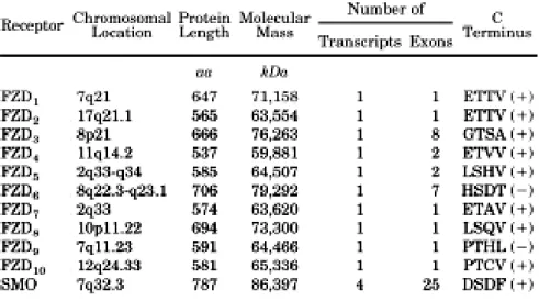
The extracellular region (Fig 9) of all Frizzled receptors consists of an N-terminal signal sequence (amino acid 1 to 36 in human FZD4) that is important for the insertion of the protein in the membrane. This is followed by a cysteine-rich domain (CRD; aa 40–161 in hFZD 4), which constitutes the binding site for Wnt ligands and Norrin (Schulte 2010). It is also hypothesized that Fzd receptors could form dimers via the CRD domain. The Fzd dimers would be sufficient to activate Wnt/ -catenin signaling. Homology analysis indicates that Fzds share only 20 to 40% of amino acid identity, but the ten cysteines that form the five disulfide bonds in the CRD domain are highly conserved between the Fzd isoforms, indicating a strong pressure of selection on these positions. The CRD domain is followed by seven transmembrane regions that form three intracellular loops, three extracellular loops, and a C-terminus of variable length. These intracellular domains are essential because they mediate various protein-protein interactions and post-translational processing through phosphorylation, ubiquitinylation, nitrosylation, or hydroxylation. The K-T-x-x-x-W domain that is very close to the membrane is highly conserved in the family. This sequence serves as the domain for interaction with the DEP domain of Disheveled. Additionally, Fzd proteins have an additional C-terminal typical PDZ binding domain for interaction with proteins such as PSD95 and with the PDZ domain of Dvl, this binding domain is conserved between most members of the family(Fig 10) (Schulte 2010)
Figure 10: Conservation of the PDZ binding domain (TXV-COOH) in the Fzd family; functional and non functional PDZ binding domain is noted as (+) or (-) respectively (Schulte and Shenoy 2011)
ii) Phenotypes of Knockout Mice for Fzd receptors
Loss of Fzd receptors leads to severe phenotypes where various territories are affected. Current works indicate that Fzd4 would be the only Fzd receptor shown to be essential for angiogenesis in adult, while Fzd5 would be essential for placental angiogenesis.
Gene Functions Phenotype of Knockouts Mouse Associated diseases
FZD1 Viable
FZD2 Viable
FZD3 Activates PKC
Activates -catenin
Defect in guidance of axons
With Fzd6: Midbrain morphogenesis defect FZD4 Activates PKC
Becomes internalized after adding Wnt5a
Activates Wnt reporter with norrin
Activates Wnt reporter with LRP5
Cerebellar, Auditory, and Esophageal Defects
Impaired Corpora Lutea Formation
Defects in vascular growth, endothelial defects
With Fzd8: Kidney phenotype
Retinal angiogenesis in familial exudative vitreoretinop athy
FZD5 Essential for yolk sac and
placental angiogenesis
Paneth cell phenotype
Neural potential in the developing Xenopus retina
Mammalian ocular development FZD6 Activates PKC Hair patterning, tissue polarity
With Fzd3: Midbrain morphogenesis defect
FZD7 Viable
FZD8 Activates Wnt with Respondin
With Fzd4: Kidney phenotype
FZD9 B cell development
Hippocampal defects
FZD10
iii) Frizzled 4
Figure 12: Diagram of Fzd-4 showing mutations associated to vitreoretinopathy (Robitaille et al. 2002, Robitaille et al. 2011)
Fzd4 is the only representative of frizzled family members that binds an additional ligand Norrin structurally different from Wnt ligands. Fzd4 and Norrin are major regulators of the vascular development of vertebrate retina. Mutations in the frizzled-4 gene have been notably associated with autosomal dominant familial exudative vitreoretinopathy (Robitaille et al. 2002). The main process in the disease is the destabilization of retinal vessels, causing retinal ischemia. Abnormal vessels then develop at the edges of the ischemic retina causing subretinal exudation and formation of scar tissue that produces tractional forces on the retina, leading to retinal detachment. The disease appeared to be linked with missense mutations of Fzd4 coding gene in the region coding for the CRD domain or truncation mutations in the internal domains (Nikopoulos et al. 2010), indicating that these domains are essential for the function of the protein (Fig 12). In 2004, Xu et al. indicated that vascular defects in fzd4 and
specific and that FEVR-Associated Fz4 mutants bind Norrin but were unable to transduce signal, they were able to place Norrin as one of the activator of Fzd4 during retinal vascularization(Xu et al. 2004). Furthermore, Ye et al found that endothelial specific deletion of fzd4 lead to retardation of vascular development of the retina, reduction in retinal capillaries and alteration of vessels of the vitreal surface that penetrate the retina that formed end in ball-like structures (Ye et al. 2010). Inhibition of Fzd4 using a blocking antibody would also lead to abnormal vascularization of the retina (Paes et al. 2011), confirming the role of the protein in the process of retinal vaculature. Fzd4 is also important in other organs. Loss of Fzd4 is also associated to cerebellar, auditory, and esophageal dysfunction (Wang et al. 2001). In a similar manner, it was demonstrated recently that Fz4 is required for blood barrier maintenance and plasticity in the central nervous system, as loss of Fzd4 leads to leakage in the cerebellum (Wang et al. 2012). These evidence shows that Fzd4 is vital for angiogenesis. That’s why our laboratory has focused part of its research on this protein.
d) Coreceptors of Fzd proteins
Figure 13: Competition between Wnt ligands would regulate activation of Wnt pathways by modulating phosphorylation of LRP5/6 and Ror2 (adapted from Grumolato et al 2010)
Wnt factors can activate the canonical and non canonical Wnt pathways through common mechanisms. Canonical Wnt3a and non canonical Wnt5a ligands bind Fzd receptor and recruit the same components. However, canonical Wnt ligands would induce phosphorylation of the Fzd receptor LRP5/6, while non canonical ligands would induce phosphorylation of the Fzd coreceptor Ror2 (Ser864) (Liu et al. 2008). Interestingly, it was shown more recently that canonical and noncanonical Wnts ligands inhibit each other by competing for Fzd binding at the cell surface leading to this differential phosphorylation of Ror2 and LRP5/6 (Grumolato et al. 2010) (Fig 13).
i) LRP5/6
Figure 14: Structure of LRP5 (He et al 2005)
LRP5 and 6 are highly homologous proteins. LRP5 contains a large extracellular domain containing four beta-propeller motifs and four epidermal growth factor-like repeats that form the binding sites for extracellular ligands. These domains are followed by three LDLR type A domains. The intracellular domain of LRP5 contains 5 PPPSP motifs. These motifs are phosphorylated upon binding of Wnt ligands and lead to binding of Ror2 to Axin, leading to activation of the canonical pathway (Grumolato et al. 2010). Zeng et al proposed a similar model in which Wnt-induced Fz-Lrp5/6 complex formation would lead to recruitment of the axin-Gsk3 complex, Gsk3 would then induce Lrp5/6 phosphorylation to initiate B-catenin signaling(Zeng et al. 2008). Interestingly, the phosphorylated forms of LRP5/6 have been shown to be internalized in clathrin-dependant vesicles with Dvl/ Fzd upon activation of the Wnt pathway, suggesting that LRP5/6 has a role for signal transduction that initiate in the early endosome (Bilic et al. 2007, Yamamoto et al. 2008). LRP5/6 is essential for angiogenesis as loss of the protein can also cause familial exudative vitreoretinopathy in humans, independently of loss of Fzd4 or Norrin
(Toomes et al. 2004). As LRP5 knockout mice display incomplete vasculature in the deeper plexus of the retina while FZD4 and Norrin knockout mice develop severe defects in both the superficial and deep plexus of the retina, Xia et al hypothesised that LRP5 would mediate Wnt signaling in Muller cells present in the deep plexus to regulate vascularization of this layer of the retina (Xia 2010). This demonstration correlates with recent findings showing that Wnt signaling is the main signaling pathway implicated in the formation of the deep retinal plexus.
ii) Ror2
Figure 15: Structure of the Ror2 protein (adapted from Scopus)
ROR2 is a member of the receptor tyrosine kinase-like orphan receptor (ROR) family. The protein is composed of an extracellular part composed of an Immunoglobulin C-2 Type domain, a Fzd domain and a Kringle domain. The Fzd domain would be essential for binding of Wnt ligands, while function of the immunoglobulin and Kringle domain are unknown. Interestingly, the tyrosine kinase domain of Ror2 is functional. The tyrosine kinase activity would be essential for signal transduction in the non canonical pathway, as activation of c-Jun/ Dishevelled would be dependant on this activity, but no targets have been found yet (Mikels et al. 2009, Ford et al. 2013). Ror2 is phosphorylated on serine/threonine residues in its c-Terminal cystein rich domain upon stimulation of cultured cells with Wnt5a, but not with Wnt3a. Phosphorylation of Ror2 would be GSK3 dependant as blockade of GSK3 would block Ror2 induced migration of endothelial cells (Yamamoto et al. 2007). Phosphorylation of Ror2 on this domain would depend on various kinases such as Casein Kinase 1-ϵ (CKIϵ) or Phosphokinase C
PKC-Casein Kinase 1 would be directly responsible for phosphorylation of Ror2 while PKC- would activate kinases upstream of Casein Kinase 1 (Kani et al. 2004). Interestingly, use of the PKC- pseudosubstrate that blocks the kinase activity of PKC-blocks the phosphorylation of Ror2 and the
activation of non canonical Wnt pathway (Nomachi et al. 2008). Using the PKC- pseudosubstrate and
ror2 targetting siRNA, it was shown that Ror2 activity was essential for formation of the lamellipodia
and reorientation of microtubule-organizing center in Wnt5a-induced cell migration. The proposed model is that Ror2 would associate with the actin-binding protein filamin A to regulate reorganization of the actin cytoskeleton to induce migration of endothelial cell; and would be required for c-Jun / JNK activation that is essential for activation of the small GTPases Rho and Rac (Nomachi et al. 2008).
iii) Ryk
Figure 16: Structure of the Ryk protein (adapted from Scopus)
Ryk is an atypical member of the family of growth factor receptor protein tyrosine kinases, whose activity is not regulated by phosphorylation. The protein has a leucine-rich extracellular domain with a WIF-type Wnt binding region, a single transmembrane domain, and an intracellular tyrosine kinase domain. This protein is involved in stimulating Wnt signaling in processes such as the regulation of axon pathfinding. Ryk functions as a coreceptor with Fzd and binds Dvl to activate the canonical Wnt pathway (Lu et al. 2004). More recently, it was shown that the E3 ubiquitin ligase Mindbomb1 would reduce cell surface levels of Ryk leading to activation of the Wnt canonical pathway, indicating that Ryk would activate the non canonical pathway(Berndt et al. 2011).
e) Downstream effectors of Wnt signaling
i) Dishevelled adaptor proteins
Dvl is the cytoplasmic link between Frizzled and downstream components of the Wnt signaling pathway. Dvl has no known enzymatic activity and functions as a scaffold protein bridging the receptors and downstream signaling components.
Figure 17: Structure of Dvl (adapted from Scopus)
The DIX domain is essential for -catenin activation, the DEP domain is implicated in the activation of the non canonical pathway, while the PDZ domain is required for both. Dvl can bind Axin2 via its DIX domain – blocking Axin-induced degradation of β-catenin (Fiedler et al. 2011). The PDZ domain is required for the interaction with the C-terminal regions of Vangl1 and 2. Dvl interacts with Fzd4 (Chen et al. 2003) via both the PDZ and DEP domains (Fig 17). In presence of Wnt5a, cytoplasmic Dvl is recruited to the membrane via endocytic vesicles where it is phosphorylated (Bryja et al. 2007). Human and murine orthologues share more than 95% sequence identity and are each 40-50% identical to Drosophila Dvl where a single member of the family is found. Sequence similarity is concentrated around the DIX domain, the PDZ domain and the DEP domain, indicating that the three domains are essential. Three members of the Dishevelled family exist in mammalians (Dvl-1 to 3). Dvl2 is strongly expressed in majority of mammalian cells, while levels of Dvl1 and 3 are much lower. Dvl1 and 3 play the major role for the activation of both Wnt pathways. The three Dvl isoforms are essential during development as knock-out of Dvl2 or 3 leads to cardiovascular defects while the knock-out mouse for Dvl1 and 2 presents an open neural tube, a characteristic of knock-out mice of genes implicated in the Wnt/PCP pathway (Wang et al. 2006).
Gene Functions Phenotype of knockouts mouse Associated diseases in human
Dvl1
Activates Wnt canonical and calcic pathway
Overexpression leads to cardiac hypertrophy and is rescued by inhibition of CamKII
Social interaction and sensorimotor gating abnormalities
With Dvl2: Open neural tube
With Dvl2: Convergent extension phenotype Activity of LRRK2 that is mutated in Parkinson is regulated by Dvl1/2/3 Dvl2
Cardiovascular outflow tract defect. With dvl1:open neural tube
With dvl1: convergent extension phenotype
Dvl3
Activates canonical
and PCP pathway Cardiac outflow tract defects
Figure 18: List of Dvl proteins (adapted from Wnt homepage, Stanford)
Figure 19: Positive (left) and negative (right) regulators of Dvl activity (Gao et al 2010)
Dvl is phosphorylated upon activation of the Wnt pathways by Casein kinase 1-ϵ, or PAR-1 depending on context (Tian-Qiang Sun and Williams 2001, Bryja et al. 2007). Several proteins have been reported to regulate stability of Dvl by facilitating its poly-ubiquitination and/or degradation. The HECT-type ubiquitin ligase NEDL1 induces degradation of Dvl1. KHL12 recruits the ubiquitin complex Cullin-3 to Dvl, causing its degradation. Prickle 1 also negatively regulates the Wnt/PCP pathway by enhancing Dvl3 degradation (Gao and Chen 2010).
ii) β –catenin
Figure 20: The -catenin destruction complex (Kimelman and Xu 2006)
-catenin, protein composed of armadillo repeats was first identified as a scaffold protein and is one of the key components of the canonical Wnt signaling pathway. In the absence of the Wnt signal, β – catenin levels are down-regulated by a complex, composed of Axin/ CKI-α (casein kinase 1-α) / APC(Adenomatous Polyposis Coli) and GSK-3 β (glycogen synthase kinase 3) (Fig20 – 1). Axin and APC act as scaffold protein bringing GSK3 and its substrate (β-catenin) into close physical proximity (Fig 20 – 2and3). This positions the N-terminus of β-catenin near CK1 and GSK3. CK1 phosphorylates β-catenin at Ser45 and GSK3 phosphorylates β-catenin at Thr41, Ser37 and Ser33 (Fig20 – 3and4). The
E3 ubiquitin ligase -TRCP1 then binds the phosphorylated N-terminus of β -catenin, causing its degradation (Fig 20- 6). Transcription of β –catenin target genes is then repressed as the transcription factors LEF/TCF form a complex with groucho, inducing histone deacetylation of the target genes of β – catenin (Saadeddin et al. 2009). In the presence of Wnt ligand, Fzd recruits Dvl which in turn brings to the membrane the axin–GSK3 complex that promotes phosphorylation of LRP5/6 by GSK3 and CK1 (Zeng et al. 2008). Phosphorylated LRP5/6 leads to inhibition of GSK3/Axin/APC complex; catenin is not ubiquitinylated and is dephosphorylated by cadherin 2 on Ser37 or Thr41. This active form of -catenin translocates to the nucleus where it complexes with transcription factors LEF/ TCF to favor transcription of genes such as Cyclin D (Sadot E et al. 2002). The membrane bound catenin has a different function. It is part of a complex of proteins that constitute adherens junctions by interacting with Ve-cadherin. Therefore, it is also implicated in processes of adhesion and permeability. Mutations in the gene coding for -catenin are a cause of colorectal cancer, pilomatrixoma, medulloblastoma, and ovarian cancer. catenin is typically a protein that can localize to Nuclear and Adhesion Complex (NACos)(Balda 2003) that stabilizes junctions when located at the adherens junction and act as pro-proliferative transcription factor when located to the nucleus.
iii) C-JUN
c-Jun is a transcription factor of the MAPK family composed of a basic region leucine zipper domain, that recognizes and binds to the enhancer heptamer motif 5'-TGA[CG]TCA-3'. It it part of the SMAD3/ SMAD4/ JUN/ FOS complex which forms at the AP1 promoter site. The SMAD3/SMAD4 heterodimer acts syngernistically with the JUN/FOS heterodimer to activate transcription in response to Wnt signaling. C-Jun is phosphorylated by CAMK4, PRKCD, DYRK2 or HIPK2 and notably at Ser-63 by PLK3 upon Wnt5a activation, leading to increase of its DNA-binding activity. The phosphorylation of c-Jun at this site will be used as an indicator of the activation of the Wnt non canonical pathway in this work. C-Jun is also phosphorylated at Thr-239, Ser-243 and Ser-249 by GSK3; reducing its ability to bind DNA (Wong et al. 2004, Gan et al. 2008, Saadeddin et al. 2009). Various works have shown a role of c-Jun in development and specifically in vascular development. Jun-null mice die between
midgestation and late gestation and exhibit impaired hepatogenesis, altered fetal liver erythropoiesis, and generalized edema (Hilberg et al. 1993), indicating an essential function of Jun in hepatogenesis and vascular permeability. Jun is also essential for placental and yolk sac vascularization (Schorpp-Kistner 1999, Schreiber 2000). Behrens et al. 1999 demonstrated that phosphorylation of Jun on ser63 is required for apoptosis. Jun can induce oncogenic transformation and was also found to be highly expressed in breast cancers (Johnson et al. 1999). Jun is also one of the first proteins to be upregulated after stroke, when the blood brain barrier is disrupted (McColl et al. 2008).
iv) RhoA
The small guanosine triphosphatase (GTP) Ras homolog family member A (Rho A) is a major regulator of actin remodeling during cell morphogenesis and motility. Rho activates its effector ROCK1, which phosphorylates and activates LIM kinase, which in turn, phosphorylates cofilin, inhibiting its actin-depolymerizing activity (Maekawa et al 1999). RhoA was shown to be implicated in migration as exoenzyme C3, a Rho inhibitor, inhibits corneal epithelial migration (Nakamure et al 2001). Pertz et al (2006) used a fluorescent biosensor to visualize the spatiotemporal dynamics of RhoA activity during cell migration. In randomly migrating cells, RhoA activity is concentrated at the leading edge while PDGF-induced membrane protrusions have low RhoA activity, potentially because PDGF strongly activates Rac, which had been shown to antagonize RhoA activity, showing that different stimuli induce distinct patterns of signaling during migration. Many elements link RhoA to Wnt signaling. Binding of DAAM1 to Dvl and the small GTPase Rho has been shown to coordinate Wnt signaling cues (Habas et al 2001), while the RhoGEF p114 was shown to transduce Wnt signaling, leading to RhoA activation(Tsuji et al. 2010). These elements place RhoA directly downstream of Wnt/PCP signaling.
2) Wnt signaling as a regulator of vascular development
a) Wnt signaling is implicated in angiogenesis during development and
in pathology
i) During embryonic development
Figure 21: Canonical Wnt signaling is up-regulated after Wnt5a KO (Topol et al 2003)
Wnt-5a is strongly expressed in the limb bud during development and its expression gradually fades (Yamaguchi et al., 1999). In the Wnt-5a KO embryos, the fingers are missing, due to massive activation of the canonical pathway, detected as an increase of β-catenin protein level, indicating that Wnt-5a would regulate the balance between the Wnt pathways (Fig 21) (Topol et al. 2003).
Yolk sac and placenta vascularization
Figure 22: a- Expression of various components of Wnt signaling in placenta and yolk sac; b- Vascular pattern is normal in Fzd5-/- embryo but vasculature remains superficial in placenta/
Wnt signaling is also important for sac yolk angiogenesis. Yolk sac angiogenesis occurs between E8.5 and E10.5. Fzd5 is strongly expressed in vessels of the yolk sac at E9.5 (Fig 22a) and Fzd5-/- embryo die in utero around E10.5, due to defects in yolk sac angiogenesis, as indicated by a poorly developed vascular bed when compared to Wt mice (Fig 22b). Vasculogenesis in the placenta is also defective as embryonic vessels do not invade the placental labyrinth in Fzd5-/- mouse (Fig 22c). Because Wnt5a and Wnt10b co-localize with Fzd5 in the developing yolk sac, these two Wnts are likely physiological ligands for Fzd5-dependent signaling for endothelial growth in the yolk sac (Fig 22a) (Tomo-o Ishikawa and Shin-ichi Nishikawa 2001).
Figure 23: a- The yolk sac of the junB KO embryo at E9.5 is translucid and shows no vessels; b- Morphological phenotypes of JunB deficient embryos (Schorpp-Kistner 1999).
Interestingly, the non canonical pathway is essential for yolk sac angiogenesis as deletion of jun or other members of the AP1 pathway such as c-Fos lead to vascular defects of the yolk sac and placenta leading to growth retardation of the embryo as from E7.5dpc (Schorpp-Kistner 1999, Schreiber 2000). This body of evidence shows that the PCP pathway is required for angiogenesis in the placenta and yolk sac, implying that the pathway is essential for embryonic development.
Brain vascularization
Vascularization of the central nervous system starts at embryonic day 9 via angiogenesis from a previously developed primitive vascular plexus (Paolinelli et al. 2011). The newly formed vessels acquire specific characteristics in the brain as they establish unique barrier characteristics called blood– brain barrier. The blood brain barrier (BBB) has a protective role as it prevents the flow of molecules from the blood to the tissue via expression of tight junction proteins that seal the space between endothelial cells.