HAL Id: tel-02932167
https://tel.archives-ouvertes.fr/tel-02932167
Submitted on 7 Sep 2020
HAL is a multi-disciplinary open access
archive for the deposit and dissemination of sci-entific research documents, whether they are pub-lished or not. The documents may come from teaching and research institutions in France or abroad, or from public or private research centers.
L’archive ouverte pluridisciplinaire HAL, est destinée au dépôt et à la diffusion de documents scientifiques de niveau recherche, publiés ou non, émanant des établissements d’enseignement et de recherche français ou étrangers, des laboratoires publics ou privés.
Control of meiotic divisions in oocytes : a novel role for
cyclin B3
Nora Bouftas
To cite this version:
Nora Bouftas. Control of meiotic divisions in oocytes : a novel role for cyclin B3. Subcellular Processes [q-bio.SC]. Sorbonne Université, 2019. English. �NNT : 2019SORUS176�. �tel-02932167�
1
Thèse de doctorat de Sorbonne Université
Ecole doctorale Physiologie Physiopathologie et Thérapeutique
Présentée par
Nora Bouftas
Control of meiotic divisions in oocytes :
a novel role for cyclin B3
Soutenue le 26 Septembre 2019
Devant le jury compose de:
Evelyn Houliston
Présidente
Anna Castro
Rapporteur
Lionel Pintard
Rapporteur
Marie-Emilie Terret
Examinatrice
Helfrid Hochegger
Examinateur
Katja Wassmann
Directrice de thèse
Summary
:
Throughout my PhD, I focused on the study of female meiotic cell cycle regulation and more specifically, the two meiotic divisions and chromosome segregation in oocytes. Meiosis is a tightly regulated process that must be completed in an orderly manner to obtain gametes with the correct number of chromosomes. The two divisions occurring during meiosis (called meiosis I and II) have different patterns of segregation. Importantly, S-phase must be suppressed between the two divisions. This leads to the creation of haploid gametes. All the steps of meiotic cell division are regulated by cyclins associated to their binding catalytic partners Cdks. Female mammalian meiosis presents a lot of challenges. It was shown that it is an error-prone process where errors in segregation occur creating aneuploid gametes. Aneuploidy occurs more frequently during female meiosis compared to male meiosis. In addition, incidence of aneuploidy increases in correlation with age. Understanding the mechanisms involved in the regulation of female mammalian meiosis is therefore essential. Progression through the meiotic cell divisions in mammalian oocytes (also named meiotic maturation) is characterized by two arrests: oocytes formed during embryonic development arrest at prophase I until hormonal stimulation induces resumption of meiosis. The second arrest is marked at metaphase II, also known as CSF arrest. This arrest is lifted only upon fertilization. During my PhD, I investigated the role of a unique cyclin, cyclin B3, through the use of cyclin B3 KO mice. I found that lack of cyclin B3-Cdk1 activity induces an arrest at metaphase I. This is due to high cyclin B1 levels and Cdk1 activity as well as inactive separase. APC/C activity was also affected since endogenous APC/C substrates were not efficiently degraded. Furthermore, the role of cyclin B3 was found to be conserved, as cyclin B3 from other species was able to rescue mouse cyclin B3 KO oocytes.As I further explored the possible roles of cyclin B3, and with the use of Xenopus oocytes, I was able to show that cyclin B3 is able to inhibit CSF arrest. The mechanism of this inhibition is still being explored, but my recent data suggests that cyclin B3 is able to induce the degradation of the APC/C inhibitor Emi2. Furthermore, oocytes lacking cyclin B3 put a precocious CSF arrest into place, leading to the metaphase I arrest observed. Mimicking fertilization or inhibiting one of the components required for CSF arrest rescues this metaphase I arrest. Hence, my PhD work has shown that cyclin B3 is essential for female meiosis to correctly activate the APC/C in meiosis I and to prevent precocious CSF arrest in meiosis I instead of meiosis II.
3
Abbreviations
AMP
Adenosine MonoPhosphate
APC/C
Anaphase Promoting Complex/Cyclosome
Arpp19
cAMP-regulated phosphoprotein 19
Bub
Budding uninhibited by benomyl
CamKII
Calmodulin-dependent protein Kinase II
Cdc
Cell division cycle
Cdk
Cyclin dependent kinase
CKI
Cyclin dependent Kinase Inhibitor
CPC
Chromosomal Passenger Complex
CPEB
Cytoplasmic Polyadenylation Element Binding
CSF
CytoStatic Factor
C-ter
C-terminal
Dbox
Destruction box
Emi
Early mitotic inhibitor
GFP
Green Fluorescent Protein
GVBD
Germinal Vesicle Breakdown
GV
Germinal Vesicle
G phase
Gap
KD
Knockdown
KO
Knockout
Mad
Mitotic arrest deficient
MAPK
Mitogen Activated Protein Kinase
MCAK
Mitotic Centromere Associated Kinase
MCC
Mitotic Checkpoint Complex
MPF
M phase Promoting Factor
Mps1
Monopolar spindle 1
MTOC
MicroTubule Organizing Center
M phase
Mitosis or Meiosis
N-ter
N-terminal
PBE
Polar Body Extrusion
PKA
Protein Kinase A
Plk1
Polo like kinase 1
PP2A
Phosphatase 2A
p90Rsk
p90 Ribosomal S6 Kinase
Rec8
Recombination protein
RFP
Red Fluorescent Protein
SAC
Spindle Assembly checkpoint
Scc
Sister chromatid cohesion
SCF
Skp-Cullin-Fbox
Sgo
Shugoshin
Sr
Strontium
S phase
Synthesis
WT
Wild Type
XErp1
Xenopus Emi1-related protein 1
YFP
Yellow Fluorescent Protein
5
Figures Index
Figure 1: Increase of trisomies in relation to maternal age
Figure 2: Different phases of the cell cycle
Figure 3: Mitosis vs. Meiosis
Figure 4: Cyclin-Cdk complexes drive the cell cycle
Figure 5: APC/C co-activators Cdh1 and Cdc20 during the cell cycle
Figure 6: Mammalian meiotic maturation
Figure 7: Oogenesis during mammalian development
Figure 8: Prophase I arrest (GV) vs. resumption of meiosis (GVBD)
Figure 9: Active SAC inhibits APC/C
Cdc20activity during meiosis I
Figure 10: The cohesin complex and its subunits
Figure 11: Protection and deprotection of centromeric cohesin during meiosis
Figure 12: APC/C activity is necessary for anaphase onset during meiosis
Figure 13: Metaphase II arrest is partly controlled by the Mos-MAPK pathway
Figure 14: Emi2 regulation during metaphase II arrest
Figure 15: Fertilization triggers anaphase II onset
Figure 16: Xenopus oocyte meiotic maturation
Figure 17: Cyclin B3 compared to A and B type cyclins
Figure 18: Cyclin B3 size in different species
Figure 19: Cyclin B3 inhibits the CSF pathway
Table of Contents
Introduction ... 8
Aneuploidy ... 9 1. Cell cycle ... 10 A) Steps of the cell cycle ... 10 B) Mitotic division ... 11 C) Meiosis: specialized cell cycle ... 11 2. Regulation of the cell cycle ... 13 A) Cyclin-Cdks ... 13 B) M phase ... 14 i) A type cyclins ... 14 ii) B type cyclins ... 14 C) MPF ... 15 D) APC/C regulation and proteolysis by the 26S proteasome in mitosis ... 16 i) Mechanism of APC/C activity ... 16 ii) APC/C co-activators Cdh1 and Cdc20 in mitosis ... 17 3. Meiotic maturation in mouse oocytes ... 18 A) Summary of meiosis in mouse oocytes ... 18 B) Oogenesis: the development of the oocyte ... 19 C) Resumption of meiotic maturation ... 20 i) Prophase I arrest: GV stage ... 20 ii) Lift of prophase I arrest: GVBD ... 21 D) Prometaphase: starting meiotic maturation ... 21 i) Spindle formation and KT-MT attachments ... 22 ii) Spindle Assembly Checkpoint ... 23 E) Metaphase I to anaphase I transition ... 24 i) Cohesin, the glue that keeps chromosomes together ... 24 ii) Two step removal of cohesin ... 25 1. Protection of centromeric cohesin ... 25 2. Deprotection: removing centromeric cohesin ... 26 iii) Cyclin B1 vs. Cyclin B2 regulation during meiosis I ... 27 F) Exit of meiosis I: Anaphase I ... 28 G) Meiosis II ... 30 i) Entry into meiosis II ... 30 ii) Regulation of Metaphase II arrest: CSF pathway ... 30 1. Mos-MAPK pathway ... 31 2. Regulation of Emi2, a potent APC/C inhibitor ... 33 H) Exit from meiosis II and fertilization ... 35 4. APC/C activity during meiosis ... 36 A) Co-activators Cdh1 and Cdc20 in meiosis ... 36 B) Substrates during meiotic maturation ... 37 5. Xenopus as an alternative model system ... 39 6. Cyclin B3 ... 40 A) Characteristics of the structure of Cyclin B3 ... 40 B) Role and conservation in different organisms ... 41 7. Project ... 44
Results ... 45
Part I: Cyclin B3 promotes anaphase I onset in oocyte meiosis ... 46
7 Part II: Cyclin B3 prevents a precocious CSF arrest in meiosis I oocytes ... 51
Discussion ... 61
How does cyclin B3 affect APC/C activity in meiosis I? ... 62 Special characteristics of cyclin B3 during meiotic maturation ... 63 Cyclin B3-Cdk1 activity down regulates overall Cdk1 activity in mouse oocytes ... 64 Precocious CSF arrest in cyclin B3 KO mouse oocytes ... 65 Conserved role of cyclin B3 in mouse and Xenopus oocyte maturation ... 66 How does cyclin B3 inhibit CSF arrest ... 67
Bibliography ... 71
Résumé en français ... 88
Other publications ... 89
Introduction
9
Aneuploidy
The cell cycle is a complex series of steps that produce all the cells of the body. The division of cell components occurs during the two types of M phase: mitosis and meiosis. Meiosis is the specialized type of cell cycle that forms the gametes that are capable of undergoing fertilization. Its proper regulation is needed to obtain a healthy embryo and offspring. Mistakes during meiosis and mitosis can lead to cells with the incorrect number of chromosomes making them aneuploid. It is known that aneuploidy can be an important characteristic of different cancers. Errors during meiosis can have devastating consequences on the future embryo.
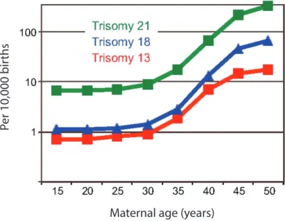
Aneuploidies observed during meiosis are in majority due to errors during female meiosis. Only 1-2% of spermatozoa were found to be aneuploid compared to 10-30% of oocytes. This marked difference clearly shows that female meiosis is more error prone. The incidence of aneuploidy even increases exponentially with the increase of maternal age (Fig. 1) (MacLennan et al. 2015; Templado, Uroz, and Estop 2013; Nagaoka, Hassold, and Hunt 2012; Hassold and Hunt 2001). This was already observed in early studies (Penrose 1933). However, this is not the case in male meiosis. No correlation of increase of age and aneuploidy were found during male meiosis. This is probably due to the fact that the mechanisms that regulate correct meiotic progress are more robust in male germ cells that lead more frequently to germ cell loss rather than increase of the incidence of aneuploidy (Vrooman et al. 2014). In addition, male meiosis is shorter in timing compared to female meiosis where for humans, oocytes are arrested for years before they can resume meiosis and meiotic maturation is a very lengthy process.
The percentages of aneuploidies observed in correlation with women’s age range from around 35% up to 80% in some studies (Capalbo et al. 2017; Hassold and Hunt 2001). Very few types of aneuploidies are viable in humans. It seems that embryos are very sensitive to gene dosage and addition or removal of a chromosome more often than not lead to embryonic lethality. Therefore, aneuploidy in somatic cells is very dangerous for the development of the future embryo. These types of aneuploidies are a major cause of miscarriage and it was observed that more than half of miscarriages during the first trimester are due to aneuploidy in somatic cells. However, there are a few viable aneuploidies due to a gain of a chromosome (trisomies). These include trisomy 13 (Patau
Per 10,000 bir
ths
Maternal age (years)
Figure 1:
Increase of trisomies in relation to maternal age. The three trisomies
indicated in this graph are the viable form of trisomy for humans. Number of
trisomies per 10,000 births was recorded. The incidence of each trisomy
significantly increases as maternal age increases. Adapted from Jones and Lane
2013.
10 syndrome), trisomy 18 (Edwards syndrome), and trisomy 21 (Down syndrome) (Fig. 1). The first two show graver consequences, as life expectancy is very low, with only 10% of individuals reaching the age of 10. This difference with trisomy 21 is probably due to the smaller size of chromosome 21 that contains fewer genes (Potapova and Gorbsky 2017). Loss or gain of sex chromosomes also presents more mild phenotypes in humans. Loss of the X chromosome in women (Turner syndrome) and gain of chromosome X in men (Klinefelter’s syndrome) leads to infertility. Gain of the Y chromosome presents no obvious defects and individuals don’t present issues with fertility. The Y chromosome has very few genes, and these genes are not needed for viability, which could explain this mild phenotype upon a supplemented gene copy (Potapova and Gorbsky 2017).
These findings are of concern in modern societies, as we know that today women have their first child at a later age. Aneuploidies during female meiosis can lead to spontaneous abortions, miscarriages, and congenital diseases. It is important to first understand how different mechanisms are put in place during female mammalian meiosis to then comprehend what goes wrong during human oocyte maturation. 1. Cell cycle A) Steps of the cell cycle The cell cycle is comprised of 2 Gap phases (G1 and G2), with a replication stage (Sphase), all three considered to constitute interphase, and finally the mitotic phase (M phase). After duplication of genetic material and cell growth occurring during interphase, the different components are divided into two cells at M phase (Fig. 2). Each phase has to be correctly completed and follow the previous phase to complete an orderly cell cycle.
During G1 phase, cells complete a growth phase that includes the synthesis of the components necessary to move on to the next phase, such as mRNA, proteins and ribosomes. At G1 entry, the cell must make a decision on whether it will begin a new cell cycle or not. The parameters of this decision depend on the cell type. This phase, like all others in the cell cycle, is controlled by a specific checkpoint that verifies the correct growth of the cell to analyze if it can move to S phase or halt at a quiescent G0 phase until conditions are more favorable. S phase is the replication stage, where DNA is replicated in order to have two copies of the cell’s genetic information. This phase is also controlled by a
M-phase
G2-phase
S-phase
G1-phase
Figure 2:
Different phases of the cell cycle. The cell cycle is comprised of four
phases. Two Gap phases, G1 and G2 phase, as well as an S phase make up
interphase. The fourth phase is the M-phase. Each phase must follow each other in
a regulated and orderly manner. G1 phase allows growth of the cell and
preparation for replication phase. During S phase, the genetic information of the
cell is duplicated. G2 phase permits the cell to continue growing and synthesizing
proteins and other components necessary for M-phase. Finally, during M-phase,
DNA is divided and the cell gives rise to daughter cells.
11 checkpoint verifying that DNA synthesis occurred correctly and the DNA damage checkpoint. G2 phase follows right before the cell enters mitosis. At this stage the cell continues to grow and synthesize protein as it prepares to enter mitosis. The transition from G2 to M phase is controlled by a DNA damage checkpoint that verifies that the genetic material is not impaired. Correct completion of these steps allows the cell to transition to M phase. During M phase, the DNA segregates into daughter cells. There exist two types of M phase called mitosis and meiosis. Both of them are controlled by the Spindle Assembly Checkpoint (SAC) that delays M phase exit if errors are observed. Consequently, each step of the cell cycle is monitored through different checkpoints (Elledge 1996). It is primordial to regulate each step in order to avoid deregulation that can lead to numerous disorders. B) Mitotic division Mitosis is a cell division that occurs in all somatic cells. During mitosis, a mother cell divides its genetic material and components of the cell into two daughter cells. Mitosis is divided into 5 phases: prophase, prometaphase, metaphase, anaphase and telophase. Cells enter mitosis in prophase where chromosomes begin to condense to be prepared for segregation. During prometaphase, chromosomes are condensed, the nuclear envelope breaks down and the spindle begins to form. Importantly, at metaphase, the microtubules emanating from the spindle must be correctly attached to a structure in the centromeric region called the kinetochore. At this stage, sister chromatids are linked together by a ring-like structure called cohesin and attached to the spindle in a bipolar manner. The chromosomes are aligned at the metaphase plate. Anaphase is marked by the segregation of chromatids due to cleavage of the cohesin. This frees sister chromatids from each other and due to the spindle pull, sister chromatids are segregated to opposite poles. The next phase is telophase where the DNA begins to decondense once again and forms a new nuclear envelope around both clusters of DNA in each daughter cell. This process is ended by a final step of cytokinesis where the membranes separate and reform to produce two individual daughter cells (Fig. 3).
C) Meiosis: specialized cell cycle
The production of haploid gametes is achieved by a specialized cell cycle, meiosis. During meiosis, the cell must divide its DNA during two different divisions with only one preceding S
Mitosis
Mieosis
PROPHASE METAPHASE ANAPHASE TELOPHASE PROPHASE METAPHASE I METAPHASE II ANAPHASE I ANAPHASE II TELOPHASEMitosis vs. meiosis. Two types of M-phases exist, mitosis and meiosis.
Both contain similar phases: prophase, prometaphase (not shown), metaphase,
anaphase and telophase. Mitotic cells undergo one round of division while in
meiosis there are two rounds of division with no intermediate S phase allowing
the creation of haploid gametes. In meiosis I, chromosomes segregate while in
meiosis II sister chromatids segregate.
12 phase. The phases described for mitosis are conserved and present during meiosis. Unlike mitosis, the challenge of the regulation of meiosis is to correctly achieve two different patterns of division resulting in a haploid gamete ready for fertilization. The first division requires segregation of homologous chromosomes while the second leads to sister chromatid segregation (Fig. 3). Female and male meiosis has a similar end goal: the creation of a euploid (with a correct number of chromosomes) gamete ready for fertilization. However, there are marked differences between the two divisions, especially for mammals. Male meiosis is less error prone than female meiosis. This could be due to the fact that male gametes (spermatozoa) are produced throughout their lifetime. This is not the case for mammalian oocytes. Male meiosis can take approximately 26 days in humans versus decades for women (S. Lane and Kauppi 2019). The stock of available oocytes is present since birth and therefore ages considerably. The oocyte is also of significant size and will be the host cell for the development of the future embryo. During female mammalian meiosis, the two cell divisions are asymmetric. Half of the genetic material is discarded into a smaller cell called the polar body. This asymmetry permits the oocyte to retain most of the nutrients and cytoplasmic components needed to complete meiotic maturation, fertilization and the next steps in embryonic development. In female mammalian meiosis, there are two main arrests observed during maturation; prophase I arrest and metaphase II arrest. The first must be maintained for up to decades and the second is vital for correct ploidy as it is only lifted at fertilization. Therefore, different and additional regulatory mechanisms are present for correct completion of female meiosis.
2. Regulation of the Cell cycle A) Cyclin-Cdks of the cell cycle Cdk (Cyclin dependent kinase) levels govern entry and exit from the different phases of the cell cycle. Cdks are serine/threonine kinases that regulate all steps of the cell cycle. More than 20 families of Cdks have been identified so far (Malumbres et al. 2009). They are characterized by a binding domain for cyclins, an ATP-binding catalytic pocket and a T-loop motif (Lim and Kaldis 2013). For Cdks to become active, they must be partnered with their regulatory subunits called cyclins. Cyclin-Cdk complexes can be formed and complete their roles in part due to the presence of different motifs found on cyclins. An example of this is the MRAIL motif found on the α helix of the cyclin box. This motif was first described in cyclin A-Cdk complexes where different observations were made. Mutating this motif was shown to disturb cyclin-Cdk binding at times and at other times abolish substrate recognition without affecting cyclin-Cdk binding, demonstrating the importance of these regulatory motifs (Bendris et al. 2011; Schulman, Lindstrom, and Harlow 1998; Jeffrey et al. 1995). Different cyclin-Cdk complexes need to be formed at different time points of the cell cycle to allow correct progression throughout the cell cycle (Murray 2004). The complex needed to progress through G1 phase is cyclin D-Cdk4/6. Entry into S phase necessitates the activity of cyclin E-Cdk2 while the rest of S phase is controlled by cyclin A-Cdk2. To successfully transition from G2 to M phase, cyclin A-Cdk1 must be involved. Finally, during M phase, cyclin B-Cdk1 seems to play the pivotal role (Fig. 4) (Satyanarayana and Kaldis 2009; Sánchez and Dynlacht 2005; Malumbres and Barbacid 2005). This does not mean that no other complexes are involved in the process. For example, depending on the species, more than one cyclin-Cdk complex is necessary for M phase. This is the case for female mammalian meiosis, which will be discussed in depth in other sections. An additional regulator that influences the activity of cyclin-Cdk complexes is the CKIs (Cdk inhibitors). CKIs are present and play a role in the regulation through the inhibition of Cdk activity. Cyclins and Cdks were also found to have functions outside the regulation of the cell cycle. These include roles in transcription, metabolism, epigenetic regulation, neuronal functions and stem cell processes. These roles can be fulfilled by the activity of the complex, or by the individual subunits, demonstrating surprisingly independent roles from each other
M
G2
S
G1
Cyclin B-Cdk1
Cyclin D-Cdk4/6
Cyclin E-Cdk2
Cyclin A-Cdk2
Cyclin A-Cdk1
Figure 4:
Cyclin-Cdk complexes drive the cell cycle. Different complexes are
needed at different stages of this cycle. G1 phase progression is governed by cyclin
D-Cdk4/6 while transition into S phase requires cyclin E-Cdk2. S phase is regulated
with activity of cyclin A-Cdk2. Progression through G2 phase and into M-phase is
controlled by cyclin A-Cdk1 and M phase needs cyclin B-Cdk1 activity. Each
complex plays an important role in each phase to complete the cell cycle correctly.
(Lim and Kaldis 2013). It has been hypothesized that the only factor that drives different phases of the cell cycle is the levels and threshold of Cdk activity rather than specific interactions with different cyclins. This was hypothesized due to the fact that in fission yeast only one cyclin-Cdk complex was needed to complete the cell cycle (Gutiérrez-Escribano and Nurse 2015; Coudreuse and Nurse 2010; Fisher and Nurse 1996; Richardson et al. 1992). This idea doesn’t seem to fit what is observed today particularly during female meiosis, where different M phase cyclins are essential for proper cell cycle progression. B) M phase cyclins: mitotic and meiotic cyclins
M phase cyclins are present during mitosis and meiosis. Some play different roles while others are redundant or can compensate for each other. M phase cyclins are important regulators of Cdk1 or Cdk2 activity, making their regulation vital for correct progression through both types of M phase.
i) A type cyclins
There are two A-type cyclins in higher vertebrates called cyclin A1 and cyclin A2. Cyclin A1 was found to be required for male meiosis since cyclin A1 knockout (KO) male mice are viable but sterile, while females were both viable and fertile (van der Meer et al. 2004; Liu et al. 1998; R. Yang, Morosetti, and Koeffler 1997; Sweeney et al. 1996). Therefore, cyclin A1 plays an important role in male fertility and meiosis. On the other hand KO of cyclin A2 provokes embryonic lethality (Kalaszczynska et al. 2009). Cyclin A2 is needed at two different stages of the cycle, which separates this cyclin from other mammalian cyclins. It is necessary for both S phase and entry into mitosis (G2/M phase). Although it is not necessary for all tissues, it is a ubiquitinously expressed cyclin. Cyclin A2 is able to bind to both Cdk1 and Cdk2 (Wolgemuth 2011). During mammalian meiosis, cyclin A2 is present in prophase I oocytes and in metaphase II arrested oocytes. It is degraded during prometaphase and reappears in meiosis II. Its exact role is still being defined as two different techniques, KO or antibody injection and overexpression experiments, seem to uncover conflicting roles (Q.-H. Zhang et al. 2017; Touati et al. 2012). ii) B type cyclins B-type cyclins are essential mitotic regulators. They are expressed temporally and secure the correct timing of each step of mitosis and meiosis. In mammals, three B-type cyclins have been studied: cyclin B1, cyclin B2 and cyclin B3. This is not the case in all other species. For
15 example, in amphibians such as Xenopus, four B-type cyclins are thought to be present in their cells, cyclin B1, B2, B4 and B5 (Hochegger et al. 2001). Our preliminary results suggest that cyclin B3 is also present in Xenopus oocytes and therefore that five B-type cyclins are present. Cyclin B1 is essential for mitosis and meiosis, while cyclin B3 was recently found to be essential for female meiosis. Although, cyclin B2 does play a non-negligible role in meiosis, cyclin B2 is not vital for mitosis and meiosis since cyclin B2 KOs are viable and fertile (although sub-fertile) (Daldello et al. 2019). Cyclin B1 is therefore the only B-type cyclin needed for mitosis. Cyclin B2 is not vital for either mitosis or meiosis but was shown to have a similar function to cyclin B1 and able to fulfill its function if expressed at its place (J. Li et al. 2018). Cyclin B3, the third B-type cyclin, can be considered as a separate type of cyclin. It shares characteristics with both A and B-type cyclins. Its particular characteristics will be further developed in its section (Part 6). It is mainly expressed in the germline in mammals. Each B-type cyclin contributes to correct meiotic maturation in mammals.
C) MPF
MPF (M phase promoting factor) produces the kinase activity necessary for mitosis and meiosis. It is an essential component of the cell cycle. Experiments completed on the frog
Rana pipiens have led to the discovery of the MPF. Injection of the cytoplasm of a
metaphase arrested oocyte into prophase I arrested oocytes led to induction of maturation and resumption of meiosis. This component was therefore called the Maturation Promoting Factor or MPF (Masui and Markert 1971). This factor must be active and present for entry into mitosis and meiosis while its inhibition is necessary for the exit and entry into other steps of the cell cycle. Uncovering what makes up the MPF took many years after its discovery. Using Xenopus cell extracts, it was possible to determine that MPF is composed of a kinase and a subunit needed for its activity (Lohka, Hayes, and Maller 1988). It was also shown that the kinase was the same as one known to be essential for mitosis in S. pombe named Cdc2. In most species, the kinase is now called Cdk1 with its sub-unit cyclin B. These important findings led to the obtainment of a Nobel Prize in physiology or medicine by three groups (Nurse 1990; T. Hunt 1989; Hartwell et al. 1973). Cyclin B usually refers to both cyclin B1 and cyclin B2. Cyclin B2 is capable of playing the role of cyclin B1 at least in meiosis (J. Li et al. 2018). Today, due to its known function, MPF is mostly known as M-phase promoting factor. To allow the proper substrate phosphorylation during M phase, the MPF requires the
help of another kinase to impede the phosphatase activity, which dephosphorylate the substrates of cyclin B1-Cdk1. This additional kinase is believed to be Greatwall (or Mastl for mammals), which phosphorylates and therefore activates Arpp19 and ENSA. These proteins are needed to inhibit the activity of the phosphatase PP2A (Castro and Lorca 2018). This maintains the phosphorylations put in place by cyclin B1-Cdk1 and allows proper entry into mitosis and meiosis. D) APC/C regulation and proteolysis by the 26S proteasome in mitosis i) Mechanism of APC/C activity
Another important regulator of the cycle is the E3 ubiquitin ligase Anaphase Promoting Complex or cyclosome (APC/C). It targets substrates for their ubiquitination. The multi-ubiquitination of the substrates leads to their degradation by the 26S proteasome. The APC/C is composed of several subunits, between 15-21, depending on the species (Pines 2011). The APC/C is part of the RING family of ubiquitin ligases but unlike other Ring family ubiquitin ligases it does not complete on its own the ubiquitination of the selected substrates. Rather, it acts by linking the ubiquitin conjugating E2 co-enzymes to the substrate selected. This allows E2 to place a ubiquitin on the lysines of the substrates. The first E2 enzyme must initiate the ubiquitination. E2 C known as Ube2C/UbcH10 is recruited to the APC/C in proximity to the substrates. A second ubiquitin-conjugating enzyme completes the elongation process of the ubiqutination chain, E2 S (Ube2S) (Brown et al. 2016; Wickliffe et al. 2011; Williamson et al. 2009). Multiple ubiquitinations make the substrate susceptible to the 26S proteasome that in turn degrades the substrate. This system is the one used for degradation of substrates during part of the cell cycle by the APC/C. The regulation of the APC/C is primordial to provoke orderly degradation of its substrates. This orderly destruction of substrates controls the outcome of specific steps during the cell cycle (Fung and Poon 2005). For example, cyclin B1 and securin must be degraded in a timely manner for correct segregation and exit from M phase. There are primary sites on substrates that are recognized by the APC/C, called degrons. These are well established and studied and include D-box, KEN box, and ABBA box motifs. But these motifs do not alone determine how and when the substrates are recognized and targeted for degradation. Several components are taken into account when it comes to timing of substrate recognition and degradation (Davey and Morgan 2016).
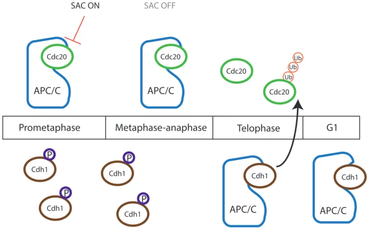
17 ii) APC/C co-activators Cdh1 and Cdc20 in mitosis The APC/C plays a role throughout certain cell cycle steps, targeting different substrates at different time points with its coactivators, Cdh1 or Cdc20 (Nakayama and Nakayama 2006). Through ubiquitylation by the APC/C, its substrates are then targeted for degradation by the 26S proteasome. The co-activators associated with APC/C recruit and bind substrates with the help of the sub-unit APC10 (da Fonseca et al. 2011; Buschhorn et al. 2011). There is an order in the activation of either co-activator in mitosis. Cdh1 is thought to have a more diverse range of substrates compared to Cdc20 that targets fewer substrates more specifically. Cdc20 is activated earlier in mitosis while Cdh1 is bound to APC/C activating it at mitosis exit and G1 phase (Fig. 5). Cdc20 is important for APC/C activity during mitotic exit. In mitosis, phosphorylation by Cdk1 promotes association of Cdc20 and APC/C (Kraft et al. 2003). On the contrary, Cdh1 is phosphorylated and inactivated by Cdk1. Once Cdk1 activity decreases, Cdh1 is no longer inhibited allowing it to activate the APC/C and target its substrates (Fig. 5). One of its substrates is Cdc20 that also contains a KEN box allowing its degradation and take over by Cdh1. APC/CCdh1 is active until G1/S transition to prevent
precocious cyclin accumulation and entry into S-phase (Watson et al. 2019; Yamano 2019; Alfieri, Zhang, and Barford 2017; Pines 2011).
It was shown that both Cdh1 and Cdc20 are vital to obtain viable and healthy mice. Cdh1 null mice were embryonically lethal due to placental defects leading to death (M. Li et al. 2008). Cdc20 null mice showed an even more precocious death arresting at the two-cell stage due to a prolonged metaphase arrest that led to embryonic lethality (M. Li, York, and Zhang 2007). Knockdown of Cdh1 in human and mouse somatic cells showed that although it is not needed for completion of mitosis, it is necessary for G1 phase and S-phase regulation (Sigl et al. 2009). However, knockdown of Cdc20 in HeLa cells led to mitotic arrest inducing cell death demonstrating its essential role in mitosis (Eichhorn et al. 2013). These studies suggest that the two co-activators can not substitute for each other in activating the APC/C and targeting substrates. Therefore, each one is essential and needed to progress through the cell cycle.
APC/C activity must be repressed at certain time periods during the cell cycle. APC/C activity is inhibited during S-phase and G2 phase to allow appropriate accumulation of cyclins. This is completed by the inhibitor Emi1, expressed during S-phase and degraded during mitosis (Reimann et al. 2001). Emi1 inhibits the APC/C by acting as a pseudo substrate and also
APC/C
Cdc20 SAC ON Cdc20 SAC OFF Cdc20 Ub Ub Ub Cdc20APC/C
APC/C
Cdh1 Cdh1 Cdh1 Cdh1 Cdh1P
P
P
P
APC/C
Cdh1Prometaphase
Metaphase-anaphase
Telophase
G1
Figure 5:
APC/C co-activators Cdh1 and Cdc20 during the cell cycle. During certain
phases of the cell cycle, the APC/C plays an essential regulatory role by targeting
substrates to 26S proteasome dependent degradation through ubiquitination.
Co-activators that bind to the APC/C are needed for its activity at different time
points. When cells enter mitosis, APC/C binds to its activator Cdc20. Its activity is
inhibited by the Spindle Assembly Checkpoint (SAC). High Cdk1 activity during
metaphase leads to the phosphorylation of another co-activator Cdh1 inhibiting its
binding to the APC/C. At the metaphase-to-anaphase transition, APC/C
Cdc20is fully
active targeting its substrates to degradation provoking a decrease of Cdk1 activity.
This decrease allows Cdh1 to become active and bind to the APC/C. APC/C
Cdh1targets
one of its substrates, Cdc20, for degradation. APC/C
Cdh1is then active at the end of
18 through inhibition of the chain elongation by Ube2S. This allows deubiquitylating enzymes to remove the initial ubiquitins on the substrates preventing their degradation (W. Wang and Kirschner 2013). During the M phase, the APC/C is inhibited by the Spindle Assembly Checkpoint (SAC) allowing correct spindle formation, chromosome congression and kinetochore to microtubule (KT-MT) attachments. The SAC inhibits APC/C activity by sequestering Cdc20 through its binding to SAC components. This mechanism adds another fine-tuned regulation of APC/C substrate degradation (Sullivan and Morgan 2007; Musacchio and Salmon 2007). 3. Meiotic maturation in mouse oocytes A) Summary of meiosis in mouse oocytes Many studies on gamete aneuploidy and analyses of aging oocytes have shown that female mammalian meiosis is error prone. In mouse oocytes, the meiotic process is complex and tightly regulated. Oocytes are formed during embryonic development and are arrested in prophase I for prolonged periods of time. This stage can be recognized by the presence of a germinal vesicle (GV) in the oocyte. Therefore, this phase is also called the GV stage (Fig. 6). All the machinery needed for completing meiosis is present in the oocyte at this stage. A growth period of the oocytes in the ovaries is important for their abilities of completing successful meiotic maturation. During this growth period, the oocytes accumulate all the transcripts necessary to complete meiosis (Holt, Lane, and Jones 2013). The nutrients and transcripts accumulated as well as the important growth makes oocytes ready and competent to complete meiotic maturation. Smaller oocytes tend to not be able to complete meiosis even if they are adept to exit prophase I arrest and resume meiosis.
The long period of arrest might be one of the causes for the errors observed specifically in females. This arrest is lifted due to hormonal stimulation (starting from puberty for humans). Oocytes can then resume meiosis marked by germinal vesicle breakdown (GVBD). During prometaphase, chromosomes condense and the spindle begins to form. At metaphase, the bipolar spindle is fully formed holding chromosomes that are aligned at the equatorial plate. Chromosomes segregate during anaphase. The first polar body is extruded (PBE1) in an asymmetric manner with half of the genetic material discarded in it. The oocytes enter meiosis II immediately while preventing the start of a possible S phase. All oocytes then
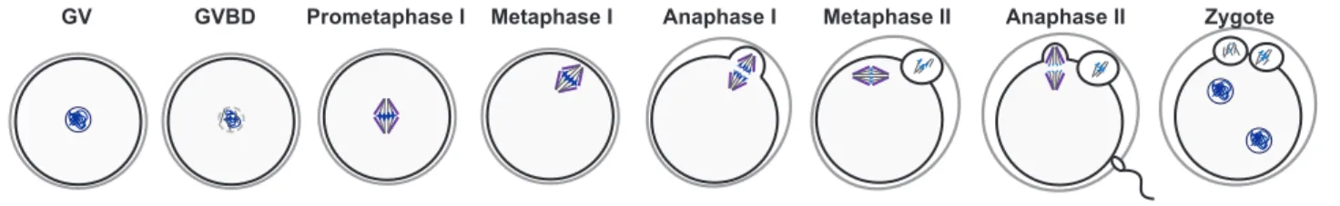
GV GVBD Prometaphase I Metaphase I Anaphase I Metaphase II Anaphase II Zygote
Figure 6:
Mammalian meiotic maturation. Mouse oocytes are formed during
embryonic development and arrest at prophase I also called germinal vesicle
stage (GV). With hormonal stimulation, oocytes resume meiosis with germinal
vesicle breakdown (GVBD). Chromosomes condense and the spindle forms
during prometaphase and chromosomes align with a fully formed spindle at
metaphase. During anaphase I, chromosomes segregate and the first polar body
is extruded. Oocytes then immediately enter meiosis II and arrest at metaphase II
also known as CSF arrest. Fertilization triggers anaphase II onset with chromatid
segregation and the second polar body extrusion. The zygote can then form with
the male and female pronuclei.
19 arrest at metaphase II, also known as CSF arrest. At this stage, the spindle is formed for a second time and the chromosomes stay aligned during the arrest. Fertilization provokes the lift of the metaphase II arrest and sister chromatid segregation. A second polar body is extruded (PBE2) and the paternal genetic material from the sperm enters the oocyte. The two pronuclei (female and male) fuse and form the zygote (Fig. 6) (Terret and Wassmann 2008). Following this, embryonic development will commence. If all of these steps are correctly completed, meiosis will give rise to euploid gametes and a healthy embryo.
B) Oogenesis: the development of the oocyte
Oocytes are formed during female embryonic development in mammals. At first, diploid primordial germ cells (PGCs) are found in embryos that later develop into the germline for both male and female mammals (H. Zhang et al. 2014). The development of the PGCs into fully formed gametes is controlled by different growth factors and cytokines. They must stay undifferentiated and avoid differentiating into somatic cells. These PGCs migrate to the developing ovaries (named genital ridges) in the female embryo, undergo multiple mitotic cycles and begin to express gamete specific genes leading to their differentiation. Then, they complete a final replication phase and begin meiosis and arrest at prophase. This final replication phase is a particular replication and is essential to put in place all the components that are necessary to correctly undergo the two meiotic divisions, namely loading cohesin onto the chromosomes and replicating chromosomes (Wear, McPike, and Watanabe 2016; McLaren 2003). At this stage oogenesis begins (MacLennan et al. 2015). Oocytes can then grow surrounded by their follicles that grow and develop resulting in preovulatory follicles (Fig. 7) (Piotrowska et al. 2018).
At the start of oocyte production during fetal development, a large stock of oocytes is created. The number of these immature oocytes is estimated at around 7 million (Thomson, Fitzpatrick, and Johnson 2010). The decrease of this stock continues as follicle formation proceeds. This process known as atresia is observed in both mice and humans (P. A. Hunt and Hassold 2008). The decrease continues after birth until puberty and this process is also observed with increase of age especially when the age of menopause is reached. Typically, only one oocyte is ovulated in women and fertilizable once a month. Therefore, very few of
PGCs
Mitosis Meiosis
Primordial follicle
Preovulatory
Oogenesis during mammalian development. During mammalian
embryonic development, primordial germ cells (PGCs) are formed and later
develop into germ cells. In female embryos, PGCs undergo several mitotic
divisions and begin to differentiate to enter meiosis and form the primordial
follicle. The oocytes arrest at prophase I following replication and recombination.
They then enter a growth phase where they increase in size and development of
their surrounding cells. When they complete their growth at the preovulatory
stage, oocytes are sensitive to hormonal stimulation and are competent to
resume meiosis. Adapted from Piotrowska et al 2018
20 the initial pool of immature oocytes are ovulated for both female mice and for women (Thomson, Fitzpatrick, and Johnson 2010).
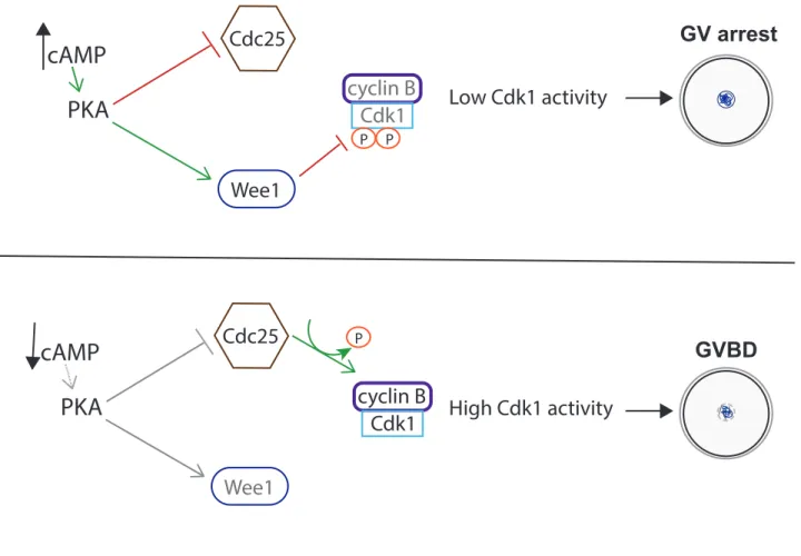
During meiosis, prophase is divided into 4 stages: leptotene, zygotene, pachytene and diplotene. It is during these phases that recombination is established between paternal and maternal homologous chromosomes. Recombination is initiated by double strand breaks prompted by the endonuclease Spo11 (Ohkura 2015; Marston and Amon 2004). Crossing-over of some selected double strand breaks allows the recombination and higher genetic diversity increasing the variety within a population. Moreover, recombination sites called chiasmata are crucial for maintaining homologous chromosomes together during meiosis I (MacLennan et al. 2015). C) Resumption of meiotic maturation i) Prophase I arrest: GV stage Vertebrate oocytes arrest at prophase I. For humans, this arrest can last up to decades. The main goal of the prophase I arrest is to maintain low Cdk1 activity (Adhikari and Liu 2014). In order to do so, a complex mechanism is put in place to ensure this arrest. The communication between the oocyte and the granulosa cells surrounding it is essential for GV stage maintenance. Indirectly regulated through contact with the granulosa cells, high cyclic adenosine 3’, 5’ –monophosphate (cAMP) levels are integral in keeping the oocytes in a GV stage. If the oocytes are separated from the granulosa cells, cAMP levels drop and oocytes resume meiosis. In order to keep a low level of Cdk1 activity, cAMP binds to the kinase Protein Kinase A (PKA) activating it. PKA can then phosphorylate its substrates such as Wee1/Myt1 kinases and the phosphatase family Cdc25 (Holt, Lane, and Jones 2013). In mouse oocytes, it is known that Cdc25B and Wee1B with Myt1 are essential during prophase I arrest. Wee1B with Myt1 phosphorylate Cdk1 on residues Thr14 and Tyr15 inhibiting its activity. On the other hand, Cdc25B dephosphorylates Cdk1 to active it. During GV stage, high levels of PKA activate Wee1B/Myt1 kinases and inhibit Cdc25B. This allows Cdk1 activity level to stay low inhibiting GVBD (Fig. 8) (Jeong Su Oh, Han, and Conti 2010; Pirino, Wescott, and Donovan 2009; Y. Zhang et al. 2008).
Another regulatory mechanism of GV stage arrest is APC/C bound to its co-activator Cdh1. Knockdown of Cdh1 leads to accelerated GVBD underlining its importance (Rattani et al.
PKA
cAMP
cyclin B Cdk1 Wee1 P Cdc25 P Low Cdk1 activity GV arrestPKA
cAMP
cyclin B Cdk1 Wee1 Cdc25 P High Cdk1 activity GVBDFigure 8:
Prophase I arrest (GV) vs. resumption of meiosis (GVBD). At GV stage,
oocytes are arrested at prophase I mainly through the inhibition of cyclin B-Cdk1
activity. Cyclic adenosine 3’, 5’-monphosphate (cAMP) levels are high during GV
stage which maintains PKA active. PKA maintains the inhibitory kinase Wee1 active
and inhibits the Cdk1-activating phosphatase Cdc25. Wee1 inhibits Cdk1 activity
through phosphorylation on two residues (Thr14 and Tyr15). This maintains low
Cdk1 activity and arrest at GV stage. Hormonal stimulation induces a decrease in
cAMP and PKA activity. Cdc25 is no longer inhibited and is able to
dephosphorylate Cdk1 allowing increase of Cdk1 activity to trigger GVBD.
21 2017). Cyclin B1 levels must be correctly controlled in GV oocytes. Therefore, it is believed that cyclin B1 must be targeted for degradation to avoid possible increase of cyclin B1 leading to increase of Cdk1 activity and premature GVBD. APC/CCdh1 itself is also regulated in GV mouse oocytes. Emi1 acts as an inhibitor of APC/C activity while the phosphatase Cdc14 promotes its activity. The localization of all of these components also plays a significant role. The inhibitory factors APC/Cdh1 and Wee1B are found mainly in the nucleus while cyclin B1-Cdk1 and Cdc25B seem to be mainly cytoplasmic (Holt, Lane, and Jones 2013). Another recent study has shown that cyclin B2 could be the main cyclin needed for GVBD in mouse oocytes. Mouse oocytes devoid of cyclin B2 have a large delay in GVBD timing (Daldello et al. 2019; J. Li et al. 2018) while cyclin B1 KO oocytes enter meiosis at a regular timing with no significant delay (J. Li et al. 2018). Therefore, it seems that several regulatory pathways are put in place in mammalian oocytes to maintain this prolonged GV arrest to prevent any precocious resumption of meiosis.
ii) Lift of prophase I arrest: GVBD
The lift of the prophase I arrest is induced by hormonal stimulation. Once hormonal stimulation occurs, cAMP levels drop in the oocyte. This leads to a decrease in PKA activity and therefore less activation of Wee1B tipping the balance in favor of the phosphatase Cdc25B. More Cdk1 is activated through the dephosphorylation of inhibitory phosphorylation. In addition, prior to GVBD, a shift occurs where the cyclinB1/B2-Cdk1 complexes as well as Cdc25B translocate to the nucleus. More Cdc25B is present to activate Cdk1. Cyclin B1/2 translocation to the nucleus is thought to dominate over APC/CCdh1
ubiquitination leading to an increase in cyclin B1/B2 levels and higher Cdk1 activity (Holt, Lane, and Jones 2013). All these factors lead to a surge in overall Cdk1 activity and therefore resumption of meiosis marked by the GVBD (Fig. 8). Following envelope breakdown, chromosomes begin to condense and the spindle begins to form as the oocyte transitions into the next steps of meiotic maturation. D) Prometaphase: starting meiotic maturation Once oocytes resume meiosis, cyclin B1-Cdk1 activity is essential to obtain a correct spindle and timely chromosome alignment. Following GVBD, the spindle begins to form and chromosomes condense. During meiosis I, a pair of homologous chromosomes are
connected each made up of two sister chromatids. Chromosomes are connected by their recombination sites that form the chiasmata, and sister chromatids by a ring-like protein complex named cohesin. These components are important to maintain a correct ploidy in the oocyte because they maintain chromosomes and sister chromatids together until anaphase I and II respectively. Cohesin is found on the arms and around the centromere. These two localizations determine the two patterns of segregation observed during meiosis. An important aspect is that during the first meiotic division arm cohesin is targeted for cleavage by the cysteine protease separase. Therefore, in meiosis I, the centromeric cohesin must be protected (see below) (Wassmann 2013).
i) Spindle formation and KT-MT attachments
The nucleation of microtubules from the centrosomes leads to the formation of the bipolar spindle during mitosis. The centrosomes are previously duplicated during S phase. The microtubules emanate from these centrosomes each made up of two centrioles and pericentriolar material. The microtubules begin to polymerize and search in the cytoplasm for chromosomes. They then attach to them at the kinetochores. This mechanism is known as “search and capture” (Heald and Khodjakov 2015; Kirschner and Mitchison 1986). In mammalian oocytes, the meiotic spindle differs from the mitotic spindle due to one key feature. No centrosomes are present in meiosis, meaning that another mechanism is needed. The spindle forms from a structure called acentriolar MicroTubule Organizing Centers (aMTOCs). They lack centrioles and microtubules emanate from them during meiosis as aMTOCs form clusters and are organized to form the two poles for the spindle (Dumont et al. 2007).
During the formation of the spindle, it is important to correct wrong KT-MT attachments. These include lateral attachments, where KTs aren’t attached to the end of the MT, but rather at the side. Meiosis I merotelic attachments, where each KT is attached to a MT coming from opposite poles, are also corrected (note that this configuration is not wrong in mitosis). To correct wrong attachments, a complex called the CPC (Chromosome passenger complex) made up of the proteins Aurora B/C, Incenp, Borealin and Survivin are present. The CPC induces destabilization of the KT-MT attachments leading to their detachments. This allows another MT to attempt a correct attachment on that KT (Carmena et al. 2012). As stated previously, the activity of cyclin B1-Cdk1 progressively increases throughout meiosis I
23 and reaches its peak activity around metaphase. This slow increase has been found to be important for correct KT-MT attachments, congression and alignment of chromosomes. It was shown that a decrease in Cdk1 activity led to a delay in stable KT-MT attachments while overexpression of cyclinB1, leading to higher Cdk1 activity, provoked a precocious stabilization of these attachments. As a consequence, errors in segregation occur in anaphase I due to lagging chromosomes (Davydenko, Schultz, and Lampson 2013).
ii) Spindle Assembly Checkpoint
Similarly to mitosis, the Spindle Assembly Checkpoint (SAC) plays an important role in regulating correct chromosome segregation. An active SAC delays anaphase to allow correct attachment of microtubules to chromosomes. Initially, the existence of the SAC during meiosis was debated. This was due to the fact that errors in segregation occur with such high incidence in human oocytes (20-30% in healthy women). Therefore, a possible explanation for these errors was thought to be an absence of a functional SAC. However, this is not the case. This mechanism is well characterized during meiosis I and its function is still debated in meiosis II. It was shown that the SAC is functional during meiosis I thanks to several studies. They have shown that in absence of SAC proteins, meiosis I is accelerated and aneuploidy occurs. If one of the core components of the SAC is depleted, chromosomes segregate precociously with untimely APC/C activation and accelerated degradation of cyclin B1 and securin even in absence of microtubules (due to nocodazole treatment). This clearly demonstrates the importance of the activity of the SAC during meiosis I (Touati et al. 2015; Hached et al. 2011; McGuinness et al. 2009; Niault et al. 2007; Wassmann, Niault, and Maro 2003).
As oocytes resume meiosis, the bipolar spindle begins to form as the chromosomes condense. The microtubules begin to attach to the chromosomes. The two sister kinetochores are oriented to the same pole in order to segregate together. This orientation is essential for correct segregation in meiosis I. When kinetochores are not attached, components of the SAC are recruited to the unattached kinetochore. The recruitment is done in a sequential manner with the kinase Mps1 first being recruited to the unattached kinetochores. Mps1 can then recruit Bub3 and Bub1 through phosphorylations. Finally BubR1 is recruited. These core components can then recruit the complex composed of Mad1-Mad2. Each of the components described here are essential for a functional SAC.
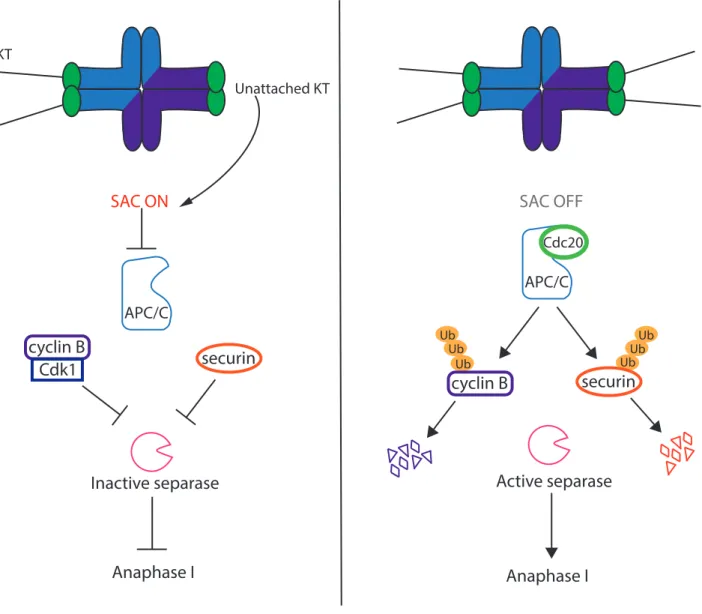
Mad2 exists in two conformations: open and closed. When Mad2 is not bound, it is in an open conformation. Once it binds to Mad1, Mad2 is converted to a closed conformation. Mad2 bound to Mad1 is then capable of converting other open Mad2 proteins into the closed conformation (M. Yang et al. 2008; Mapelli et al. 2007). This allows Mad2 to bind to Cdc20 (De Antoni et al. 2005) and furthermore, to Bub1 and Bub3 forming the mitotic checkpoint complex (MCC) (Sudakin, Chan, and Yen 2001). The MCC is a potent APC/C inhibitor leading to the inhibition of ubiquitination of APC/C substrates and therefore inhibition of anaphase onset (Fig. 9).
Interestingly, the SAC in meiosis seems to be less robust than in mitosis. In mitotic cells, one unattached kinetochore is sufficient to activate the SAC (Rieder et al. 1995). However, this does not seem to be case in meiosis. The APC/C substrates cyclin B1 and securin begin to be degraded during meiosis I before all kinetochores are correctly attached (S. I. R. Lane, Yun, and Jones 2012). This indicates that the SAC is not sensitive enough to detect incorrect attachments of some kinetochores. This could be due to the size of the cell with an important volume of the cytoplasm. In this case, the signal of the SAC on the kinetochores would be diluted in the cell and therefore too weak to be able to inhibit correctly the APC/C. Some studies have hypothesized that the size of a cell impacts the strength of the SAC. When the size of the cell is decreased, this leads to a more robust SAC while increasing the size induces an accelerated anaphase (Kyogoku and Kitajima 2017; Galli and Morgan 2016; Hoffmann et al. 2011). However, this does not seem to be the case for embryonic divisions in the mouse where the size of the cell doesn’t correlate with the robustness of the SAC (Vázquez-Diez, Paim, and FitzHarris 2019). Therefore, the reasons for leaky SAC control in oocytes are still not clear. E) Metaphase I to anaphase I transition i) Cohesin, the glue that keeps chromosomes together In both mitosis and meiosis, cohesin holds sister chromatids together. It is essential for many processes during the cell cycle and crucial for correct chromosome segregation. In mammals, this ring is composed of 4 sub-units; SMC1 and SMC3 (“structural maintenance of chromosomes”), one HEAT repeat domain sub-unit STAG1, STAG2 or STAG3 and a α-kleisin subunit (Brooker and Berkowitz 2014). The kleisin subunit is the target that is cleaved by
MPS1 BUB1 UNATTACHED
KT
BUB3 BUBR1 MAD1 MAD2 OPEN CDC20APC/C
MAD1 MAD2 CLOSED BUB3 BUBR1 MAD2CLOSEDCDC20
Figure 9:
MAD2
CLOSED
MCC
Active SAC inhibits APC/C
Cdc20activity during meiosis I. During mitosis or
meiosis, the SAC is active when kinetochores (KTs) are unattached. This generates the
recruitment of different proteins to the KT. Mps1 is recruited to the unattached KT and
then recruits Bub1, Bub3 and BubR1. They can then recruit dimers of Mad1-Mad2.
Mad2 protein is present in two conformations: closed when binding to Mad1 and
open when free. Free open Mad2 can interact with the closed Mad2, become
converted into closed Mad2 to bind to Cdc20 and form the MCC to inhibit the APC/C
until KTs are correctly attached.
MAD2
separase allowing opening of the ring and separation of chromosomes. There are subunits that are specific to either mitosis or meiosis. The meiotic α-kleisin subunits are Rad21L, present early in meiosis, (Herrán et al. 2011; Lee and Hirano 2011) as well as Rec8, while Scc1 is the kleisin present during mitosis (Fig. 10) (Petronczki, Siomos, and Nasmyth 2003). They are substrates of separase and are therefore cleaved by separase during anaphase (Petronczki, Siomos, and Nasmyth 2003). Cohesin is put in place during DNA replication. This implies that during meiosis, cohesin is present on the DNA since foetal development. During meiosis, like in mitosis, cohesin holds sister chromatids together. In addition, it also plays a role in stabilizing the chiasmata created during recombination that holds the homologous chromosomes together in meiosis I. Furthermore, in mice it was shown that there is no turnover of cohesin (Tachibana-Konwalski et al. 2010). Therefore, cohesin is holding chromosomes together for extended periods of times especially in mammals. It was suggested that an additional cause of high aneuploidy rates in female mammalian meiosis is cohesin fatigue. In older mice, the level of cohesin found in oocytes is significantly decreased compared to younger females (Jessberger 2012; Chiang et al. 2010; Lister et al. 2010). The decreased levels of cohesin in older mice could partly explain how aneuploidy rates increase with age. ii) Two step removal of cohesin A particularity of meiosis is the two different segregation patterns observed in MI and MII with no S phase in between. In meiosis I, cohesin holding together sister chromatids and stabilizing the physical connection between homologous chromosomes is present on both chromosome arms and the centromere. To correctly segregate chromosomes during meiosis I, separase must only cleave arm cohesin. Therefore, centromeric cohesin must be “protected” at this stage. Oocytes can then enter MII with sister chromatids still held together at the centromere. This allows correct bipolar attachment of sister kinetochores. Cohesin located at the centromere can then be “deprotected” in order to allow separase to cleave it inducing sister chromatid segregation at anaphase II (Wassmann 2013). These two patterns of segregation must therefore be tightly regulated. 1. Protection of centromeric cohesin
The arm cohesion removal occurs during the first meiotic segregation and depends on separase activity (Kudo et al. 2006; Terret et al. 2003). It was shown in yeast that in order to
SMC3
SMC1α,
SMC1β
STAG1, STAG2,
STAG3
Scc1,
RAD21L
,
Rec8
Figure 10:
The cohesin complex and its subunits. Cohesin is a ring like complex
that holds sister chromatids together during mitosis and meiosis. Its cleavage allows
separation of the sister chromatids during anaphase. It is made up of SMC1 and SMC3
(structural maintenance of chromosomes), STAG1, STAG2, or STAG3 (HEAT repeat
domain sub-unit) and a
α-kleisin sub-unit. The sub-units expressed specifically during
meiosis are in green. Adapted from Brooker and Berkowitz 2014
be cleaved, the cohesin sub-unit Rec8 must be phosphorylated. This phospohorylation is counteracted by the phosphatase PP2A that is recruited by the protein shugoshin (Sgo) at the centromere during meiosis I (Fig. 11) (Katis et al. 2010; Ishiguro et al. 2010; Riedel et al. 2006). Two isoforms of shugoshin are present in mammalian cells; Sgo1 and Sgo2. It was found that Sgo2 is essential for meiosis while Sgo1 seems to play a more crucial role in mitosis (Rattani et al. 2013; Lee et al. 2008; Llano et al. 2008; McGuinness et al. 2005). During mitosis a mechanism called the prophase pathway removes arm cohesion independent of separase. Sgo1 present at the centromeres protects cohesin from this form of cleavage to prevent precocious sister chromatid segregation (McGuinness et al. 2005). Sgo2 mutant mice are sterile underlining its importance during meiosis. Lack of Sgo2 leads to loss of centromeric cohesin protection and precocious sister chromatid segregation at anaphase I. This leads to random second segregation and aneuploid gametes (Llano et al. 2008). Furthermore, its exact localization in the centromeric region is also essential for correct protection and segregation. Recently, it was shown that the centromeric localization of Sgo2 and not at the pericentromere seems to be the primary pool of protective Sgo2. Sgo2 could have different roles during meiosis that could be distinguished by its exact localization (Yakoubi et al. 2017). It plays an important role in bi-orientation through its interaction with MCAK and inhibition of Aurora B/C. Sgo2 is also able to bind to Mad2 leading to inactivation of the SAC (Rattani et al. 2013). As previously stated, Sgo2 acts as a recruiting platform for PP2A. PP2A was also found to be crucial for the protection of centromeric cohesin. Inhibition of PP2A using Okadaic Acid (OA) provokes precocious sister chromatid segregation similarly to Sgo2 depletion (Mailhes et al. 2003). These findings indicate that without the protection of Sgo2-PP2A, separase is able to cleave all cohesin. This outcome must be avoided or it will lead to severe aneuploidies. 2. Deprotection: removing centromeric cohesin The second important step during meiosis is the chromatid segregation during anaphase II. This is accomplished by separase that cleaves centromeric cohesin. Therefore, at this stage, centromeric cohesin must be “deprotected”. There are a few hypotheses on how this is done. One hypothesis is the tension-sensing model. In this model, the different KT-MT attachment in meiosis I and II determines how centromeric cohesin is deprotected only in meiosis II. Because sister kinetochores are attached in a bipolar way in meiosis II, meaning
P P P P A2 PP 2o gS A2 PP 2o gS P P
Metaphase I
Anaphase I
Sgo2 PP2A I2PP2A P PMeiosis II
Anaphase II
Figure 11:
Protection and deprotection of centromeric cohesin during meiosis.
Cohesin at the centromeres must be protected during meiosis I and “deprotected”
during meiosis II. Cohesin, and more specifically Rec8, is believed to necessitate
phosphorylation to be cleaved by separase. Rec8 is protected from phosphorylation
by the complex Sgo2-PP2A. Sgo2 recruits PP2A to the centromere and PP2A
dephosphorylates Rec8.This protects Rec8 from separase cleavage. At the arms, Rec8
is not protected leading to its phosphorylation and cleavage and chromosome
segregation at anaphase I onset. During meiosis II, PP2A-Sgo2 should be inhibited to
deprotect centromeric cohesin and allow its phosphorylation and cleavage. This
inhibition is thought to be performed by I2PP2A. Once centromeric cohesin is
deprotected, separase cleaves it at anaphase onset provoking sister chromatid
segregation.
pulled away from each other, this physically dislocates Sgo2-PP2A away from the centromere making it accessible to phosphorylation and separase cleavage (Nerusheva et al. 2014; Lee et al. 2008; Gómez et al. 2007). A second model involves the protein SET/I2PP2A. SET/I2PP2A has several functions such as acting as a histone chaperone and interestingly as a PP2A inhibitor even though this role is highly controversial. It is found on the centromere and co-localizes with PP2A and Rec8. It was shown that SET/I2PP2A knockdown leads to defective sister segregation in anaphase II. When SET/I2PP2A is knocked down, an obvious lack of sister chromatid segregation is observed during anaphase II. Therefore, without SET/I2PP2A, centromeric cohesin stays protected (Fig. 11) (Chambon et al. 2013). The mechanism by which SET/I2PP2A deprotects centromeric cohesin is still unknown at this stage, and currently examined in the group. iii) Cyclin B1 vs Cyclin B2 regulation during meiosis I As evidenced by the KOs of cyclin B1 or cyclin B2, both cyclins share a lot of similarities but also differences in timing of expression. It was shown that when cyclin B1 is depleted only in oocytes, they are able to complete meiosis I similarly to control oocytes. This was found to be caused by an increase in cyclin B2 expression. Ccnb1 -/- oocytes therefore compensate by expressing more cyclin B2. This increase is sufficient for correct GVBD as well as normal anaphase I timing. Therefore, cyclin B2 is able to take over as part of the MPF when cyclin B1 is not present only during meiosis I. After meiosis I exit, oocytes fail to enter meiosis II because oocytes are not able to re-accumulate enough cyclin B2 and Cdk1 activity when cyclin B1 is absent. Exogenous expression of cyclin B2 at this time is sufficient to rescue this phenotype indicating that it is capable of fulfilling the function of cyclin B1 but that the regulation of its expression is different from that of cyclin B1. This difference in expression could explain why mice lacking cyclin B1 in oocytes are sterile (J. Li et al. 2018). On the other hand, Ccnb2-/- mice are viable and fertile but do give a smaller number of litter of smaller size. In these oocytes, GVBD is hindered and asynchronous. They present other defects such as late anaphase onset and in a proportion of oocytes, failure to complete and exit meiosis I. There are nonetheless oocytes that are able to complete it and enter meiosis II. The defects observed in these oocytes are thought to be due to lower MPF activity. Unlike Ccnb1-/- oocytes, lack of cyclin B2 doesn’t induce an increase in cyclin B1 expression suggesting that