423
D
isturbed cholesterol homeostasis is intimately linked with human diseases, most notably atherosclerosis and ensuing cardiovascular complications. Despite an increase in our understanding of the basic mechanisms underlying athero-genesis and the availability of therapeutic modalities to treat dyslipidemia, cardiovascular complications remain the lead-ing cause of death in Western countries.1 Atherosclerosis is a lipid-driven disease that is also characterized by the pres-ence of low-grade inflammation in the vascular wall.2 Within the atherosclerotic plaque, macrophages are able to, among others, internalize modified low-density lipoprotein, promote removal of excess cholesterol from the developing atheroscle-rotic plaque, and respond to local and systemic inflammatory cues.3 Owing to their ability to integrate lipid and inflam-matory signaling, the central role played by macrophages in atherosclerosis is well recognized. This also emphasizes the need to elucidate the genetic programs and genes governing macrophage function in atherogenesis.The liver X receptors α (LXRα, NR1H3) and β (LXRβ, NR1H2) are central transcriptional regulators of cholesterol metabolism.4,5 LXRs are sterol-responsive nuclear receptors that are activated under conditions of elevated cellular sterol load. In macrophages, their activation leads to induction of a transcriptional program that is aimed toward reducing the cel-lular cholesterol burden and concomitantly inhibiting inflam-matory signaling. This is largely achieved through LXRs’ ability to (1) enhance cholesterol transport through their tar-get genes ABCA1 (ATP-binding cassette transporter A1),6–8 ABCG1 (ATP-binding cassette transporter G1),9 and APOE,10 (2) limit uptake of lipoprotein-derived cholesterol by induc-ing expression of the E3 ubiquitin ligase IDOL (inducible degrader of the low-density lipoprotein [LDL] receptor),11,12 and (3) transrepress inflammatory signaling induced by inflammatory cues.13–15 Accordingly, Lxrαβ−/− macrophages
accumulate cholesterol in vivo and are hyper-responsive to inflammatory stimuli.13,16 Reciprocally, pharmacological
Arterioscler Thromb Vasc Biol is available at http://atvb.ahajournals.org DOI: 10.1161/ATVBAHA.116.308434
Objective—The sterol-responsive nuclear receptors, liver X receptors α (LXRα, NR1H3) and β (LXRβ, NR1H2), are key determinants of cellular cholesterol homeostasis. LXRs are activated under conditions of high cellular sterol load and induce expression of the cholesterol efflux transporters ABCA1 and ABCG1 to promote efflux of excess cellular cholesterol. However, the full set of genes that contribute to LXR-stimulated cholesterol efflux is unknown, and their identification is the objective of this study.
Approach and Results—We systematically compared the global transcriptional response of macrophages to distinct classes of LXR ligands. This allowed us to identify both common and ligand-specific transcriptional responses in macrophages. Among these, we identified endonuclease–exonuclease–phosphatase family domain containing 1 (EEPD1/KIAA1706) as a direct transcriptional target of LXRs in human and murine macrophages. EEPD1 specifically localizes to the plasma membrane owing to the presence of a myristoylation site in its N terminus. Accordingly, the first 10 amino acids of EEPD1 are sufficient to confer plasma membrane localization in the context of a chimeric protein with GFP. Functionally, we report that silencing expression of EEPD1 blunts maximal LXR-stimulated Apo AI-dependent efflux and demonstrate that this is the result of reduced abundance of ABCA1 protein in human and murine macrophages.
Conclusions—In this study, we identify EEPD1 as a novel LXR-regulated gene in macrophages and propose that it promotes cellular cholesterol efflux by controlling cellular levels and activity of ABCA1.
Visual Overview—An online visual overview is available for this article. (Arterioscler Thromb Vasc Biol. 2017;37:423-432.
DOI: 10.1161/ATVBAHA.116.308434.)
Key Words: ABCA1 ◼ cholesterol efflux ◼ cholesterol metabolism ◼ LXR ◼ macrophages ◼ nuclear receptors
Received on: September 9, 2016; final version accepted on: January 2, 2017.
From the Department of Medical Biochemistry, Academic Medical Center, University of Amsterdam, The Netherlands (J.K.N., D.S.K., S.S., E.C.L.C., M.M., A.S., N.Z.); and Université Clermont Auvergne, CNRS, Inserm, GReD, Clermont–Ferrand, France (J.-M.A.L., S.B.).
The online-only Data Supplement is available with this article at http://atvb.ahajournals.org/lookup/suppl/doi:10.1161/ATVBAHA.116.308434/-/DC1.
Correspondence to Noam Zelcer, PhD, Department of Medical Biochemistry, Academic Medical Center, University of Amsterdam, Meibergdreef 9, 1105 AZ Amsterdam, The Netherlands. E-mail n.zelcer@amc.uva.nl
© 2017 The Authors. Arteriosclerosis, Thrombosis, and Vascular Biology is published on behalf of the American Heart Association, Inc., by Wolters Kluwer Health, Inc. This is an open access article under the terms of the Creative Commons Attribution Non-Commercial-NoDervis License, which permits use, distribution, and reproduction in any medium, provided that the original work is properly cited, the use is noncommercial, and no modifications or adaptations are made.
Regulates ABCA1 Abundance and Cholesterol Efflux
Jessica Kristine Nelson, Duco Steven Koenis, Saskia Scheij, Emma Clare Laura Cook,
Martina Moeton, Ana Santos, Jean-Marc Adolphe Lobaccaro, Silvere Baron, Noam Zelcer
424 Arterioscler Thromb Vasc Biol March 2017
engagement of LXRs promotes reverse cholesterol transport and decreases atherosclerotic plaque development in ApoE−/−
and Ldlr−/− mice fed an atherogenic diet.17 LXR activity in macrophages, liver, and the intestine has been reported to con-tribute to their antiatherosclerotic function.16,18–22
LXRs are ligand-dependent transcription factors, and their activation, and hence stimulation of cholesterol efflux, requires receptor-ligand binding. Their endogenous ligands are oxysterols, including 22(R)-, 24(S)-, and 27-hydroxy-cholesterol, and intermediates of the cholesterol biosynthetic pathway, most notably desmosterol.23–25 Similarly, endocyto-sis of (modified) lipoproteins or efferocytoendocyto-sis increases the cellular cholesterol and oxysterol pool and also promotes LXR signaling.26,27 High-affinity synthetic agonists have been also developed to therapeutically target LXRs, with sev-eral reported to have preferential activation of, for example, LXRβ over LXRα resulting in a differential transcriptional response.28 Although these different classes of agonists acti-vate LXRs, natural and synthetic agonist markedly differ with respect to their inhibitory effect on the sterol-regulatory element-binding proteins pathway. Oxysterols and interme-diates of the cholesterol biosynthetic pathway prevent pro-cessing and maturation of sterol-regulatory element-binding proteins to their transcriptionally active form, whereas syn-thetic ligands do not.29,30 This implies that the LXR-induced transcriptional response to these ligands should be distinct. Furthermore, whether the distinct endogenous ligands induce a differential LXR transcriptional response has not been thor-oughly investigated.
To systematically evaluate the LXR response in macro-phages, we treated cells with a panel of distinct LXR ligands. Transcriptional profiling allowed us to identify both over-lapping and ligand-specific LXR-dependent transcriptional responses to these ligands. Among LXR-responsive genes, we identified endonuclease–exonuclease–phosphatase fam-ily domain containing 1 (EEPD1/KIAA1706) as a previously unrecognized direct transcriptional target of LXRs in macro-phages. We report here that EEPD1 promotes LXR-stimulated cholesterol efflux by regulating abundance of ABCA1 at the plasma membrane.
Materials and Methods
Materials and Methods are available in the online-only Data Supplement.
Results
To map the LXR ligand–induced transcriptional program in human macrophages, we performed RNA sequencing on sterol-depleted THP1 cells—a human monocytic leu-kemia cell line that can be readily differentiated to macro-phages with phorbol 12-myristate 13-acetate—treated with 4 different classes of LXR ligands: GW3695 (synthetic), 22R-hydroxycholesterol (22R-HC; oxysterol), desmosterol (cholesterol biosynthesis intermediate), and acetylated-LDL (Ac-LDL; modified lipoprotein). Sterol-depleted cells were used as a baseline since a lack of sterols is known to reduce basal LXR-dependent signaling, thus allowing for greater sen-sitivity in detecting LXR-induced transcriptional changes.
We identified 1171 protein-coding transcripts that were differentially expressed with an adjusted false discovery rate
P value of <0.05 in response to sterol depletion. Of these, 555 transcripts were upregulated, and 616 transcripts were down-regulated (Figure IIA in the online-only Data Supplement). Subsequent treatment of sterol-depleted THP1 macrophages with 1 µmol/L GW3965, 5 µmol/L 22R-HC, 5 µmol/L desmo-sterol, or 50 µg/mL Ac-LDL resulted in a significant change in expression of 1267 to 1856 genes, depending on the ligand used, with only a modest number of genes (287) being regu-lated by all ligands tested (Figure 1A). Hierarchical clustering of our RNA-seq data sets revealed that GW3965 treatment resulted in a transcriptional profile that was distinct from the other ligands, whose expression profiles clustered more closely together (Figure 1B). Gene ontology analysis revealed that gene clusters that were similarly regulated by all ligands were enriched for genes involved in the cellular response to cytokines and cell activation by extracellular ligands. On the other hand, gene clusters that showed differential regu-lation between the ligands (primarily GW3965 versus the other ligands) were enriched for genes involved in the posi-tive regulation of immune system processes, wound healing, and lipid biosynthesis (Figure 1B). Pathway analysis using ingenuity pathway analysis revealed a similar profile, with GW3965 treatment resulting in a transcriptional profile that was less enriched for genes involved in cholesterol biosynthe-sis when compared with either the other ligands versus sterol depletion or sterol depletion versus cholesterol-rich medium (Figure IIB in the online-only Data Supplement). Similarly, principal component (PC) analysis showed that there was little variance between the biological replicates of each of the experimental conditions and that each ligand induced a distinct gene expression profile when compared with the sterol-depleted baseline, with GW3965 treatment being the most different from all other conditions (Figure 1C). Pathway analysis using Metascape on the top 100 most variable genes in each of the principal components showed that most of the variation between the data sets was caused by genes involved in lipid metabolism (PC1 and PC2) and, to a lesser extent, the inflammatory response (PC3; Figure IIC in the online-only Data Supplement).
To filter out genes that were most strongly induced by all 4 of the ligands tested in our screen, we applied a stricter cutoff in which only protein-coding transcripts that showed a 1.5-fold change in either direction with an false discovery
Nonstandard Abbreviations and Acronyms
22R-HC 22R-hydroxycholesterol
ABCA1 ATP-binding cassette transporter A1 ABCG1 ATP-binding cassette transporter G1 Ac-LDL acetylated-LDL
EEPD1 endonuclease-exonuclease-phosphatase family domain con-taining 1
IDOL inducible degrader of the LDL receptor LDL low-density lipoprotein
LXR liver X receptors LXRE LXR-responsive element
rate–adjusted P value of <0.001 were considered (Figure 2A). Using this cutoff, we identified several established LXR-responsive genes, such as ABCA1,6 ABCG1,9 LXRα,31
SMPDL3A,32 and IDOL.11 ApoE, which is an established LXR target in macrophages,10 did not meet this cutoff but was also included in the subsequent evaluation. Several addi-tional genes that have not been previously described as LXR targets, including EEPD1, PPF1A2, IQSEC1, and PBX4, were also identified. We confirmed these observations from our RNA-seq screen using qPCR, which demonstrated that the LXR ligands induce expression of both the established and novel LXR-regulated genes (Figure 2B). Furthermore, as expected, desmosterol, 22R-HC, and Ac-LDL inhibited expression of sterol-regulatory element-binding protein–reg-ulated genes (eg, HMGCR and LDLR), whereas GW3965 did not (Figure 2A and 2B).
The initial screen and validation involved treating the cells with the different ligands for 18 hours. This implies that the increase in expression of these genes may not reflect them being direct transcriptional targets of LXR, but rather be an
indirect effect of LXR activation that is mediated by another protein. To test this possibility, we treated cells with GW3965 for 6 hours together with cycloheximide, a protein synthesis inhibitor, reasoning that if induction is indirect it should be abolished by this treatment. Of the genes tested, induction of
PPF1A2, IQSEC1, and PBX4 expression by LXR activation was abolished by cycloheximide, suggesting that their induc-tion is indirect (Figure 3A). In contrast, inducinduc-tion of the other genes studied was unaffected by cycloheximide, a finding that is in line with several of them reported to be direct LXR tran-scriptional targets and also consistent with their rapid maxi-mal response to LXR activation (≈3 hours; Figure IIIA in the online-only Data Supplement).
Among the novel LXR-responsive genes identified in our screen, EEPD1 is one of the few that were induced by all the ligands tested. Similar to the canonical LXR targets, Abca1,
Abcg1, and Idol, expression of Eepd1 was increased in response to different LXR ligands in bone marrow–derived macrophages from wild-type cells (Figure 3B). Regulation of Eepd1 expres-sion by the ligands was strictly dependent on LXRs because Figure 1. Transcriptional profiling of liver X receptors (LXR) activation in human THP1 macrophages. A, THP1 macrophages were sterol
depleted and treated with 1 µmol/L GW3965, 5 µmol/L 22R-hydroxycholesterol (22R-HC), 5 µmol/L desmosterol, or 50 µg/mL acety-lated-low-density lipoprotein (Ac-LDL) for 18 h. The Venn diagram details the overlap in differentially regulated genes. B, Hierarchical
clustering of differentially expressed genes in response to the different LXR ligands. The top 3 Gene ontology (GO) biological processes that were enriched in each the major gene clusters are indicated. C, Principal component analysis (PCA) plot of the changes in gene
expression in response to LXR ligands and sterol depletion. Axis titles show the percentage of variance explained by each of the princi-pal components.
426 Arterioscler Thromb Vasc Biol March 2017
it was blunted in macrophages derived from Lxrαβ−/− mice
(double knockout mice). In human macrophages derived from peripheral blood monocytes, expression of EEPD1 was induced by 2 synthetic LXR ligands and akin to other established LXR-regulated genes was sensitive to sterol depletion (Figure 3C). Having established that EEPD1/Eepd1 is expressed in macro-phages, we determined its expression in a panel of mouse tissues (Figure 3D). We observed expression of Eepd1 in all tissues that were examined, with a particularly high expression in metaboli-cally active and in macrophage-rich tissues (eg, skeletal muscle, white adipose tissue, and spleen). We therefore anticipated that similar to most other LXR-regulated genes, EEPD1 would be regulated by LXR activation in multiple cell types. To test this hypothesis, we investigated the regulation of EEPD1/Eepd1 by LXR in several human and murine cell lines that originate from different tissues. In these cells, we found that EEPD1/Eepd1 was only induced in macrophage-like cells (Figure 3E). This was not simply because of aberrant LXR signaling in these cells since in response to LXR ligand, all were able to activate the canonical LXR target ABCA1/Abca1 (Figure IIIB in the online-only Data Supplement). Consistent with LXR-dependent regulation, we identified a potential LXR-responsive element (LXRE) within intron 2 of EEPD1 by analyzing a previously
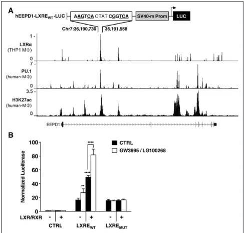
reported LXRα ChIP-seq study (Figure 4A).33 In human pri-mary macrophages, this LXRE is adjacent to a macrophage lineage–specifying PU.1 peak. In addition, this genomic region is enriched for H3K27Ac and H3K4me1 histone modifica-tions, all of which were absent in human adipocytes, skeletal muscle, and HepG2 cells (Figure IV in the online-only Data Supplement). These observations suggest that the macrophage-specific regulation of EEPD1 by LXRs is the result of a per-missive epigenetic landscape surrounding the LXRE in intron 2 that is not present in other cell types. The corresponding LXRE-containing genomic region could drive expression of a luciferase reporter in response to transfection of LXR/RXR, and furthermore when the cells were cotreated with synthetic LXR/RXR ligands (Figure 4B). Mutating the predicted LXRE in this context ablated the response to both LXR/RXR and the ligands. Collectively, these results show that EEPD1 is a direct, macrophage-specific LXR target gene.
The EEPD1 gene encodes a 569 amino acids protein that contains several distinct functional domains. Its N-terminal region contains 2 adjacent helix–hairpin–helix motifs, a motif which is often associated with DNA binding. The C-terminal region contains a large (EEP) domain (Figure 5A). The helix–hairpin–helix motifs present in EEPD1 could suggest Figure 2. Transcriptional profiling identifies liver X receptors (LXR)–regulated genes in THP1 macrophages. A, Heat map showing the fold
change (log2-transformed) in expression of the 39 most differentially expressed transcripts between the different LXR ligands with sterol depletion as baseline. Genes in bold were further validated and characterized. B, THP1 differentiated macrophages were treated for 18 h
in sterol-depleted medium with vehicle, 1 µmol/L GW3965, 5 µmol/L desmosterol, 5 µmol/L 22R-hydroxycholesterol (22R-HC), or 10 µg/ mL acetylated-low-density lipoprotein (Ac-LDL). Subsequently, expression of the indicated genes was determined by qPCR. Each bar represents the mean±SD (n=4). *P<0.05, **P<0.01, ***P<0.001, ****P<0.0001.
association of the protein with DNA and involvement in DNA-binding related processes (eg, DNA repair). However, in silico analysis of its amino acid sequence revealed that EEPD1 is unique among the EEP superfamily of proteins in that it contains highly evolutionary-conserved myristoylation and palmitoylation lipid modification sites, which are known to serve as membrane anchors (Figure 5A). To evaluate the cel-lular localization of EEPD1, we generated an EEPD1wt-eGFP (enhanced green fluorescent protein) expression construct and studied its localization in COS7 cells (Figure 5B). Consistent with the presence of the highly conserved N-terminal myris-toylation and palmimyris-toylation sites, wild-type EEPD1 localized exclusively to the plasma membrane in both live and fixed cells. Remarkably, abolishing the single myristoylation site (EEPD1G2A-eGFP) resulted in drastically altered localization of EEPD1 (Figure 5B). A similar shift in cellular localization was also observed when we treated RAW264.7 macrophages that express an inducible EEPD1wt-eGFP construct with 2-hydroxy-myristic acid, a potent inhibitor of the myristoyl conjugating enzyme N-myristoyltransferase (Figure VA in the online-only Data Supplement). Mutation of the predicted palmitoylation site (EEPD1C7A-eGFP), or of both sites simultaneously (EEPD1G2A/C7A-eGFP), also resulted in a loss of association of EEPD1 with the plasma membrane, further emphasizing the importance of these lipid anchors in EEPD1 localization
(Figure 5B). It should be noted that under these conditions, we were unable to observe EEPD1 localized in the nucleus. However, because these lipid modifications are dynamic,34 we reasoned that either LXR activation or the cellular sterol status may influence the localization of EEPD1. We tested this pos-sibility by determining the localization of EEPD1 in cells after sterol depletion and treatment with the LXR ligand GW3965. Under both conditions, we observed no shift of EEPD1 from the plasma membrane towards an intracellular compartment or the nucleus (Figure VB in the online-only Data Supplement). Finally, to further substantiate the role of the proposed N-terminal lipid modifications on EEPD1 localization, we engineered chimeric constructs consisting of the first 10 amino acids of EEPD1 fused to eGPF (EEPD1(1–10)-eGFP), with or without the predicted myristoylation and palmitoylation sites. We found that the localization of these constructs was similar to that of the corresponding full-length or mutated EEPD1 pro-tein variants (Figure 5C), thereby demonstrating that these first 10 amino acids of EEPD1 are both necessary and sufficient to confer plasma membrane localization. Unfortunately, the com-mercial antibodies we tested were unable to detect endogenous EEPD1 by immunostaining, but we could detect endogenous EEPD1 protein in crude membrane fractions from THP1 cells by immunoblotting (Figure 5D). Moreover, the level of endog-enous EEPD1 protein in these crude membrane fractions was Figure 3. Endonuclease–exonuclease–phosphatase family domain containing 1 (EEPD1) is a direct liver X receptors (LXR) target gene in
human and mouse macrophages. A, THP1 macrophages were treated for 6 h with 1 µmol/L GW3965 or vehicle control in the presence of
10 µg/mL cycloheximide. Expression of the indicated genes was determined by qPCR and displayed as relative mRNA expression com-pared with vehicle-treated controls. Each bar represents the mean±SD (n=4). B, bone marrow–derived macrophages (BMDMs) from
wild-type (WT) and Lxrαβ−/− (double knockout mice [DKO]) mice were cultured in sterol-depleted medium for 18 h and subsequently treated
with 1 µmol/L GW3965, 5 µmol/L desmosterol, 5 µmol/L 22R-hydroxycholesterol (22R-HC) or vehicle control for 6 h. Expression of the indicated genes was determined, and each bar represents the mean±SD (n=4). C, Human peripheral blood monocyte cells (PBMCs) were
differentiated to macrophages and treated with vehicle, 1 µmol/L GW3965, or 1 µmol/L T0901317. Expression of EEPD1 was determined, and each bar represents the mean±SD (n=3). D, The indicated tissues were isolated from WT mice (n=3) and expression of Eepd1
deter-mined by qPCR. Each bar represents the mean±SD E, The indicated human (left) and murine (right) cell lines were treated with 1 µmol/L
GW3965 or vehicle control for 18 h. EEPD1/Eepd1 mRNA expression was determined by qPCR, and each bar represents the mean±SD (n=4). *P<0.05, **P<0.01, ***P<0.001, ****P<0.0001
428 Arterioscler Thromb Vasc Biol March 2017
increased by LXR activation, as could be anticipated from it being an LXR transcriptional target. In aggregate, these experi-ments demonstrate that EEPD1 localizes to the plasma mem-brane and that this is dependent on lipid modifications of the first 10 amino acids.
Having established that EEPD1 is an LXR target in mac-rophages, we then aimed to elucidate its function. Since a major role of LXR in macrophages is to promote cholesterol efflux, we evaluated the role of EEPD1 in this process. We effectively silenced EEPD1/Eepd1 in THP1 and J774 macro-phages, respectfully, using independent siRNAs that reduced the basal, as well as the LXR-inducible expression of EEPD1/
Eepd1 mRNA and EEPD1 protein levels (Figure VIA through VIC in the online-only Data Supplement). Importantly, effec-tive silencing of EEPD1/Eepd1 did not alter the induction of
ABCA1/Abca1 or of other LXR-regulated genes in response to LXR ligand (Figure VIA through VIC). However, LXR-stimulated Apo A1-dependent cholesterol efflux was attenuated in EEPD1/Eepd1-silenced THP1 and J774 cells (Figure 6A and 6B). In contrast, efflux toward high-density lipoprotein remained unchanged, in line with no changes in ABCG1 protein levels (Figure VII in the online-only Data Supplement). Our results therefore point toward EEPD1 playing a role in promoting cholesterol efflux from macrophages. Because ABCA1/Abca1 expression remained unchanged in EEPD1/Eepd1-silenced cells, we evaluated the level of ABCA1 protein. Consistent with reduced efflux, we determined that silencing EEPD1 reduced the LXR-stimulated level of cellular ABCA1 content by ≈50% in both macrophage cell types (Figure 6C and 6D). This reduc-tion seemed specific, as the level of 2 established cholesterol transporters, ABCG1/Abcg1 and SR-BI, and of the transferrin
receptor remained unchanged (Figure 6C and 6D; Figure VIA in the online-only Data Supplement). Taken together, our results point toward EEPD1 being an LXR-regulated target gene that is important for maintaining ABCA1 protein levels and promoting cholesterol efflux from macrophages.
Discussion
Macrophages are central determinants of atherosclerosis.3 Therefore, studies aimed at elucidating the genes governing their handling of lipids and inflammation are central to under-standing their role in the vascular wall in diseased states. As such, the most important finding of our study is that using global transcription analysis, we have identified a novel LXR-regulated gene, EEPD1, which by post-transcriptionally regulating ABCA1 abundance is a determinant of cholesterol efflux from macrophages.
THP1 macrophages are commonly used as a model for human-derived macrophages and have been previously used to evaluate the transcriptional response to LXR ligands.30,35 Our study is distinct from these in that we simultaneously evalu-ated the transcriptional LXR program in response to distinct classes of ligands. An important aspect of our approach is that it allowed us to differentiate the response between synthetic and endogenous ligands. One obvious and expected finding was the absence of inhibition of the sterol-regulatory element-binding protein pathway by the synthetic ligand GW3965, which also underlies hepatosteatosis and increased lipogenesis in livers of mice treated with this compound.36 Less obvious was the lack of a large overlap between the transcriptional response of cells to the different classes of ligands, with each ligand eliciting a distinct profile. Although in some cases, this may represent
Figure 4. A liver X receptors–responsive
element (LXRE) in intron 2 of endonu-clease–exonuclease–phosphatase fam-ily domain containing 1 (EEPD1) drives LXR-dependent expression. A, An LXR
ChIP-seq experiment was analyzed and used to identify an active LXRE in human THP1 cells (GSM700470). Similarly, PU.1 binding sites (GSM785501) and activate enhancer regions marked by H3K27Ac (GSM785500) were evaluated in human monocyte-derived macrophages (human MΦ). The wild-type LXRE-containing region was cloned into pGL2-SV40 firefly luciferase (LXREWT). The underlined
nucle-otides were altered to create a mutant LXRE (LXREMUT). B, HEK293T cells were
transfected with the indicated luciferase reporters with or without LXR and RXR expression plasmids. Subsequently, cells were treated with 1 µmol/L GW3965 and 100 nmol/L LG100268 for 24 h. In all luciferase experiments, the transfection efficiency was normalized using Renilla luciferase, which was cotransfected. Each bar represents the mean±SD rela-tive to vehicle-treated control cells (CTRL; n=6). **P<0.01, ****P<0.0001.
quantitative differences (eg, in our experimental setting Ac-LDL elicited a smaller change in LXR-dependent gene expression), others changes may reflect ligand-specific effects. For example, our RNA-seq analysis identified strong induction of glycolysis-associated genes, among others of PDK4, which is not observed with the other ligands (data not shown). This observation is in line with the notion that LXR ligands with specific transactiva-tion profiles can be developed for LXR so as to, for example, potentially mitigate a lipogenic gene program.28,36,37
Our study also identified a set of genes in macrophages that are pan-regulated by all the LXR ligands we tested, among them the novel target gene, EEPD1. Our studies support the notion that EEPD1 is a direct LXR target gene as we demon-strate that its regulation requires LXRs, it is rapidly induced by LXR ligands, and this induction also occurs in the face of pro-tein synthesis inhibition, ruling out a secondary transcriptional response. We also provide compelling evidence showing that
EEPD1/Eepd1 is regulated in an LXR-dependent manner in murine and human macrophage cell lines and primary cells. Furthermore, by analyzing a reported ChIP-seq study of LXRα in THP1 macrophages,35 we identified an LXR-associated peak within intron 2 of EEPD1, which is absent in adipocytes, and demonstrate that this genomic region harbors a functional LXRE. In human macrophages, the LXRE coincides with a strong peak of the macrophage lineage–specifying PU.1 transcription factor, as well as enrichment of H3K27Ac and H3K4me1 histone modifications that are absent in other cell types. These findings are consistent with a permissive epigen-etic landscape surrounding the LXRE in intron 2 of EEPD1 in macrophages. In conclusion, we propose that EEPD1 is a bonefide LXR-responsive target gene in macrophages.
LXRs are central determinants of macrophage cholesterol metabolism, largely owing to their ability to promote reverse cholesterol efflux.38 Enhanced cholesterol efflux is critically Figure 5. Endonuclease–exonuclease–phosphatase family domain containing 1 (EEPD1) is anchored to the plasma membrane. A,
Sche-matic representation of the structure of EEPD1 (569 amino acids) depicting the 2 N-terminal helix–hairpin–helix (HhH) and the C-terminal exonuclease–endonuclease–phosphatase (EEP) domains. Box, Evolutionary conservation of the first 20 amino acids of EEPD1 with the predicted myristoylation and palmitoylation sites indicated. B, COS7 cells were transfected with wild-type (WT) or mutated EEPD1-GFP
constructs as indicated. Representative images from fixed and live cells were taken 48 h after transfection. C, COS7 cells were
trans-fected with WT or mutated EEPD1(1–10)-GFP constructs, and representative images are shown. B and C, Scale bar is 5 µm. D, THP1
mac-rophages were grown in either sterol-containing or sterol-depleted medium for 16 h with or without 1 µmol/L GW3965 or vehicle control. Subsequently, crude membrane fractions were prepared and analyzed by immunoblotting as indicated. Endogenous EEPD1 levels were quantified and normalized to the level of TOM20 (an abundant and stable mitochondrial membrane protein) and displayed as the average of 3 independent experiments.
430 Arterioscler Thromb Vasc Biol March 2017
dependent on transcriptional regulation of ABCA1 and
ABCG1 by LXRs.6,9,39 Accordingly, loss of LXRs or of these cholesterol efflux transporters has dramatic consequences on the accumulation of cholesterol in macrophages.6,40–42 However, recent studies emphasize the atheroprotective activity of LXRs in the intestine and liver and question the importance of ABCA1- and ABCG1-dependent cholesterol efflux from macrophages in this setting.19–22 Nevertheless, the identification of EEPD1 as a transcriptional target of LXRs in macrophages and the demonstration that this gene is necessary for maximal LXR-stimulated Apo A1-dependent cholesterol efflux contribute to our understanding of LXRs function in these cells. We propose that the underlying cause for decreased efflux from EEPD1-silenced cells is reduced cellular abundance of ABCA1 and decreased ABCA1 den-sity on the plasma membrane. Because silencing of EEPD1 does not impair the level of ABCA1 mRNA or its induction by LXR stimulation, the decrease of ABCA1 protein in
EEPD1/Eepd1-silenced macrophages likely involves a post-transcriptional event.
ABCA1 is reported to have a relatively short half-life, estimated in murine macrophages to be ≈1 hour.43 However, despite its inherent instability, there is ample evidence dem-onstrating that LXR activation robustly increases ABCA1 in macrophages in a time- and dose-dependent manner.6,9,11
This suggests that next to transcriptional regulation, LXRs may also promote stabilization of ABCA1. Our results are consistent with the idea that LXR-dependent regulation of EEPD1 contributes to stabilization of ABCA1. The under-lying mechanism for this is still unclear but may involve modification of cellular membrane lipids, a function that is emerging as an important determinant of LXR function in cells.44,45 Members of the EEP-containing family of pro-teins, to which EEPD1 belongs, catalyze cleavage of phos-phodiester bonds found in nucleic acids, phospholipids, and perhaps also proteins. Specifically, several members of this family have lipid phosphatase activity, mainly toward inositol phosphates. As inositol phosphates are important regulators of intracellular trafficking,46 EEPD1 could control ABCA1 abundance by modulating the level of specific inositol phos-phate species to promote residence of ABCA1 in the plasma membrane or prevent its trafficking toward degradation pathways. However, we point out that this would need to be rather specific because EEPD1 silencing does not change the level of ABCG1, SR-BI, or transferrin receptor. An alterna-tive possibility is that EEPD1 may directly regulate the sta-bility of ABCA1. A cytoplasmic proline/glutamate/serine/ threonine–containing sequence in ABCA1 has been dem-onstrated to act as a phosphorylation-dependent switch that controls stability of ABCA1.47 Accordingly, phosphorylation Figure 6. Endonuclease–exonuclease–phosphatase family domain containing 1 (EEPD1) silencing decreases Apo A1-dependent
choles-terol efflux and ABCA1 abundance. A, THP1 or (B) J774 macrophages were transfected with control (nontargeting [NT]) or EEPD1/Eepd1
siRNAs 48 h. Subsequently, cells were treated with or without 1 µmol/L GW3965 for 4 h followed by an additional 18 h with medium con-taining 2 µCi/mL [3H]Cholesterol and 50 µg/mL acetylated-low-density lipoprotein (Ac-LDL). Cholesterol efflux was initiated by incubating
the cells with or without 10 µg/mL Apo A1 for 6 h. Cholesterol efflux is expressed as the percentage of the radioactivity released from cells into the medium relative to the total radioactivity in cells and medium combined. Results were calculated as net efflux (efflux with Apo A1 minus 0.2% bovine serum albumin alone). Each bar represents the mean±SD of 4 independent experiments done in duplicate. C, THP1 or
(D) J774 macrophages were transfected as described above and subsequently treated with 1 µmol/L GW3965 or vehicle control for 18 h.
Total cell lysates were immunoblotted as indicated. The level of ABCA1 was determined by densitometry after normalization to actin and the average indicated (C, n=3; D, n=4). *P<0.05, **P<0.01, ***P<0.001, ****P<0.0001
of the proline/glutamate/serine/threonine domain promotes calpain-mediated degradation of ABCA1 and attenuates Apo A1-dependent cholesterol efflux.48 Although speculative, if EEPD1 acts as a phosphatase, it could directly stabilize ABCA1 at the plasma membrane by preventing its phosphor-ylation-dependent degradation. We have been thus far unable to demonstrate binding between EEPD1 and ABCA1 (data not shown), yet this does not preclude the possibility that such an interaction could be weak or transient. Future stud-ies to address the functional interaction between EEPD1 and ABCA1 are clearly warranted.
Reports on the possible physiological roles of EEPD1 are scarce. Next to our study, which is the first to identify EEPD1 as an LXR target and ascribe it a function in cellular sterol homeostasis, 2 other studies from the Hromas group recently reported that EEPD1 has a function in DNA repair in the nucleus.49,50 Wu et al49 compellingly demonstrated that loss of EEPD1 in several cell types facilitates repair of stressed replication forks induced by DNA-damaging chem-icals and that it does so by promoting homologous recom-bination. In our studies, we have not observed localization of EEPD1 in the nucleus under any of the conditions evalu-ated. Rather, endogenous EEPD1 was enriched in crude membrane fractions, and heterologous EEPD1 specifically localized to the plasma membrane. Furthermore, in line with the predicted lipid modifications of the N-terminal sequence of EEPD1, we found that the first 10 amino acids are suf-ficient to localize GFP exclusively to the plasma membrane. These findings are at odds with the nuclear localization of EEPD1 reported by Wu et al.49 We note, however, that for some of these studies, overexpression of a construct encod-ing N-terminally tagged EEPD1 was used, a modification that would mask the native lipidation sites in the N-terminal sequence of EEPD1. Accordingly, similar to the EEPD1 lipi-dation mutations, an N-terminally tagged EEPD1 does not localize exclusively to the plasma membrane (Figure VIII in the online-only Data Supplement). Reconciling the proposed functions of EEPD1 is difficult at present, yet the sterol-dependent regulation of EEPD1 by LXRs is consistent with it having a function in cholesterol homeostasis, similar to other LXR-regulated genes. However, we cannot rule out the possibility that in different contexts, cell types, or tissues, EEPD1 adopts a different function. In that respect, the fact that we only identify sterol-dependent regulation of EEPD1 in macrophages, but not in other cell types, including those studied by Wu et al,49 is consistent with the possibility that EEPD1 has cell-type–specific functions that will need to be addressed in future studies.
In conclusion, our study identifies EEPD1 as a sterol-responsive gene that is regulated by LXRs in macrophages. We propose that EEPD1 acts as part of the LXR-regulated program to promote ABCA1-dependent cholesterol efflux from macrophages and that it does so by maintaining stability of ABCA1. Whether EEPD1 plays a role in the development of atherosclerosis remains to be investigated.
Acknowledgments
We thank members of the Zelcer group, Menno de Winther, and Irith Koster for their comments and suggestions.
Sources of Funding
N. Zelcer is an Established Investigator of the Dutch Heart Foundation (2013T111) and is supported by an ERC Consolidator grant (617376) from the European Research Council.
Disclosures
None.
References
1. Mozaffarian D, Benjamin EJ, Go AS, et al; American Heart Association Statistics Committee and Stroke Statistics Subcommittee. Heart disease and stroke statistics–2015 update: a report from the American Heart Association.
Circulation. 2015;131:e29–e322. doi: 10.1161/CIR.0000000000000152.
2. Lusis AJ. Atherosclerosis. Nature. 2000;407:233–241. doi:
10.1038/35025203.
3. Moore KJ, Tabas I. Macrophages in the pathogenesis of atherosclerosis.
Cell. 2011;145:341–355. doi: 10.1016/j.cell.2011.04.005.
4. Peet DJ, Turley SD, Ma W, Janowski BA, Lobaccaro JM, Hammer RE, Mangelsdorf DJ. Cholesterol and bile acid metabolism are impaired in mice lacking the nuclear oxysterol receptor LXR alpha. Cell. 1998;93:693–704. 5. Zelcer N, Tontonoz P. Liver X receptors as integrators of metabolic and
inflammatory signaling. J Clin Invest. 2006;116:607–614. doi: 10.1172/ JCI27883.
6. Venkateswaran A, Laffitte BA, Joseph SB, Mak PA, Wilpitz DC, Edwards PA, Tontonoz P. Control of cellular cholesterol efflux by the nuclear oxy-sterol receptor LXR alpha. Proc Natl Acad Sci USA. 2000;97:12097– 12102. doi: 10.1073/pnas.200367697.
7. Repa JJ, Turley SD, Lobaccaro JA, Medina J, Li L, Lustig K, Shan B, Heyman RA, Dietschy JM, Mangelsdorf DJ. Regulation of absorption and ABC1-mediated efflux of cholesterol by RXR heterodimers. Science. 2000;289:1524–1529.
8. Wang N, Tall AR. Regulation and mechanisms of ATP-binding cassette trans-porter A1-mediated cellular cholesterol efflux. Arterioscler Thromb Vasc
Biol. 2003;23:1178–1184. doi: 10.1161/01.ATV.0000075912.83860.26. 9. Kennedy MA, Venkateswaran A, Tarr PT, Xenarios I, Kudoh J, Shimizu N,
Edwards PA. Characterization of the human ABCG1 gene: liver X recep-tor activates an internal promoter that produces a novel transcript encoding an alternative form of the protein. J Biol Chem. 2001;276:39438–39447. doi: 10.1074/jbc.M105863200.
10. Laffitte BA, Repa JJ, Joseph SB, Wilpitz DC, Kast HR, Mangelsdorf DJ, Tontonoz P. LXRs control lipid-inducible expression of the apolipopro-tein E gene in macrophages and adipocytes. Proc Natl Acad Sci USA. 2001;98:507–512. doi: 10.1073/pnas.021488798.
11. Zelcer N, Hong C, Boyadjian R, Tontonoz P. LXR regulates choles-terol uptake through Idol-dependent ubiquitination of the LDL receptor.
Science. 2009;325:100–104. doi: 10.1126/science.1168974.
12. Sorrentino V, Zelcer N. Post-transcriptional regulation of lipoprotein receptors by the E3-ubiquitin ligase inducible degrader of the low-density lipoprotein receptor. Curr Opin Lipidol. 2012;23:213–219. doi: 10.1097/ MOL.0b013e3283532947.
13. Joseph SB, Castrillo A, Laffitte BA, Mangelsdorf DJ, Tontonoz P. Reciprocal regulation of inflammation and lipid metabolism by liver X receptors. Nat Med. 2003;9:213–219. doi: 10.1038/nm820.
14. Ghisletti S, Huang W, Ogawa S, Pascual G, Lin ME, Willson TM, Rosenfeld MG, Glass CK. Parallel SUMOylation-dependent pathways mediate gene- and signal-specific transrepression by LXRs and PPARgamma. Mol Cell. 2007;25:57–70. doi: 10.1016/j.molcel.2006.11.022.
15. Ogawa S, Lozach J, Benner C, Pascual G, Tangirala RK, Westin S, Hoffmann A, Subramaniam S, David M, Rosenfeld MG, Glass CK. Molecular determinants of crosstalk between nuclear receptors and toll-like receptors. Cell. 2005;122:707–721. doi: 10.1016/j.cell.2005.06.029. 16. Tangirala RK, Bischoff ED, Joseph SB, Wagner BL, Walczak R, Laffitte BA,
Daige CL, Thomas D, Heyman RA, Mangelsdorf DJ, Wang X, Lusis AJ, Tontonoz P, Schulman IG. Identification of macrophage liver X receptors as inhibitors of atherosclerosis. Proc Natl Acad Sci USA. 2002;99:11896–11901. 17. Joseph SB, McKilligin E, Pei L, et al. Synthetic LXR ligand inhibits
the development of atherosclerosis in mice. Proc Natl Acad Sci USA. 2002;99:7604–7609. doi: 10.1073/pnas.112059299.
18. Levin N, Bischoff ED, Daige CL, Thomas D, Vu CT, Heyman RA, Tangirala RK, Schulman IG. Macrophage liver X receptor is required for antiatherogenic activity of LXR agonists. Arterioscler Thromb Vasc Biol. 2005;25:135–142.
432 Arterioscler Thromb Vasc Biol March 2017
19. Zhang Y, Breevoort SR, Angdisen J, Fu M, Schmidt DR, Holmstrom SR, Kliewer SA, Mangelsdorf DJ, Schulman IG. Liver LXRα expression is cru-cial for whole body cholesterol homeostasis and reverse cholesterol trans-port in mice. J Clin Invest. 2012;122:1688–1699. doi: 10.1172/JCI59817. 20. Kappus MS, Murphy AJ, Abramowicz S, Ntonga V, Welch CL, Tall AR,
Westerterp M. Activation of liver X receptor decreases atherosclerosis in Ldlr⁻/⁻ mice in the absence of ATP-binding cassette transporters A1 and G1 in myeloid cells. Arterioscler Thromb Vasc Biol. 2014;34:279–284. doi: 10.1161/ATVBAHA.113.302781.
21. Breevoort SR, Angdisen J, Schulman IG. Macrophage-independent regula-tion of reverse cholesterol transport by liver X receptors. Arterioscler Thromb
Vasc Biol. 2014;34:1650–1660. doi: 10.1161/ATVBAHA.114.303383. 22. Lo Sasso G, Murzilli S, Salvatore L, D’Errico I, Petruzzelli M, Conca P,
Jiang ZY, Calabresi L, Parini P, Moschetta A. Intestinal specific LXR acti-vation stimulates reverse cholesterol transport and protects from athero-sclerosis. Cell Metab. 2010;12:187–193. doi: 10.1016/j.cmet.2010.07.002. 23. Janowski BA, Willy PJ, Devi TR, Falck JR, Mangelsdorf DJ. An oxysterol
signalling pathway mediated by the nuclear receptor LXR alpha. Nature. 1996;383:728–731. doi: 10.1038/383728a0.
24. Spann NJ, Garmire LX, McDonald JG, et al. Regulated accumulation of desmosterol integrates macrophage lipid metabolism and inflammatory responses. Cell. 2012;151:138–152. doi: 10.1016/j.cell.2012.06.054. 25. Yang C, McDonald JG, Patel A, Zhang Y, Umetani M, Xu F, Westover EJ,
Covey DF, Mangelsdorf DJ, Cohen JC, Hobbs HH. Sterol intermediates from cholesterol biosynthetic pathway as liver X receptor ligands. J Biol
Chem. 2006;281:27816–27826. doi: 10.1074/jbc.M603781200. 26. A-Gonzalez N, Bensinger SJ, Hong C, et al. Apoptotic cells promote their
own clearance and immune tolerance through activation of the nuclear receptor LXR. Immunity. 2009;31:245–258.
27. Tabas I. Macrophage death and defective inflammation resolution in ath-erosclerosis. Nat Rev Immunol. 2010;10:36–46. doi: 10.1038/nri2675. 28. Hong C, Tontonoz P. Liver X receptors in lipid metabolism: opportunities
for drug discovery. Nat Rev Drug Discov. 2014;13:433–444. doi: 10.1038/ nrd4280.
29. Goldstein JL, DeBose-Boyd RA, Brown MS. Protein sensors for mem-brane sterols. Cell. 2006;124:35–46. doi: 10.1016/j.cell.2005.12.022. 30. Ignatova ID, Angdisen J, Moran E, Schulman IG. Differential regulation
of gene expression by LXRs in response to macrophage cholesterol load-ing. Mol Endocrinol. 2013;27:1036–1047. doi: 10.1210/me.2013-1051. 31. Laffitte BA, Joseph SB, Walczak R, Pei L, Wilpitz DC, Collins JL,
Tontonoz P. Autoregulation of the human liver X receptor alpha promoter.
Mol Cell Biol. 2001;21:7558–7568.
32. Noto PB, Bukhtiyarov Y, Shi M, McKeever BM, McGeehan GM, Lala DS. Regulation of sphingomyelin phosphodiesterase acid-like 3A gene (SMPDL3A) by liver X receptors. Mol Pharmacol. 2012;82:719–727. doi: 10.1124/mol.112.078865.
33. Pehkonen P, Welter-Stahl L, Diwo J, Ryynänen J, Wienecke-Baldacchino A, Heikkinen S, Treuter E, Steffensen KR, Carlberg C. Genome-wide land-scape of liver X receptor chromatin binding and gene regulation in human macrophages. BMC Genomics. 2012;13:50. doi: 10.1186/1471-2164-13-50. 34. Aicart-Ramos C, Valero RA, Rodriguez-Crespo I. Protein palmitoylation
and subcellular trafficking. Biochim Biophys Acta. 2011;1808:2981–2994. doi: 10.1016/j.bbamem.2011.07.009.
35. Feldmann R, Fischer C, Kodelja V, Behrens S, Haas S, Vingron M, Timmermann B, Geikowski A, Sauer S. Genome-wide analysis of LXRα activation reveals new transcriptional networks in human atherosclerotic foam cells. Nucleic Acids Res. 2013;41:3518–3531. doi: 10.1093/nar/gkt034. 36. Schultz JR, Tu H, Luk A, Repa JJ, Medina JC, Li L, Schwendner S, Wang
S, Thoolen M, Mangelsdorf DJ, Lustig KD, Shan B. Role of LXRs in control of lipogenesis. Genes Dev. 2000;14:2831–2838.
37. Wagner BL, Valledor AF, Shao G, Daige CL, Bischoff ED, Petrowski M, Jepsen K, Baek SH, Heyman RA, Rosenfeld MG, Schulman IG, Glass CK. Promoter-specific roles for liver X receptor/corepressor complexes in the regulation of ABCA1 and SREBP1 gene expression. Mol Cell Biol. 2003;23:5780–5789.
38. Tall AR, Yvan-Charvet L, Terasaka N, Pagler T, Wang N. HDL, ABC transporters, and cholesterol efflux: implications for the treat-ment of atherosclerosis. Cell Metab. 2008;7:365–375. doi: 10.1016/j. cmet.2008.03.001.
39. Lawn RM, Wade DP, Garvin MR, Wang X, Schwartz K, Porter JG, Seilhamer JJ, Vaughan AM, Oram JF. The Tangier disease gene product ABC1 controls the cellular apolipoprotein-mediated lipid removal path-way. J Clin Invest. 1999;104:R25–R31. doi: 10.1172/JCI8119.
40. Yvan-Charvet L, Ranalletta M, Wang N, Han S, Terasaka N, Li R, Welch C, Tall AR. Combined deficiency of ABCA1 and ABCG1 promotes foam cell accumulation and accelerates atherosclerosis in mice. J Clin Invest. 2007;117:3900–3908. doi: 10.1172/JCI33372.
41. Out R, Hoekstra M, Hildebrand RB, Kruit JK, Meurs I, Li Z, Kuipers F, Van Berkel TJ, Van Eck M. Macrophage ABCG1 deletion disrupts lipid homeostasis in alveolar macrophages and moderately influences atherosclerotic lesion development in LDL receptor-deficient mice.
Arterioscler Thromb Vasc Biol. 2006;26:2295–2300. doi: 10.1161/01. ATV.0000237629.29842.4c.
42. Out R, Hoekstra M, Habets K, Meurs I, de Waard V, Hildebrand RB, Wang Y, Chimini G, Kuiper J, Van Berkel TJ, Van Eck M. Combined deletion of macrophage ABCA1 and ABCG1 leads to massive lipid accumulation in tissue macrophages and distinct atherosclerosis at relatively low plasma cholesterol levels. Arterioscler Thromb Vasc Biol. 2008;28:258–264. doi: 10.1161/ATVBAHA.107.156935.
43. Oram JF, Lawn RM, Garvin MR, Wade DP. ABCA1 is the cAMP-induc-ible apolipoprotein receptor that mediates cholesterol secretion from macrophages. J Biol Chem. 2000;275:34508–34511. doi: 10.1074/jbc. M006738200.
44. Ito A, Hong C, Rong X, Zhu X, Tarling EJ, Hedde PN, Gratton E, Parks J, Tontonoz P. LXRs link metabolism to inflammation through Abca1-dependent regulation of membrane composition and TLR signaling. Elife. 2015;4:e08009. doi: 10.7554/eLife.08009.
45. Rong X, Albert CJ, Hong C, Duerr MA, Chamberlain BT, Tarling EJ, Ito A, Gao J, Wang B, Edwards PA, Jung ME, Ford DA, Tontonoz P. LXRs regulate ER stress and inflammation through dynamic modulation of membrane phospholipid composition. Cell Metab. 2013;18:685–697. doi: 10.1016/j.cmet.2013.10.002.
46. Di Paolo G, De Camilli P. Phosphoinositides in cell regulation and mem-brane dynamics. Nature. 2006;443:651–657. doi: 10.1038/nature05185. 47. Wang N, Chen W, Linsel-Nitschke P, Martinez LO, Agerholm-Larsen B,
Silver DL, Tall AR. A PEST sequence in ABCA1 regulates degradation by calpain protease and stabilization of ABCA1 by apoA-I. J Clin Invest. 2003;111:99–107. doi: 10.1172/JCI16808.
48. Martinez LO, Agerholm-Larsen B, Wang N, Chen W, Tall AR. Phosphorylation of a pest sequence in ABCA1 promotes calpain degrada-tion and is reversed by ApoA-I. J Biol Chem. 2003;278:37368–37374. doi: 10.1074/jbc.M307161200.
49. Wu Y, Lee S-H, Williamson EA, et al. EEPD1 rescues stressed replica-tion forks and maintains genome stability by promoting end resecreplica-tion and homologous recombination repair. PLoS Genet. 2015;11:e1005675. 50. Chun C, Wu Y, Lee SH, Williamson EA, Reinert BL, Jaiswal AS,
Nickoloff JA, Hromas RA. The homologous recombination component EEPD1 is required for genome stability in response to developmental stress of vertebrate embryogenesis. Cell Cycle. 2016;15:957–962. doi: 10.1080/15384101.2016.1151585.
Highlights
• Transcriptional profiling of macrophages defines a distinct liver X receptor (LXR) response to different classes of LXR ligands.
• Endonuclease–exonuclease–phosphatase family domain containing 1 (EEPD1) is a previously unrecognized transcriptional target of LXR in human and murine macrophages.
• EEPD1 governs post-transcriptional abundance of ABCA1 and is required for maximal LXR-stimulated cholesterol efflux to Apo A1.