HAL Id: hal-02019554
https://hal.archives-ouvertes.fr/hal-02019554
Submitted on 10 May 2020
HAL is a multi-disciplinary open access
archive for the deposit and dissemination of
sci-entific research documents, whether they are
pub-lished or not. The documents may come from
teaching and research institutions in France or
abroad, or from public or private research centers.
L’archive ouverte pluridisciplinaire HAL, est
destinée au dépôt et à la diffusion de documents
scientifiques de niveau recherche, publiés ou non,
émanant des établissements d’enseignement et de
recherche français ou étrangers, des laboratoires
publics ou privés.
Tânia C. Gonçalves, Evelyne Benoit, Michel Partiseti, Denis Servent
To cite this version:
Tânia C. Gonçalves, Evelyne Benoit, Michel Partiseti, Denis Servent. The NaV1.7 Channel Subtype
as an Antinociceptive Target for Spider Toxins in Adult Dorsal Root Ganglia Neurons. Frontiers in
Pharmacology, Frontiers, 2018, 9, pp.1000. �10.3389/fphar.2018.01000�. �hal-02019554�
doi: 10.3389/fphar.2018.01000
Edited by: Yuri N. Utkin, Institute of Bioorganic Chemistry (RAS), Russia Reviewed by: Sandrine Cestèle, UMR7275 Institut de Pharmacologie Moléculaire et Cellulaire (IPMC), France Maria Elena De Lima, Universidade Federal de Minas Gerais, Brazil *Correspondence: Denis Servent denis.servent@cea.fr
Specialty section: This article was submitted to Pharmacology of Ion Channels and Channelopathies, a section of the journal Frontiers in Pharmacology Received: 22 June 2018 Accepted: 14 August 2018 Published: 04 September 2018 Citation: Gonçalves TC, Benoit E, Partiseti M and Servent D (2018) The NaV1.7 Channel Subtype as an Antinociceptive Target for Spider Toxins in Adult Dorsal Root Ganglia Neurons. Front. Pharmacol. 9:1000. doi: 10.3389/fphar.2018.01000
The Na
V
1.7 Channel Subtype as an
Antinociceptive Target for Spider
Toxins in Adult Dorsal Root Ganglia
Neurons
Tânia C. Gonçalves
1,2, Evelyne Benoit
2,3, Michel Partiseti
1and Denis Servent
2*
1Sanofi R&D, Integrated Drug Discovery – High Content Biology, Paris, France,2Service d’Ingénierie Moléculaire des
Protéines, CEA de Saclay, Université Paris-Saclay, Gif-sur-Yvette, France,3Institut des Neurosciences Paris-Saclay, UMR
CNRS/Université Paris-Sud 9197, Gif-sur-Yvette, France
Although necessary for human survival, pain may sometimes become pathologic if
long-lasting and associated with alterations in its signaling pathway. Opioid painkillers
are officially used to treat moderate to severe, and even mild, pain. However, the
consequent strong and not so rare complications that occur, including addiction and
overdose, combined with pain management costs, remain an important societal and
economic concern. In this context, animal venom toxins represent an original source
of antinociceptive peptides that mainly target ion channels (such as ASICs as well as
TRP, Ca
V, K
Vand Na
Vchannels) involved in pain transmission. The present review
aims to highlight the Na
V1.7 channel subtype as an antinociceptive target for spider
toxins in adult dorsal root ganglia neurons. It will detail (i) the characteristics of these
primary sensory neurons, the first ones in contact with pain stimulus and conveying the
nociceptive message, (ii) the electrophysiological properties of the different Na
Vchannel
subtypes expressed in these neurons, with a particular attention on the Na
V1.7 subtype,
an antinociceptive target of choice that has been validated by human genetic evidence,
and (iii) the features of spider venom toxins, shaped of inhibitory cysteine knot motif,
that present high affinity for the Na
V1.7 subtype associated with evidenced analgesic
efficacy in animal models.
Keywords: voltage-gated sodium channels, NaV1.7 channel subtype, spider toxins, pain, dorsal root ganglia neurons, electrophysiology
Abbreviations:ASIC, acid-sensitive ionic channel; BGB, blood-ganglia-barrier; BNB, blood-nerve-barrier; CaVchannel,
voltage-gated calcium channel; CNS, central nervous system; DRG, dorsal root ganglia; EC50, effective concentration
necessary for increasing the response by 50%; GDNF, glial cell line-derivated neurotrophic factor; GMT, gating modifier toxin; GPCR, G-protein-coupled receptor; HnTx, hainantoxin; HwTx, huwentoxin; IC50, effective concentration necessary
for decreasing the response by 50%; iPSCs, induced pluripotent stem cells; JzTx, jingzhaotoxin; KVchannel, voltage-gated
potassium channels; NaSpTx, spider NaVchannel toxins; NAT, natural antisense transcript; NaVchannel, voltage-gated
sodium channel; NGF, nerve growth factor; PaurTx, Phrixotoxin; PcTx-1, psalmotoxin-1; PNS, peripheral nervous system; ProTx, protoxin; PSN, primary sensory neuron; PTM, post-translational modification; Ret, “rearranged during transfection” proto-oncogene; RUNX, Runt-related transcription factor; SSN, secondary sensory neuron; TRP channel, transient receptor potential channel; TSN, tertiary sensory neuron; TTX, tetrodotoxin.
INTRODUCTION
According to the International Association for the Study of
Pain, at least 10% of the world’s population suffer from
pain since 1 over 10 adults has experienced or had (acute,
chronic, intermittent or combined) pain with a median of
suffering time around 7 years (
Goldberg and McGee, 2011
).
The unpleasant sensation of pain is necessary to maintain
the body integrity. However, it is often accompanied by
long-term complications not only limited to comorbidities, as
depression, but also including social and economic concerns
as inability to work, social isolation and intrusive thoughts,
leading to costs of more than 600 billion US dollars annually
(
Holmes, 2016
). Pain care is thus a global public health
priority whose management must be regulated in its totality by
policies.
Nowadays, mild to moderate pain may be treated effectively
with a combination of physical modalities (e.g., ice, rest
and splints) and non-opioid analgesics (e.g., non-steroidal
anti-inflammatory drugs, acetaminophen or other adjuvant
medications). In contrast, the health system is pushed into its
limits to treat debilitating chronic pain because the therapy is
ineffective and/or associated with devastating effects. Indeed,
management of chronic and severe pain, especially related to
cancers or neuropathies, often requires opioids (
Savage et al.,
2008
). Unfortunately, the opioid abuse and overdose often
lead to death, which stimulates industries and academics to
find an alternative with acceptable undesired effects (
Negus,
2018
).
In this context, the likely promising target for therapeutic
treatment to fight pain and avoid central side-effects is
the neuron located in the periphery dorsal root ganglia
(DRG) which conveys pain from the skin and tendons to
the central nervous system (CNS). The DRG neurons are
well-known to express various families of transmembrane
proteins, including ion channels, G-protein-coupled receptors
(GPCRs) and gap junctions/pannexins (
Pan et al., 2008
;
Spray and Hanani, 2017
;
Yekkirala et al., 2017
). Among
the ion channel family, the most extensively studied targets
for pain treatment are voltage-gated calcium (Ca
V) and
sodium (Na
V) channels. In particular, it is well established
that small molecules that target Na
Vchannels attenuate
chronic and debilitating pain in humans, as exemplified by
tetrodotoxin (TTX). However, due to a lack of selectivity,
pronounced side-effects have been described, such as nausea,
dizziness, oral numbness and tingling, limiting thus the
therapeutic development of this molecule (
Hagen et al.,
2017
). During the last decade, the attraction of scientists
for the Na
V1.7 channel subtype has greatly increased, due
to its validation by human genetic diseases as a pain
target. Many studies have been reported in the literature to
describe gating modulators or pore blockers that affect the
functional properties of this subtype (
Vetter et al., 2017
).
Therefore, the present review will focus on the fascinating
spider venom toxins which represent an original source of
proteins possessing complex structures associated with specific
electrophysiological effects and prone to be more selective
for the Na
V1.7 channel subtype mainly expressed in DRG
neurons.
PRIMARY SENSORY NEURONS AS
FRONT DOOR FOR PAIN
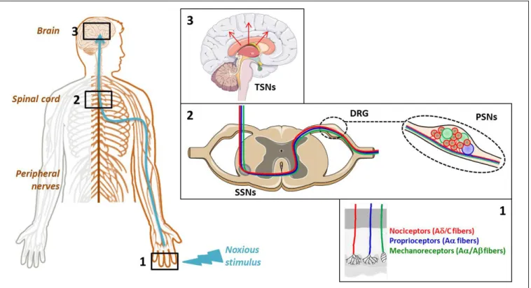
The cellular elements involved in pain transmission from the
peripheral to the CNS are detailed in Figure 1. The noxious
information is first detected by the nociceptors of peripheral and
visceral tissue, and then conveyed by the dendrites of primary
sensory neurons (PSNs). The nociceptors are located at the
level of free nerve endings of A
δ and C fibers of PSNs that
respond to noxious stimuli and are widely found throughout
skin and internal tissue. Three main types of pain receptors
exist: the thermal, the mechanical and the polymodal receptors,
activated by temperature, high pressure and mechanical, thermal
or/and chemical stimuli, respectively (Figure 1, Box 1). The
PSNs are pseudo-bipolar neurons which send their axons,
components of dorsal roots, to the laminas I, II and V of
the dorsal horn of spinal cord and establish synapses with the
dendrites of secondary sensory neurons (SSNs) (Figure 1, Box
2). The SSNs, in turn, bring the noxious information to the
hypothalamus and connect to tertiary sensory neurons (TSNs)
whose cell bodies constitute, in part, the brain cortex (Figure 1,
Box 3). At each CNS level, the information is integrated and
modulated by different ascending/descending control systems
such as the medullary control, named “gate control,” and
the diffuse inhibitory control including the noradrenergic and
serotoninergic pathways induced by nociception from the higher
centers to the dorsal horn, giving the affective, sensory and
cognitive dimensions to the human experience of pain (
Porreca
and Navratilova, 2017
).
The neuron bodies of PSNs constitute the 31 pairs of DRG,
coming out all along the spinal marrow: 8 cervical (C1-C7, note
that the first cervical spinal nerve is born above C1 and the
eighth one below C7), 12 thoracic (T1–T12), 5 lumbar (L1–
L5), 5 sacred (S1–S5), and 1 coccygeal (Co) which is vestigial.
The cranial sensory (trigeminal or Gasser’s) ganglion (nerve
V) conveys facial skin sensitivity, the spiral (or cochlear) and
vestibular (or Scarpa’s) ganglia (nerve VIII) serve the hearing
and balance senses, respectively, and the geniculate ganglion
(nerve VII) transfers facial sensations, with the contribution of
the superior and inferior (or petrous) ganglia of glossopharyngeal
nerve (nerve IX) and the superior (or jugular) and inferior (or
nodose) ganglia of vagus nerve (nerve X).
Dorsal root ganglia present a rich capillary bed in cell body
area (Figure 2), with the particularity of high fenestrations
between two endothelial cells being permeable to low and high
molecular weight compounds (
Petterson and Olsson, 1989
;
Parke
and Whalen, 2002
;
Jimenez-Andrade et al., 2008
;
Berta et al.,
2017
). In contrast to the cell body area, the nerve fiber area
wrapped by the epineural sheath, i.e., the dura mater continuum
in peripheral nervous system (PNS), presents a blood-nerve
barrier similar to the CNS blood-brain barrier (BNB), with a
lot of tight junctions between cells that prevent the passage of
unwanted drugs (
Jimenez-Andrade et al., 2008
;
Liu et al., 2018
).
FIGURE 1 | Cellular elements involved in pain transmission from the peripheral to the central nervous system (CNS). (Box 1) The pain (thermal, high pressure, mechanical, chemical) information is first detected by the receptors located at the level of free nerve endings of primary sensory neuron (PSN) fibers. (Box 2) Then, it is conveyed by the dendrites of these neurons, components of dorsal root ganglia (DRG), to the dorsal horn of spinal cord where it is transmitted to the dendrites of secondary sensory neurons (SSNs). (Box 3) Finally, it is brought to the hypothalamus via the tertiary sensory neurons (TSNs) whose cell bodies constitute, in part, the brain cortex.
FIGURE 2 | Schematic representation of morphological characteristics of ganglion and nerve capillaries. Ganglion capillaries differ from nerve ones by the presence of fenestration and absence of narrow tight junctions. Nerve endothelial cells are surrounded by pericytes.
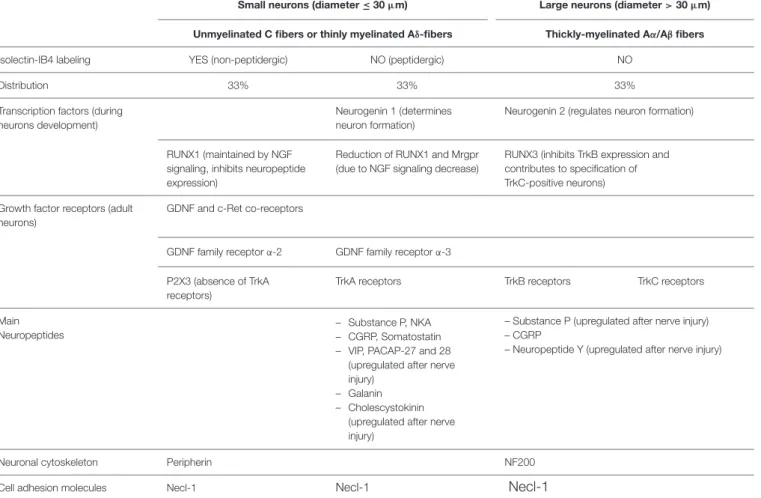
Soma of PSNs relaying the sensory information are part
of the DRG which also contain other different cell types
such as glial cells, endothelial cells and macrophages. Two
groups of DRG neurons may be distinguished using light and
electronic microscopy: the small dark neurons (cross-sectional
area ≤ 800
µm
2and diameter ≤ 30
µm) composed of high
threshold, slowly-conducting unmyelinated (C) and/or thinly
myelinated (A
δ) nerve fibers, and the large light neurons
(cross-sectional area
> 800 µm
2and diameter
> 30 µm)
constituted of low threshold, fast-conducting thickly-myelinated
(A
α and Aβ) nerve fibers (
Elliott and Elliott, 1993
;
Taddese
et al., 1995
;
Ho and O’Leary, 2011
). The small DRG neurons
that convey mainly pain message are subdivided into two groups:
the non-peptidergic and the peptidergic neurons, depending
on isolectin-IB4 labeling (Table 1). This subdivision of small
neurons results from the expression level of runt-related
transcription factor 1 (RUNX1), responsible for neuropeptide
expression, regulated by the nerve growth factor (NGF) signaling
during cell growth and differentiation (
Luo et al., 2007
). In
adult DRG neurons, RUNX and neurogenin transcription factors
regulate the expression of (i) glial cell line-derivated neurotrophic
factor (GDNF) and tyrosine kinase c-Ret co-receptors (allowing
the GDNF-ligand expression required for cell post-natal survival
and indicative of non-peptidergic neurons), and (ii) the
tropomyosin receptor-kinase receptors (TrkA, B and C which
bind NGF or brain-derived neurotrophic factor,
neurotrophin-4 and neurotrophin-3, respectively). The expression of growth
factor receptors is therefore of great help to better characterizing
adult DRG neurons (
Ernsberger, 2009
). Although only the
small DRG neurons which are not labeled by isolectin-IB4 are
peptidergic, the high dense-core vesicles of large neurons may
also contain peptides, depending on both the vesicle size and the
nerve condition, i.e., normal or injured (
Wiesenfeld-Hallin and
Xu, 2001
). The peptidergic neurons deliver not only substance
P and calcitonin gene-related peptide, but also somatostatin,
vasoactive intestinal peptide and cholecystokinin. When released
TABLE 1 | Characteristics of DRG neurons.
Small neurons (diameter ≤ 30µm) Large neurons (diameter> 30 µm) Unmyelinated C fibers or thinly myelinated Aδ-fibers Thickly-myelinated Aα/Aβ fibers Isolectin-IB4 labeling YES (non-peptidergic) NO (peptidergic) NO
Distribution 33% 33% 33%
Transcription factors (during neurons development)
Neurogenin 1 (determines neuron formation)
Neurogenin 2 (regulates neuron formation)
RUNX1 (maintained by NGF signaling, inhibits neuropeptide expression)
Reduction of RUNX1 and Mrgpr (due to NGF signaling decrease)
RUNX3 (inhibits TrkB expression and contributes to specification of TrkC-positive neurons) Growth factor receptors (adult
neurons)
GDNF and c-Ret co-receptors
GDNF family receptorα-2 GDNF family receptorα-3 P2X3 (absence of TrkA
receptors)
TrkA receptors TrkB receptors TrkC receptors
Main Neuropeptides
– Substance P, NKA – CGRP, Somatostatin – VIP, PACAP-27 and 28
(upregulated after nerve injury)
– Galanin – Cholescystokinin
(upregulated after nerve injury)
– Substance P (upregulated after nerve injury) – CGRP
– Neuropeptide Y (upregulated after nerve injury)
Neuronal cytoskeleton Peripherin NF200
Cell adhesion molecules Necl-1 Necl-1
Necl-1
The table illustrates the characteristics of DRG neurons regarding isolectin IB4-labeling, growth factor receptors, transcription factors, main neuropeptides, neuronal cytoskeleton composition, and cell adhesion molecules. GDNF, glial cell line-derivated neurotrophic factor; Trk, Tropomyosin receptor-kinase; P2X3, P2X purinergic receptor subunit 3; RUNX, Runt-related transcription factor; NGF, Nerve Growth Factor; Mrgpr, Mas-related G- protein coupled receptor; NKA, neurokinin A; CGRP, Calcitonin Gene-Related Peptide; VIP, Vasoactive Intestine Peptide; PACAP, Pituitary Adenylate Cyclase-Activating Polypeptide; NF, Neurofilament; Necl, Nectin-like molecule. The increased gradient of Necl-1expression between small non-peptidergic, small peptidergic and large neurons are represented by an increasing size of letters.
in the CNS areas associated with pain transmission, these
neuropeptides affect the expression pattern of SSNs, PSNs and
peripheral organs (
Moraes et al., 2014
). The type of cytoskeleton
neurofilaments present in DRG neurons is correlated with both
the axonal diameter and the conduction velocity of action
potential: intermediate neurofilament peripherin (57 kDa) is
expressed in slowly-conducting unmyelinated (C) and/or thinly
myelinated (A
δ) nerve fibers whereas the heavy neurofilament
NF200 (200 kDa) is expressed in fast-conducting
thickly-myelinated (A
α and Aβ) nerve fibers. The expression of
the cell adhesion nectin-like molecule 1, interacting with the
cytoskeleton, reflects the myelination level of nerve fibers (
Ho
and O’Leary, 2011
).
Because of sequencing advances, a large scaled and more
precise genetic characterization of DRG is now possible to
better identifying the function and underlying mechanisms
of each neuron. Therefore, an innovative approach to get
rid of pain sensation, without affecting other physiological
pain (or itch) pathways, would be to inhibit/remove only
the population of DRG neurons that are responsible
for the noxious disturbance (
Liem et al., 2016
;
Li et al.,
2018
).
ELECTROPHYSIOLOGICAL STUDIES OF
DRG NEURONS IN VITRO
Different types of tissues or individual cells can be used to
perform electrophysiological studies of DRG neurons
in vitro,
each of them offering advantages and disadvantages. Hence,
the primary cell cultures of rodent models (rats or mice)
provide freshly isolated DRG neurons, however dissociated
using enzymatic treatments which may disturb, in some extent,
their functioning and thus their electrophysiological recordings.
However, the two enzymatic procedures needed to replate the
cells (i.e., detach and again deposit them on glass-slides) 24 h
after their dissociation, in order to slow down extensive neurite
growth that could limit adequate electrophysiological recordings,
represent an aggressive cell treatment but were reported to have
no marked effect on the neuronal action potential (
Caviedes et al.,
1990
). In any case, a delay of 4–7 days between cell dissociation
and recordings is primordial to obtain adequate membrane
conditions for experiments. A more physiological alternative to
avoid cell dissociation and thus enzymatic procedures is to use
DRG explants, i.e., slices of DRG previously inserted in 2%
agar. Under these conditions, neurons are kept in their native
environment and their plasma membrane is not altered (
Scholz
et al., 1998
;
Scholz and Vogel, 2000
). However, the maximal
life-time of DRG explants, as that of primary cell cultures, is of
about 2 weeks.
Another possibility is thus the use of immortalized DRG
neurons which offer the advantage of being maintained in
cultures for long periods of time by changing freshly-made
medium daily. The principle consists in immortalizing
DRG neurons from human fetuses or rodents by using a
tetracycline-responsive v-myc oncogene (
Sah et al., 1997
;
Raymon et al., 1999
), a medium previously conditioned
with the rat thyroid cell line UCHT1 (
Allen et al., 2002
), or
telomerase reverse transcriptase expression vectors added in
the medium (
Chen et al., 2007
). Immortalized DRG neurons
may also be directly obtained from transgenic rats harboring the
temperature-sensitive large T-antigen gene (
Nishiya et al., 2011
).
Immortalized human DRG neurons became an advance 30 years
ago because of human tissue short supply. This type of more
homogeneous cell lines is of great interest for high throughput
screening of antinociceptive compounds.
Recently, the development of the induced pluripotent stem
cell (iPSC) technology opens up new perspectives in personalized
medicine, drug discovery or cell therapy. In the context of pain
studies, iPSCs, derived for example from mesenchymal cells
of a patient with inherited pain disease, are dedifferentiated
to acquire the neuronal phenotype (bipolar cells) with the
appropriate external medium containing neural growth factors.
Then, the cell cultures will allow performing electrophysiological
studies and pharmacological validation of a drug directly on
targets presenting the mutation responsible for the patient pain
phenotype (
Cao et al., 2016
;
Sommer, 2016
;
Yang Y. et al., 2018
).
VOLTAGE-GATED SODIUM CHANNELS
EXPRESSED IN DRG NEURONS
Na
Vchannels are crucial transmembrane proteins for the
communication of excitable cells in vertebrate and invertebrate
organisms, due to their important role in action potential genesis
and propagation. In terms of discovery, these channels are the
founding members of a superfamily comprising more than 140
members grouped into eight families (voltage-gated Na, K and Ca
channels, Ca-activated K channels, cyclic nucleotide-modulated
ion channels, transient receptor potential (TRP) channel,
inward-rectifying K channels and two-pore K channels) which, after
the GPCRs, constitute the second largest group of signaling
molecules encoded by the human genome (
Yu and Catterall,
2004
).
The fundamental functional features that allow Na
Vchannels
to perform their role in cellular electrical signaling include a
high selective permeation of Na ions and a gating system whose
opening and closing are controlled by both the time and the
membrane potential. Currently available data indicate that these
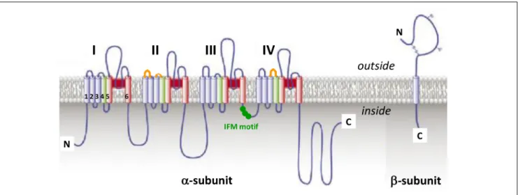
channels consist of a pore-forming
α-subunit (glycoprotein of
220–260 kDa) which is formed by four homologous domains
(designated DI to DIV), each comprising six hydrophobic
transmembrane
α helices segments (designated S1–S6) connected
by extra- and intra-cellular loops (Figure 3). The channel pore
formation is attributed to the hairpin-like P loops connecting S5
and S6 segments (extracellular part of the pore) and to the S6
segments (intracellular part of the pore) of each domain. The
channel activation (opening) is associated with the S4 segments
of each domain, containing repeated motifs of positively charged
amino acid residues (arginine) followed by two hydrophobic
residues, which lead to the opening of the pore by moving
outward under the influence of the membrane electric field to
initiate protein conformational change. The channel inactivation
(closing), meanwhile, is associated with the intracellular loop
connecting DIII and DIV domains, including the isoleucine,
phenylalanine and methionine (IFM) motif (
Catterall, 2000
;
Goldin et al., 2000
;
Payandeh et al., 2011
). Ten
α-subunits of the
mammalian Na
Vchannel, referred as Na
V1.1-1.10 (the first and
second numbers indicating the gene subfamily and the specific
channel isoform, respectively), have been identified so far. These
subunits, which exhibit 40–70% sequence homology and closely
related structures, can be distinguished according to their specific
expression in tissues and their sensitivity to TTX, a well-known
blocker of Na
Vchannels (Table 2). The structure, functional
characteristics and phylogenetic relationships of the various Na
Vchannel subtypes have been largely detailed in the literature
(
Catterall, 2000
;
Goldin et al., 2000
;
Goldin, 2001, 2002
;
Catterall
et al., 2005b, 2007
).
The expression, pharmacology and functioning of Na
Vchannels can be altered by post-translational modifications
(PTMs) of
α-subunits, such as acetylation, phosphorylation,
glycosylation and palmitoylation that occur after translation of
mRNAs into peptidic chains or during secretory pathways. These
PTMs greatly contribute to the development of chronic pain
syndromes and may also modulate the toxin sensitivity of Na
Vchannels (
Liu et al., 2012
). In acquired, but not in inherited,
pain syndromes, various signaling pathway activations may
alter expression and functioning of Na
Vchannels (
Laedermann
et al., 2015
). In mammals, the
α-subunit is associated with an
auxiliary
β-subunit (glycoprotein of 30–40 kDa), consisting of
a single transmembrane
α helices segment, a long N-terminal
extracellular immunoglobulin type V domain and a short
C-terminal intracellular domain (Figure 3), which may in
particular modulate the channel functioning, regulate its
trafficking and expression at the membrane surface and/or link
it to cytoskeleton proteins (
Catterall, 2000
;
Xie et al., 2016
).
Therefore, it is likely that the
β-subunit type and presence or
absence in overexpression systems, or even in native tissues,
will have big impact on Na
Vchannel readout during molecule
screening experiments, in particular. Hence, the functional
behavior of Na
V1.8 subtype has been reported to be highly
dependent on the type of
β-subunit expressed under normal
and disease conditions (
Vijayaragavan et al., 2004
). Among the
four auxiliary
β-subunits (β1-β4) identified so far, only β2- and
FIGURE 3 | The voltage-gated sodium channel. Schematic representations ofα-subunit and auxiliary β-subunit of NaVchannels, in which cylinders are
transmembraneα helices. In red: S5 and S6 pore-forming segments, in green: S4 voltage-sensor segment, and in blue: S1, S2, and S3 segments. IFM, isoleucine, phenylalanine and methionine residues. The orange loops in DII and DIV domains correspond to spider toxins binding sites (adapted fromCatterall et al., 2007).
TABLE 2 | Expression in tissues and TTX sensitivity of NaVchannel subtypes.
NaVsubtype Gene Expression in tissues TTX sensitivity
NaV1.1 SCN1A – PNS (DRG)
– CNS (hippocampus, neocortex, cerebellum, retinal ganglion, microglia) – Keratinocytes
Yes
NaV1.2 SCN2A – PNS (DRG; unmyelinated or pre-myelinated axons and dendrites) – CNS (hippocampus, neocortex; cerebellum, astrocytes) – Fibroblast, isletβ-cells, odontoblasts, osteoblasts
Yes
NaV1.3 (fetal) SCN3A – PNS (early postnatal periods, adult DRG when nerve injury or inflammation, Schwann cells)
– CNS (hippocampus, neocortex) – Fibroblasts, isletβ-cells
Yes
NaV1.4 SCN4A – Skeletal muscle Yes
NaV1.5 SCN5A – Heart
– Skeletal muscle (denervated or fetal)
No
NaV1.6 SCN8A – PNS (DRG, nodes of Ranvier of motoneurons, Schwann cells)
– CNS (Purkinje, pyramidal and granule neurons, nodes of Ranvier and axon initial segment of axons, astrocytes, microglia)
– Cancer cells, endothelial cells, fibroblasts, keratinocytes, macrophages
Yes
NaV1.7 SCN9A – PNS (DRG and sympathetic ganglion neurons, neuroendocrine cells) – CNS (olfactory sensory neurons)
– Smooth myocytes
– Prostate and breast tumor cells, human erythroid progenitor cells, fibroblasts, immune cells
Yes
NaV1.8 SCN10A – PNS (DRG)
– CNS (Purkinje neurons, astrocytes, Müller glia)
– Endothelial cells, fibroblasts, keratinocytes, T lymphocytes
No
NaV1.9 SCN11A – PNS (DRG)
– CNS (hypothalamus, astrocytes, Müller glia)
– Cancer cells, endothelial cells, fibroblasts, T lymphocytes
No
NaV1.10 (NaV1.x, NaV2.1-2.3) SCN7A – Lung, uterus, heart – PNS (DRG, Schwann cells)
– CNS (thalamus, hippocampus, cerebellum, median preoptic nucleus)
No
The table illustrates the expression in tissues and sensitivity to TTX of NaVchannelα-subunits (adapted fromGoldin, 2001;Trimmer and Rhodes, 2004;Black and
β4-subunit have been reported to be covalently linked, by their
N-terminal domain, to Na
Vchannel
α-subunits (
Namadurai
et al., 2015
). Recently,
β4-subunit has been highlighted as a
painkiller target because of its action of regulating fast resurgent
Na currents in sensory neurons associated with pain disorders
(
Xie et al., 2013
;
Lewis and Raman, 2014
;
Barbosa et al., 2015
).
Seven over the ten Na
Vchannel subtypes (Na
V1.1, 1.3,
1.6–1.10) which are expressed in DRG neurons are detailed
in Table 3. All these subtypes are thus potentially involved
in conveying noxious stimuli and may represent a target for
pain treatment. Indeed, the Na
V1.6–1.9 subtypes, as main
actors of pain anatomical and physiological integrities, have
been genetically proved to be linked to human pain disorders.
However, the high expression of Na
v1.7 subtype in DRG neurons
(see Figure 4) and its multiple reported mutations inducing
genetic-painful and painless disorders, largely documented in the
literature, make this subtype one of the most promising target
to alleviate pain. The contribution of Na
V1.1 and 1.10 subtypes
to pain message was evidenced by pharmacological approaches,
and the Na
V1.3 (fetal) subtype was reported to be overexpressed
during injury-induced pain. It is worth noting that Na
V1.2 is the
only subtype that does not transmit pain message in the PNS,
although present in DRG neurons. In the CNS, mutations in the
sequence coding for this subtype have been reported to induce
epileptogenic and/or neurodevelopmental disorders (
Liao et al.,
2010
;
Hackenberg et al., 2014
;
Ben-Shalom et al., 2017
;
Wolff
et al., 2017
).
Na
V1.1, encoded by the SCN1A gene, is a TTX-sensitive,
fast-activating and inactivating Na
Vchannel. Its expression is
located in the CNS, PNS (more precisely in DRG neurons) and
keratinocytes (
Trimmer and Rhodes, 2004
;
Black and Waxman,
2013
). This subtype was recently reported as a potential pain
target involved in neuropathic pain (Irritable Bowel Syndrome,
visceral hypersensitivity) and in acute pain and mechanical
allodynia, due to the correlation between its activity and pain
behaviors in rodent models using the activating spider toxin
Hm1a and the selective inhibitory small molecule ICA-121431
(
Osteen et al., 2016, 2017
;
Salvatierra et al., 2018
). The important
function of Na
V1.1 in the CNS is highlighted by more than
500 mutations in its coding sequence that cause epileptic
syndromes (Febrile Seizure, Generalized Epilepsy with Febrile
Seizures +, and Severe Myoclonic Epilepsy of Infancy also known
as Dravet syndrome) (
Catterall et al., 2010
). Moreover, three
of these mutations are also correlated to familial hemiplegic
migraine, and several copy number variants have been linked
to neurodevelopmental disorders such as intellectual disability,
autism and psychiatric disease (
Dichgans et al., 2005
;
Fry et al.,
2016
;
Xiong et al., 2016
).
Na
V1.3, encoded by the SCN3A gene, is also a TTX-sensitive,
fast-activating and inactivating Na
Vchannel (
Cummins et al.,
2001
). This fetal subtype is normally expressed in early postnatal
periods. However, it is also expressed at very low levels in
adult sensory primary afferents, and is rapidly upregulated
in DRG after peripheral axotomy by sciatic nerve transection
or chronic constriction (
Waxman et al., 1994
;
Black et al.,
1999
;
Dib-Hajj et al., 1999
) or by tight ligation of the spinal
nerve (
Boucher et al., 2000
;
Kim et al., 2001
), and in painful
diabetic neuropathy (
Tan et al., 2015
;
Yang et al., 2016
). Na
V1.3
promotes the spontaneous ectopic discharge observed during
nerve injury. In particular, its over-expression after spinal cord
injury leads to rhythmic oscillatory burst firing, alternating with
single spikes and silent periods, in second order dorsal horn
sensory neurons, and to spindle wave firing mode in thalamus
(ventral posterior lateral) neurons with identifiable peripheral
receptive fields (
Hains et al., 2003
;
Lai et al., 2003
). The central
neuropathic pain is also explained by Na
V1.3 upregulation which
induces neuronal hyperexcitability and alters the process of
somatosensory information (
Hains et al., 2003
;
Hains et al., 2005,
2006
). Recently, loss-of-function of the SCN3A gene, resulting
in reduced expression or deficient trafficking to the plasma
membrane of the protein, was reported to contribute to increased
seizure susceptibility (
Lamar et al., 2017
).
Na
V1.6, encoded by the SCN8A gene, is a TTX-sensitive
fast-activating and inactivating Na
Vchannel expressed in the
PNS (DRG neurons, nodes of Ranvier of motoneurons, Schwann
cells), in the CNS (Purkinje, pyramidal and granule neurons,
nodes of Ranvier and initial segment of axons, astrocytes,
microglia) and in non-neuronal tissues such as cancer cells,
endothelial cells, fibroblasts, keratinocytes and macrophages
(
Trimmer and Rhodes, 2004
;
Black and Waxman, 2013
;
Israel
et al., 2017
). This subtype is upregulated in various peripheral
pain pathways including oxaliplatin-induced cold allodynia
(
Deuis et al., 2013
), type-2 diabetic neuropathic pain (
Ren et al.,
2012
) and inflammatory pain (
Xie et al., 2013
). The Na
V1.6
α-subunit, covalently linked to the β4-subunit, can underlie
excitatory, persistent and resurgent currents which induce
repetitive firing and abnormal spontaneous activity of sensory
neurons (
Lewis and Raman, 2014
;
Barbosa et al., 2015
;
Xie et al.,
2016
). A Na
V1.6-gene mutation resulting in gain-of-function
has been reported to potentiate transient and resurgent Na
currents, leading to increased excitability in trigeminal neurons,
exacerbating thus the pathophysiology of vascular compression
and contributing to idiopathic trigeminal neuralgia (
Grasso et al.,
2016
;
Tanaka et al., 2016
). In contrast to Na
V1.1 and 1.2, Na
V1.6
is involved in seizure resistance (
Makinson et al., 2014
). The
knock-down of Na
V1.6 in the brain was shown to compensate
the Na
V1.1-gene mutation-induced imbalance of excitation over
inhibition involved in epileptogenic disorders, which motivates
the necessity to find specific Na
V1.6 inhibitors to treat debilitating
or fatal form of epilepsy such as the Dravet syndrome (
Catterall,
2012
;
Anderson et al., 2017
). Finally, more than ten human
de
novo mutations of Na
V1.6 gene have been identified in patients
with two types of CNS disorders, epileptic encephalopathy and
intellectual disability (
O’Brien and Meisler, 2013
).
Na
V1.7, encoded by the SCN9A gene, is a TTX-sensitive
fast-activating and inactivating Na
Vchannel. It is expressed in
the somatosensory system (mainly in C- and A
β-type DRG
neurons) and in the sympathetic ganglion neurons (myenteric
and visceral sensory neurons) of PNS, but only in the olfactory
sensory neurons of CNS. This subtype is also present in smooth
myocytes (
Jo et al., 2004
;
Israel et al., 2017
;
Vetter et al., 2017
),
and in non-excitable cells such as prostate and breast tumor cells,
human erythroid progenitor cells, fibroblasts and immune cells
T ABLE 3 | Electr ophysiological pr operties and disor ders associated with Na V channel subtypes expr essed in DRG neur ons and involved in pain. Na V subtype Activation (m) and inactivation (h) gating pr operties 1 Pain signs Genetic pain disor ders Pain-unr elated disor ders Time- dependence V oltage-dependence Na v 1.1 Fast (Tp = 1.23 ms) Vm0 .5 = − 20 mV Vh0 .5 = − 52 mV – Acute pain – Mechanical allodynia – V isceral hypersensitivity – Irritable Bowel syndr ome – Epileptic syndr omes – Familial hemiplegic migraine – Neur odevelopmental disor ders Na v 1.3 (fetal) Fast (Tp = 1.08 ms) Vm0 .5 = − 20 mV Vh0 .5 = − 58 mV – Painful nerve injury – Painful diabetic neur opathy – Central neur opathic pain – Incr eased seizur e susceptibility Na v 1.6 Fast (Tp = 1.03 ms) Vm0 .5 = − 19 mV Vh0 .5 = − 56 mV – Oxaliplatin-induced cold allodynia – Painful diabetic neur opathy – Inflammatory pain – Painful neur opathy (idiopathic trigeminal neuralgia) – Seizur e resistance – Epileptic encephalopathy – Intellectual disability – Cer ebellar atr ophy , behavioral deficits and ataxia ⇒ Resurgent/persistent curr ents Na v 1.7 Fast (Tp = 1.09 ms) Vm0 .5 = − 33 mV Vh0 .5 = − 62 mV – Painful diabetic neur opathy – Inflammatory pain – Acute noxious mechanosensation – Congenital insensivity to pain – Her editary sensory and autonomic neur opathy – Genetic painful neur opathies – Anosmia and hyposmia – Epileptic syndr omes – Autism spectrum disor der – Irritating, itchy cough ⇒ T reshold curr ent Na v 1.8 Slow (Tp = 1.06 ms) Vm0 .5 = − 6 mV Vh0 .5 = − 35 mV – Painful neur opathy (AIDS, diabetes, cancer) – Inflammatory pain – Maintenance of bone cancer pain – Painful neur opathy (small fiber neur opathy , inherited erythr omelalgia) – Multiple scler osis – Car diac conduction abnormalities ⇒ Persistent curr ents Na v 1.9 V ery slow (Tp > > 1 ms) Vm0 .5 = − 48 mV Vh0 .5 = − 31 mV – Inflammation-induced hyperalgesia and peripheral sensitization – Inflammatory , heat and mechanical pain hypersensitivity – Maintenance of bone cancer pain – Per ception of cold pain – V isceral pain – Congenital insensivity to pain – Her editary sensory and autonomic neur opathy – Genetic painful neur opathies – Hirschprung’ s disease (mega colon motility) – Bladder motility dysfunction – Essential tr emor associated with familial episodic pain ⇒ T reshold curr ent Na V 1.10 (Na V 1.x, 2.1–2.3) Na-dependence (threshold value = 150 mM) – Bone cancer -r elated pain – Chr onic hyper natr emia – Epileptogenic pr ocess 1Adapted from Deuis et al. (2017a ) and Inserra et al. (2017 ) . Tp, time to peak current; Vm0.5 , test-pulse voltage corresponding to 50% maximal activation; Vh0.5 , pre-pulse voltage corresponding to 50% steady-state inactivation.
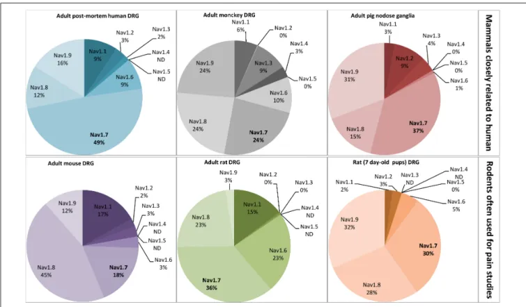
FIGURE 4 | Relative proportion of NaVchannelα-subunits detected in mammalian dorsal root ganglia (DRG) neurons by RT-PCR. DRG neurons are from some representative mammals of different orders reported in the literature: primates, artyodactyla and rodents. The first order, including human and monkey, is closely related to the pig, belonging to the second order. The rodents, more distant from the human, represent the model often used for pain studies. The adult
post-mortem human DRG neurons were obtained from healthy donors. All the data are from DRG neurons of adult mammals except those from 7 day-old rats. ND, non-determined. Adapted fromRaymond et al. (2004),Berta et al. (2008),Ho and O’Leary (2011),Muroi and Undem (2014), andChang et al. (2018).
a threshold channel since it is involved in the action potential
(i.e., pain message) triggering by regulating the resting membrane
potential of DRG. The implication of Na
V1.7 in neuropathic
(diabetes) and inflammatory pain, as well as in acute noxious
mechanosensation, has been explained by gene upregulation
or variants (
Dib-Hajj et al., 2013
;
Blesneac et al., 2018
). In
addition, multiple Na
V1.7 genetic mutations have been linked
to painless or painful phenotypes. Hence, congenital SCN9A
loss-of-function mutations, such as congenital insensivity to pain
and type IID of hereditary sensory and autonomic neuropathy,
can induce genetic diseases with a complete absence of pain. In
contrast, the SCN9A gain-of-function mutations cause genetic
painful neuropathies such as small fiber neuropathy, primary
erythromelalgia and paroxysmal extreme pain disorder (
de Lera
Ruiz and Kraus, 2015
;
Vetter et al., 2017
). The Na
V1.7 expression
in the CNS is responsible for anosmia and hyposmia, always
linked to painless phenotypes, and epilepsy (presence of different
variants in patients showing seizures and Dravet syndrome, and
of two SCN9A mutations related to epilepsy phenotype), as well
as to autism spectrum disorder (
Dib-Hajj et al., 2013
;
Mulley
et al., 2013
;
Rubinstein et al., 2018
;
Yang C. et al., 2018
). Na
V1.7
has also been reported to be the major Na
Vsubtype in irritating,
itchy cough conveyed by DRG neurons (
Muroi and Undem,
2014
;
Sun et al., 2017
).
Na
V1.8, encoded by the SCN10A gene, is a TTX-resistant
Na
Vchannel that exhibits slow activation and inactivation,
as well as rapid repriming kinetics in C- and A
β-type DRG
neurons. With its slow kinetics and high activation threshold,
this subtype corresponds to 80–90% of the inward current
necessary to the rising phase of action potentials (
Renganathan
et al., 2001
;
Patrick Harty and Waxman, 2007
). It is ectopically
expressed in the CNS Purkinje neurons during multiple sclerosis
disorder, and becomes thus a target of choice to develop a
treatment for this disorder (
Han et al., 2016
). Na
V1.8 mRNAs
were also detected and quantified in astrocytes, Müller glia,
endothelial cells, fibroblasts, keratinocytes and T lymphocytes
(
Black and Waxman, 2013
). This subtype has been reported
to contribute to neuropathic pain, notably associated with
acquired immunodeficiency syndrome, diabetes and cancer, as
well as to inflammatory pain (
Thakor et al., 2009
;
Qiu et al.,
2012
;
Belkouch et al., 2014
;
Liu X. D. et al., 2014
). Moreover,
SCN10A gain-of-function mutations are associated, not only
with the above mentioned neuropathic pain, but also with small
fiber neuropathy and inherited erythromelalgia (
Faber et al.,
2012
;
Huang et al., 2016
;
Kist et al., 2016
). Finally, genetic
variations of SCN10A have been reported to correlate with
cardiac conduction abnormalities in patients suffering from
hypertrophic cardiomyopathy-like atrial fibrillation and Brugada
syndrome (
Zimmer et al., 2014
;
Behr et al., 2015
;
Iio et al.,
2015
).
Na
V1.9, encoded by the SCN11A gene, is a TTX-resistant
Na
Vchannel with very slow activation and inactivation kinetics.
This subtype is also a threshold channel but exhibits different
biophysical properties, compared with Na
V1.7 subtype. Roughly
80% of small-diameter sensory DRG neurons but only a few
large-diameter ones and trigeminal ganglia (including C-type
nociceptive cells) were reported to express Na
V1.9 mRNAs (
Dib-Hajj et al., 1998
). The expression pattern of this subtype is
merely limited to the PNS, despite spots of expression in the
CNS (hypothalamus, astrocytes, Müller glia), endothelial cells,
fibroblasts, and T lymphocytes. It was also detected in some
cancers such as lymphoma and small cell lung cancer (
Black and
Waxman, 2013
;
Israel et al., 2017
). Na
V1.9 plays a major role
(i) in inflammatory, heat and mechanical pain hypersensitivity,
as revealed in both (sub) acute and chronic inflammatory pain
models, (ii) in the maintenance of bone cancer pain (with the
Na
V1.8 subtype), (iii) in the perception of cold pain under normal
and pathological conditions, and (iv) in visceral pain (
Lolignier
et al., 2011
;
Qiu et al., 2012
;
Dib-Hajj et al., 2015
;
Lolignier et al.,
2015
;
Hockley et al., 2016
;
Lolignier et al., 2016
). More recently,
multiple Na
V1.9 genetic mutations were linked to painless or
painful phenotypes, making this subtype the second target of
interest (after the Na
V1.7 subtype) to treat pain. Hence, on
one hand, congenital SCN11A loss-of-function mutations, such
as congenital insensitivity to pain and type VII of hereditary
sensory and autonomic neuropathy, were reported to result in
genetic diseases with a complete absence of pain (
Leipold et al.,
2013
;
Woods et al., 2015
;
Phatarakijnirund et al., 2016
;
Huang
et al., 2017
;
King et al., 2017
). On the other hand, the SCN11A
gain-of-function mutations lead to genetic painful neuropathies
such as small fiber neuropathy and rare inheritable pain disorders
(
Zhang et al., 2013
;
Huang et al., 2014
;
Han et al., 2015
;
Leipold
et al., 2015
;
Kleggetveit et al., 2016
;
Okuda et al., 2016
;
Han et al.,
2017
). Finally, Na
V1.9 expression has also been linked to the
Hirschprung’s disease (affecting the mega colon motility), and
implicated in the development of inflammation-based bladder
motility dysfunction and in essential tremor associated with
familial episodic pain (
Ritter et al., 2009
;
O’Donnell et al., 2016
;
Leng et al., 2017
).
Na
V1.10, encoded by the SCN7A gene and also named Na
V1.x
or Na
V2.12.3 (according to the species), is an atypical subtype
associated with leak currents and considered as descendant
of Na
Vchannel
α1-subunits despite, notably, a less than 50%
sequence homology and marked discrepancies in S4 segments
and the intracellular loop connecting DIII and DIV domains
(
Goldin et al., 2000
;
Yu and Catterall, 2003
;
Nehme et al.,
2012
). In addition and in contrast to other Na
Vchannel
subtypes, Na
V1.10 is not activated by the membrane potential
but is sensitive to extracellular concentration of Na ions with a
threshold value of 150 mM (
Hiyama et al., 2002
). It is expressed
in the lung, uterus and heart, in the PNS neurons (e.g., medium
to large-sized DRG neurons, non-myelinating Schwann cells) and
in the CNS (e.g., thalamus, hippocampus, cerebellum, median
preoptic nucleus) (
Fukuoka et al., 2008
;
Garcia-Villegas et al.,
2009
) In particular, this subtype is clearly present in the primary
regions implicated in hydromineral homeostasis, such as the
subfornical organ, the vascular organ of the lamina terminalis
and the median eminence which control the Na-intake behavior
by changing neuronal excitability (
Watanabe et al., 2006
;
et al., 2015
;
Kinsman et al., 2017
). It is involved in autoimmunity
process causing chronic hypernatremia (
Hiyama et al., 2010
)
and in epilectogenic process (
Gorter et al., 2010
). Recently, the
inhibition or suppression of Na
V1.10 was reported to reduce
pain behaviors in a bone cancer-induced model by decreasing the
excitability of DRG neurons (
Ke et al., 2012
).
Using electrophysiological studies of DRG neurons
in vitro
for drug-discovery research may be limited by the relative
proportions of targeted Na
Vchannel subtypes, as exemplified
by the plant alkaloid paclitaxel whose effects differ between
the models used (
Chang et al., 2018
). Indeed, the relative
proportion of Na
Vchannel subtypes varies between small- and
large-diameter DRG neurons, the first one expressing more
TTX-resistant and less TTX-sensitive subtypes than the second
one in both rodent and human neurons (
Djouhri et al., 2003
;
Zhang et al., 2017
). In addition, the relative proportion of
Na
Vsubtypes varies according to the species studied. This is
illustrated in Figure 4 by the relative quantification of each Na
Vchannel subtype mRNA in small-diameter DRG neurons, the
most documented in the literature because of their interest to
treat pain, in various mammalian species. As expected from their
importance in pain process, Na
V1.6–1.9 subtypes are relatively
more expressed than Na
V1.1–1.3 subtypes, and the expression of
the pain-unrelated Na
V1.4 and 1.5 subtypes, when detected, is
extremely low and their function unknown (
Ho et al., 2013
).
The DRG neurons from rodent models are preferentially
used for pain studies, compared with those from human,
because they are rapidly available, easy to manipulate, cheap and
exhibit well-conserved anatomical and physiological properties.
However, adult mice and rat differ in their relative proportions of
Na
Vsubtypes: more than 50% of mouse DRG Na
Vsubtypes are
TTX-resistant (i.e., 45% of Na
V1.8 and 12% of Na
V1.9) whereas
it is the opposite in rat DRG neurons (i.e., 15% of Na
V1.1,
23% of Na
V1.6 and 36% of Na
V1.7) (
Berta et al., 2008
;
Chang
et al., 2018
). Interestingly, the level of expression of Na
Vsubtypes
is greatly influenced by the age of mammal, i.e., the neuron
maturation, as exemplified by the high expression of Na
V1.9
subtype in pup rat DRG neurons which is replaced by Na
V1.1
and 1.6 subtype expression in adult rat DRG neurons (
Ho and
O’Leary, 2011
). PCR analysis of the seven Na
Vsubtypes expressed
in DRG neurons reveals that post-mortem human DRG neurons
from healthy donors show relatively high expression of Na
V1.7
(49%) and low expression of Na
V1.8 (12%), whereas the mouse
DRG neurons present high expression of Na
V1.8 (45%) and low
expression of Na
V1.7 (18%), the adult rat DRG neurons having
an intermediate expression of Na
V1.7 (36%) and Na
V1.8 (23%)
(
Chang et al., 2018
).
The mammals closely related to human (i.e., adult
monkey and pig) roughly conserve the Na
Vsubtype
expression pattern of post-mortem human DRG neurons,
i.e., Na
V1.7 ≥ Na
V1.9 ≥ Na
V1.8 (
Raymond et al., 2004
;
Muroi
and Undem, 2014
). Although the adult post-mortem human
terms of physiology to estimate the relative proportions of Na
Vsubtypes in living humans (
Zhang et al., 2017
;
Chang et al., 2018
),
and even if some mammalian models seem closed to human, the
message needs to be always shaded when extrapolated to human.
Moreover, RT-PCR consists in averaging Na
Vsubtype mRNAs
present in nucleus of cell population, and does not represent
strictly the level of functional Na
Vsubtypes located in cell plasma
membranes.
In several mammals DRG neurons, alternative splicing of Na
Vα-subunit genes has been detected, resulting in the expression
of multiple proteins. However, the functional significance of this
process has not been completely elucidated (
Dietrich et al., 1998
;
Schirmeyer et al., 2014
). Some variants seem to lead to subunits
showing redundant or no obvious pharmacological and/or
functional differences, compared with the wild-type subunit
(
Schirmeyer et al., 2014
). However, different pharmacological
and functional properties between variant and wild-type subunits
are evidenced in the literature, such as their sensitivity to
drugs/toxins (
Dietrich et al., 1998
;
Tan et al., 2002
;
Thompson
et al., 2011
;
Boullot et al., 2017
), their functional specificity
regarding tissue/cell localization (
Song et al., 2004
), and their
involvement in membrane excitability
via the regulation of
translational repression (
Lin and Baines, 2015
). Some alternative
splice events are unique to DRG neurons. Hence, significant
changes in the splicing patterns of Scn8a and Scn9a genes
were observed in a rat model of neuropathic pain, leading
to down-regulation of all transcripts (
Raymond et al., 2004
).
Moreover, four alternative splice variants of SCN9A gene were
reported to be expressed in human DRG neurons. The difference
between two of them at the exon 5 level (exons 5A and 5N)
results in two different amino acid residues, located in the S3
segment of DI domain acid. One of them, negatively charged,
may be involved in modifications of Na
Vchannel activation
and de-activation, impacting thus the paroxysmal extreme pain
disorder disease phenotype (mutation I1461T). The two other
alternative splice variants differ at the exon 11 level, leading to
the presence (11L) or absence (11S) of an 11-amino acid sequence
in the intracellular loop connecting DI and DII domains of Na
Vchannels, an important region for protein kinase A regulation
which will thus influence neuronal excitability and pain sensation
(
Chatelier et al., 2008
;
Jarecki et al., 2009
). Recently, a (NAT)
was reported to be a potential candidate gene for patients with
inherited (primary erythromelalgia, paroxysmal extreme pain
disorder, and painful small fiber neuropathy) or acquired chronic
pain disorders linked to the SCN9A locus, taking into account
that the sense gene must not contain mutations which lead to
sense gene-NAT pairing. This is the first example of a new therapy
based on increased native antisense mRNAs to treat chronic pain
in humans (
Koenig et al., 2015
).
ANALGESIC SPIDER TOXINS
TARGETING THE NA
V
1.7 CHANNEL
SUBTYPE
Arachnids (araneae order) are the most diverse group of
venomous animals with more than 46,000 extant species
subdivided in araneomorph (crossing fangs) and mygalomorph
(parallel fangs) suborders. Theraphosidae, the most studied and
represented family in Arachnoserver 3.0 database, belongs to the
latter suborder, with approximatively 470 species, a bit more than
one quarter of all species (
Pineda et al., 2018
). Each spider venom
contains from 100s to 1000s peptides (
Escoubas, 2006
), meaning
that more than 10 million spider-venom peptides with an original
sequence remain to be discovered since only approximatively
0.01% of these toxins have been explored until now (
Klint et al.,
2012, 2015b
). The major components of most spider venoms are
small disulfide-rich peptide toxins (
Saez et al., 2010
).
Because of their major role in action potential genesis and
propagation in CNS, PNS, heart, smooth and skeletal muscles,
Na
Vchannels are crucial for vital functions and are thus targeted
by various groups of toxins that interact with at least six specific
channel receptor-sites (
Cestele and Catterall, 2000
;
Catterall
et al., 2007
;
Gilchrist et al., 2014
;
Israel et al., 2017
). Toxins
that alter these channels may affect one or more of their three
essential properties: activation, inactivation and ion selectivity.
In that regard, toxins that have been isolated from different
venomous animals (such as spiders, scorpions, cone snails, sea
anemones and centipedes) may be classified as pore blockers
and/or gating modifiers (
Israel et al., 2017
). The main source
of the approximately 20 analgesic peptide toxins targeting the
Na
V1.7 subtype is the venoms of tarantula constitutive of the
theraphosidae family (Figure 5) (
Klint et al., 2015b
;
Vetter et al.,
2017
). It is worth nothing that this family also contains many
Na
Vchannel activators (
Deuis et al., 2017b
), such as Hm1a toxin
which has been reported to induce a painful behavior when
injected in rodents (
Jami et al., 2017
). A small amount of these
toxins also target other ion channel types located at the level of
DRG neurons and, thus, taking part into pain processing such as
TRP channels A1 antagonized by Protoxin (ProTx)-I and Ph
α1β,
acid-sensitive ionic channel (ASIC)1a inhibited with high affinity
by psalmotoxin (PcTx)-1, and N-type Ca
Vchannels targeted by
Ph
α1β, although with less potency than for TRPA1 (
de Souza
et al., 2013
;
Gui et al., 2014
;
Osmakov et al., 2014
;
Tonello et al.,
2017
).
Spider toxins targeting the Na
V1.7 subtype with an IC
50less
than 500 nM are considered as analgesic toxin inhibitors (
Klint
et al., 2015a
), and belong to the three first classes of spider
Na
Vchannel toxins (NaSpTx), based on their primary structure
and disulfide framework (Figure 5). It is worth noting that this
classification also includes spider toxins which target not only
the Na
V1.7 channel subtype but also other subtypes of ionic
channels, as exemplified by the
ω-TRTX-Gr2a toxin (GpTx-1)
which was initially reported as a Ca
V3.1 subtype blocker after
isolation from the Chilean tarantula,
Grammostola rosea, venom
(
Ono et al., 2011
). The NaSpTx peptides are gating modifier
toxins (GMTs) because they alter channel gating by stabilizing
voltage-sensors (mainly S3–S4 segments of DII domain) in a
closed, or resting, configuration state (Table 4) (
Klint et al.,
2012
). The Na
V1.7 analgesic spider toxin inhibitors are shaped
by inhibitory cystine knot (ICK) scaffold due to 6 cysteines,
arranged into a ring composed of two disulfide-bridges crossed by
a third one (
Saez et al., 2010
). These peptides share a conserved
amphipathic surface profile characterized by a high proportion
FIGURE 5 | Sequence alignment obtained by Multalin (version 5.4.1) of different potential analgesic toxins sorted by NaVspider toxins families, using their UniProtKB identifiers. The consensus sequence is shown above each alignment, with the disulfide bond connectivity. In dark blue, highly conserved amino acid residues (100%), and in light blue, poorly conserved amino acid residues (>50%). The Greek letter(s) before the toxin name is associated to its type of action: µ for NaV channel inhibition,β for shift in the voltage-dependence of NaVchannel activation,ω for CaVchannel inhibition, andκ for KVchannel inhibition. ProTx, protoxin; HnTx, hainantoxin; CcoTx, ceratotoxin; HwTx, huwentoxin; JzTx, jingzhaotoxin; aa, amino acid residues; NaSpTx, spider NaVchannel toxin.
of hydrophobic/aromatic amino acid residues, such as Trp, Tyr
and Phe, surrounded by charged amino acids which constitute a
dipole potential with negative (Asp and Glu) and positive (Lys
and Arg) zones (
Jung et al., 2005
;
Cai et al., 2015
). Finding more
selective GMTs than pore-blockers of Na
V1.7 subtypes is likely
because the voltage-sensors are more variable in terms of amino
acid sequence than the pore region of Na
Vchannels (
Catterall
et al., 2005a
;
Payandeh et al., 2011
).
ProTx-III,
ceratotoxin-1,
GpTx-1,
Cd1a,
huwentoxin
(HwTx)-IV, hainantoxin (HnTx)-IV, Hd1a, HnTx-III and HnTx-I
are Na
V1.7 potential analgesic peptide toxins, composed of
33–35 amino acid residues, that belong to NaSpTx-1 family,
with nanomolar affinities (IC
50between 2.1 and 440 nM)
for this Na
Vsubtype. According to their electrophysiological
properties, these toxins act as pore blockers of the Na
V1.7
subtype and, except for ProTx-III and ceratotoxin-1, induce
minor alterations (less than 5 mV) in the voltage-dependence
of its activation and steady-state inactivation (Figure 5 and
Table 4
). Various mutants of ProTx-III and ceratotoxin-1were
produced, showing a 10–20-mV shift in the voltage-dependence
of Na
V1.7 activation without any change in its fast and
steady-state inactivation (
Bosmans et al., 2006
;
Cardoso
et al., 2015
), in agreement with their interaction with the
receptor-site 4 of Na
Vchannels (i.e., S3–S4 segments of DII
domain). Models of docking toxins on Na
Vchannels have
been reported, placing toxin peptides in the cleft between the
channel S1–S2 and S3–S4 transmembrane
α-helices (
Minassian
et al., 2013
;
Cai et al., 2015
;
Murray et al., 2016
). Even the
main channel amino acid residues involved in toxin-channel
interactions were located in the extracellular loop connecting
S3 and S4 segments of DII domain, some residues of the
extracellular loop connecting S1 and S2 segments of DII
T ABLE 4 | Selectivity pr ofile, electr ophysiological characteristics and channel binding site of Na V 1.7 potential analgesic peptide toxins repr esentative of Na V spider toxins (NaSpTx) families. GpTx-I (NaSpTx-1) HwTx-IV (NaSpTx-1) Pn3a (NaSpTx-2) Pr oTx-II (NaSpTx-3) ω -theraphotoxin-Gr2a µ -theraphotoxin-Hh2a µ -theraphotoxin-Pn3a β /ω -theraphotoxin-Tp2a Wild-type Wild-type m3-HwTx-IV (E1G-E4G-Y33W) Wild-type Wild-type GP-Pr oTX-II/JNJ63955918 (W7Q-W30L) Selectivity pr ofile (IC 50 ) 1 hNa V 1.7 4.4 nM 17–33 nM 0.4–3 nM 0.9 nM 0.3 nM 10 nM mNa V 1.7 1.5 nM rNa V 1.7 4.4 nM hNa V 1.1 41 nM 8.1 nM 37 nM 16 nM 10 µ M hNa V 1.2 44 nM 11.9 nM 124 nM 41 nM 1.6 µ M hNa V 1.3 20 nM 190 nM 7.2 nM 210 nM 102 nM hNa V 1.4 301 nM 4–10 µ M 369 nM 144 nM 39 nM 5 µ M hNa V 1.5 4.2 µ M > 10 µ M > 1 µ M 800 nM 79–398 nM > 10 µ M hNa V 1.6 52–83.3 nM 6.8 nM 129 nM 26 nM 1 µ M hNa V 1.8 > 10 µ M > 10 µ M > 1 µ M > 10 µ M 146 nM hNa V 1.9 2.4 µ M rK V 2.1 > 300 nM hK V 11.1 (Erg) > 10 µ M > 10 µ M hCa V 1.2 > 10 µ M > 10 µ M Inhibition hCa V 2.2 > 10 µ M hCa V 3.1 9.8 nM Inhibition hα 7 nAChR > 10 µ M hα 3 nAChR > 10 µ M TTX-S Na V (DRG) m: 6.3 nM r-m: 30–130 nM r: 300 nM r: 78% inhibition by 300 nM TTX-R Na V (DRG) r-m: > 10 µ M r: no effect of 300 nM Electr ophysiology 2 Shift in mV − 0.9 mV − 1.7 mV + 21.3 mV + 31.1 mV + 15.3 mV Shift in hV − 5.9 mV − 1.8 mV − 2.7 mV unchanged − 6 mV Fast inactivation Unchanged Unchanged Na V dependent change Inhibited Unchanged Activation kinetics Unchanged Slowed Inactivation kinetics Unchanged Unchanged Slowed τON 25–34 s (1 µ M) 167 s (30 nM) 2.5 s (1 µ M) < 1 min (300 nM) τOFF Reversible 88 s 40 s Channel binding site (TTX-S Na V subtypes) Receptor -site 4 (S1–S2/S3–S4 of DII) Receptor -site 4 (S1–S2/S3–S4 of DII) Receptor -sites 3 and 4 (S3–S4 of DII/DIV) Receptor -sites 3 and 4 (S3-S4 of DII/DIV) 1IC 50 , effective concentration necessar y for decreasing the response by 50%; h, human; m, mouse, r, rat. 2m V , voltage-dependence of activation; hV , voltage-dependence of inactivation; τON , time constant of toxin effect, τOFF , time constant of toxin wash-out. TTX-S, tetrodotoxin-sensitive; TTX-R, tetrodotoxin-resistant.