Strongyloides stercoralis larvae excretion patterns before and
after treatment
F. SCHÄR1,2, J. HATTENDORF1,2, V. KHIEU1,2,3, S. MUTH3, M. C. CHAR3, H. P. MARTI2,4
and P. ODERMATT1,2*
1
Department of Epidemiology and Public Health, Swiss Tropical and Public Health Institute, Basel, Switzerland 2
University of Basel, Basel, Switzerland 3
National Center for Parasitology, Entomology and Malaria Control, Ministry of Health, Cambodia 4
Department of Medical Services and Diagnostics, Swiss Tropical and Public Health Institute, Basel, Switzerland (Received 29 October 2013; revised 12 December 2013; accepted 17 December 2013; first published online 17 February 2014)
S U M M A R Y
The variability of larval excretion impedes the parasitological diagnosis of Strongyloides stercoralis in infected individuals. We assessed the number of larvae excreted per gram (LPG) stool in 219 samples from 38 infected individuals over 7 consecutive days before and in 470 samples from 44 persons for 21 consecutive days after ivermectin treatment (200μg kg− 1BW). The diagnostic sensitivity of a single stool sample was about 75% for individuals with low-intensity infections (41 LPG) and increased to 95% for those with high-intensity infections (510 LPG). Doubling the number of samples examined per person increased sensitivity to more than 95%, even for low-intensity infections. There was no indication of a cyclic excretion of larvae. After treatment, all individuals stopped excreting larvae within 3 days. Larvae were not detected during any of the following 18 days (total 388 Baermann and 388 Koga Agar tests). Two stool samples, collected on consecutive days, are recommended in settings where low or heterogeneous infection intensities are likely. In this way, taking into account the possible biological variability in excretion, the efficacy of ivermectin treatment can be assessed as soon as 4 days after treatment.
Key words: Strongyloides stercoralis, larvae excretion, sensitivity, treatment, ivermectin.
I N T R O D U C T I O N
Strongyloides stercoralis, a soil-transmitted nematode found worldwide (Genta,1989; Bethony et al.2006), is arguably the most neglected of all neglected helminth infections (Olsen et al.2009; Schär et al.
2013). Infectious S. stercoralis (L3) larvae are found
in humid soil contaminated with fecal material. From there, the larvae penetrate the intact skin of the human host. In resource-poor countries, environ-mental, socio-economic and behavioural factors, and inadequate sanitary conditions provide the ideal circumstances for high S. stercoralis transmission rates.
Parasitological diagnosis of S. stercoralis infection relies on identifying larvae in stool. The Baermann technique (Baermann,1917) and Koga Agar culture (Koga et al. 1991) are recommended for diagnosis, preferably used in combination and carried out on multiple stool samples (Khieu et al.2013). Infections with low numbers of excreted larvae are particularly challenging to diagnose and can easily be missed. For other intestinal helminths, detailed egg excretion pattern studies have been performed, e.g. for liver
flukes (Lovis et al.2012), but information is missing for S. stercoralis.
The efficacy of ivermectin in treating S. stercoralis has been demonstrated many times (Marti et al.1996; Toma et al. 2000; Adenusi et al. 2003; Heukelbach et al. 2004; Igual-Adell et al. 2004; Ordonez and Angulo, 2004; Suputtamongkol et al. 2011; Khieu et al. 2013). However, little is known about the parasite clearance time and cessation of larvae excretion after ivermectin treatment (Uparanukraw et al.1999) in endemic settings. Yet, this information is crucial for planning and executing follow-up examinations after treatment.
We assessed the excretion pattern of S. stercoralis larvae by examining stool samples daily for 7 days before and for 21 days after ivermectin treatment (200μg kg− 1BW, single oral dose (SOD)) in 38 and 44 infected patients, respectively.
M A T E R I A L S A N D M E T H O D S
Ethical considerations
This study was undertaken in Preah Vihear (March
2010) and Takeo provinces (March 2011) in
Cambodia. The study protocol was approved by the Ethics Committee of the Canton of Basel (EKBB, 21/09 dated 29 January 2009), Switzerland and by * Corresponding author: Swiss Tropical and Public
Health Institute, Socinstrasse 57, Postfach 4002 Basel, Switzerland. E-mail: [email protected]
the National Ethics Committee Health Research (NECHR; 033 dated 20 March 2009), Ministry of Health, Cambodia. All participants were informed of the purpose and process of the study. Written informed consent was obtained from all individuals prior to enrolment. All diagnosed infections were treated at the end of the study, according to the National Cambodian treatment guidelines.
Enrolment
Individuals diagnosed with S. stercoralis during a community-based helminth survey were randomly selected. Either the examined person alone (in Preah Vihear) or the examined person and all persons living in the same household (in Takeo) were enrolled.
Field procedures
Participants provided a stool sample each morning, for 7 days (Fig. 1). Each day they received a new labelled stool container to use for the following day. The collected samples were transported at ambient temperature and within 1 h to a health centre field laboratory where the laboratory examinations were conducted (2–3 h after defecation).
Participants that tested positive for S. stercoralis at least once during the 7 days were treated with a SOD of ivermectin (200μg kg− 1 body weight) on day 7. Daily stool collection continued for another 21 days after treatment was given.
Laboratory procedures
Upon the sample’s arrival in the laboratory, a
quantitative Baermann test was performed
(Baermann,1917). In brief, the stool container with the sample was weighed. A Baermann test was set up and the container was weighed again. The stool
sample (about 15–25 g) was placed in a wire-mesh in a funnelfilled with tap water. Artificial light was used to stimulate larval migration out of the stool into the funnel. After 2 h, the water containing the larvae was collected and centrifuged for 5 min at 2000 rpm. The sediment was examined under a microscope (400 × magnification) and S. stercoralis larvae were counted.
For each stool sample, a Koga Agar culture (Koga et al.1991) was also performed. In brief, a hazelnut-sized piece of stool was placed on a soft Agar plate. It was then incubated for 48 h at 28 °C. The plates were then rinsed and the liquid centrifuged for 2 min at 2000 rpm. The sediment was examined under a microscope for the presence of larvae and adults.
Statistical analysis
Data were entered in Microsoft Excel, cross-checked against original data sheets and analysed using the statistical software, R. Strongyloides stercoralis infec-tion intensity was calculated as larvae per gram stool (LPG). The relationship between diagnostic sensi-tivity and infection intensity (i.e. the mean LPG per individual) was visually assessed using a LOWESS smoother. Diagnostic sensitivity was calculated for both examining a stool sample on a single day and for examining samples collected on 2 consecutive days. Data from participants providing samples on 6 or more days before treatment were analysed for periodicity in the excretion pattern. Algorithms to detect periodicity of unknown time intervals usually require longer time series. However, every harmonic excretion pattern would have fewer local maxima compared with random noise. We compared the observed local maxima in the larvae excretion counts within individuals with the number of local maxima in randomly generated numbers. A local maximum was identified when the LPG on a certain day was higher than the counts of the previous day and the following day.
After treatment, the infection rate was calculated for each day as the number of patients in which at least one S. stercoralis larva was observed divided by the number of patients treated and followed-up. For each observation day, the larvae reduction rate (LRR) was calculated. We used the mean number of larvae excreted by each individual before treatment as the reference value. The overall LRR was calculated by first dividing the mean LPG for each day post treatment by the overall LPG. The result of the division was subtracted from 1 to get the LRR.
R E S U L T S
Enrolment and compliance
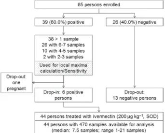
Altogether, 65 people were enrolled in the study (Fig. 1). First, a baseline examination was performed. Fig. 1. Flowchart: enrolment and follow-up of study
Individuals testing positive for S. stercoralis were randomly selected, as were individuals who tested negative (to form a control group). The overall mean age of participants was 26·7 years (range: 3–81 years). There were 32 (49·2%) males and 33 (50·8%) females. A total of 800 stool samples with an average weight of 19·7 g (range: 4·3–44·7 g) were examined with the Baermann method. All diagnostic results obtained by the Baermann method were confirmed by Koga Agar culture.
Larval excretion pattern before treatment
Thirty-nine (60·0%) people were infected with S. stercoralis. During the baseline examination, 18 (27·7%) provided 7 samples each, 14 (21·5%) provided 6 samples each and 32 (49·2%) provided 2–5 samples each. One person (2·6%) provided only 1 sample and was excluded from the excretion pattern analysis. The mean number of stools provided by healthy individuals was significantly lower than that provided by infected persons (4·2 vs 5·6 stool samples, P = 0·008). Altogether, 26 participants were negative, yet one person who tested negative at the initial examination presented a S. stercoralis infection on the third day. That person was subse-quently moved into the treatment group.
For the larval excretion pattern analysis, the 38 infected individuals who provided more than one
sample (median: 6 samples) were included.
Considerable variation in larvae excretion densities was observed in the pre-treatment examinations. The highest and lowest average densities per person observed were 151·2 LPG (range: 28·8–410·6 LPG) and 0·03 LPG (range 0–0·07 LPG), respectively.
Twenty-three patients (60·5%) tested positive for larvae each day before treatment, 9 (23·7%) had 1 day where no larvae were excreted, 4 (10·5%) had 2 days with no larvae present and 2 (5·3%) had 3 negative examinations.
The diagnostic sensitivity of a single-day examin-ation was 75% for individuals with an average LPG of 1 or less. Higher average LPGs increased diag-nostic sensitivity. An LPG of 10 or more resulted in sensitivities of more than 95% (Fig. 2). When stool samples collected on 2 consecutive days were analysed, the sensitivity increased remarkably. The sensitivity of two samples collected on consecutive
days was 97·0% (LPG≤1) and close to 100%
(LPG510), respectively.
We found a total of 65 local maxima in the 26 individuals with 6 or more stool examinations before treatment, which corresponds exactly to the expected number of local maxima in randomly generated Fig. 2. Diagnostic sensitivity of examining a single stool sample (black dots) and two samples collected on consecutive days (grey dots) by mean LPG count. Small random noise was added to alleviate overplotting. Lines represent the corresponding locally weighted scatterplot smoothing (LOWESS) curves.
numbers (median 65, 95% CI: 60–71), indicating no cyclic larvae release.
Larval excretion pattern after ivermectin treatment
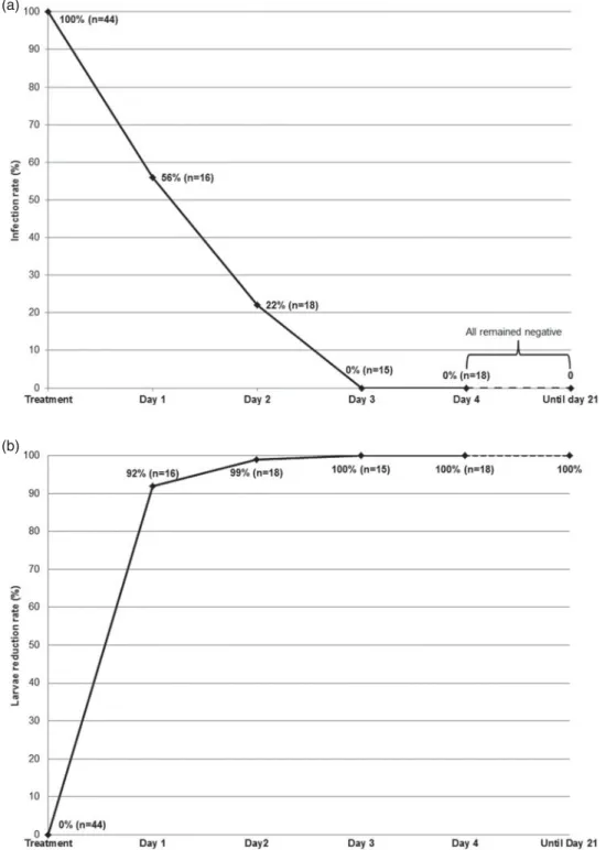
On the first day of follow-up after treatment, 16 (33·3%) participants provided a stool sample, 9 (56·3%) of which were found to be positive for larvae (Fig. 3a). On the second day, 18 (37·5%) provided a sample, of which 4 (22·2%) still tested positive. On the third day, none of the 15 (31·3%) samples provided yielded any S. stercoralis. From day 3 to
21 days after treatment, an average of 17 samples (35·4%) was returned each day, with a range of 11 to 24 samples per day. A total of 310 stool samples were received and analysed with Baermann and Koga Agar culture. Not a single larva was detected. All the results obtained by Baermann method were confirmed by the Koga Agar culture method.
From all the samples provided post-treatment, the LRR was calculated. The LRR increased dramati-cally to 92 and 99%, 24 and 48 h after treatment, respectively (Fig. 3b). From the third day onwards, all stool examinations remained negative.
(a)
(b)
Fig. 3. (a) Strongyloides stercoralis infection rate after ivermectin treatment. (b) Larvae reduction rate after ivermectin treatment.
D I S C U S S I O N
Strongyloides stercoralis remains one of the most neglected and under-reported helminthic infec-tions (Olsen et al. 2009; Schär et al. 2013). The irregular excretion of larvae, as well as the very low sensitivity of standard O&P examination procedures for S. stercoralis, requiring time-consuming special methods, are the main factors contributing to this under-reporting. Our results show that there is considerable variation in the numbers of S. stercoralis larvae excreted. Individuals with high-intensity in-fections (more than 10 larvae per gram stool) tended to present a positive result on all days of examination. Uparanukraw and colleagues (Uparanukraw et al.
1999) also documented variation of larval excretion, yet the underlying mechanisms accounting for this variation remain unclear (Jones, 1950; Jones and Abadie, 1954; Leighton and MacSween, 1990). As expected, we showed that diagnostic sensitivity in-creases with higher infection intensity. Diagnosing low-intensity chronic infections remains a major challenge and are likely to be missed by a single examination.
To further increase diagnostic sensitivity and to minimize false-negative results, it is important to analyse multiple stool samples on consecutive days. We demonstrated that sensitivity increases substan-tially if 2 samples are analysed. For several other helminthic infections, others have recommended that at least 3 samples on consecutive days be examined (Marti and Koella,1993; Dreyer et al.1996; Knopp et al.2008). For S. stercoralis, it is essential to target eradication of the pathogen rather than reducing infection intensity, as the ability of auto-infection can sustain the infection in the host. Grove reported patients testing positive after 2 years, even if the initial follow-up after treatment failed to show an S. stercoralis infection (Grove,1982).
When comparing the local maxima of larvae excreted with the maxima of random numbers, we could not establish a difference, which indicates that there is no cyclic pattern underlying the excretion of larvae. We also compared the diagnostic sensitivity of analysing 2 samples on consecutive days with the diagnostic sensitivity of analysing 2 samples with a sampling-free day in-between. As we did not detect a cyclic pattern, we expected the sensitivities to be approximately the same. This is reflected in our results. The diagnostic sensitivity of two samples with a sampling-free day in-between was 99% (compared with 97% on consecutive days). This difference is likely due to chance, given the small sample size. Another possible explanation might be the number of low-intensity infections, of which some had 2 consecutive days with no larvae excreted. Consequently, one would expect a further increase in sensitivity when analysing stool samples obtained on 3 consecutive days.
Ivermectin treatment proved to be very effective. In all infected participants, larvae excretion subsided. We investigated, for the first time, what happens directly after treatment. In our study, all individuals who received treatment (200μg kg− 1, SOD) stopped excreting larvae 72 h after treatment, at the latest. Larvae could not be detected in any of the partici-pants during the remaining 19 days following treatment. However, the absence of larvae does not ensure that the parasite has been eradicated from the host, which, in contrast to other helminths, should be the target of S. stercoralis treatment, as a few surviving larvae may lead parthenogenetically again to high infection densities (Satoh and Kokaze,2004; Requena-Mendez et al. 2013). However, current diagnostic parasitological methods do not allow us to verify this and prolonging the follow-up period is not an option in endemic areas, as the possibility of re-infection will still exist.
The standard Baermann method does not quantify the infection and only reports the presence or absence of the pathogen. We modified the Baermann method to calculate larvae per gram stool. Counting larvae in a Baermann examination can be challenging, es-pecially in cases of heavy infections, with many larvae present. As the larvae are alive and moving, there is a possibility of missing or double-counting larvae. The counts might, therefore, differ slightly from the true number, yet this difference is likely to be small and the calculated LPG values reflect the intensity of infection appropriately.
Compliance tends to be an issue in these studies, as very often participants are not able to defecate every day. It is very important to ensure that the stool samples provided are from the same person. If the person was not able to defecate, the stool container was returned empty. Furthermore, it was important that the stool samples provided were fresh to ensure that the larvae would still be alive.
Studies that focus on soil-transmitted helminths, including S. stercoralis, should apply the best available coprological diagnostic methods, like the Baermann method and/or Koga Agar culture. This combination of diagnostic methods, although work-and material-intensive, would help to get more accurate numbers on the global prevalence of S. stercoralis. Today, most studies on helminths either do not include S. stercoralis or use unsuitable diagnostic approaches, which, due to the factors stated above, leads to a severe under-reporting of S. stercoralis worldwide (Schär et al.2013).
In our study, we showed the importance of examining multiple stool samples on consecutive days because of the irregular excretion of larvae and the danger of missing low-intensity infections. Furthermore, we showed that treatment with iver-mectin leads to the absence of larvae excreted within 3 days after treatment. This has implications for planning and conducting follow-up studies. Our data
indicate that the clearance of larvae is rapid. Even if a possible biological variability in excretion is taken into account, follow-up periods in ivermectin trials can be short, i.e. 4 days.
A C K N O W L E D G E M E N T S
The authors would like to thank the staff at the National Center for Parasitology, Entomology and Malaria Control for all of their valuable help, as well as the participants who took part in this survey. We thank Mrs Amena Briët for the English editing of this manuscript.
A U T H O R C O N T R I B U T I O N S
FS, HPM and PO conceived and designed the study; FS and VK collected data under the supervision of SM; FS and HPM coordinated thefield laboratory activities; VK, MCC and SM coordinated thefield work in Cambodia; JH, FS, HPM and PO analysed data and interpreted the results; FS wrote the manuscript with PO; PO supervised the first author in all aspects of the study; all authors have read and approved thefinal version of manuscript.
F I N A N C I A L S U P P O R T
This study was supported by the UBS Optimus
Foundation.
C O N F L I C T O F I N T E R E S T
The authors declare no conflict of interest.
R E F E R E N C E S
Adenusi, A. A., Oke, A. O. and Adenusi, A. O. (2003). Comparison of ivermectin and thiabendazole in the treatment of uncomplicated Strongyloides stercoralis infection. African Journal of Biotechnology2, 465– 469.
Baermann, G. (1917). Eine einfache Methode zur Auffindung von Ankylostomum (Nematoden) Larven in Erdproben. Mededeel mit H. Geneesk Laboratories Weltevreden, Feestbundel, Batavia. 41–47. Bethony, J., Brooker, S., Albonico, M., Geiger, S. M., Loukas, A., Diemert, D. and Hotez, P. J. (2006). Soil-transmitted helminth infec-tions: ascariasis, trichuriasis, and hookworm. Lancet367, 1521–1532. doi: 10.1016/S0140-6736(06)68653-4.
Dreyer, G., Fernandes-Silva, E., Alves, S., Rocha, A., Albuquerque, R. and Addiss, D. (1996). Patterns of detection of Strongyloides stercoralis in stool specimens: implications for diagnosis and clinical trials. Journal of Clinical Microbiology34, 2569–2571.
Genta, R. M. (1989). Global prevalence of strongyloidiasis: critical review with epidemiologic insights into the prevention of disseminated disease. Reviews in Infectious Diseases11, 755–767.
Grove, D. I. (1982). Treatment of strongyloidiasis with thiabendazole: an analysis of toxicity and effectiveness. Transactions of the Royal Society of Tropical Medicine and Hygiene76, 114–118.
Heukelbach, J., Wilcke, T., Winter, B., Sales de Oliveira, F. A., Saboia Moura, R. C., Harms, G., Liesenfeld, O. and Feldmeier, H. (2004). Efficacy of ivermectin in a patient population concomitantly infected with intestinal helminths and ectoparasites. Arzneimittelforschung 54, 416–421.
Igual-Adell, R., Oltra-Alcaraz, C., Soler-Company, E., Sanchez-Sanchez, P., Matogo-Oyana, J. and Rodriguez-Calabuig, D. (2004). Efficacy and safety of ivermectin and thiabendazole in the treatment of
strongyloidiasis. Expert Opinion on Pharmacotherapy5, 2615–2619. doi: 10.1517/14656566.5.12.2615.
Jones, C. A. (1950). Clinical studies in human strongyloidiasis. I. Semeiology. Gastroenterology16, 743–756.
Jones, C. A. and Abadie, S. H. (1954). Studies in human strongyloidiasis. II. A comparison of the efficiency of diagnosis by examination of feces and duodenalfluid. American Journal of Clinical Pathology 24, 1154–1158. Khieu, V., Schär, F., Marti, H., Sayasone, S., Duong, S., Muth, S. and Odermatt, P. (2013). Diagnosis, treatment and risk factors of Strongyloides stercoralis in schoolchildren in Cambodia. PLoS Neglected Tropical Diseases 7, e2035. doi: 10.1371/journal.pntd.0002035.
Knopp, S., Mgeni, A. F., Khamis, I. S., Steinmann, P., Stothard, J. R., Rollinson, D., Marti, H. and Utzinger, J. (2008). Diagnosis of soil-transmitted helminths in the era of preventive chemotherapy: effect of multiple stool sampling and use of different diagnostic techniques. PLoS Neglected Tropical Diseases 2, e331. doi: 10.1371/journal. pntd.0000331.
Koga, K., Kasuya, S., Khamboonruang, C., Sukhavat, K., Ieda, M., Takatsuka, N., Kita, K. and Ohtomo, H. (1991). A modified agar plate method for detection of Strongyloides stercoralis. American Journal of Tropical Medicine and Hygiene45, 518–521.
Leighton, P. M. and MacSween, H. M. (1990). Strongyloides stercoralis. The cause of an urticarial-like eruption of 65 years’ duration. Archives of Internal Medicine150, 1747–1748.
Lovis, L., Mak, T. K., Phongluxa, K., Aye Soukhathammavong, P., Vonghachack, Y., Keiser, J., Vounatsou, P., Tanner, M., Hatz, C., Utzinger, J., Odermatt, P. and Akkhavong, K. (2012). Efficacy of praziquantel against Schistosoma mekongi and Opisthorchis viverrini: a randomized, single-blinded dose-comparison trial. PLoS Neglected Tropical Diseases6, e1726. doi: 10.1371/journal.pntd.0001726.
Marti, H. and Koella, J. C. (1993). Multiple stool examinations for ova and parasites and rate of false-negative results. Journal of Clinical Microbiology 31, 3044–3045.
Marti, H., Haji, H. J., Savioli, L., Chwaya, H. M., Mgeni, A. F., Ameir, J. S. and Hatz, C. (1996). A comparative trial of a single-dose ivermectin versus three days of albendazole for treatment of Strongyloides stercoralis and other soil-transmitted helminth infections in children. American Journal of Tropical Medicine and Hygiene55, 477–481. Olsen, A., van Lieshout, L., Marti, H., Polderman, T., Polman, K., Steinmann, P., Stothard, R., Thybo, S., Verweij, J. J. and Magnussen, P. (2009). Strongyloidiasis – the most neglected of the neglected tropical diseases? Transactions of the Royal Society of Tropical Medicine and Hygiene103, 967–972. doi: 10.1016/j.trstmh.2009.02.013. Ordonez, L. E. and Angulo, E. S. (2004). Efficacy of ivermectin in the treatment of children parasitized by Strongyloides stercoralis. Biomedica24, 33–41.
Requena-Mendez, A., Chiodini, P., Bisoffi, Z., Buonfrate, D., Gotuzzo, E. and Munoz, J. (2013). The laboratory diagnosis and follow up of strongyloidiasis: a systematic review. PLoS Neglected Tropical Diseases 7, e2002. doi: 10.1371/journal.pntd.0002002.
Satoh, M. and Kokaze, A. (2004). Treatment strategies in controlling strongyloidiasis. Expert Opinion on Pharmacotherapy5, 2293–2301. doi: 10.1517/14656566.5.11.2293.
Schär, F., Trostdorf, U., Giardina, F., Khieu, V., Muth, S., Marti, H., Vounatsou, P. and Odermatt, P. (2013). Strongyloides stercoralis: global distribution and risk factors. PLoS Neglected Tropical Diseases7, e2288. doi: 10.1371/journal.pntd.0002288.
Suputtamongkol, Y., Premasathian, N., Bhumimuang, K., Waywa, D., Nilganuwong, S., Karuphong, E., Anekthananon, T., Wanachiwanawin, D. and Silpasakorn, S. (2011). Efficacy and safety of single and double doses of ivermectin versus 7-day high dose albendazole for chronic strongyloidiasis. PLoS Neglected Tropical Diseases5, e1044. doi: 10.1371/journal.pntd.0001044.
Toma, H., Sato, Y., Shiroma, Y., Kobayashi, J., Shimabukuro, I. and Takara, M. (2000). Comparative studies on the efficacy of three anthelminthics on treatment of human strongyloidiasis in Okinawa, Japan. Southeast Asian Journal of Tropical Medicine and Public Health31, 147–151.
Uparanukraw, P., Phongsri, S. and Morakote, N. (1999). Fluctuations of larval excretion in Strongyloides stercoralis infection. American Journal of Tropical Medicine and Hygiene60, 967–973.