HAL Id: hal-03063758
https://hal.archives-ouvertes.fr/hal-03063758
Submitted on 18 Dec 2020HAL is a multi-disciplinary open access archive for the deposit and dissemination of sci-entific research documents, whether they are pub-lished or not. The documents may come from teaching and research institutions in France or abroad, or from public or private research centers.
L’archive ouverte pluridisciplinaire HAL, est destinée au dépôt et à la diffusion de documents scientifiques de niveau recherche, publiés ou non, émanant des établissements d’enseignement et de recherche français ou étrangers, des laboratoires publics ou privés.
The α-arrestin family of ubiquitin ligase adaptors links
metabolism with selective endocytosis
Jennifer Kahlhofer, Sébastien Léon, David Teis, Oliver Schmidt
To cite this version:
Jennifer Kahlhofer, Sébastien Léon, David Teis, Oliver Schmidt. The α-arrestin family of ubiqui-tin ligase adaptors links metabolism with selective endocytosis. Biology of the Cell, Wiley, 2020, �10.1111/boc.202000137�. �hal-03063758�
This article has been accepted for publication and undergone full peer review but has not been through the copyediting, typesetting, pagination and proofreading process, which may lead to differences between this version and the Version of Record. Please cite this article as doi:
10.1111/boc.202000137.
This article is protected by copyright. All rights reserved.
The -arrestin family of ubiquitin ligase adaptors links metabolism
with selective endocytosis.
Jennifer Kahlhofer
1, Sebastien Leon
2, David Teis*
1, Oliver Schmidt*
1*equal contribution, corresponding authors
1
Institute for Cell Biology, Biocenter, Medical University Innsbruck, Austria.
2
Université de Paris, CNRS, Institut Jacques Monod, Paris, France. Corresponding authors:
Dr. David Teis
Medical University Innsbruck, Biocenter, Institute for Cell Biology Innrain 80/82, CCB Building
A-6020, Innsbruck, Austria Email: david.teis@i-med.ac.at
phone: +43 512 9003 70191
Dr. Oliver Schmidt
Medical University Innsbruck, Biocenter, Institute for Cell Biology Innrain 80/82, CCB Building
This article is protected by copyright. All rights reserved.
2 Email: oliver.schmidt@i-med.ac.at
phone: +43 512 9003 70189
Abstract: The regulation of nutrient uptake into cells is important, as it allows to either increase biomass for cell growth or to preserve homeostasis. A key strategy to adjust cellular nutrient uptake is the reconfiguration of the nutrient transporter repertoire at the plasma membrane by the addition of nutrient transporters through the secretory pathway and by their endocytic removal. In this review we focus on the mechanisms that regulate selective nutrient transporter endocytosis, which is mediated by the -arrestin protein family. In the budding yeast Saccharomyces cerevisiae, 14 different -arrestins (also named arrestin-related trafficking adaptors, ARTs) function as adaptors for the ubiquitin ligase Rsp5. They instruct Rsp5 to ubiquitinate subsets of nutrient transporters to orchestrate their endocytosis. The ART proteins are under multilevel control of the major nutrient sensing systems, including amino acid sensing by the general amino acid control (GAAC) and target of rapamycin (TOR) pathways, and energy sensing by 5’-adenosine-monophosphate-dependent kinase (AMPK). The function of the six human -arrestins is comparably under-characterized. Here, we summarize the current knowledge about the function, regulation and substrates of yeast ARTs and human -arrestins, and highlight emerging communalities and general principles.
-Arrestins are an evolutionary conserved family of ubiquitin ligases adaptor proteins. -Arrestin - ubiquitin ligase complexes selectively ubiquitinate distinct plasma membrane proteins (including nutrient transporters) to initiate their endocytosis and degradation. The activity of these ubiquitin ligase adaptors is controlled by various nutrient signaling pathways. Thereby, cellular nutrient import is adjusted to the metabolic state of cells to maintain homeostasis.
This article is protected by copyright. All rights reserved.
3 Keywords: ubiquitin ligase, ubiquitin, metabolism, arrestin, -arrestin, endocytosis, nutrient
transporter
The arrestin protein family
The members of the arrestin family harbor a characteristic arrestin fold, consisting of a highly curved, bi-lobed -sandwich structure (Alvarez, 2008; Aubry et al., 2009) (reviewed in (Aubry and Klein, 2013)). In humans, the arrestin family has 14 members: six -arrestins also called arrestin-domain-containing proteins (ARRDCs), four visual/-arrestins, and four VPS26 (vacuolar protein sorting-associated protein 26)-like proteins (Shi et al., 2006; Alvarez, 2008). The function of VPS26 proteins was reviewed elsewhere (Chen et al., 2019). The focus of this review will be on the -arrestins. Yet, before we turn to them, we briefly introduce visual/-arrestins, since most of our knowledge of the arrestin family derives from studying their function.
The visual/-arrestins are key regulators of G-protein coupled receptor (GPCRs) signaling and only found in metazoans (Alvarez, 2008). The human -arrestin family includes two visual or cone arrestins (Arrestin-1 (Arr1) and Arr4), which are expressed in photoreceptors, and two non-visual arrestins (Arr2 and Arr3, also known as -arrestin-1 and -2, respectively), which are ubiquitously expressed. The functions of visual and -arrestins in the inactivation, internalization, trafficking and signaling of GPCRs, including the light receptor rhodopsin, are well characterized (reviewed in (Shenoy and Lefkowitz, 2011; Hilger et al., 2018)). Detailed structural analysis of all four /visual-arrestin subtypes and of -/visual-arrestin-GPCR complexes (Granzin et al., 1998; Hirsch et al., 1999; Han et
al., 2001; Milano et al., 2002; Sutton et al., 2005; Zhan et al., 2011; Shukla et al., 2013, 2014; Zhou et al., 2017) revealed similarities in their molecular structure and in the binding mechanism of arrestins
with their cognate GPCRs (Zhuo et al., 2014). -Arrestins harbor a central polar core flanked by cup-shaped N- and C- arrestin fold domains (Hirsch et al., 1999; Han et al., 2001). In their basal state, -arrestins are kept inactive by a network of interactions between residues within the polar core, and by interactions of the N-domain with the C-terminal-tail. Activated and phosphorylated GPCRs allow binding of their C-termini to the N-domain of -arrestins, thereby disrupting the basal state and triggering high-affinity receptor binding (reviewed in (Chen et al., 2018)). The polar core is specific for the visual and -arrestins and not present in other members of the arrestin family (Alvarez, 2008). Contrary to visual and cone arrestins, -arrestins contain a C-terminal clathrin-binding box (Goodman et al., 1996) and an AP-2 binding site (Laporte et al., 1999) that mediate GPCR endocytosis (reviewed in (Shenoy and Lefkowitz, 2005)).
This article is protected by copyright. All rights reserved.
4 -Arrestins represent the evolutionary more ancestral branch of the arrestin family, as they are readily present in fungi, worms and protists. Despite being more prevalent in evolution, the function of -arrestins was characterized more recently mainly through work in fungi (Herranz et al., 2005; Lin et al., 2008; Nikko et al., 2008). The genome of the budding yeast Saccharomyces cerevisiae encodes 15 arrestin proteins: 14 -arrestins and a single Vps26 protein, but no -arrestins (Alvaro et
al., 2014). The -arrestins appear to function as adaptors for ubiquitin ligases that mediate substrate
specificity towards membrane proteins (Aubry et al., 2009). They contain arrestin-like domains, but lack the -arrestin N-domain helix (Alvarez, 2008), which is part of the three-element interaction motif and involved in maintaining the inactive state of -arrestins (Sente et al., 2018). -Arrestins also lack clathrin binding sites, but typically harbor one or more proline-rich PPxY (PY) motifs in their C-terminal regions (reviewed in (Becuwe et al., 2012a)) (Figure 1A). These PY motifs interact with WW domains, which are found in a variety of proteins including E3 ubiquitin ligases of the conserved Nedd4 family (neural precursor cell expressed developmentally down-regulated protein 4) (Andoh et
al., 2002; Lin et al., 2008; Rauch and Martin-Serrano, 2011). Compared to -arrestins, the molecular
functions of -arrestins are less understood, and their individual physiological roles are only beginning to be elucidated. However, it is now becoming clear that many -arrestins are controlled by metabolic signaling pathways, and thereby play important roles by linking cellular metabolism with selective endocytosis of nutrient transporters to promote adaptation to nutrient fluctuations.
Arrestin related trafficking adaptors (ARTs) - -arrestins of budding yeast
Unicellular organisms, like budding yeasts, are frequently exposed to acute changes in nutrient availability. These cells therefore must regulate the import of nutrients by remodeling their plasma membrane proteome, including nutrient transporters or permeases, to adapt their growth to changing environmental conditions (Haguenauer-Tsapis and André, 2004). Nutrient transporter endocytosis is controlled by ubiquitination (reviewed in (Lauwers et al., 2010)). The covalent attachment of ubiquitin to specific lysine residues in membrane proteins is highly selective and serves as a molecular tag along the endocytic pathway. It serves first as a signal for internalization, likely through the interaction with the endocytic machinery at the plasma membrane (reviewed in (Kaksonen and Roux, 2018)). After endocytosis, ubiquitination mediates intracellular sorting events, including sorting into the multivesicular body (MVB) pathway by the endosomal sorting complexes required for transport (ESCRT) machinery, which is a prerequisite for lysosomal degradation of membrane proteins (Katzmann et al., 2001) (reviewed in (Migliano and Teis, 2018)). In yeast, a single HECT-type (homologous to the E6AP carboxyl terminus) ubiquitin ligase, called Rsp5, is responsible for the ubiquitination of nutrient transporters and hence for their endocytosis (Hein et al., 1995; Galan et al., 1996; Wang et al., 1999). The first protein identified to function together with Rsp5 in protein ubiquitination was Bul1 (Binds ubiquitin ligase 1) (Yashiroda et al., 1996, 1998), a protein
This article is protected by copyright. All rights reserved.
5 later shown to be related to the -arrestins (Merhi and André, 2012). In yeast, the -arrestins are more frequently called arrestin-related trafficking adaptors (ARTs) (Lin et al., 2008). 14 ARTs are encoded in the genome of Saccharomyces cerevisiae: Art1 (Ldb19), Art2 (Ecm21), Art3 (Aly2), Art4 (Rod1), Art5, Art6 (Aly1), Art7 (Rog3), Art8 (Csr2), Art9 (Rim8), Art10, Bul1, Bul2, Bul3 and Spo23 (reviewed in (O’Donnell and Schmidt, 2019)). Their arrestin domains are not easily predicted from the primary sequence, because they are interspersed at varying positions by large, probably unstructured loops, which modulate the function and location of ARTs in ways that are currently only partially understood (Baile et al., 2019). It is now becoming clear that most, if not all, ARTs act as adaptors that confer substrate specificity for Rsp5. Rsp5 is a member of the conserved Nedd4 family of ubiquitin ligases and displays a modular organization comprising an N-terminal C2 domain, three WW domains, and a C-terminal catalytic HECT ubiquitin ligase domain (reviewed in (Rotin and Kumar, 2009)). The C2 domain binds phospholipids, mediates membrane interaction (Plant et al., 2000), and has a role in MVB sorting (Dunn et al., 2004). Each WW domain of Rsp5 can bind directly to PY motifs (Hesselberth et al., 2006), and so far, all ARTs were found to interact with Rsp5 through their PY motifs, allowing the formation of specific ART-Rsp5 complexes. Whether the three WW domains of an individual Rsp5 molecule can simultaneously engage more than one ART is currently unclear. Hence, at least 14 distinct ART-Rps5 complexes can be formed. These different ART-Rsp5 complexes determine the selectivity of endocytosis of most nutrient transporters and G-protein coupled receptors in response to various stimuli (summarized in Table 1).
Decoding ART-Rsp5 specificity
Some ART-Rsp5 complexes function in a partially redundant manner with overlapping substrate specificity, whereas other ART-Rsp5 complexes are highly selective depending on the biological context or stimulus (Table 1). For example, Art1-Rsp5 mediates the endocytosis of the methionine transporter Mup1, the lysine transporter Lyp1 and the arginine transporter Can1 in response to excess of their respective amino acid substrates (Lin et al., 2008; MacGurn et al., 2011). Yet, upon amino acid and nitrogen starvation, the same set of amino acid transporters is downregulated by Art2-Rsp5 (Ivashov et al., 2020). Remarkably, Art1-Rsp5 and Art2-Rsp5 bind to different seemingly acidic sorting signals in these transporters (see below). Thus, the complementary use of Art1-Rsp5 and Art2-Rsp5 for the downregulation of amino acid transporters is one example of how ART-Rsp5 complexes act on nutrient transporters in a both stimulus-specific, as well as transporter-specific manner (Lin et al., 2008; Nikko and Pelham, 2009). A similar situation exists for the high-affinity glucose transporters Hxt6 and Hxt7. In the presence of glucose, they are endocytosed by the glucose-activated arrestin Art4 (Nikko and Pelham, 2009), whereas their endocytosis during prolonged glucose limitation depends on the glucose starvation-specific arrestin Art8 (Hovsepian et
al., 2017). Comparably, the endocytosis of the lactate transporter Jen1 in response to glucose also
requires Art4, whereas cycloheximide treatment triggers Jen1 endocytosis in an Art4-independent but Bul1-dependent manner (Hovsepian et al., 2018). The versatility of the ART-Rsp5 systems for
This article is protected by copyright. All rights reserved.
6 transporter ubiquitination upon various physiological stimulations probably accounts for the described redundancy in ART function (Lin et al., 2008; Nikko and Pelham, 2009).
Localization of ART-Rsp5 complexes
ART-Rsp5 complexes are sometimes sequentially activated, and can act at different internal compartments to regulate the fate of nutrient transporters along the endocytic pathway (Soetens et
al., 2001; O’Donnell et al., 2010; Becuwe and Léon, 2014; Hager et al., 2018; Hovsepian et al., 2018;
Martínez-Márquez and Duncan, 2018). Art1 can re-localize from the cytosol and/or the trans-Golgi network (TGN) to the plasma membrane in response to endocytic signals (MacGurn et al., 2011; Baile et al., 2019). A signal-induced recruitment to discrete regions of the plasma membrane was also observed for Art9 after alkaline pH shift (Herrador et al., 2015). Art3 localizes in a constitutive manner to both the plasma membrane and internal compartments (endosomes and TGN) (O’Donnell
et al., 2010). At the plasma membrane, Art3 localizes to endocytic sites (Boeke et al., 2014). This may
explain its involvement in the endocytosis of many cargoes (Hatakeyama et al., 2010; O’Donnell et
al., 2010, 2013; Crapeau et al., 2014; Prosser et al., 2015; Wawrzycka et al., 2019; Nishimura et al.,
2020; Sen et al., 2020). Consistently, Art3 interacts with subunits of clathrin adaptors of both the plasma-membrane (AP-2) or the TGN (AP-1) (O’Donnell et al., 2010). Its paralog Art6 is restricted to internal compartments and interacts only with AP-1, but shows some functional overlap with Art3 (O’Donnell et al., 2010). Both Art3 and Art6 regulate the recycling of the general amino-acid permease Gap1 from endosomes in some conditions, whereas they regulate its internalization from the plasma membrane in others (Crapeau et al., 2014). An example of sequential and location-specific ART function was described for the endocytosis of the lactate transporter Jen1 upon exposure to glucose. While both Bul1 and Art4 can mediate Jen1-ubiquitination at the plasma membrane to initiate endocytosis, Art4 at the TGN is essential for the subsequent post-endocytic sorting of Jen1 into the MVB pathway (Becuwe et al., 2012b; Becuwe and Léon, 2014; Hovsepian et
al., 2018). The role of ART proteins in protein sorting at the TGN is also supported by early work on
Bul1 and Bul2 (Helliwell et al., 2001) and a more recent study on Art1 (Martínez-Márquez and Duncan, 2018). How ARTs are recruited to their site of action is not fully understood. In particular it is unclear if ARTs interact directly with the headgroups of specific lipids in their target membranes (e.g. phosphatidyl-inositol-phosphates). In addition, it is also not clear how ARTs selectively recognize their substrates at these various compartments. For example, the domains involved in localization and substrate specificity of Art1 are distinct, suggesting that substrate binding is not the primary means of Art1 translocation to the plasma membrane (Baile et al., 2019).
This article is protected by copyright. All rights reserved.
7 ART-Rsp5-mediated endocytosis of certain nutrient transporters (Mup1, Can1, Fur4, Thi7, Jen1) requires conformational changes from the outward open to the inward open state, which are induced by substrate transport (Cain and Kaiser, 2011; Keener and Babst, 2013; Ghaddar et al., 2014; Gournas et al., 2017; Talaia et al., 2017; Savocco et al., 2019). Similar results regarding the mechanism for substrate-induced endocytosis have been obtained in Aspergillus nidulans (Gournas
et al., 2010; Papadaki et al., 2019). Some nutrient transporters, including Can1 and Mup1, are
clustered in the sphingolipid- and ergosterol-rich membrane compartment occupied by Can1 (MCC) (Malínská et al., 2003; Grossmann et al., 2007; Spira et al., 2012). MCCs are associated with eisosomes, which form furrow-like invaginations of the plasma membrane (Walther et al., 2006). MCCs may serve as a storage compartment, which stabilizes nutrient transporters in their inactive state (Grossmann et al., 2008; Gournas et al., 2018; Moharir et al., 2018; Appadurai et al., 2020) (reviewed in (Babst, 2020)).
In the presence of their substrate, nutrient transporters are rapidly exchanged between MCCs and the surrounding lipid domains (Brach et al., 2011). Substrate flux through Can1 and Mup1 (i.e. arginine or methionine transport) induces conformational changes in these transporters that drive lateral movement out of the MCC (Gournas et al., 2017, 2018; Busto et al., 2018). In case of Mup1, the substrate-induced conformational change leads to exposure an extended acidic patch in the N-terminal cytosolic tail that is recognized by activated Art1-Rsp5 complexes (Guiney et al., 2016). This acidic patch is in close proximity to the first transmembrane domain and the ubiquitination sites of Mup1 (Figure 1B, upper panel). The N-terminus of Mup1 is required but not sufficient for degradation, indicating the existence of a second interaction site. Indeed, a C-terminal plug region after the last transmembrane domain of Mup1 is displaced upon substrate transport and is also critically required for Art1-dependent internalization (Busto et al., 2018). In this model, a basic region in the arrestin C-domain of Art1 interacts electrostatically with the cytosolic acidic patch and possibly the core and/or plug of Mup1, which orients Rsp5 to ubiquitinate the nearby N-terminal lysine residues (Guiney et al., 2016; Busto et al., 2018). Very similar findings have been obtained for the arginine permease Can1 (Gournas et al., 2017, 2018): in absence of arginine, Can1 accumulates in MCCs in an outward-open conformation. Transport of arginine facilitates a shift to an inward-facing conformation that promotes the re-localization of Can1 out of MCCs and causes the exposure of a short acidic N-terminal sequence close to the first transmembrane domain, which serves as the recognition site of activated Art1-Rsp5 (Keener and Babst, 2013; Guiney et al., 2016; Gournas et al., 2017, 2018; Busto et al., 2018). Also in the uracil transporter Fur4, a conformation-sensor, termed the loop-interacting domain (LID), mediates exposure of a degron for Rsp5-dependent ubiquitination (Keener and Babst, 2013).
Further metabolic cues can induce the selective endocytosis of a wide range of nutrient transporters independently of substrate binding. Amino acid and nitrogen limitation, for example, triggers endocytosis of Mup1 and Can1, but requires the action of Art2 (Jones et al., 2012; Müller et
This article is protected by copyright. All rights reserved.
8 contains a C-terminal basic motif, which directs Rsp5 to an acidic patch in the C-terminal tails of Mup1 and Can1 (Figure 1B, lower panel). This leads to ubiquitination of nearby lysine residues, which are distinct from those targeted by Art1-Rsp5. Noteworthy, the C-terminal tails of several other Art2 substrates also contain acidic amino acid sequences that could serve as recognition motif for Art2 (Ivashov et al., 2020). In addition, this region is also subject to substantial phospho-regulation, which might additional regulate these interactions (see below). Hence, both Art1 and Art2 use basic patches in their extended arrestin domains to detect acidic sorting motifs and thereby instruct Rsp5 to ubiquitinate nearby lysine residues with remarkably high selectivity. A context-dependent involvement of different ART-Rsp5 complexes was also observed for the endocytosis of Gap1. Upon rapamycin treatment, Art3-Rsp5 or Art6-Rsp5 interact with the C-terminal region of Gap1 to ubiquitinate a single N-terminal lysine (K16), whereas the Bul1/Bul2-Rsp5 complexes can interact with either the N- or C-terminal tail depending on the stimulus (amino acids or rapamycin) and target both K9 and K16 (Crapeau et al., 2014).
Further studies described the importance of acidic residues in cargo proteins for their ART-mediated ubiquitination. The arsenite transporter Acr3 requires an N-terminal acidic tail for its Art3/Art4-dependent endocytosis (Wawrzycka et al., 2019). An acidic patch located in the C-terminal tail of Jen1 triggers its downregulation upon activation of glucose signaling through recruitment of Art4 (Fujita et al., 2018). This region acts as a portable signal for glucose-induced, Art4-mediated endocytosis as it can be transferred to another transporter (Mup1) to impose both glucose-induced regulation and Art4-dependency. This and the fact that Art4 is also in charge of other glucose-regulated substrates such as the glycerol/H+ symporter Stl1 (Becuwe and Léon, 2014) or glucose transporter Hxt6 (Nikko and Pelham, 2009; Llopis-Torregrosa et al., 2016) suggests not only that Art4 is mobilized during glucose response, but also that Art4, similar to Art2, can act independently of substrate-induced conformational changes.
Another layer of regulation is provided by the phosphorylation of plasma membrane proteins, which often promotes ubiquitination and subsequent endocytosis (Table 2) (Marchal et al., 1998, 2000; Opekarová et al., 1998; Nikko et al., 2008; Paiva et al., 2009; Iesmantavicius et al., 2014; Ivashov et al., 2020; Tumolo et al., 2020). The yeast casein kinase 1 (Yck1/2)-dependent phosphorylation on N-terminal serine/threonine sites of the uracil permease Fur4 is a pre-requisite for its ubiquitination on a nearby lysine residues (Marchal et al., 2000). However, it is still unknown if and how these phosphorylation events modulate interactions with ART-Rsp5 complexes. In the presence of its ligand -factor, the GPCR Ste2 undergoes conformational changes that promote phosphorylation by Yck1/2 (Hicke and Riezman, 1996; Hicke et al., 1998), which may induce the binding of Art4 and Art7 to its C-terminus (Alvaro et al., 2014). An appealing common mechanism for the stimulating role of substrate phosphorylation in endocytosis may thus involve the addition of negative charges to the acidic patches in substrate proteins, which might help to generate and / or stabilize the interaction with ART proteins.
This article is protected by copyright. All rights reserved.
9 It will be interesting to see in the future if the concept of an electrostatic interaction between an ART basic region and an acidic domain on transporters can be generalized, and if the exposure of acidic patches in the N- or C- terminal tails of nutrient transporters provides degrons that are recognized by all 14 dedicated ART-Rsp5 complexes.
Multilevel regulation of -arrestins in budding yeast.
ARTs are subject to complex regulation processes including dynamic modifications by (1) ubiquitination, (2) phosphorylation, and also (3) transcriptional regulation (Figure 2).
(1) Regulation of ART activity by ubiquitination/deubiquitination
Following the description of the ART family in yeast (Lin et al., 2008) and examination of published proteomic datasets (Andoh et al., 2002; Ho et al., 2002; Peng et al., 2003; Gavin et al., 2006; Hesselberth et al., 2006; Kee et al., 2006; Krogan et al., 2006; Gupta et al., 2007) it became clear that most if not all ARTs are Rsp5 interactors and ubiquitinated in vivo. ARTs are typically mono-/oligo-ubiquitinated by Rsp5, which depends on interaction of their PY motifs with WW domains of the Rsp5. This was shown for Art1, where ubiquitination of K486 (requiring the WW2 domain of Rsp5) is needed for the endocytic downregulation of Can1 (Lin et al., 2008). Art4 is ubiquitinated in response to glucose exposure, which is important for its function in the endocytosis of Jen1 (Becuwe et al., 2012b). Downregulation of Gap1 upon growth on preferred nitrogen sources is accompanied by Rsp5-dependent ubiquitination of Bul1 and Bul2 (Merhi and André, 2012), and Rsp5 also ubiquitinates Art3 (Hatakeyama et al., 2010). Since most ubiquitination sites have not been experimentally determined, it is unclear if the positions of functionally relevant ubiquitinated residues are conserved between different ARTs. However, it seems that the ubiquitination of Art8, which is required for its function in endocytosis of high-affinity glucose transporters (Hovsepian et
al., 2017), occurs in the same region that was described for Art4, i.e. in the N-terminal part of the
arrestin-C domain (Becuwe et al., 2012b). Interestingly, for a number of ART proteins, the most frequently detected ubiquitinated lysine residues map to a conserved sequence motif in this region (Figure 3 A, B). This seems to be similar in human -arrestins (Figure 4 A, B). Further work is needed to clarify if this represents a critical ubiquitin acceptor lysine for Rsp5-dependent activation of ARTs.
ARTs can also become poly-ubiquitinated by Rsp5 on distinct acceptor sites. This attachment of Lys(63)-linked polyubiquitin chains induces the proteasomal degradation of several ARTs, which is antagonized by the deubiquitinating enzymes (DUBs) Ubp2 and Ubp15 (Kee et al., 2005, 2006; Ho et
This article is protected by copyright. All rights reserved.
10 Interestingly, for the majority of ARTs, it is unclear how ubiquitination contributes to their function on a mechanistic level. In case of Art1, ubiquitination is required for its endocytic function and subcellular localization, as a non-ubiquitinatable mutant of Art1 accumulates diffusely in the cytosol (Lin et al., 2008). Ubiquitination of ARTs can also modulate substrate interaction, as it is the case for Art9. Art9 mediates the interaction of the pH sensor Rim21 with the ESCRT machinery (Herrador et al., 2010). At the moment, this seems to be a specific feature of a few members of the -arrestin family, including Art9 and human ARRDC1, which contain “late domains” similar to those found in some viral proteins (e.g. Gag of HIV) in charge of recruiting ESCRT proteins to viral egress sites (Rauch and Martin-Serrano, 2011). Mono-ubiquitination of Art9 by Rsp5 promotes the binding of the ESCRT-I subunit Vps23, which in turn prevents poly-ubiquitination of Art9 by Rsp5 (Herrador
et al., 2013). The role of Art9 poly-ubiquitination is not entirely clear, but it may trigger its
proteasomal degradation.
There are also several instances in which ubiquitination does not seem to be required for ART function. For example, stress conditions trigger Gap1 endocytosis without affecting the ubiquitination of the ARTs involved (Bul1/Bul2 and Art3/Art6) (Crapeau et al., 2014). Similarly, the ubiquitin-conjugation site on Art4 is not required for the endocytosis of low-affinity glucose transporters upon treatment with the metabolic inhibitor 2-deoxyglucose (O’Donnell et al., 2015), and Art1 ubiquitination seems dispensable for the endocytosis of Ste2 and the subsequent downregulation of pheromone signaling (Alvaro et al., 2014). Therefore, it remains unclear how ubiquitination contributes mechanistically to ART function in some but not all endocytosis events.
(2) Regulation of ARTs by phosphorylation/dephosphorylation
In addition to ubiquitination, phosphorylation of ARTs provides an important regulatory mechanism. Phosphorylation typically appears to inhibit ART function and may be the primary mechanism by which ARTs are inactivated. Several ARTs are directly or indirectly under the control of major nutrient sensing kinases, including the target of rapamycin complex 1 (TORC1) and the 5’adenosine-monophosphate-activated protein kinase (AMPK) Snf1, and thus their activity is coupled to cellular metabolism. Hence, major nutrient sensing pathways switch ARTs from an inactive, phosphorylated and (sometimes) deubiquitinated form to their active, dephosphorylated and (often) ubiquitinated form (Figure 2) (Nikko and Pelham, 2009; MacGurn et al., 2011; Becuwe et
al., 2012b; Merhi and André, 2012; Becuwe and Léon, 2014; Llopis-Torregrosa et al., 2016;
Hovsepian et al., 2017).
Phosphorylation of ARTs sometimes creates binding sites for the 14-3-3 proteins Bmh1/2, and most ARTs interact with 14-3-3 proteins (Kakiuchi et al., 2007). Somehow, the binding of 14-3-3 to
This article is protected by copyright. All rights reserved.
11 ARTs hinders ubiquitination or may even promote their deubiquitination, although the underlying mechanism is unclear (Becuwe et al., 2012b; Merhi and André, 2012; Becuwe and Léon, 2014; Hovsepian et al., 2017). In other instances, ubiquitination and phosphorylation are uncoupled, as is the case for Art1, which seems to be primarily regulated at the phosphorylation level (MacGurn et
al., 2011). Moreover, (de)phosphorylation sometimes regulates the subcellular localization of ARTs
(MacGurn et al., 2011; Becuwe and Léon, 2014). Phosphomimetic mutations at selected serine or threonine residues in Art1 prevent its interaction with Mup1 (Lee et al., 2019). Art1 phosphorylated on these sites remains cytosolic or associated with the Golgi. Upon dephosphorylation, Art1 is able to associate with the plasma membrane and promote Mup1 endocytosis. The cyclin and cyclin-dependent kinase pair, Clg1-Pho85 promotes phosphorylation of these sites, whereas Pho80-Pho85 activity impedes phosphorylation at the same sites, yet both kinases seem not to phosphorylate Art1 directly (Baile et al., 2019). Phosphorylation of Art9 in the inter-arrestin hinge region by the plasma membrane-associated casein kinase 1 (Yck1/2) inhibits its translocation to the plasma membrane and attenuates signalling by the Rim21 alkaline pH sensor (Herrador et al., 2015). Art6 is dephosphorylated by the calcium-regulated phosphatase calcineurin (phosphoprotein phosphatase 2B), which then allows the endocytosis of Dip5 (Hatakeyama et al., 2010; O’Donnell et al., 2013). Thus, a consensus model emerges in which phosphorylation of ART protein renders them inactive and therefore ART-mediated endocytosis often requires dephosphorylation events in response to nutrient availability or other cellular cues.
TORC1 signaling is one of the critical signaling pathways known to regulate ART proteins. TORC1 stimulates cell growth by promoting anabolic processes (protein synthesis, lipid synthesis), and by repressing catabolic processes, such as autophagy (reviewed in (Eltschinger and Loewith, 2016)). TORC1 signaling also promotes Art1-dependent endocytosis of amino acid transporters in response to substrate excess. The substrate-induced endocytosis of the Art1 substrates Can1, Mup1, Lyp1, Fur4 and Tat2 was shown to be controlled by TORC1 signaling (Galan et al., 1996; Schmidt et al., 1998; MacGurn et al., 2011). Of note, Art1-mediated endocytosis is exclusive for those amino acid transporters that are actively engaged in substrate transport. This specificity is achieved because substrate flux causes conformational changes, which leads to the exposure of the sorting signal for Art1 (as discussed above). In response to amino acids influx, activated TORC1 phosphorylates its downstream kinase Npr1 (nitrogen permease reactivator 1) and thereby inhibits its activity. This allows Art1 activation to downregulate the respective amino acid transporter. Art1 activation additionally requires the action of the Ppz1/2 phosphatases. At the plasma membrane, they dephosphorylate Art1 at its Npr1-dependent phosphorylation sites, which promotes interaction of Art1 with its substrates (Lee et al., 2019). Conversely upon TORC1 inactivation, Npr1 becomes active and phosphorylates Art1, thereby preventing its recruitment to the plasma membrane and hence Art1-mediated endocytosis (MacGurn et al., 2011). Substrate-induced endocytosis can additionally be mediated by Art2 (Fur4, Tat2) and by Art3 (Dip5), and Npr1-dependent phosphorylation events were also detected on these ART proteins (O’Donnell et al., 2010; MacGurn et al., 2011; Gournas et
al., 2017, 2018). Thus, it is possible that multiple ART proteins are co-regulated by TORC1-Npr1
This article is protected by copyright. All rights reserved.
12 feedback loop, which couples nutrient transporter abundance to the influx of nutrients and prevents excessive accumulation of certain nutrients. Noteworthy, the substrate-induced endocytosis of the thiamine transporter Thi7, which is mediated by Art2-Rsp5, requires the Sit4 phosphatase (phosphoprotein phosphatase 2A) and TORC1 activity, but is independent of the Npr1 kinase, suggesting the existence of additional mechanisms (Savocco et al., 2019).
Npr1 also negatively regulates Bul1- and Bul2-mediated endocytosis of Gap1, by creating binding sites for the yeast 14-3-3 proteins and thus preventing their interaction with Rps5. This contributes to the stabilization of Gap1 at the plasma membrane during nitrogen starvation. Upon nitrogen repletion, Bul1/2 are dephosphorylated, presumably by Sit4, which leads to their dissociation from 14-3-3 proteins and activation (Merhi and André, 2012).
Of note, also the inactivation of TORC1 signaling, both by rapamycin treatment and by amino acid and nitrogen starvation, is accompanied by selective nutrient transporter endocytosis via a distinct mechanism (Schmidt et al., 1998; Jones et al., 2011; Müller et al., 2015). Nutrient transporter endocytosis in response to amino acid and nitrogen starvation differs in many characteristics from the exclusive substrate induced endocytosis. It is slower and targets multiple transporters simultaneously but still occurs with high selectivity, which is conferred by Art2 for amino acid transporters (Mup1, Can1, Lyp1) and Art4 for glucose transporters (Hxt3) (Ivashov et al., 2020). Interestingly, a fraction of nutrient transporters might be protected from starvation-induced endocytosis because they laterally segregate into MCCs, which increase in number and size upon starvation. (Gournas et al., 2018; Laidlaw et al., 2020).
The yeast AMPK Snf1 is essential for the adaption to glucose limitation and for the utilization of carbon sources that are less preferred than glucose, such as lactate. When yeast cells are grown in lactate medium, Snf1 becomes activated and phosphorylates Art4 (Shinoda and Kikuchi, 2007), leading to its inactivation. This prevents the Art4-dependent ubiquitination and degradation of the lactate transporter Jen1 and thereby stimulates lactate influx (Fujita et al., 2018). A similar mechanism prevents the Art4-dependent down-regulation of the high-affinity glucose transporter Hxt6 at glucose-limiting growth conditions (Llopis-Torregrosa et al., 2016). Once glucose becomes available, Art4 is dephosphorylated by the glucose-activated phosphoprotein phosphatase 1 (PP1) Glc7/Reg1, and subsequently ubiquitinated by Rsp5. This activates Art4 and promotes ubiquitination and internalization of Jen1 by Rsp5 (Becuwe et al., 2012b). Activated Art4 also controls the fate of internalized Jen1 at the TGN and mediates its sorting to the vacuole for degradation (Becuwe and Léon, 2014). Treatment of cells with the non-metabolizable glucose analog 2-deoxyglucose (2DG) causes ubiquitin-dependent endocytosis of Hxt1 and Hxt3. This involves the two paralogs Art4 and Art7, both of which are regulated by Snf1 and PP1 (O’Donnell et al., 2015). Snf1-mediated phosphorylation further regulates Art4-mediated down-regulation of the pheromone receptor Ste2.
This article is protected by copyright. All rights reserved.
13 In presence of glucose, Art4 is phosphorylated and inactivated by residual Snf1 activity and by the TORC2-Ypk1 signaling axis at multiple sites. This prevents the association of Ste2 with the Art4-Rsp5 complex (Alvaro et al., 2014, 2016). Ste2 internalization for the adaptation to pheromone signaling requires dephosphorylation of Art4 by the calcium-dependent phosphatase calcineurin. Thereby, the calcineurin-dependent activation of Art4 ensures removal of Ste2 from the cell surface of mating cells only in response to influx of calcium that occurs at a late stage in pheromone signaling (Alvaro
et al., 2014). Thus, as exemplified by Art4, regulation of ART phosphorylation patterns can control
context-dependent endocytosis of different cargoes.
Finally, the glucose-mediated activation of the cyclic adenosine-monophosphate (cAMP)-dependent protein kinase A (Bcy1-Tpk1/2/3) leads to the phosphorylation of Art8 on serine/threonine residues located in its N-terminal region. This triggers 14-3-3 recruitment and subsequent Art8 deubiquitination and inactivation in glucose replete conditions (Hovsepian et al., 2017).
Taken together, metabolic protein kinases and phosphatases modulate directly the activity of ART proteins towards their substrates at the plasma membrane and thereby link cellular metabolism with selective endocytosis.
(3) Transcriptional regulation of ARTs
ARTs can also be regulated by differential gene expression. The activation of the general amino acid control (GAAC) pathway leads to the upregulation of Art2 mRNA and protein levels (Ivashov et
al., 2020). Upon amino acid starvation, the protein kinase Gcn2 is activated by unloaded tRNAs, and
subsequently phosphorylates the eukaryotic translation initiation factor 2a (eIF2a). This decreases protein synthesis globally, but allows the preferential translation of a subset of mRNAs including that of the transcription factor Gcn4. Gcn4 induces the transcription of numerous genes required for amino acid metabolism (reviewed in (Hinnebusch, 2005)), including ART2 (Schuldiner et al., 1998; Venters et al., 2011; Ivashov et al., 2020) (Figure 1B, lower panel). Interestingly, unscheduled upregulation of Art2 drives the endocytosis and lysosomal degradation of amino acid transporters even in the presence of sufficient amino acids (Ivashov et al., 2020). Hence, Art2 appears to be primarily activated via increased gene expression, and accordingly its levels must be tightly controlled. Nevertheless, Art2 also seems to have housekeeping functions when expressed at basal levels, since it is also involved in the substrate- and cycloheximide-induced endocytosis of various transporters (Lyp1, Smf1, Fur4, Tat2, Thi7, Nrt1 and Thi72) (Lin et al., 2008; Nikko et al., 2008; Nikko and Pelham, 2009; Savocco et al., 2019).
This article is protected by copyright. All rights reserved.
14 In a conceptually similar process, the expression of Art8, the closest paralog of Art2, is regulated in response to glucose availability. Upon glucose deprivation, the yeast AMPK Snf1 phosphorylates the transcriptional repressors Mig1 and Mig2, causing their release from DNA (reviewed in (Broach, 2012)). This allows for increased ART8 transcription and subsequent up-regulation of Art8 protein levels (Bendrioua et al., 2014; Hovsepian et al., 2017). Once upregulated, Art8 drives remodeling of the glucose transporter repertoire at the plasma membrane, including the lysosomal degradation of the high-affinity transporters Hxt2, Hxt4, Hxt6 and Hxt7. Thus, transcriptional regulation of ARTs by nutrient sensing pathways is a means to control their abundance and restrict their activity to specific metabolic conditions.
Emerging rules for ART activation and function in yeast
Based on current evidence, a model for the metabolic control of the endocytic downregulation of nutrient transporters in yeast by ART-Rsp5 complexes is emerging. This model can be summarized as follow:
(1) The nutrient transporter must exist in a conformation that exposes a (acidic) sorting motif. This may be assisted by further modifications such as phosphorylation.
(2) The cognate ART needs to be expressed and/or activated by metabolic signaling.
(3) The active ART binds the ubiquitin ligase Rsp5 through interaction of its PY motifs with at least one of the three WW motifs of Rsp5.
(4) The active ART-Rsp5 complex uses a basic patch in its arrestin domain to recognize the exposed (acidic) sorting motif on their substrate transporters.
(5) By binding to their sorting motifs, ART proteins orient Rsp5 towards specific lysine residues close to the acidic sorting motifs, thereby mediating highly selective ubiquitination. This ubiquitination initiates selective nutrient transporter endocytosis, and often also mediates endosomal sorting into the MVB pathway for lysosomal degradation.
To thoroughly decode ART-Rps5 substrate specificity, future work needs to decipher whether all ART-Rsp5 complexes follow this rather general set of rules or act differently in a context dependent manner.
This article is protected by copyright. All rights reserved.
15
The -arrestin family in humans
In the following section we attempted to summarize the current knowledge of -arrestins in humans and mammalian model systems. In these organisms, the molecular mechanism by which -arrestins function is relatively under-characterized, and little is known about their substrates and regulation. Many studies have instead focused on the physiological roles of -arrestins in disease contexts, and mouse models have been generated and phenotypically characterized (Aubry et al., 2009; Shea et al., 2012).
Interaction of -arrestins with different ubiquitin ligases
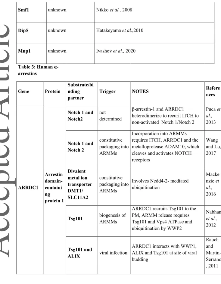
The human genome encodes six -arrestins (TXNIP and ARRDC1-5) (reviewed in (Aubry and Klein, 2013)) (Table 3). All of them, with exception of ARRDC5, harbor PY motifs that may bind WW domains in HECT-type ubiquitin ligases. ARRDC1, ARRDC3, ARRDC4 and TXNIP appear to interact with the Nedd4-like ubiquitin ligases Nedd4 (the ortholog of Rsp5), ITCH, WW-domain-containing protein 1 (WWP1) and WWP2, whereas ARRDC2 interacts with WWP1, but not with WWP2 and ITCH (Nabhan et al., 2010; Zhang et al., 2010; Rauch and Martin-Serrano, 2011; Shea et al., 2012; Qi et al., 2014b; Liu et al., 2016). Noteworthy, the phosphorylation status of the tyrosine resides within the PY motifs of -arrestins may influence their choice of binding partners, since phosphorylation of a PY motif of TXNIP abolished the binding to ITCH but instead favored the recruitment of Src-homology 2 (SH2) domain-containing proteins in vitro (Liu et al., 2016) and in vivo (Spindel et al., 2014). TXNIP phosphorylation also promotes its ubiquitination and proteasomal degradation (Wu et al., 2013; Waldhart et al., 2017) as detailed in the next section. Hence, the regulation of -arrestins by phosphorylation and ubiquitination is not restricted to yeast.
Interactions of -arrestins with the membrane trafficking machinery
Some ARRDCs interact with the ESCRT-I protein Tumor susceptibility gene 101 (TSG101) (Nabhan
et al., 2012; Anand et al., 2018), or with the ESCRT-III-associated apoptosis-linked gene-2 interacting
protein X (ALIX) (Dores et al., 2015), which confers to them specific properties that will be discussed below (see part on Arrestin-mediated vesicles). ARRDC3, ARRDC4 and TXNIP interact with clathrin (Shea et al., 2012; Wu et al., 2013). TXNIP can additionally interact with the AP-2 adaptor complex (Wu et al., 2013).
This article is protected by copyright. All rights reserved.
16 The localization of human ARRDCs was evaluated in various cellular models (Oka et al., 2006b; Patwari et al., 2009; Nabhan et al., 2010, 2012; Vina-Vilaseca et al., 2011; Shea et al., 2012; Han et
al., 2013; Dores et al., 2015). ARRDCs have been found in various compartments along the endocytic
pathway including the plasma membrane, early - and late endosomes and lysosomes, and in cytosolic (or even nuclear) pools. The mechanisms that controls their localization have sometimes been tackled. In the case of ARRDC3, at least one PY motif and the arrestin-like domain are required for its localization to endosomes. Moreover, endosomal localization of ARRDC3 positively correlates with its ubiquitination (Han et al., 2013; Tian et al., 2016). The C-terminal, PY motif-containing region is also important for the proper localization of ARRDC1 (Rauch and Martin-Serrano, 2011). These results suggest that ARRDCs have functions in endocytic membrane protein trafficking, but the following sections will show that they have additional cellular and physiological functions.
Trafficking, signaling and metabolic functions of Thioredoxin-interacting protein
(TXNIP)
TXNIP, also called vitamin D3-upregulated protein-1 (VDUP1) or thioredoxin binding protein 2
(TBP-2), is an extensively studied protein. It is a multi-functional protein involved in the response to various stresses such as oxidative or ER stress, but is also involved in apoptosis, DNA damage response, and inflammation (reviewed in (Yoshihara, 2020)). TXNIP was first identified as a negative regulator of thioredoxin (TRX) proteins (Nishiyama et al., 1999; Junn et al., 2000), small proteins with key roles in redox reactions (reviewed in (Holmgren, 1995)). TXNIP can function as redox protein that negatively regulates TRX via the formation of an inter-molecular disulfide bond between oxidized TXNIP and reduced TRX, that requires two critical cysteine residues of TXNIP that are not present in other human -arrestins (Patwari et al., 2006; Polekhina et al., 2013; Hwang et al., 2014). Interestingly, the structure of TXNIP (amino acids 3-317, bound to TRX) reveals a compact fold composed of arrestin N- and C-domains (Hwang et al., 2014), which is different from the yeast arrestin domains that contain large and probably unstructured insertions (Baile et al., 2019). In TXNIP, the arrestin N- and C-domains adopt an S-shape, which also differs from the typical shape of -arrestins (Polekhina et al., 2013; Hwang et al., 2014; Liu et al., 2016). TRX/TXNIP complexes, called redoxisomes, function in numerous cellular pathways, including transcriptional regulation, cell signaling, apoptosis and inflammation (Qin et al., 1995; Hatai et al., 2000; Meuillet et al., 2004; Zhang et al., 2004; Saxena et al., 2010; Zhou et al., 2010). The unique role of TXNIP in redox signaling pathways has been discussed elsewhere (reviewed in (Yoshihara et al., 2014; Matsuzawa, 2017)). Yet, TXNIP has further TRX-independent functions. Under conditions of hypoxia and exercise, TXNIP is induced and binds to REDD1 (regulated in development and DNA damage responses 1), a negative regulator of mechanistic target of rapamycin (mTOR) signaling (Jin et al., 2011; Qiao et al., 2015). The formation of the REDD1/TXNIP complex promotes the formation of reactive oxygen species (ROS), suppresses mTOR signaling and activates autophagy. How these processes are related to the function of TXNIP as a ubiquitin ligase adaptor remains to be clarified.
This article is protected by copyright. All rights reserved.
17 A key function of TXNIP, which is independent of TRX binding, is the regulation of glucose and lipid metabolism (Patwari et al., 2009). TXNIP negatively regulates glucose uptake by acting as an adaptor for clathrin-mediated endocytosis of GLUT1 and GLUT4, the two major glucose transporters. TXNIP localizes to the plasma membrane, where it directly interacts with GLUT1 and GLUT4 (Wu et
al., 2013; Waldhart et al., 2017) and uses a di-leucine motif in its c-terminal tail to interact with
components of the endocytic machinery (either clathrin or AP-2) (Wu et al., 2013). Additionally, TXNIP suppresses GLUT1 mRNA expression through an unknown mechanism (Wu et al., 2013). The TXNIP-mediated regulation of GLUT1 and GLUT4 endocytosis is tightly coupled to intracellular glucose homeostasis and ATP availability. Upon conditions of energy stress (e.g. ATP depletion), AMPK restores energy homeostasis (reviewed in (Jeon, 2016)) in part by stimulating glucose uptake (Wu et al., 2013), reviewed in (O’Donnell and Schmidt, 2019)). For the efficient uptake of glucose, GLUT1 and GLUT4 must remain at the plasma membrane and therefore TXNIP must be inactivated, which occurs through its AMPK-mediated phosphorylation on Serine 308 followed by proteasomal degradation. Ubiquitination by a HECT-E3 ligase may be involved, since TXNIP binding to ITCH via its PY motifs also leads to its ubiquitination and proteasomal degradation (Zhang et al., 2010; Wu et al., 2013; Liu et al., 2016). The ensuing GLUT1 accumulation at the plasma membrane leads to an increased glucose influx, which restores ATP levels and thus shuts down AMPK signaling. By providing a homeostatic regulation from AMPK to TXNIP, cells control acute and long-term glucose uptake in response to energy stress (Wu et al., 2013). Similar results were obtained upon insulin stimulation. Insulin stimulates glucose uptake primary into muscle and adipose tissues by activating the phosphatidylinositol 3-kinase (PI3K) pathway and its downstream protein kinase AKT, which drives a rapid fusion of GLUT4 storage vesicles with the plasma membrane (Summers et al., 1998). This is amplified by an inhibition of GLUT4 endocytosis through the activation of AKT, which phosphorylates TXNIP at Serine 308 to induce its degradation (Waldhart et al., 2017). Finally, destabilization of the TXNIP transcript in response to remodeling of the extracellular matrix contributes to stabilizing GLUT1 at the plasma membrane. This allows for an increase in both glucose uptake and glycolysis, providing energy to support cell migration (Sullivan et al., 2018).
Consistent with the role of TXNIP in regulating GLUT1- and GLUT4-dependent glucose uptake, several studies showed that TXNIP-deficient mice are hypoglycemic and hypoinsulinemic (Schulze et
al., 2004; Sheth et al., 2005; Oka et al., 2006a; Chutkow et al., 2008), and that disruption of TXNIP in
obese mice improved glucose intolerance (Yoshihara et al., 2010). Furthermore, TXNIP deletion enhances glucose uptake in skeletal muscle and adipose tissue (Parikh et al., 2007; Waldhart et al., 2017). A body of evidence suggests that TXNIP is subjected to complex transcriptional regulation by glycolytic flux (Stoltzman et al., 2008; Cha-Molstad et al., 2009; Yu et al., 2010), hypoxia (Wong and Hagen, 2013; Görgens et al., 2017) or even at the level of RNA stability (Sullivan et al., 2018). Interestingly, TXNIP expression is strongly upregulated in diabetic patients (Parikh et al., 2007).
This article is protected by copyright. All rights reserved.
18 Intriguingly, and contrary to its inhibitory action on glucose transport, TXNIP binds to and stimulates the function of the primary fructose transporters GLUT2 and GLUT5 (Dotimas et al., 2016). Feeding of mice with an energy-rich diet triggers increased fructose uptake in a TXNIP-dependent manner. This involves both an increased expression of GLUT5 and its facilitated trafficking to the cell surface through interaction with the Rab11a GTPase (Shah et al., 2020). Collectively, this indicates that upregulation of TXNIP may contribute to the development of metabolic syndromes.
TXNIP has also been reported to function as a tumor suppressor, and its expression is commonly silenced by genetic or epigenetic events in tumor cells (reviewed in (Zhou et al., 2011b)). The tumor-suppressive functions of TXNIP may, at least in part, be due to its inhibition of TRX (Butler et al., 2002; Chen et al., 2008; Zhou et al., 2011a). However, oncogenic activation of PI3K/AKT signaling promotes glucose uptake in part through the downregulation of TXNIP expression. This suggests that the glucose homeostatic functions of TXNIP (i.e. downregulation of glucose uptake) might also antagonize the Warburg effect and the associated metabolic changes in cancer cells (Hong et al., 2016).
Trafficking functions of other ARRDCs
A clear example of ARRDC-dependent trafficking was described for ARRDC3 which mediates the lysosomal trafficking of the activated GPCR PAR1 (protease-activated receptor 1) via the MVB pathway (Dores et al., 2015). This occurs through interaction of ARRDC3 with ALIX, and ARRDC3 mediates ubiquitination of ALIX through recruitment of WWP2. ALIX ubiquitination may be required for coupling PAR1 to the ESCRT-III complex subunit charged multivesicular body protein 4 (CHMP4) (Dores et al., 2015). Similar to ARRDC3, ARRDC4 may also function as an adaptor for recruiting E3 ubiquitin ligases to GPCRs, as observed for the vasopressin receptor 2 (V2R) (Shea et al., 2012).
ARRDC3 has also been linked to the regulation of adrenergic signaling through its interaction with the GPCR β2-adrenergic receptor (2AR). ARRDC3 interacts via its PY motifs with Nedd4 and mediates association of the latter with activated β2AR (Nabhan et al., 2010; Patwari et al., 2011; Shea et al., 2012). Contrary to early findings (Nabhan et al., 2010), ARRDC3 does not seem to have a direct role in receptor internalization, but rather functions sequentially with -arrestin 2 (Han et al., 2013). First, -arrestin 2 is essential for Nedd4-mediated ubiquitination and endocytosis of β2AR from the plasma membrane (Goodman et al., 1996; Shenoy et al., 2008), and ARRDC3 is later recruited to Nedd4-bound receptors on early endosomes (Han et al., 2013). The interaction with ARRDC3 promotes retention of the β2AR on endosomes by preventing its association with the sorting nexin family member 27 (SNX27), which mediates recycling of β2AR back to the plasma
This article is protected by copyright. All rights reserved.
19 membrane (Temkin et al., 2011; Tian et al., 2016). The crystal structure of the N-terminal lobe of ARRDC3 revealed a large electropositive region (‘basic patch’), which seems to be important for β2AR binding (Qi et al., 2014a), similar to the proposed / putative mechanism of substrate binding in several yeast ARTs (Guiney et al., 2016; Ivashov et al., 2020) and -arrestins (Mayer et al., 2019).
ARRDC1 is involved in the regulation of NOTCH signaling. Interestingly, -arrestins heterodimerize with ARRDC1 to recruit ITCH to inactive NOTCH receptors and mediate their ubiquitination and lysosomal degradation (Puca et al., 2013). Such a hetero-dimerization between members of the - and -subfamilies of arrestins was observed previously (Shea et al., 2012) and increases the combinatorial diversity for the regulation of plasma membrane proteins (reviewed in (Puca and Brou, 2014)).
The roles of other ARRDCs in metabolism
Beyond TXNIP, roles in metabolic regulation were also demonstrated for other ARRDCs (reviewed in (Patwari and Lee, 2012; O’Donnell and Schmidt, 2019)). A genome-wide linkage study for obesity-related genes revealed that ARRDC3 is associated to obesity in male human individuals. ARRDC3 deficiency in mice prevented age-related obesity and increased insulin sensitivity (Patwari et al., 2011; Shea et al., 2012). Also, liver-specific ARRDC3 deletion increased hepatic insulin sensitivity, which was associated with increased insulin receptor protein levels, higher glycogen levels, and lower endogenous glucose production (Batista et al., 2020). Mice with decreased ARRDC3 levels were protected from obesity due to increased energy expenditure. How ARRDC3 is linked mechanistically to the regulation of metabolism is not clear (Patwari et al., 2011). Expression analyses showed that ARRDC3 is subjected to metabolic regulation in various tissues (adipose tissues or skeletal muscles) (Patwari et al., 2011; Batista et al., 2020), and both ARRDC2 and ARRDC3 expression are regulated by fasting/feeding in murine skeletal muscle (Gordon et al., 2019). Co-immunoprecipitation studies revealed that ARRDC3 interacts with the insulin receptor via its C-terminal-tail, which contains the PY motifs but also a tyrosine residue known to be phosphorylated in certain tumors (Batista et al., 2020). How ARRDC3 influences insulin signaling mechanistically will require further studies, but current knowledge suggests that ARRDC3 is part of a negative feedback loop.
Similarly to TXNIP, ARRDC4 overexpression decreased glucose uptake in vitro in primary human skin fibroblasts, suggesting it could also be involved in the regulation of glucose metabolism (Patwari
et al., 2009). Mutational analysis showed that this required the arrestin domain, but not the
C-terminal PY motifs (Patwari et al., 2009). If and which glucose transporters are subject to ARRDC4-mediated down-regulation is currently unknown.
This article is protected by copyright. All rights reserved.
20
ARRDCs in cancer susceptibility and development
A link between the loss of ARRDC3 function and cancer progression was established in various models. Analysis of human tumor samples and different cancer cell lines revealed that ARRDC3 expression is repressed in a subset of breast cancers (Draheim et al., 2010; Cai et al., 2014; Soung et
al., 2014, 2017) and in prostate cancer (Zheng et al., 2017). Part of this downregulation involves
epigenetic silencing of the gene (Soung et al., 2014) or microRNA-based repression (Yao et al., 2016). The mechanism by which ARRDC3 represses tumorigenesis is not yet clear (Nabhan et al., 2010; Arakaki et al., 2018). ARRDC3 overexpression represses proliferation, migration and invasion of cancer cells and mitigates in vivo tumorigenicity in a mouse xenograft model, whereas downregulation of ARRDC3 has opposite effects and leads to a drastic increase in tumor size. These effects are dependent on the presence of integrin (ITG) β4 (Draheim et al., 2010). ARRDC3 was shown to bind ITG β4 and to prevent its recycling from endosomes, instead promoting its Nedd4-mediated ubiquitination and lysosomal degradation (Draheim et al., 2010; Soung et al., 2018). In addition, ARRDC3 may affect breast cancer by controlling degradation of the protease-activated receptor 1 (PAR1) (Dores et al., 2015; Arakaki et al., 2018), and renal cell carcinoma by promoting Itch-mediated degradation of the yes-associated protein 1 (YAP1), a co-transcription factor and activator of the Hippo pathway (Xiao et al., 2018). Similar observations were made in colorectal cancer (Shen et al., 2018). Collectively, these data indicate the importance of ARRDC3 in the control of cancer-related signaling pathways.
New functions of ARRDCs in intercellular communication through
arrestin-mediated vesicles (ARMMs)
ARRDC1 is localized to the plasma membrane through its N-terminal arrestin domain (Nabhan et
al., 2012). Additionally, ARRDC1 contains a PSAP motif that allows its interaction with the ESCRT-I
protein TSG101. The PSAP motif is similar to those found in the “late domains” used by viruses for dependent budding (Rauch and Martin-Serrano, 2011). ARRDC1 also interacts with the ESCRT-III-associated protein ALIX (Rauch and Martin-Serrano, 2011).
Similar to the mechanism of viral budding, ARRDC1-mediated recruitment of ESCRTs to the plasma membrane drives the formation of extracellular microvesicles, named ARMMs (arrestin-mediated microvesicles) (Nabhan et al., 2012; Anand et al., 2018). These microvesicles, also referred to as ectosomes, are distinct from multivesicular body-derived exosomes, since they bud directly
This article is protected by copyright. All rights reserved.
21 from the plasma membrane and lack late endosomal markers (reviewed in (Cocucci and Meldolesi, 2015)). Their budding is driven by the ARRDC1-mediated recruitment of TSG101 and the AAA-ATPase Vps4 to the plasma membrane (Nabhan et al., 2012). ARMM release from the PM is further promoted by the recruitment of the ubiquitin ligase WWP2 by ARRDC1 and its ubiquitination (Nabhan et al., 2012). Interestingly, ARRDC2 and ARRDC3 also interact with the ESCRT-III-associated protein ALIX through a proline-rich region and with WWP1 (Rauch and Martin-Serrano, 2011; Shea et
al., 2012), but so far there is no evidence suggesting that they are capable of ARMM formation.
ARMMs contain ARRDC1 and also some of its cargoes, suggesting an unexpected mechanism by which clearance of transporters from the plasma membrane does not involve internalization of the protein, but rather its release into the extracellular space (Nabhan et al., 2012; Mackenzie et al., 2016)). It is proposed that this may be – in analogy to exosomes - a means to allow intercellular communication (Wang and Lu, 2017). Indeed, active NOTCH receptors are present in ARMMs, and their sequestration requires ARRDC1-mediated ubiquitination by ITCH (Puca et al., 2013; Wang and Lu, 2017). Similarly, the divalent metal ion transporter DMT1 (SLC11A2) is sorted to ARMMs and released into the gut lumen after its ubiquitination. This is mediated by ARRDC1 and ARRDC4, which independently stimulate ARMM formation (Mackenzie et al., 2016). Interestingly, selective targeting to ARMMs may not be limited to membrane proteins, because hepatocellular carcinoma (HCC)-derived ectosomes contain the pyruvate kinase M2 (PKM2) isoform, an enzyme which catalyzes the final step of glycolysis and is highly expressed in cancer cells (Hou et al., 2020). The sorting mechanism of this soluble enzyme to ARMMs is not yet deciphered, but it depends on prior sumoylation as well as on ARRDC1, with whom PKM2 interacts. The ARMM-mediated release of PKM2 from cells might contribute to a remodeling of the tumor microenvironment (Hou et al., 2020).
Interestingly, a recent study used ARMMs as a tool for the packaging and intracellular delivery of macromolecules. The fusion of selected cargoes to ARRDC1 or to WW domains resulted in their packaging into ARMMs and release. Remarkably ARMMs were capable to transfer their cargo into recipient cells, where it carried out its expected biological functions (Wang et al., 2018). Therefore, ARMMs may become an important tool for the intracellular delivery of therapeutic macromolecules.
Conclusion
For decades, researchers realized that the regulation of nutrient transport systems allows cells to adapt nutrient uptake to a changing environment. Yet, the molecular mechanisms that implement context specific re-configuration of these nutrient transporter systems were not fully understood. As pointed out in this review, the role of -arrestins as ubiquitin ligase adaptors could link cellular metabolism with the control of nutrient transporter endocytosis. Since -arrestins are regulated by
This article is protected by copyright. All rights reserved.
22 metabolic signaling and, in turn, also regulate metabolism they may be integral to regulatory loops that maintain cellular homeostasis.
Therefore, it will be essential to continue the effort of understanding how -arrestins integrate into the global metabolic homeostasis in unicellular organisms, but also how they function in tissues of multicellular organism and in the context of pathologies.
Acknowledgements
This work was supported by EMBO/Marie Curie (ALTF 642–2012; EMBOCOFUND2010, GA-2010– 267146) and ‘Tiroler Wissenschaftsfond’ to OS, Austrian Science Fund (P29583) to DT and Agence Nationale pour la Recherche (‘P-Nut’, ANR-16-CE13-0002-01) to SL. JK is a Recipient of a DOC Fellowship of the Austrian Academy of Sciences at the Institute of Cell Biology, Medical University of Innsbruck.
This article is protected by copyright. All rights reserved.
23 References:
Abe, F, and Iida, H (2003). Pressure-induced differential regulation of the two tryptophan permeases Tat1 and Tat2 by ubiquitin ligase Rsp5 and its binding proteins, Bul1 and Bul2. Mol Cell Biol 23, 7566–7584.
Alvarez, CE (2008). On the origins of arrestin and rhodopsin. BMC Evol Biol 8, 222.
Alvaro, CG, Aindow, A, and Thorner, J (2016). Differential Phosphorylation Provides a Switch to Control How α-Arrestin Rod1 Down-regulates Mating Pheromone Response in Saccharomyces cerevisiae. Genetics 203, 299–317.
Alvaro, CG, O’Donnell, AF, Prosser, DC, Augustine, AA, Goldman, A, Brodsky, JL, Cyert, MS, Wendland, B, and Thorner, J (2014). Specific alpha-arrestins negatively regulate Saccharomyces cerevisiae pheromone response by down-modulating the G-protein-coupled receptor Ste2. Mol Cell Biol 34, 2660–2681.
Anand, S, Foot, N, Ang, C-S, Gembus, KM, Keerthikumar, S, Adda, CG, Mathivanan, S, and Kumar, S (2018). Arrestin-Domain Containing Protein 1 (Arrdc1) Regulates the Protein Cargo and Release of Extracellular Vesicles. Proteomics 18, 1800266.
Andoh, T, Hirata, Y, and Kikuchi, A (2002). PY motifs of Rod1 are required for binding to Rsp5 and for drug resistance. FEBS Lett 525, 131–134.
Appadurai, D et al. (2020). Plasma membrane tension regulates eisosome structure and function. Mol Biol Cell 31, 287–303.
Arakaki, AKS, Pan, W-A, Lin, H, and Trejo, J (2018). The α-arrestin ARRDC3 suppresses breast carcinoma invasion by regulating G protein-coupled receptor lysosomal sorting and signaling. J Biol Chem 293, 3350–3362.
Aubry, L, Guetta, D, and Klein, G (2009). The arrestin fold: variations on a theme. Curr Genomics 10, 133–142.
Aubry, L, and Klein, G (2013). True arrestins and arrestin-fold proteins: a structure-based appraisal. Prog Mol Biol Transl Sci 118, 21–56.
Babst, M (2020). Regulation of nutrient transporters by metabolic and environmental stresses. Curr Opin Cell Biol 65, 35–41.