Development of a miniaturized microscope for depth- scanning imaging at subcellular resolution in freely
behaving animals
Thèse
Arutyun Bagramyan
Doctorat en physique Philosophiæ doctor (Ph. D.)
Québec, Canada
Development of a miniaturized microscope for depth-scanning imaging at subcellular resolution in
freely behaving animals
Thèse
Arutyun Bagramyan
Sous la direction de:
Tigran Galstian, directeur de recherche
Résumé
Le fonctionnement du cerveau humain est fascinant. En seulement quelques millisecon- des, des milliards de neurones synchronisés perçoivent, traitent et redirigent les informa- tions permettant le contrôle de notre corps, de nos sentiments et de nos pensées. Mal- heureusement, notre compréhension du cerveau reste limitée et de multiples questions phys- iologiques demeurent. Comment sont exactement reliés le fonctionnement neuronal et le comportement humain ?
L’imagerie de l’activité neuronale au moyen de systèmes miniatures est l’une des voies les plus prometteuses permettant d’étudier le cerveau des animaux se déplaçant librement.
Cependant, le développement de ces outils n’est pas évident et de multiples compromis techniques doivent être faits pour arriver à des systèmes suffisamment petits et légers. Les outils actuels ont donc souvent des limitations concernant leurs caractéristiques physiques et optiques. L’un des problèmes majeur est le manque d’une lentille miniature électrique- ment réglable et à faible consommation d’énergie permettant l’imagerie avec un balayage en profondeur.
Dans cette thèse, nous proposons un nouveau type de dispositif d’imagerie miniature qui présente de multiples avantages mécaniques, électriques et optiques par rapport aux sys- tèmes existants. Le faible poids, la petite dimension, la capacité de moduler électriquement la distance focale à l’aide d’une lentille à cristaux liquides (CL) et la capacité d’imager des structures fines sont au cœur des innovations proposées. Dans un premier temps, nous présenterons nos travaux (théoriques et expérimentaux) de conception, assemblage et op- timisation de la lentille à CL accordable (TLCL, pour tunable liquid crystal lens). Deux- ièmement, nous présenterons la preuve de concept macroscopique du couplage optique en- tre la TLCL et la lentille à gradient d’indice (GRIN, pour gradient index) en forme d’une tige. Utilisant le même système, nous démontrerons la capacité de balayage en profondeur dans le cerveau des animaux anesthésiés. Troisièmement, nous montrerons un dispositif d’imagerie (2D) miniature avec de nouvelles caractéristiques mécaniques et optiques per- mettant d’imager de fines structures neuronales dans des tranches de tissus cérébraux fixes.
Enfin, nous présenterons le dispositif miniaturisé, avec une TLCL intégrée. Grâce à notre système, nous obtenons≈100µm d’ajustement électrique de la profondeur d’imagerie qui permet d’enregistrer l’activité de fines structures neuronales lors des différents comporte- ments (toilettage, marche, etc.) de la souris.
Abstract
The functioning of the human brain is fascinating. In only a few milliseconds, billions of finely tuned and synchronized neurons perceive, process and exit the information that drives our body, our feelings and our thoughts. Unfortunately, our understating of the brain is limited and multiple physiological questions remain. How exactly are related neural func- tioning and human behavior ?
The imaging of the neuronal activity by means of miniaturized systems is one of the most promising avenues allowing to study the brain of the freely moving subjects. However, the development of these tools is not obvious and multiple technical trade-offs must be made to build a system that is sufficiently small and light. Therefore, the available tools have different limitations regarding their physical and optical characteristics. One of the major problems is the lack of an electrically adjustable and energy-efficient miniature lens allowing to scan in depth.
In this thesis, we propose a new type of miniature imaging device that has multiple mechan- ical, electrical and optical advantages over existing systems. The low weight, the small size, the ability to electrically modulate the focal distance using a liquid crystal (LC) lens and the ability to image fine structures are among the proposed innovations. First, we present our work (theoretical and experimental) of design, assembling and optimization of the tunable LC lens (TLCL). Second, we present the macroscopic proof-of-concept optical coupling be- tween the TLCL and the gradient index lens (GRIN) in the form of a rod. Using the same system, we demonstrate the depth scanning ability in the brain of anaesthetized animals.
Third, we show a miniature (2D) imaging device with new mechanical and optical features allowing to image fine neural structures in fixed brain tissue slices. Finally, we present a state-of-the-art miniaturized device with an integrated TLCL. Using our system, we obtain a ≈100µm electrical depth adjustment that allows to record the activity of fine neuronal structures during the various behaviours (grooming, walking, etc.) of the mouse.
Contents
Résumé iii
Abstract iv
Contents v
List of Tables vii
List of Figures viii
Acknowledgements xii
Foreword xiii
Introduction 1
I.1 Imaging, the power of light. . . 1
I.2 Fluorescence, imaging the brain . . . 2
I.3 In vivo, benchtop systems. . . 3
I.4 In vivo, miniaturized systems . . . 5
I.5 Summary of existing limitations . . . 8
I.6 Liquid crystals . . . 10
1 Dynamic control of polarisation mismatch and coma aberrations in rod GRIN assemblies 20 1.1 Résumé . . . 20
1.2 Abstract. . . 21
1.3 Introduction . . . 21
1.4 Coma aberration . . . 22
1.5 Polarization aberration . . . 25
1.6 Discussions and conclusions . . . 28
1.7 Appendix . . . 29
2 Motion-free endoscopic system for brain imaging at variable focal depth us- ing liquid crystal lenses 31 2.1 Résumé . . . 31
2.2 Abstract. . . 32
2.3 Introduction . . . 32
2.4 Materials and methods/experimental . . . 35
2.5 Results and discussion . . . 41
2.6 Conclusion . . . 50
3 Lightweight 1-photon miniscope for imaging in freely behaving animals at subcellular resolution 51 3.1 Résumé . . . 51
3.2 Abstract. . . 51
3.3 Introduction . . . 52
3.4 Design and fabrication . . . 52
3.5 Results and discussion . . . 56
3.6 Conclusion . . . 59
4 Focus tunable microscope for subcellular imaging in freely behaving animals 60 4.1 Résumé . . . 60
4.2 Abstract. . . 61
4.3 Introduction . . . 61
4.4 Results . . . 62
4.5 Discussion . . . 70
4.6 Appendix . . . 72
Conclusion 76 5.1 Summary . . . 76
5.2 Conclusions . . . 79
5.3 Future developments . . . 79
Publication 81
Bibliography 82
List of Tables
I.1 Comparison between the TLCL and the EWL . . . 18 1.1 Values of drive parameters used for obtaining minimal polarization mismatch 26 2.1 Resolution table of three different imaging configurations . . . 44
List of Figures
I.1 Functioning and biological application of the fluorescent proteins . . . 2
I.2 Benchtop 2-photon microscope design adapted forin vivoimaging . . . 4
I.3 Miniaturized 1-photon microscope design . . . 5
I.4 Miniaturized multi-photon microscope designs . . . 7
I.5 Different phases of matter . . . 10
I.6 Different states of matter, thermotropic LCs . . . 11
I.7 Elongated elliptic model of NLCs . . . 12
I.8 Electrical alignment of LC molecules . . . 14
I.9 Tunable LC cell . . . 15
I.10 Resistivity and distribution of the electrical field . . . 16
I.11 Rubbing and the alignment LC molecules . . . 17
I.12 Miniaturised depth scanning devices . . . 18
1.1 Optical power, RMS aberrations and coma versus control frequency . . . 23
1.2 Demonstration of the strong coma correction using 2 TLCLs . . . 24
1.3 Schematic demonstration of one possible TLCL stacking option and polariza- tion mismatch correction . . . 26
1.4 Characterization of the impact of polarization mismatch on the quality of images 28 1.5 Polarimetry set-up . . . 29
1.6 Shack-Hartmann set-up . . . 29
2.1 Schematic representation of the TLCL . . . 34
2.2 The optical probe and experimental set-up . . . 40
2.3 Characterization of the TLCL . . . 42
2.4 Characterization of the optical resolution of the assembly in the water . . . 43
2.5 Imaging granule neurons in the adult olfactory bulb in thick brain sections . . 45
2.6 Imaging dendrites and spines with the optical probe assembly . . . 47
2.7 Motion-free variable-depthin vivoimaging of interneurons in the adult mouse cortex . . . 48
2.8 Motion-free variable-depthin vivoimaging of interneurons in the adult OB . . 49
3.1 Design of the proposed M1S . . . 53
3.2 Optical probe assembly . . . 54
3.3 Schematics of the developed system . . . 55
3.4 Resolution characterization . . . 56
3.5 Imaging with the developed M1S system . . . 58
4.1 Tunable liquid crystal lens design and characterisation . . . 64
4.2 Schematic presentation of the ensemble of the imaging system . . . 65
4.3 Electrical focal shift and characterisation of optical parameters of the mDS1s . 66 4.4 Time-lapse imaging of Ca2+ activity from GCaMP6s-labeled neurons of the
motor cortex . . . 68 4.5 Electrical focus adjustment and time-lapse imaging of Ca2+activity from neu-
ronal structures in the motor cortex of a freely behaving animal . . . 69 4.6 Characterization of the TLCL . . . 72 4.7 Portability test of the mDS1s . . . 73
To my beloved mother, father, sister and my beautiful girlfriend who helped me all the way through this long Journey.
The best way out of a difficulty is through it.
Will Rogers
Acknowledgements
It has been a long journey. Now that I look at all these years that I have spent to complete this work, I clearly see that I could not do it without the help of many people who stand alongside me and supported me. For that, I want to thank them.
First, I would like to thank Professor Tigran Galstian for his support and supervision. This work had plenty of difficulties, repeated ups and downs and without the constant support of Prof. Galstian, nothing would have been possible. His scientific knowledge, rigour and passion are inspiring and I truly consider myself lucky, to be around and to learn as much as I can from the best.
I would like also to thank technicians Éric Rousseau, Stéphane Gagnon and Patrick Larochelle for their help. Their professionalism and wise advises were key to the success of the project.
Thank you for your patience, curiosity and generosity, always giving 100% of what was pos- sible. After all these hours we passed together, all this work, I truly consider you as my friends rather than simple colleagues. Thank you !
I would like to thank my friends in both departments: physics and biology. Here I think- ing about Loïc Tabourin, Louis Bégel, Justin Stevens, Marc-Antoine Boulé, Oleksandr Sova, Anastasiia Pusenkova, Ali Rastqarfarajzadeh, Cedric Bressan, Sarah Malvaut, Archana Gen- gatharan, Delphine Hardy, Bakhshetyan Karen, Ruggiero Francavilla, etc. Thank you guys for your support and everyday energy ! Our laughs, brainstorming moments, dinner evenings, sports and many other activities were amazing. I wish you success in finishing your studies and hope you will find a job that completes you and makes you happy. All the best !!
Finally, but probably most importantly, I would like to thank my family. My father, my mother, my sister and my girlfriend who were of indescribable support. They made me believe in myself, gave me confidence and support so I can pursue my studies, going this far. Each difficulty I had, each time I was down, they were present, giving me the energy and motivation to continue. For their unconditional love and support, I would like to kindly thank them. Love you guys !
Foreword
This thesis presents the development of a miniaturized epifluorescence microscope system with integrated liquid crystal lens enabling electrical modulation of the focal distance. The detailed optical design, fabrication, characterization and the experimental utility of the de- vice will be presented. Various mechanical and optical novelties will also be discussed along- side the potential application ideas. This thesis has five chapters. The first chapter is the Introduction, where I present the subject, existing limitations and our motivation for the development of a miniaturized depth scanning imaging system. The historical review of an- terior miniaturized systems and the description of the liquid crystal lens are also presented to help better understand the upcoming chapters.
Each of 4 chapters corresponds to an article published or submitted for publication dur- ing my doctorate studies. Note that the original text content has been preserved while the numbering of legends of the equations, tables, figures and references has been adapted to the layout of the thesis. This work was carried out under the supervision of Tigran Gals- tian at the Center for Optics, Photonics and Laser at Laval University. The contribution of collaborators and co-autors of the papers will be explained.
Chapter 1:A. Bagramyan, T. Galstian, "Dynamic control of polarization mismatch and coma aberrations in rod-GRIN assemblies,"Optics Express, OE27, 14144–14151 (2019).
This paper presents the development of small diameter liquid crystal lens adapted for our applica- tion. The utility of the lens, correction of optical aberrations and compensation of the polarisation miss-match are presented. My role in this publication was to design-build-characterize different liquid crystal lenses under the supervision of professor T. Galstian.
Chapter 2: A. Bagramyan, A. Saghatelyan, T. Galstian, "Motion-free endoscopic system for brain imaging at variable focal depth using liquid crystal lenses,"Journal of Biophotonics10, 762–774 (2017).
Prior to the miniaturisation, the developed liquid crystal lens was integrated and tested in a macro- scopic imaging system. The experimental utility of the lens was demonstrated by electrically ad- justing the focal plane position in the brain of anesthetized, head restrained animals. My role in this publication was to design-build-characterize, the optical set-up under the supervision of
Professor T. Galstian. I also performed the surgery, imaging of the animals under the supervision of professor A. Saghatelyan. All authors participated in data interpretation and writing of the manuscript.
Chapter 3:A. Bagramyan, "Lightweight 1-photon miniscope for imaging in freely behaving animals at subcellular resolution,"IEEE Photonics Technology Letters32, 909–912 (2020).
This publication presents the development of an optimised miniaturized imaging system with fixed focal plane. The novel mechanical (weight, size) and optical features (resolution, magnification) of the device are described. My role in this publication was to design-build-characterize the novel miniaturized device. I also tested the imaging capacity of the system, analysed the data and wrote the Manuscript.
Chapter 4: A. Bagramyan, L. Tabourin, A. Rastqar, F. Bretzner, T. Galstian, "Focus tunable microscope for subcellular imaging in freely behaving animals,"submitted, Nature Methods, 2020.
This paper presents the integration of the liquid crystal lens device in the 2D system described earlier. The experimental utility of the final system is demonstrated by electrically adjusting the focal plane position in the brain of freely behaving animals. My role in this publication was to design-build-characterize the imaging capacity of the miniaturized device. Alongside A. Rastqar (master student in Neuroscience) and L. Tabourin (doctorate student in Physics) I performed the surgery and imaging of the animals under the supervision of Professor F. Bretzner. All authors participated in data interpretation and writing of the manuscript.
Introduction
I.1 Imaging, the power of light
Optical imaging is one of the most established methods for the studying of small biologi- cal features, invisible by the naked eye. Behind this success are the different properties of the light allowing to efficiently study the structural and functional characteristics of living organisms [1;2].
First, light is non-invasive. In contrast to classical surgical methods, it allows investigation in absence of direct physical contact with the object being imaged [3]. In biomedical appli- cations, this is crucial, as it allows to study living specimens in their natural environment with minimal modification of their original behaviour.
Second, light is diverse. The vast spectrum of light offers multitude of wavelengths, sub- stantially broadening the experimental repertoire and adding to the diversity of light-tissue interactions [4].
Third, light is precise. The use of optical filters, gratings, etc. allows to choose, very pre- cisely, among the different spatial characteristics (amplitude, polarisation, etc.) of the light, adding to the specificity and accuracy of light-tissue interactions.
Finally, light is fast. Biological events, such as neuronal activity, for instance, are quite rapid [5] and therefore require the use of tools with a high sampling rate.
Until recently, the human eye was the only tool allowing to capture the light, observe and analyze objects. Thus, the application repertoire of optical imaging was defined by the lim- ited capacity of human visual system (eye, brain) that was unable to take full advantage of light’s potential [6;7].
In the past decades, revolutionary advances in optics [8;9] (development of various light sources, lasers, etc.), electronics [10] (powerful computers, high-speed data storing devices, etc.) and optoelectronics [11] (camera sensors (complementary metal oxide semiconductor (CMOS) or charge-coupled devices (CCD)), photo-detectors, etc.) have contributed to the rapid growth of the potential applications in the field of light microscopy [11]. The simple
pictorial visualization of the human eye has been surpassed and today, it’s possible to effi- ciently digitize, quantify and record, for a long period, the dynamic behaviour of light and also, its interactions with the living tissue.
I.2 Fluorescence, imaging the brain
The development of the first genetically modified green fluorescent protein (GFP) [12;13], was another major breakthrough in the field of biomedical imaging. Neuroscience was par- ticularly interested by this new protein that allowed the targeting of specific neuronal struc- tures (FigureI.1c-d) within the ensemble of brain cells [14].
The operation of GFP molecules is demonstrated in the Figure I.1a. By illuminating the protein with the proper wavelength of light, it’s possible to force transitions from lower, to higher energy levels [15;16]. The return of the electron to the original orbital (desexcitation) generates light with a longer wavelength and lower energy (Figure I.1a,b). The difference between excitation and emission peaks is described by theStokes shift[17] (FigureI.1b).
Figure I.1: Functioning and biological application of the fluorescent proteins. a.
Schematic demonstration of electronic transition andb.corresponding absorption and emis- sion (fluorescence) spectra [15;16].c.Fluorescent neuron expressing GFP.d.Color diversity of fluorescent proteins [18].
By using the correct imaging configuration (e.g., epifluorescence, FigureI.3b), it’s possible to take advantage of Stokes shift to efficiently monitor neurons. To do so, GFP is inserted
within the neurons by modifying animal’s genetics (transgenic mice [19]) or by viral injec- tion [14]. Both methods will force the host brain cells to fabricate the fluorescent protein.
Once assembled, the fluorescent protein will brighten the neurons in presence of the proper excitation light (FigureI.1c). The collection, filtering and focusing of the emitted light (Fig- ure I.1a-b) will allow to visualize the target population of neurons (expressing GFP) and to contrast them from the ensemble of brain cells that are invisible in the absence of the fluorescent proteins [13].
Different variants of GFP (e.g., voltage-sensitive [20], pH-sensitive [21], calcium sensitive [22], etc.) have been developed in recent years allowing to investigate the functional charac- teristics of neurons by spatio-temporal imaging of their intra- and inter-cellular processes.
The genetically encoded calcium indicators (GECIs) like GCaMP for instance, have the par- ticularity to activate (by undergoing conformational changes) their fluorescence in the pres- ence of calcium ions (Ca2+) [22; 23]. When neurons send information along an axon (to other neurons) the difference of electrical potential across their membrane changes drasti- cally and triggers the opening of various ions channels (K+,N a+,Cl−, etc.) [24].Ca2+is one of the ions that are transported through the membrane of neurons during the activation [25].
Triggered by the presence of calcium, the fluorescence of GCaMP increases (proportionally to the amount ofCa2+molecules) and allows to monitor (≈25-30fps) the level of activation within the ensemble of the neuronal structure (soma, dendrites, axons, spines) [26].
I.3 In vivo, benchtop systems
The combination of previously mentioned progress in electronics, optics and genetics had a big impact in the fields of neuroscience and biophotonics with a rapid growth of various imaging applications and tools. The development of in vivo imaging devices (from latin :
"within the living") is particularly attractive here, allowing to study the living tissues in their natural environment, in our case, the brain [27;28].
First in vivo systems were based on conventional benchtop (macroscopic) epifluorescence set-ups with adapted mechanical holders to ensure the immobilization of the animal’s head (Figure I.2) [29;30;31;32;33]. These systems were able to image and record the neuronal activity within the living brain at relatively high frame-rates [27]. The use of small mag- nification objectives [29] allowed to increase the field of view (FOV) giving access to larger imaging zones with a higher density of neurons. Another advantage of benchtop micro- scopes was their flexibility in terms of the imaging configuration allowing to alter between the regular wide-field [34] and more advanced, high-resolution approaches (e.g., 2-photon [30], confocal [35], etc.) . Thus, these systems were capable to image with cellular [33], and, subcellular [30] resolutions allowing to study the main body of the neurons (soma) and also the small neuronal features (dendrites, axons, process, spines, etc.).
Figure I.2: Benchtop 2-photon microscope design adapted forin vivoimaging[33].
However, there were limitations from the behavioural perspective; first benchtop systems were adapted for deeply anaesthetized subjects. Even though certain investigations could be addressed here [27], the efficiency and the resemblance with real, awake animal mod- els remained questionable [36; 37;38]. Moreover, the study of important behaviours (e.g.
running, grooming, etc.) was impossible in anesthetized subjects. To solve these issues, re- searches developed advanced conditioning protocols allowing to habituate animals to the head fixed situation [39]. This was an important step forward allowing to work with con- sciously awake animals, able of responding to the external simulations (e.g., visual [40], au- ditory [41], etc.). In recent years, multiple studies emerged proposing advanced variations of benchtop systems for studying more complex situation, such as virtual reality [40; 31]
(figureI.2).
Limitations. Although promising, benchtop systems are expensive, hardly accessible, com- plex to build and suffer from major limitations caused by the head fixation of the animal.
Even though certain biological questions are studied using this method [27], the absence of the vestibular cues (induced by the rotation of the head) is susceptible to affect the naviga- tion of animals and also impact various decision making choices [42;43]. The head fixation is also known to cause neck pain, stress and fatigue that affect the behavior and modify the original activation pattern of the neuronal structures [44;45]. Finally, multiple behaviours (e.g., cognition, foraging, social interaction, mating, fighting etc.) in rodents are naturally expressed through the head movement and require investigation in the freely moving sub-
jects [46].
I.4 In vivo, miniaturized systems
To solve limitations regarding the head restrained model, portable miniaturized microscopes have been developed for neuroscience applications [28;46]. These systems are usually tiny and lightweight enough to be carried by small animals such as birds, rats and even mice. The first prototype was reported in 2011, demonstrating the ability to image multiple neurons within the brain of a freely behaving mouse [47]. Multiple adaptations of such portable devices have emerged over the last years [46; 48]. Hereafter we chose to separate them in two categories depending on their capacity to distinguish small neuronal features.
I.4.1 Imaging with cellular resolution
The first category is composed of miniaturized microscopes withcellularresolution adapted for the imaging of large structures, such as the soma (neuron’s body,≈15-50µm in diameter) [46;48]. To access more neurons and to have a better representation of the neuronal network, small magnifications (×4-×10) and large field of views are preferred here. These systems are particularly attractive for the characterization of the functional role of a specific population of cells within a large region of interest (ROI) [28].
Figure I.3: Miniaturized 1-photon microscope design. a. Schematic representation of a system adapted for the imaging of brain surface (left). Configuration allowing to image deep regions of the brain (right). b. Miniscope system developed at the University of California, Los Angeles (UCLA) [49;46].
Short technical description. This category is mainly composed of wide-field imaging de- vices [50;49;51;52] with epifluorescence imaging configuration (FigureI.3a). The excitation light sources are standard LEDs [49] (emitting in visible spectrum range) and are placed di- rectly on the miniaturized system or brought to the optical pathway by means of large core
(≥ 200µm) multi-mode optical fibers [53]. The presence of chromatic filters and dichroic mirror ensures the proper synchronisation with the excitation and emission wavelengths of the neurons marked with GFP (or other fluorescent protein, e.g., GCaMP). Achromatic lenses are usually used to guide and focus the light on the pixelized sensors such as a CMOS or a CCD. As demonstrated in FigureI.3a, the use of GRIN optical probes can enable access to deep regions of the brain (1-6 mm from the surface) [54].
Limitations. Although promising, important limitations within this category of imaging devices significantly reduce their efficiency and also limit their application repertory.
First, the lack of depth scanning mechanisms limits these systems to two dimensional (2D) imaging. This drastically reduces the efficiency of surgical procedures and also affects the quality of calcium recordings [55]. Trying to address this issue, some tunable miniaturized systems have been developed by private companies [56; 57]. The system from Doric lens [57], for instance, reports a≈ 300µm focal shift capability. However, this depth scanning system is heavy and once mounted can weight up to≈4.5±0.5 g (≈3.0 g the miniscope,≈ 0.5 g the implant,≈1g the screw(s) and dental cement). Latter "load" is significantly higher compared to the conventionally established≈10% of the animal’s weight (≈2.5 g) [28;46].
Thus, the use of such device with small animals, such as mice, is doubtful. Another depth scanning 1-photon system from Inscopix [56] seems to be significantly lighter, but to the best of our knowledge, similar to Doric’s system, its depth scanning ability has never been reportedin vivo. Consequently, the practical effectiveness of existing depth scanning devices isn’t obvious.
Second, the development of compact 1-photon devices is challenging in terms of optical and mechanical engineering. Unfortunately, available cellular imaging systems remain heavy, bulky and difficult to carry for small animals [27]. This has a negative impact from multiple perspectives. For instance, it limits the experimental repertory to adult subjects only (usu- ally bigger and stronger) making almost impossible the work with younger (smaller) animals that are particularly interesting for the study of brain development paradigms (e.g., neuro- genesis : cellular division, migration of stem cells, integration within the existing network, etc.) [58]. In addition, the exaggerated weight and size (FigureI.3b) of these imaging devices can cause stress, affect the head movement of the animal, modify the natural navigation and decision making choices [45].
Finally, the cellular resolution of available 1-photon systems isn’t sufficient to image and record from finer structures of neuronal circuitry such as axons, dendrites, spines [46].
These subcellular compartments are wherein all neuronal processing and the better under- standing of their function appears to be the next step toward a better understanding of the brain [59].
I.4.2 Imaging with subcellular resolution
The second category is composed of miniaturized microscopes withsubcellular resolution adapted for the imaging of finer neuronal structures [60; 61; 62; 63; 64; 65]. To increase the quality of calcium recording from small neuronal features, high magnification and high pixels/object ratios are required here. These systems are particularly attractive for the char- acterization of the functioning role of dendrites, processes (e.g., filopodium), axons, spines, etc. within various brain regions of freely behaving animals [59].
Figure I.4: Schematic representation of miniaturized multi-photon microscope designs.
a. System with internal MEMS placed directly on the miniaturized device. [62]. b. Sys- tem with an external scanning approach. Electrowetting lens (EWL) is placed directly on the miniaturized device. [66]. c. Scanning system based on the mechanical displacement (rotation) of the optical fiber [67].
Short technical description. In terms of optical configuration, this category is more diver- sified. It’s composed of miniaturized 2-photon [66; 62], confocal [65] and fiber-scanning devices [67; 60; 61] (FigureI.4) with different optical designs and components. The com- mon difference compared to the previous category is the choice of the light source and of the optical detector. In this kind of approach, coherent sources, such as lasers (continue or pulsed) are considered. The use of near-infrared excitation wavelengths (FigureI.4a) enables longer penetration capacity within the living tissues [68]. The optical sensors are standard photo-detectors with a single photosensitive area. To form an image in the absence of pix-
elated light detector, different scanning strategies are considered. Micro-electromechanical systems (MEMS) are usually used and commonly placed outside the miniaturized system due to their noticeable size and weight (figureI.4b) [66]. Recent studies have also reported MEMS that were placed directly on the miniaturized device (Figure I.4a) avoiding the use of multimode fiber bundles [62]. Finally, some scanning technics consider the mechanical displacement of the imaging optical fiber by means of mechanical actuators [67] (figureI.4c).
Limitations. Also within this category, there are important limitations that reduce the effi- ciency and the experimental capability of subcellular imaging devices.
First, the depth scanning isn’t obvious here. Even though the use of electrowetting lenses (EWL) was recently reported [66; 65], major drawbacks disadvantage this approach. For instance, currently available EWLs [69] have low optical power, large clear aperture, high operational voltages, big size and weight, etc. A more detailed description of these limita- tions is presented in the upcoming section.
Second, the available miniaturized subcellular devices are complex to build (mechanically and optically), expensive (pulsed light sources, MEMS, etc.) and hardly accessible. The use of custom components (e.g., dispersion corrected fibers [62], multicore fiber bundles [66;65], etc.) usually restrains the reported techniques to the original research groups which detain the manufacturing know-how.
Third, devices within this category suffer from excessive weight and size that has negative impact on the animal’s behaviour (see the previous section).
Fourth, the optical design of these systems requires the use of large diameter optical probes (more then 1.5 mm) that are invasive and harmful to the animal. The surgical procedure usually requires brain aspiration that can have major undesirable impact on the animal’s health and also impact the functioning of the brain [70].
Finally, the use of complex scanning methods and the presence of coherent light sources disables the compatibility with future wireless approaches [51;46].
I.5 Summary of existing limitations
There are different drawbacks among the existing portable miniaturized devices. Three of them are common to both categories (cellular and subcellular) and thus are fundamental issues within this application.
The first majorissue is the lack of an efficient depth scanning element enabling the electrical modulation of the focal distance. This challenge is difficult to address due to the severe physical, electrical and optical criterias : small size, lightweight, low power consumption, low operational voltages and high optical power (in the order of hundreds of dioptres).
To the best of our knowledge, no device reported in the literature is able to satisfy the en- semble of these requirements. The closest candidate is the commercially available EWL from Varioptics (now Corning [69]), but as discussed previously, this alternative fails in different aspects. First, EWLs have low optical powers that range from -5 to 15 dioptres. As will be described in the chapter 1, the direct coupling with GRIN probes (FigureI.3a, right) requires much higher optical powers, in the order of hundreds of diopters. In the absence of GRIN lenses, deep regions (e.g., hippocampus, sub-ventricular zone, etc. [71]) remain inaccessible and EWL systems can only be used to image the surface of the brain. Second, the size of the clear aperture (CA) of EWLs is another limitation. The smallest CA available (1.6 mm) is 3 times larger compared to GRIN’s diameter (0.5 mm). This difference drastically reduces the efficiency of the direct optical coupling between the 2 elements (GRIN and EWL). Third, EWLs are quite demanding in terms of the operational voltage (can reach up to 60 Volt). This is a major issue for future wireless approaches that require low power consumption [51]. Fi- nally, the mechanical characteristics (weight and size) of EWLs are borderline acceptable, furthermore complicating the use of this element within the miniaturized devices.
The second majorlimitation within the existing miniaturized cellular and subcellular de- vices is their weight and size. As mentioned previously, the available tools are big and bulky and usually hardly compatible with small animals such as young or elder mice. Even-though these systems are often used in such subjects, their impact on the animal’s condition (health) and behaviour is a problem that must be addressed in the future.
The third major limitation is that the imaging of fine neuronal structures has been so far exclusively reserved to the subcellular systems [60;61; 62;63; 64; 65]. From the practical perspective this is problematic because of the complexity, price and inaccessibility of the devices within this category. Therefore, the ability to image fine structures with 1-photon imaging system (simple, affordably and accessible) would be a big asset allowing an easier access and imaging of fine neuronal structures within freely behaving animals.
To solve these limitations, we used liquid crystal technology that is well established within the miniaturized application (e.g., cellphone cameras, medical endoscopes, etc.) [72;73;74;
75; 76]. To solve the first major limitation within the existing devices, we have developed a custom liquid crystal (LC) lens [77] adapted to the miniaturized microscopes applica- tions [28]. To solve remaining limitations, we have developed a lightweight miniaturized 1-photon system that integrates the LC lens and enables depth imaging of subcellular struc- tures [53].
I.6 Liquid crystals
I.6.1 Historical
The discovery of an intermediate state (other than the crystalline solid or the isotropic liq- uid) emerged in the 19th century, at the time when only three states of matter (solid, liq- uid, gas) were known. In 1888, while heating cholesterol benzoate, the Austrian botanist Reinitzer was surprised to observe two melting points [78]. Looking for an explanation to this strange phenomena, he contacted the physicists Otto Lehmann writing him the follow- ing letter :
Figure I.5: Different phases of matter. At the 2nd melting point, the cloudy liquid (liquid crystals) become isotropic, almost transparent [79].
The two substances (cholesterol esters) are the site of such beautiful and strange phenomena that they should, I hope, also interest you in the highest degree. [. . . ] The substance has two melt- ing points, if we can put it that way. Around 145.5°C it melts and forms a cloudy but completely fluid liquid, which around 178.5°C suddenly becomes completely transparent. By cooling it, we see the blue and purple colors appear which fade quickly by having the opaque substance (like milk) but still fluid. Continuing the cooling, the blue and purple colorings reappear and, immediately after, the substance solidifies, forming a white crystalline mass. [. . . ] The cloudiness that appears when heating is due not to the crystals but to the liquid that forms the oily streaks ..
Lehmann decided to study this strange phenomenon and using his specialized microscope, he was able to study the LCs under the polarized light at variable temperatures. A year later (1889), Lehmann published an article entitled "Uber Fliessende Krystalle" which turns out to be the first publication introducing the concept of "liquid crystal" [80]. The publication sparked huge interest at the time, but the low practical potential has quickly drawn away
from the attention from LCs.
In 1962 (73 years later), liquid crystal materials regain a new, this time, commercial interest from RCA Laboratories who focused on the development of flat panel electronic displays [81]. Within 4 years, the company was able to make a mixture of nematic liquid crystals that was stable at room temperature [82]. This was a fundamental advancement that al- lowed to fabricate the first practical display devices [83]. Since that moment, different LCs (e.g., N-4-Methoxybenzylidene-4-butylaniline, 4-Cyano-4’-pentylbiphenyl, etc.) have been developed, continuing to improve the practical utility and popularity of LCs within com- mercial applications [84]. Thus, from 1980 to 1990, the liquid crystal technology became widely spread and could be found in displays, watches, large size television screens (LCDs), etc. [76;85].
I.6.2 Types of LCs
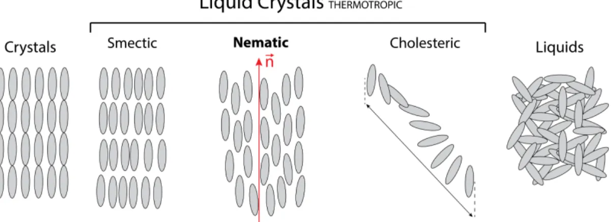
Liquid crystals can be divided into two groups: thermotropic and lyotropic. The mesophase state (between solids and liquids) of lyotropic LCs [86] occurs upon the addition of a solvent (typically water), whilst the thermotropic LCs [87] are mainly dependent on the tempera- ture. Within our application, we will focus on thermotropic LCs that can be sub-classified into calamitic (C-type) and discotic liquid (D-type) crystals depending on the shape of the molecules [88]. C-type LCs are particularly interesting due to the diversity of their self- assembled configurations that can be either nematic, smectic and cholesteric phases. Each of these phases has different properties such as the anisotropy, ferroelectricity, chirality, etc.
[89].
Figure I.6: Different states of matter. In the middle, different types of thermotropic LCs : smectic, nematic, cholesteric.
I.6.3 Nematic LCs
Nematic LCs are the most simple, the most studied and the most used in different display and imaging applications [85]. As demonstrated in the FigureI.6, the distribution of the
center of mass of nematic LC molecules is random, but the direction of these molecules is common. The average direction of these molecules can be represented by the unitary vector
~
n (Figure I.6), called the director [89]. The uniform orientation of molecules within the volume results in the formation of liquid "monocrystals".
However, if external forces are applied to the volume of molecules then, the orientation of the director may vary [89]. This can occur in different situations. For instance, during the manufacturing of the LCs cells, the presence of small impurities can induce variable forces within the cell that will produce undesired domains separated by abrupt changes of director orientations, called disclinations [90]. However, other type of forces (within the volume) can be induced in a desired way, for instance, by applying an external electrical field. This is the case in multiple applications that require orientation gradient of molecules, that is particularly attractive due to the anisotropic properties of LCs [72;91;92].
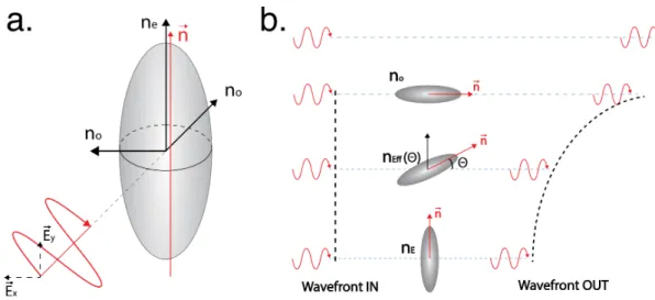
I.6.4 Optical anisotropy of LCs
LCs have different properties (e.g., refractive index, conductivity, absorbance, etc.) for differ- ent directions of light polarization (anisotropic materials). In our case, we are interested in the optical anisotropy of nematic LCs (NLCs, FigureI.6) [89]. These materials are uni-axial (with positive and negative anisotropy) and the molecule of NLCs with positive anisotropy is often represented by the elongated elliptic model (I.7) :
Figure I.7: Elongated elliptic model of uni-axe LCs. a. Uni-axial calamitic (C-type) LCs.b.
Temporal delay in function of the orientation of molecules.
As demonstrated in the FigureI.7a, the two perpendicular components (E~x, E~y) of the po- larized light beam (entering the NLCs layer) will see 2 different refractive indexes. The component (E~x) that is perpendicular to the director (~n), will perceive an ordinary refrac- tive index (n⊥) while the parallel component (E~y) will see the extraordinary refractive index component (nk) [89]. The speed of light ("v" in matter) being dependent on the refractive
indexn (v = c/n, "c" is the speed of light in vacuum), a relative phase shift 4φ will occur between the components E~x and E~Y. Consequently, the output polarisation will change to circular, elliptic or linear depending on the value of4φEq.(1) :
4φ=2πd4no
λ (1)
where d is the thickness of NLC ;λis the wavelength of the light beam;4nois the difference between the extraordinary and ordinary refractive indexes.
The temporal delay induced by the NLCs can also be used to modify the wavefront of light [93]. For our application, particularly interesting is the orientation of NLCs, demonstrated in the Figure I.7b that can change the shape of incoming wave-front. This occurs due to the variation of the angleθbetween the wave-vector of light and the director of NLC. This reorientation gradient of NLC molecules changes of the refractive index Eq.(2) [92] :
ne(θ) = n⊥×nk
q
n2k×cos(θ)2+n2⊥×sin(θ)2
(2)
wherenk andn⊥are the refractive indexes that are parallel and perpendicular to the director~n;θ is the angle between the director~nand the axis of propagation of light.
As demonstrated in FiguresI.8andI.9, the application of a non-uniform electrical field can generate the desired (e.g., spherical or parabolic, FigureI.10) wave-front (see next section).
Thus, the dynamic control of the orientation of NLC molecules is a powerful tool enabling control over the convergence of the light [92;93].
I.6.5 Electrical alignment of NLCs
As mentioned previously, the reorientation of NLC molecules can be used to shape the wave- front of the light and, for obvious reasons, this modulation must be electrical.
Apart from the anisotropic optical properties, the NLC molecules have another interesting feature : anisotropic polarisability at lower frequencies (e.g., 1 kHz) [92]. In the presence of an external electric field, the movement and the displacement of electrons (usually stronger in the longitudinal direction of molecules) results in the induction of electric dipoles. The so called "dielectric torque" (Eq.(3)) will now tend to align the molecules in the direction parallel to the field (FigureI.8) [94] :
Γ =D~×E~ (3)
whereΓ is the dielectric torque;D~ is the electrical induction;E~ is the electric field.
If the amplitude of the electric field is higher then the intrinsic elastic forces of the NLCs (that are responsible for the alignment of molecules), re-orientation of molecules will occur [92]. The minimum voltage necessary to initiate rotation of molecules is defined as the threshold voltage [72] and its value depends on the type of NLCs. Thus, the anisotropic mobility (polarisability) of electrons allows to use an external electric field to reorient the NLC molecules and to electrically modulate the extraordinary refractive index.
Figure I.8: On the left : LC molecules in absence of external electrical field. On the right : electric induction, formation of LC dipoles and alignment of LC molecules by the electrical field.
To generate a lens, NLC molecules must be reoriented in a quasi-parabolic way to obtain the desired refractive index profile of the GRIN lenses, demonstrated in the FigureI.10[94]. To achieve this, a custom NLC cell must be assembled with specific parameters (e.g., shape of the electrodes, resistivity of the cell, etc.), adapted for our application [92].
I.6.6 Tunable LC lens (TLCL)
The LC cells are usually composed of two face-to-face glass substrates with different thin layers (Figure I.9a) [92]. Each of these layers has an important role that will be described hereafter.
Distance between the substrates can be controlled by using microscopic spacers. The pres- ence of electrodes on the internal sides (toward the NLC molecules) of substrates allows to generate an electric field that reorients molecules. The shape of the electrode can differ de- pending on the application allowing to generate the desired distribution of the electric field within the volume of NLCs [72;92].
Electrode shape and distribution of the electric field
The combination of uniformly distributed electrode on substrate 1 (buttom) and the circular ring-shaped electrode on substrate 2 (top), allows to generate asphericaldistribution of the electric field (Figure I.9d) [95; 96; 97]. The corresponding spherical reorientation of NLC molecules will occur and will allow to generate a lens like index distribution of the refractive
Figure I.9: Tunable LC cell. a. Schematics of the design. b. Electrical circuit (of the TLCL) that acts as a low pass filter. c. Frequency dependence of the circuit in b. d. Distribution of the electrical field within the LC cell.
index (FigureI.10). The optical power (OP) of the generated lens (Eq.(4)) will be inversely proportional to the square of the radius of the electrode ring [92].
OP =2L4n
r2 (4)
where OP is the optical power; L is the thickness of the NLC layer;4nis the difference between the refractive indexes values in the center and the periphery of the lens.
Another important feature within the design of the NLC cell is the capacity to control the distribution of the electrical field by modulating the input voltage frequency. To better understand the concept, the NLC cell is usually modeled as a distributed electrical RC cir- cuit (FigureI.9b) with a parallelly displaced capacitor and resistance that are in series with the resistance Rhl (described in the upcoming section) [98]. The following circuit design is known to be an electrical low pass filter that discriminates the high frequencies (Figure I.9c). By carefully selecting the correct resistor(Rhl)-capacitor(C) combination, it’s possible to precisely control the frequency dependence of attenuation. Within our application, the≈ 1 kHz frequency is usually used as the cut-offfrequency (I.9c) with the amplitude of higher frequencies being (gradually) attenuated [92]. This frequency dependent transmission de- crease can be used to create a gradient of spatial distribution of the electrical field that will change the orientation of NLC molecules (FigureI.10).
As mentioned previously, to achieve a good spherical distribution of the electric field within the cell, it’s important to define the correct combination of capacitor (C) and resistor (Rhl) (FigureI.9) [98]. The value of the capacitor (C) is established by the type of the NLCs (e.g., E7, ML6080, etc.) while the resistivity (Rhl) can be controlled by carefully adjusting the thickness and concentration of different partially oxidized metallic layers deposited on the substrate (FigureI.9a) [99].
Resistivity, hiden layer (HL)
The resistivity Rhl of the NLC cell is a key parameter allowing to optimise the distribution of the electrical field (E~z) with the cell [99]. IfRhl is too low, the fieldE~z will be too strong and will never reach the zero amplitude at the center (FigureI.10a, #1) of the lens. In the opposite case, if theRhl is too high, the field will only be present at the edges of electrodes and will not propagate towards the center of the cell (Figure I.10a, #2) [92]. Therefore, the value of the resistivity must be chosen carefully to obtain the optimal distribution presented on the FigureI.10a, #3.
Figure I.10:Resistivity and the distribution of the electrical field. a.TLCLs with different resistivities. Case 1: too low; Case 2: too high; Case 3: optimal; b. Distribution of the refractive index within the LC cells in a.
Even-though several studies have been reported in the literature [99;100; 101], computa- tional simulations remain complex and to the best of our knowledge, no theoretical model can predict the exact value of the resistivity. Thus, this parameter is often determined em- pirically (by observation or experience rather than theory).
Rubbing
To create a stable and reversible reorientation of molecules within the NLC cell, it’s useful
to have a rather strong anchoring on the surface of cell subtracts. As a general rule, planar anchoring (~nparallel to the surface) is obtained by rubbing a thin layer of polyimide (figure I.9a, figureI.11) with NLCs tending to align in the direction of rubbing [102;103]. Having an anchor point helps to control the alignment within the volume and also to stabilizes the molecules at their equilibrium position (in the absence of the field) [92].
Figure I.11: Rubbing and the alignment LC molecules. a. No voltage is applied. Rubbing tends to align molecules (director being parallel to the direction of the rubbing). In the absence of rubbing, the orientation of molecules is random (right). b. AC square voltage is applied to the cell. Rubbing helps to generates a smooth reorientation gradient of molecules (left). In the absence of rubbing, different LC domains are formed (right). The "disclination"
(separations between LC domains) are indicated with black lines.
In the absence of the electric field, anchoring creates a restoring torque that forces the molecules to return to their initial orientation (Figure I.11a). This initial alignment (of- ten parallel to the substrate) enables gradual and uniform re-alignment in the presence of the external electric field [102;103]. In the opposite scenario (no rubbing) molecules will align arbitrarily (in the energetically favorable direction) on the surface of the substrate and also within the volume (FigureI.11). When an electrical field will be applied, the forced re- alignment of molecules (from random initial positions) can create separate domains, called disclinations (FigureI.11b) [90]. Latter is known to increase optical aberrations and scatter (FigureI.11b) that degrades the focusing capability of the NLC lens.
I.6.7 Alternative technologies
The optimisation of the NLC cell (e.g., diameter, resistivity, etc.) allowed us to build a cus- tom tunable LC lens (TLCL) adapted for miniaturized microscope application in a freely behaving animals [73;77]. The detailed description of the parameters can be found in chap- ters 1, 2, 3 and 4.
Compared to our TLCL (FigureI.12), the conventional mechanical systems are bulky, costly and fragile [92]. The voice coil motors are widely used in mobile devices, but they are com-
Figure I.12: Miniaturised depth scanning devices. a. EWL lens from CORNING [69]. b.
Tunable liquid crystal lens developed at Laval University.
posed of more than 100 pieces that must be manually assembled. Their cost (in high volume) is significantly higher (by an order of magnitude). They also have ringing, hysteresis that complicates the autofocus search and they are driven by current and their power consump- tion is about 100 mW [92].
Relatively small liquid lenses can also be fabricated using the electrowetting approach [104].
The comparison between the TLCL and its closest commercial EWL alternative from CORN- ING [69] is presented in TableI.1:
EWL TLCL
Optical power (D) -5 to 15 160
Clear aperture (mm) 1.6 0.5
Weight (g) 1 0.1
Dimensions (mm) 7.7 x 7.7 x 2 5.5 x 5.5 x 0.8
Voltage (V) 60 3 to 5
Delta shift (µm) 10 100
Table I.1: Comparison between the TLCL and the EWL.
The optical power of the TLCL is much higher compared to the EWL. Thus, our lens can reach up to 100µm focal shift, that is≈ ×10 times higher compared to EWL. While offering high OP (and focal shift), the TLCL has lower operational voltages compared to EWL. The latter requires up to 60 Volts to reach the maximal optical power value of 15 dpt. The high
operation voltage complicates the transition toward the wireless approach that require mod- est electrical parameters. The TLCL requires a standard AC source with 3-5 Volts output and the minimal current consumption results in very low power consumption (few microwatts).
Thus, our TLCL is better adapted for both alternatives (wired and wireless) compared to the EWL. Finally, the physical performance of the TLCL is also better with a notable 10 times difference in weight. The dimensions of the TLCL are smaller compared to EWL (FigureI.12 and Table I.1) allowing an easier integration within miniaturized imaging system [28; 46]
(e.g., U.Laval, UCLA, etc.).
The comparison between the developed custom tunable liquid crystal lens and its closest alternative [69] clearly demonstrates the multiple advantages and the better performance of our device. Thus, we believe that the developed electrical tunable device will allow to solve existing limitations and enable efficient depth scanning within the miniaturized de- vices adapted for neuroimaging in freely behaving animals.
Short description of the upcoming chapters :
The first chapter of the thesis will present the development of the TLCL, the optimization and the correction of optical aberrations (e.g., coma and polarization mismatch).
The second chapter will present the development of an optical probe that combines a TLCL and a GRIN lens. The electrical modulation of the working distance of the probe will be demonstrated theoretically and experimentally. The incorporation of the tunable probe within a homemade macroscopic epifluorescence system will allow to image different depth positions in the brain of an anesthetized mouse.
To enable the imaging in freely moving animals, the third chapter will present the develop- ment of a miniaturized 1-photon microscope. The new optical (high resolution, high mag- nification, low aberrations, etc.) and mechanical (size, weight, etc.) features of the device will be described. The imaging capacity of the developed device will be demonstrated by imaging fine neuronal structures in a fixed brain tissue preparation.
The fourth chapter will present the integration of the TLCL within the miniaturized 1- photon microscope. The final miniaturized device will be described and characterized (depth modulation capacity, resolution, magnification, etc.). The biological functionality will be demonstrated by motionless electrical depth imaging (≈100µm) of different neuronal struc- tures in the brain of freely moving animals.
Chapter 1
Dynamic control of polarisation
mismatch and coma aberrations in rod GRIN assemblies
Arutyun Bagramyan1and Tigran Galstian1,2∗.
1Center for Optics, Photonics and Laser, Department of Physics, Engineering Physics and Optics, Université Laval. 2375 Rue de la Terrasse, Québec (Qc), G1V 0A6, Canada
2 TLCL Optical Research Inc. and Lensvector inc. 2375 Rue de la Terrasse, Québec (Qc), G1V 0A6, Canada
Published in Optics Express Vol. 27, Issue 10, pp. 14144-14151 (2019)
1.1 Résumé
Nous décrivons la combinaison de lentilles à cristaux liquides (TLCL) électriquement mo- dulables avec des lentilles à focale fixe (GRIN) pour des applications endoscopiques. La fabrication et le contrôle ont été optimisés pour diminuer les aberrations de l’ensemble du système. Une attention particulière a été attribuée aux aberrations de coma et de polari- sation. L’aberration de coma a été réduite en empilant deux TLCL avec des angles de pré- inclinaison "opposés" (toutes les molécules sont dans le même plan), puis deux doublets de ce type ont été utilisés avec des molécules orientées en croisé (dans des plans perpendicu- laires) pour réduire la dépendance à la polarisation. La lentille obtenue (GRIN adaptative) permet un balayage focal supérieur à 80 µm (avec des aberrations RMS particulièrement faibles≤ 0,16µm), permettant l’observation en haute résolution de neurones à différentes profondeurs.
1.2 Abstract
We describe the use of stacked electrically tunable liquid crystal lenses (TLCLs) along with rod gradient index (GRIN) fixed focus lenses for endoscopic applications. Architectural and driving conditions are found for the optimization of total aberrations of the assembly. Par- ticular attention is devoted to the coma and polarization aberrations. The coma aberration is reduced by stacking two TLCLs with "opposed" pretilt angles (all molecules are in the same plane) and then two such doublets are used with cross oriented molecules (in perpendicular planes) to reduce the polarization dependence. Obtained adaptive rod-GRIN lens enables a focus scan over 80µm (with exceptionally low RMS aberrations≤0.16µm) making possible the high quality observation of neurons at various depths.
1.3 Introduction
Electrically tunable liquid crystal (LC) lenses (TLCLs) [105;91;93] have been considered for various applications, including aberration control [106;107;108], imaging [109] and vision [110;111;112;113;77;114;40]. Thanks to their high sensitivity to electric fields [115;94], those devices can operate at very low current and voltage values (requiring µW electrical power consumption) being thus "user friendly" for mobile applications. Namely, one of the growing needs in TLCLs is the focus tuning in miniature endoscopic probes [116;117;118]
designed to study brain activities in freely behaving small animals (typically mice) [73;119].
This application imposes very specific additional requirements on the TLCL. First of all, the majority of TLCLs are using nematic LC (NLC) materials, which are inherently polarization sensitive and can focus only one linear (extraordinary) polarization of light in the transmis- sion geometry (we call them "half-lenses"). Neural fluorescence images being recorded in non-polarized light, a combination of at least two TLCLs is needed (with ground state opti- cal axis rotated at 90owith respect to each other) to handle two orthogonal polarizations of light [109; 92]. Thus the mismatch of optical power (OP) values between those TLCLs and the corresponding mismatch of focal points (at the origin of "polarization aberrations") must be reduced to avoid the degradation of images.
In addition, the typical clear aperture (CA) of the optical system in general (and of the TLCL in particular) must be reduced (sometimes down to 0.5mm) to minimize the damage to the brain tissue. In this case, coma aberration become very important [108;120].
Finally, to reach deep zones of the brain, rather long (several mm long) gradient index (GRIN) rod lenses must be used with huge (fixed) OP values, often at the order of 2000 Diopters (D). This last requirement imposes two additional constraints: increased aberra- tions of the system because of the low quality of commercially available GRIN lenses and the need to have very high OP variation range (provided by the TLCL) to insure a noticeable shift of the image plane (see hereafter).
One possible solution for the coma problem of the Rod-GRIN/TLCL assembly was already described in [120]. The current work aims addressing differently this problem as well as the polarization aberration problems in above-mentioned assemblies (the GRIN aberrations alone were partially analyzed in [108]). Thus, we shall start by briefly reminding the coma aberration problem and by describing an alternative approach to reduce it. We shall then describe our experimental results in the framework of an imaging system with multiple GRIN/TLCL assembly and will analyze the corresponding polarization aberrations’ prob- lem. We shall present both numerical simulations and experimental observations. We shall then discuss the obtained results and conclude.
1.4 Coma aberration
For this application we have used the well-known "modal-control" lens design [95;97]. The basic unit of this TLCL is a single layer of NLC [115] that is sandwiched between two sub- strates of thickness = 0.1 mm. The optical birefringence of the NLC was∆n≡nk-n⊥≈0.18, werenkandn⊥are ordinary and extraordinary refractive indices. The dielectric anisotropy of the NLC used was∆≡k-⊥≈10. The electric field profile (inside the cell) was generated with the help of a uniform indium tin oxide (ITO) layer (on the first substrate) and a combi- nation of a hole shaped electrode (with an internal diameter of CA=0.55 mm) and a weakly conductive layer of ≈40MΩ/sq sheet resistance (on the second substrate). Both substrates were further covered by planar alignment layers (Polyimide), which were rubbed in "anti- parallel" directions to obtain a mono domain cell with approximatelyα≈3oof pretilt angle (the angle between the local averaged molecular orientation, the so-calleddirector[115], and the cell substrates). The details of cell fabrication can be found elsewhere [120;121;109].
The thickness of the NLC layer was maintained atL= 40µm by means of spacers (inserted in advance within the peripheral adhesive walls).
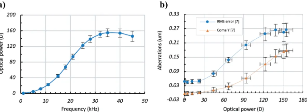
The control of the OP of this lens was performed for fixed voltage values (see hereafter) by changing the frequency of the electrical signal (square shaped AC), see, e.g. [121]. The OP and aberration values were detected by means of a Shack-Hartmann wave front sensor.
Obtained results are summarized in Fig. 1.1.
Since the TLCL is a flat electrically variable GRIN lens, its OP variation range scales quadrat- ically with the inverse of its radiusr= CA/2 (see, e.g. [120]):
OP =2∗∆n∗L
r2 (1.1)
LargeL(40µm) and smallr(0.275 mm) values explain the relatively large OPs observed ex- perimentally (Fig. 1.1(a)). Indeed, the corresponding theoretical estimation (using Eq.(1.1)) shows that the maximum possible OPmax ≈190D . However, the LC orientation is also non
Figure 1.1: a)Optical power (in diopters) versus control frequency (in kHz) andb)circles - total RMS aberrations (inµm) and triangle - separated coma aberrations versus the optical power for the TLCL.
uniform along the light propagation direction (in the non-saturated mode, the LC reorienta- tion could be described by a half sinusoid: zero at boundaries and maximal in the center of the cell since we have strong boundary alignment conditions on cell substrates) that reduces the effective modulation depth of the refractive index. Typically, we can use a factor of≈ 0.85 (for the "efficiency" of use of∆n) to obtain the experimentally achievable values. This would bring us down to ≈161D, which matches perfectly with our experimental data (Fig.
1.1(a)).
Most importantly, we can see the significant contribution of the systematic coma aberration (triangles, Fig. 1.1(b)) in total RMS aberrations of the TLCL. One possible solution to this problem (based on the stack of NLC layers) was discussed by S. Sato and co-workers [122].
Another solution (based on the segmentation of the hole patterned electrode) was discussed in [120]. The aimed-here endoscopic application would favor the first approach due to the desire to study the calcium activity in the brain [47], which requires relatively fast scans of image planes. The stacked solution may be preferred here since it allows the use of thin NLC layers and the reorientation (and thus focus scanning) time is proportional toL2. This will be certainly more expensive approach, but the price is not a critical factor for this application.
We shall thus further explore the stacked solution.
Figure 1.2demonstrates the single and stacked solutions as well as the corresponding ex- perimental results (all experiments were conducted by applying a square shaped AC signal with an amplitude ranging between 3.5 and 4.5 volts). Figure 1.2a demonstrates the ob- served strong coma in the case of a TLCL with single NLC layer (see the cell schematics, top left column). Traditional polarimetric set-up (Appendix, Fig. 1.5) was used (the TLCL was placed between two crossed polarizers, with the ground state director of NLC along the diagonal) to visualize the coma by fringe spacing (Fig. 1.2(a), the photo at the bottom left column; each bright fringe representing 2π shifts on the transmitted phase front, see, e.g.
Figure 1.2:a)Demonstration of the strong coma in the case of a TLCL with single NLC layer (left column) and its compensation by the combination of two TLCLs with “opposed” pretilt angles (right column). b)Optical power (in diopters) versus control frequency (in kHz) for 2 TLCLs with single NLC layers (squares and triangles) and for one combined TLCL (double NLC stack, circles).c)Total RMS aberrations,d)Coma aberrations.
[120]). The right column of Fig.1.2(a) shows its compensation by the combination (stack) of two TLCLs with "opposed" pretilt angles (see the cell schematics, top right column). Figure 1.2b shows the OP versus the control frequency for 2 individual TLCLs with single NLC layer (squares and triangles) and for their combination TLCL (double NLC layer with two
"antiparallel" pretilt angles, circles, [47]).
As we can see (from Fig. 1.1(a)), the contrast of fringes is slightly degraded for the double cell. We think that this is related to the increased light scattering and to the imperfect align- ment of two cells. It is interesting also to notice that the maximum achievable OP (circles, Fig1.2(b)) for the double cell TLCL is less than the addition of OP values of two individual TLCLs. This might be related to the imperfect alignment of those cells as well as due to the fact that the focusing phenomenon is more efficient (stronger) for collimated beams while the use of the second TLCL becomes less efficient due to the wavefront curvature created by the previous TLCL.
Corresponding RMS and coma aberrations are demonstrated in Fig. 1.2(c) and figure Fig.
1.2(d), respectively.
![Figure I.2: Benchtop 2-photon microscope design adapted for in vivo imaging [33].](https://thumb-eu.123doks.com/thumbv2/123doknet/2906917.75346/18.918.309.598.99.504/figure-benchtop-photon-microscope-design-adapted-vivo-imaging.webp)


![Figure I.5: Different phases of matter. At the 2nd melting point, the cloudy liquid (liquid crystals) become isotropic, almost transparent [79].](https://thumb-eu.123doks.com/thumbv2/123doknet/2906917.75346/24.918.233.685.344.654/figure-different-phases-matter-melting-crystals-isotropic-transparent.webp)