HAL Id: hal-01517346
https://hal.sorbonne-universite.fr/hal-01517346
Submitted on 3 May 2017
HAL is a multi-disciplinary open access
archive for the deposit and dissemination of sci-entific research documents, whether they are pub-lished or not. The documents may come from teaching and research institutions in France or abroad, or from public or private research centers.
L’archive ouverte pluridisciplinaire HAL, est destinée au dépôt et à la diffusion de documents scientifiques de niveau recherche, publiés ou non, émanant des établissements d’enseignement et de recherche français ou étrangers, des laboratoires publics ou privés.
new broad-spectrum antiparasitic and antibacterial
agent
Zahid Raja, Sonia André, Feten Abbassi, Vincent Humblot, Olivier Lequin,
Tahar Bouceba, Isabelle Correia, Sandra Casale, Thierry Foulon, Denis
Sereno, et al.
To cite this version:
Zahid Raja, Sonia André, Feten Abbassi, Vincent Humblot, Olivier Lequin, et al.. Insight into the mechanism of action of temporin-SHa, a new broad-spectrum antiparasitic and antibacterial agent. PLoS ONE, Public Library of Science, 2017, 12 (3), pp.e0174024. �10.1371/journal.pone.0174024�. �hal-01517346�
RESEARCH ARTICLE
Insight into the mechanism of action of
temporin-SHa, a new broad-spectrum
antiparasitic and antibacterial agent
Zahid Raja1☯, Sonia Andre´1☯, Feten Abbassi1¤, Vincent Humblot2, Olivier Lequin3,4, Tahar Bouceba5, Isabelle Correia3,4, Sandra Casale2, Thierry Foulon1, Denis Sereno6,7, Bruno Oury6,7‡, Ali Ladram1‡*
1 Sorbonne Universite´s, UPMC Univ Paris 06, CNRS, Institut de Biologie Paris-Seine (IBPS), Biogenèse des Signaux Peptidiques (BIOSIPE), Paris, France, 2 Sorbonne Universite´s, UPMC Univ Paris 06, CNRS, Laboratoire de Re´activite´ de Surface (LRS), Paris, France, 3 Sorbonne Universite´s, UPMC Univ Paris 06, Ecole Normale Supe´rieure, CNRS, Laboratoire des Biomole´cules, Paris, France, 4 Department of Chemistry, Ecole Normale Supe´rieure, PSL Research University, UPMC Univ Paris 06, CNRS, Laboratoire des
Biomole´cules, Paris, France, 5 Sorbonne Universite´s, UPMC Univ Paris 06, CNRS, Institut de Biologie Paris-Seine (IBPS), Plate-forme Interactions Mole´culaires, Paris, France, 6 Institut de Recherche pour le De´veloppement (IRD), UMR 224 IRD-CNRS-Univ Montpellier 1 et 2 Maladies infectieuses et Vecteurs: e´cologie, ge´ne´tique, e´volution et controˆle (MiVegec), Montpellier, France, 7 IRD, UMR 177 IRD-CIRAD, Interactions Hoˆtes-Vecteurs-Parasites-Environnement dans les maladies tropicales ne´glige´es dues aux Trypanosomatidae (InterTryp), Montpellier, France
☯These authors contributed equally to this work.
¤ Current address: Ecole Supe´rieure des Sciences et Techniques de la Sante´ de Monastir (ESSTSM), Universite´ de Monastir, Monastir, Tunisia.
‡ These authors also contributed equally to this work.
*ali.ladram@upmc.fr
Abstract
Antimicrobial peptides (AMPs) are promising drugs to kill resistant pathogens. In contrast to bacteria, protozoan parasites, such as Leishmania, were little studied. Therefore, the anti-parasitic mechanism of AMPs is still unclear. In this study, we sought to get further insight into this mechanism by focusing our attention on temporin-SHa (SHa), a small broad-spec-trum AMP previously shown to be active against Leishmania infantum. To improve activity, we designed analogs of SHa and compared the antibacterial and antiparasitic mechanisms. [K3]SHa emerged as a highly potent compound active against a wide range of bacteria, yeasts/fungi, and trypanosomatids (Leishmania and Trypanosoma), with leishmanicidal intramacrophagic activity and efficiency toward antibiotic-resistant strains of S. aureus and antimony-resistant L. infantum. Multipassage resistance selection demonstrated that tem-porins-SH, particularly [K3]SHa, are not prone to induce resistance in Escherichia coli. Anal-ysis of the mode of action revealed that bacterial and parasite killing occur through a similar membranolytic mechanism involving rapid membrane permeabilization and depolarization. This was confirmed by high-resolution imaging (atomic force microscopy and field emission gun-scanning electron microscopy). Multiple combined techniques (nuclear magnetic reso-nance, surface plasmon resoreso-nance, differential scanning calorimetry) allowed us to detail peptide-membrane interactions. [K3]SHa was shown to interact selectively with anionic model membranes with a 4-fold higher affinity (KD= 3 x 10⁻⁸M) than SHa. The amphipathic
α-helical peptide inserts in-plane in the hydrophobic lipid bilayer and disrupts the acyl chain
a1111111111 a1111111111 a1111111111 a1111111111 a1111111111 OPEN ACCESS
Citation: Raja Z, Andre´ S, Abbassi F, Humblot V,
Lequin O, Bouceba T, et al. (2017) Insight into the mechanism of action of temporin-SHa, a new broad-spectrum antiparasitic and antibacterial agent. PLoS ONE 12(3): e0174024.https://doi.org/ 10.1371/journal.pone.0174024
Editor: Surajit Bhattacharjya, Nanyang
Technological University, SINGAPORE
Received: December 5, 2016 Accepted: February 22, 2017 Published: March 20, 2017
Copyright:© 2017 Raja et al. This is an open access article distributed under the terms of the Creative Commons Attribution License, which permits unrestricted use, distribution, and reproduction in any medium, provided the original author and source are credited.
Data Availability Statement: All relevant data are
within the paper and its Supporting Information files.
Funding: This work was supported by the
University Pierre and Marie Curie (UPMC), Institute of Research for Development (IRD–AAP Leishmed 2010), French state funds managed by the ANR (Investissements d’Avenir program, reference ANR-11-IDEX-0004-02, within the framework of the Cluster of Excellence MATISSE), and by funds from the Convergence MECV 2011 program of
packing via a detergent-like effect. Interestingly, cellular events, such as mitochondrial membrane depolarization or DNA fragmentation, were observed in L. infantum promasti-gotes after exposure to SHa and [K3]SHa at concentrations above IC50. Our results indicate
that these temporins exert leishmanicidal activity via a primary membranolytic mechanism but can also trigger apoptotis-like death. The many assets demonstrated for [K3]SHa make this small analog an attractive template to develop new antibacterial/antiparasitic drugs.
Introduction
Antimicrobial peptides (AMPs) are a ubiquitous group of natural compounds that play a major role in the innate immune system [1,2]. Because of their ability to rapidly kill a wide range of microorganisms by inducing the lysis of their membranes and/or acting on intracellu-lar targets [3], these peptides are less susceptible to induce microbial resistance. Naturally occurring AMPs, such as those from amphibians, are considered promising candidates for the development of therapeutic drugs, including anti-infective agents to treat resistant pathogens as well as anticancer, antidiabetic and immunomodulatory agents [4,5].
Amphibian AMPs of the temporin family [6–10] represent particularly attractive potential therapeutic candidates. These peptides are synthesized from precursors of the dermaseptin superfamily and display the characteristic structural features: a highly conserved N-terminal region (signal peptide followed by an acidic propiece) and a hypervariable C-terminal region encoding the progenitor sequence of the AMP [11,12]. Mature temporins share unique prop-erties that distinguish them from other AMPs. These peptides have a small size, usually 13–14 residues [13]. However, we recently isolated an atypical member of the temporin family con-taining only 8 amino acid residues, named temporin-SHf (FFFLSRIFNH2), which is the
small-est natural linear AMP found to date, with the highsmall-est percentage of Phe residues (50%) for any known peptide or protein [14]. In contrast to most AMPs, temporins have a low net posi-tive charge (0 to +3). All temporins are C-terminally amidated and adopt an amphipathic α-helical structure in apolar media or in membrane mimetic environments [15–17]. Recently, such structure for a temporin was also observed in a media containing bacterial cells [18], using a previous circular dichroism protocol that was used for the first time to study the sec-ondary structure of AMPs (cecropin A and magainin-2) in the presence ofE. coli cells [19]. This amphipathicα-helical structure enables the temporins to interact with and destabilize microbial cytoplasmic membrane, thereby promoting membrane permeabilization and/or dis-ruption via a “carpet-like” mechanism that can involve the formation of toroidal pores, chan-nel aggregates or more complex structures depending on the concentration, length and sequence of the peptide [13,15,20,21]. At very high peptide concentrations, the membrane can be disintegrated in a detergent-like manner.
Temporins have a relatively narrow spectrum of activity, predominantly against Gram-pos-itive bacteria [7–9]. However, a few members of this family display a wider spectrum, with potent activity against Gram-negative bacteria and yeasts [22–25]. Moreover, a small number of temporins are able to kill parasites. Currently, only six temporins have been reported to have leishmanicidal activity (Table 1). Other amphibian AMPs with a larger size were also shown to act onLeishmania parasites, such as dermaseptin-S1 (34 residues), the first
discov-ered leishmanicidal peptide, or bombinins H2 (20 residues) and H4 (21 residues) [26,27]. Different AMP antiparasitic activities are observed depending on the nature of the AMP and the parasite and also on the stage of the parasite. Several antiparasitic mechanisms have UPMC. ZR and SA were supported by a fellowship
from the French Ministère de l’Enseignement Supe´rieur et de la Recherche, allocated by the Ecole Doctorale iViv (ED 387, UPMC, Paris, France). The funders had no role in study design, data collection and analysis, decision to publish, or preparation of the manuscript.
Competing interests: The authors have declared
been described [30–32], which involve disruption of the parasite membrane in the case of temporin-Ta and Tb [28] or activity against intracellular targets in the case of histatine-5 [33]. The fact that amastigotes (the intracellular form in the vertebrate host) are generally more resistant to AMPs compared to promastigotes (the extracellular form in the insect vector) sug-gests that the surface composition of theLeishmania parasites is important and that the
nega-tively charged glycocalyx of promastigotes, composed mainly of lipophosphoglycan (LPG) and proteophosphoglycan (PPG), plays a significant role in AMP activity. A recent study by Eggi-mann and collaborators indicated that PPG is a major factor for the activity of temporin-SHa againstL. mexicana promastigotes and that the lack of PPG and LPG on the surface increases
the resistance of thisLeishmania species to temporins [29]. Temporins are among the smallest natural antiparasitic peptides reported to have activity against both promastigotes and amasti-gotes. Moreover, it appears that the small size and low charge of temporins favor diffusion across the glycocalyx into the plasma membrane. Therefore, these peptides may be useful tools to elucidate the antiparasitic mechanism and are also attractive candidates to reinforce the lim-ited arsenal of chemotherapeutic agents for which there is evidence of emerging resistance, such as pentavalent antimonials [34–36] or miltefosine [37].
We previously showed that temporin-SHa (SHa) has a broad-spectrum activity toward Gram-positive and Gram-negative bacteria, yeasts andLeishmania parasites [22]. This peptide inhibited the growth ofL. infantum promastigotes and axenic amastigotes. Based on the
com-plex balance of structural and physicochemical parameters (length, secondary structure, net positive charge, hydrophobicity, helicity and amphipathicity) that govern the antimicrobial activity of AMPs [4,38–41], we designed substituted analogs of SHa ([K3]temporin-SHa: [K3] SHa; [A2,6,9]temporin-SHa: [A2,6,9]SHa; [A2,6,9, K3]temporin-SHa: [A2,6,9, K3]SHa) to improve the antibacterial/antiparasitic activity of the parent peptide.
In this study, the structure of the peptides and their cytotoxicity against several mammalian cells were determined, and a detailed comparison of the antibacterial and antiparasitic activi-ties of SHa and its analog [K3]SHa was carried out. We screened a large panel of Gram-nega-tive and Gram-posiGram-nega-tive bacteria of clinical interest and also various trypanosomatid parasites (Leishmania and Trypanosoma), including antibiotic-resistant strains of S. aureus and
anti-mony-resistantL. infantum. Multipassage resistance selection was performed to determine
whether bacterial resistance could occur against SHa and its analogs. Biochemical and bio-physical studies allowed us to compare the antiparasitic and antibacterial mechanisms of the temporins in detail. We first carried out membrane permeabilization, depolarization and time-kill assays. The temporin-induced membrane damages were then visualized by atomic force microscopy (AFM) and field emission gun-scanning electron microscopy (FEG-SEM) imaging of bacteria and parasites. We also analyzed the typical hallmarks of apoptosis inL. infantum promastigotes, such as mitochondrial depolarization and DNA fragmentation. Table 1. Temporins reported to have leishmanicidal activity.
Temporin Sequence Net chargea Reference
Ta FLPLIGRVLSGILNH2 +2 [28] Tb LLPIVGNLLKSLLNH2 +2 [28] SHa FLSGIVGMLGKLFNH2 +2 [22] SHd FLPAALAGIGGILGKLF +2 [23] Tl FVQWFSKFLGRILNH2 +3 [29] Tf FLPLIGKVLSGILNH2 +2 [29] apH 7.4. https://doi.org/10.1371/journal.pone.0174024.t001
Finally, peptide-membrane interactions were studied by surface plasmon resonance (SPR) and differential scanning calorimetry (DSC) using models of eukaryotic and prokaryotic cell branes and also by nuclear magnetic resonance (NMR) spectroscopy using bicelles as a mem-brane mimetic.
Results
Design of SHa analogs
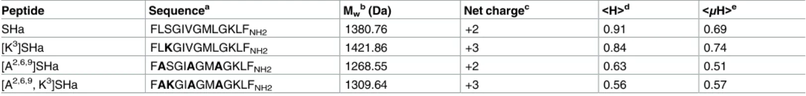
The SHa analogs were designed and synthetized by modifying the structural and physicochem-ical parameters known to control the antimicrobial activity of AMPs, such as net positive charge, hydrophobicity, helicity and amphipathicity. Specific amino acid positions in the sequence of SHa were substituted to improve the antibacterial and antiparasitic activity of SHa while reducing its cytotoxic activity. First, the net positive charge of SHa was increased to a value of +3, yielding the analog [K3]SHa (Table 2). This analog has a Lys residue in position 3, which is located on the polar face of theα-helix (Fig 1), instead of a Ser residue. Because high hydrophobicity was shown to increase cytotoxic activity [4], we also designed two analogs with reduced hydrophobicity on the apolar face of theα-helix (Fig 1). The residues Leu2,9and Val6 were replaced with Ala, yielding the analog [A2,6,9]SHa and also [A2,6,9, K3]SHa (Table 2). As shown by the Schiffer-Edmundson helical wheel projections (Fig 1), all analogs were predicted to conserve the amphipathic character of SHa.
[K
3]SHa displays more potent antimicrobial activity than SHa
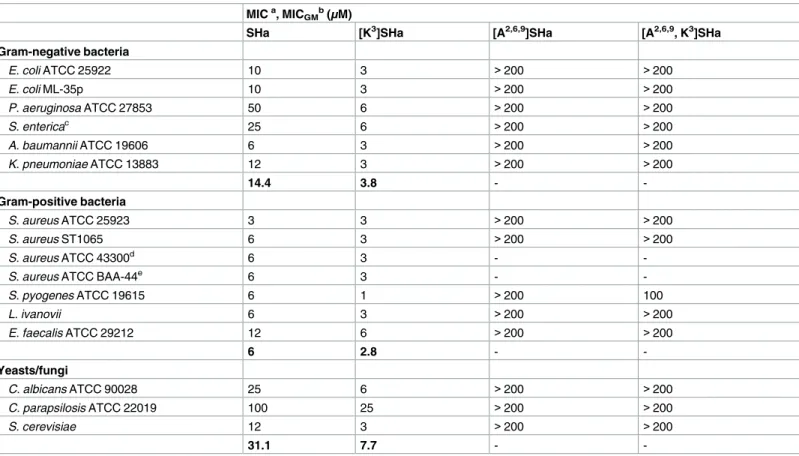
We first investigated the antimicrobial activity of SHa and analogs by determining minimal inhibitory concentrations (MICs) against a wide panel of Gram-negative and Gram-positive bacteria and yeasts/fungi (Table 3). The two analogs [A2,6,9]SHa and [A2,6,9, K3]SHa were inac-tive against all strains (MIC > 200μM). These two peptides were then used as negative
con-trols. In contrast, the analog [K3]SHa was found to be highly active against all the tested bacteria (MIC = 1–6μM) and yeasts/fungi (MIC = 3–25 μM), including the
multidrug-resis-tant strains of the clinically relevant pathogenic speciesS. aureus, ATCC 43300 and ATCC
BAA-44. A significant increase in activity was observed for this analog compared to the parent peptide, especially for Gram-negative bacteria and yeasts/fungi (Table 3). Indeed, for Gram-negative strains, MIC values decreased by approximately 8-fold forP. aeruginosa, a species
that frequently develops high intrinsic resistance to many conventional antibiotics, 4-fold for
S. enterica (serotype Enteritidis) and K. pneumoniae, 3-fold for E. coli, and 2-fold for A. bau-mannii. Moreover, [K3]SHa was 4-fold more potent against yeasts/fungi, even toward the
Table 2. Sequence and physicochemical properties of temporin-SHa and its analogs.
Peptide Sequencea M
wb(Da) Net chargec <H>d <μH>e
SHa FLSGIVGMLGKLFNH2 1380.76 +2 0.91 0.69
[K3]SHa FLKGIVGMLGKLFNH2 1421.86 +3 0.84 0.74
[A2,6,9]SHa FASGIAGMAGKLFNH2 1268.55 +2 0.63 0.51
[A2,6,9, K3]SHa FAKGIAGMAGKLF
NH2 1309.64 +3 0.56 0.57
aThe substituted residues are indicated in bold in the sequence of the analogs.
bTheoretical average molecular mass using Peptide Mass Calculator v3.2 (http://rna.rega.kuleuven.ac.be/masspec/pepcalc.htm). cpH 7.4.
dHydrophobicity and
ehydrophobic moment were calculated using HeliQuest (http://heliquest.ipmc.cnrs.fr/). https://doi.org/10.1371/journal.pone.0174024.t002
SHa-insensitive speciesC. parapsilosis (MIC = 25 μM).Table 3also indicates that [K3]SHa retained the high potency of the parent peptide againstS. aureus ATCC 25923 (MIC = 3 μM)
but was slightly more active toward other Gram-positive bacteria (2-fold) and significantly more active towardS. pyogenes (6-fold). The geometric mean of the MIC values (MICGM) was
calculated for several strains of theTable 3to provide an overall evaluation of the antimicrobial activity against Gram-negative bacteria, Gram-positive bacteria and yeast/fungi, and then to use it for the determination of the therapeutic index of SHa and [K3]SHa. MICGMvalues
indi-cated inTable 3(in bold) underline the higher antimicrobial potency of [K3]SHa, particularly against Gram-negative bacteria and yeasts/fungi. At a concentration 2-fold above the MIC,
Fig 1. Schiffer-Edmundson helical wheel representation of temporin-SHa and its analogs. SHa, temporin-SHa; [K3]SHa,
[K3]temporin-SHa; [A2,6,9]SHa, [A2,6,9]temporin-SHa; [A2,6,9, K3]SHa, [A2,6,9, K3]temporin-SHa. Apolar residues are represented
in black and polar/basic residues in gray/white. Amino acid modifications are in bold. An amphipathic character, with two well-separated polar and apolar faces, is observed. Adapted from Heliquest.
https://doi.org/10.1371/journal.pone.0174024.g001
temporin-SHa (12μM) and [K3]SHa (6μM) were both able to completely kill Gram-positive
bacteria, such asS. aureus, within 5 min (Fig 2A). However, at similar concentrations, [K3]SHa was more efficient than SHa in killing Gram-negative bacteria (Fig 2B). While a 120-min expo-sure was needed for approximately 93% killing by SHa, only 15 min was needed for the [K3] SHa analog to achieve complete lethality inE. coli cells.
Leishmanicidal activity was analyzed on both promastigotes and amastigotes (axenic and intramacrophagic). Potent and rapid antiprotozoal activities were obtained for SHa and its analog [K3]SHa (Table 4andFig 3). The effect of temporin-SHa againstL. infantum can
be visualized in real time (S1 Movie). As shown inTable 4, a growth inhibitory effect was observed with promastigotes of several species ofLeishmania responsible for visceral (L. infan-tum), cutaneous (L. major and L. amazonensis) and mucocutaneous (L. braziliensis)
leishmani-ases, with slightly higher activity for [K3]SHa (IC50values in the range of 5–10μM) compared
to the parent peptide SHa (IC50= 7–18μM). We noticed a 2-fold improvement of
leishmanici-dal activity (IC50= 8μM for SHa and 5 μM for [K3]SHa) when peptides were incubated with L. infantum promastigotes in serum-free medium. Moreover, time-kill curves revealed clear
differences in the parasite-killing efficiency of temporin-SHa (Fig 3A) and [K3]SHa (Fig 3B).
Table 3. Antibacterial activity of temporin-SHa analogs compared to the parent peptide. MICa, MICGMb(μM)
SHa [K3]SHa [A2,6,9]SHa [A2,6,9, K3]SHa
Gram-negative bacteria E. coli ATCC 25922 10 3 >200 >200 E. coli ML-35p 10 3 >200 >200 P. aeruginosa ATCC 27853 50 6 >200 >200 S. entericac 25 6 >200 >200 A. baumannii ATCC 19606 6 3 >200 >200 K. pneumoniae ATCC 13883 12 3 >200 >200 14.4 3.8 - -Gram-positive bacteria S. aureus ATCC 25923 3 3 >200 >200 S. aureus ST1065 6 3 >200 >200 S. aureus ATCC 43300d 6 3 -
-S. aureus ATCC BAA-44e 6 3 -
-S. pyogenes ATCC 19615 6 1 >200 100 L. ivanovii 6 3 >200 >200 E. faecalis ATCC 29212 12 6 >200 >200 6 2.8 - -Yeasts/fungi C. albicans ATCC 90028 25 6 >200 >200 C. parapsilosis ATCC 22019 100 25 >200 >200 S. cerevisiae 12 3 >200 >200 31.1 7.7 - -a
Values represent the means of three independent experiments performed in triplicate.
b
MICGMdenotes the geometric mean of the MIC values (indicated in bold) for all Gram-negative, Gram-positive and yeast/fungal strains. c
Serotype Enteritidis.
d
Resistant to methicillin and oxacillin.
e
Resistant to amoxicillin/clavulanic acid, cephalothin, ciprofloxacin, erythromycin, gentamicin, imipenem, oxacillin, penicillin, tetracycline, ampicillin, doxycycline, methicillin, azithromycin, ceftriaxone, clindamycin, lincomycin, perfloxacin, rifampin, and tobramycin.
While both peptides were active againstL. infantum promastigotes at 3 μM (38% and 49%
kill-ing for SHa and [K3]SHa, respectively, at 180 min), [K3]SHa induced death instantly at 12μM
(within the first min) compared to 5 min for SHa. However, at 6μM, [K3]SHa was able to rap-idly (15 min) and completely kill promastigotes, whereas a significant reduction in the number of parasites (~ 91%) was observed only after a 180-min incubation with temporin-SHa. Con-sistent with their lack of antibacterial effect, [A2,6,9]SHa and [A2,6,9, K3]SHa showed no anti-parasitic activity againstL. infantum promastigotes (IC50> 200μM,Table 4). As shown inFig
3C, no killing was observed when parasites were incubated with 96μM [A2,6,9, K3]SHa. As observed for antibiotic-susceptible and antibiotic-resistantS. aureus strains, [K3]SHa and SHa retained the ability to killL. infantum parasites susceptible and resistant to antimony with the
same efficiency (Table 4). Interestingly, leishmanicidal activity was demonstrated for [K3]SHa (IC50= 5μM) and SHa (IC50= 9μM) against intramacrophagic amastigotes as well as against
the axenic forms (IC50= 20μM) (Table 4). Other trypanosomatids, such asT. brucei gambiense
(responsible for sleeping sickness) andT. cruzi (the etiological agent of Chagas disease), were Fig 2. Time-killing curves of SHa and its analog [K3]SHa against S. aureus ST1065 (A) and E. coli
ML-35p (B). Bacteria (106cfu/ml) were incubated in phosphate-buffered saline (PBS) with temporins at concentrations 2-fold above the MIC obtained for S. aureus ST1065 (6μM for [K3]SHa and 12μM for SHa).
The negative control corresponds to bacteria incubated in PBS without peptide (w/o peptide). The data are shown as the means±SEM from a single experiment carried out in triplicate and are representative of three independent experiments.
https://doi.org/10.1371/journal.pone.0174024.g002
susceptible to temporins (Table 4). The activity againstT. brucei gambiense was quite similar
for SHa and [K3]SHa (IC50~ 15μM) but T. cruzi was slightly more susceptible to the peptide
analog (~ 2-fold). The trypanocidal effect of temporin-SHa is shown in theS2 Movie.
As a positive control, we also evaluated the leishmanicidal effect of dermaseptin B2, a potent 33-residue AMP isolated from frog skin (Phyllomedusa bicolor) that kills microorganisms by
the carpet mechanism [42]. A high leishmanicidal activity againstL. infantum promastigotes
was observed for this peptide (IC50= 2.5μM).
[K
3]SHa displays a better therapeutic index
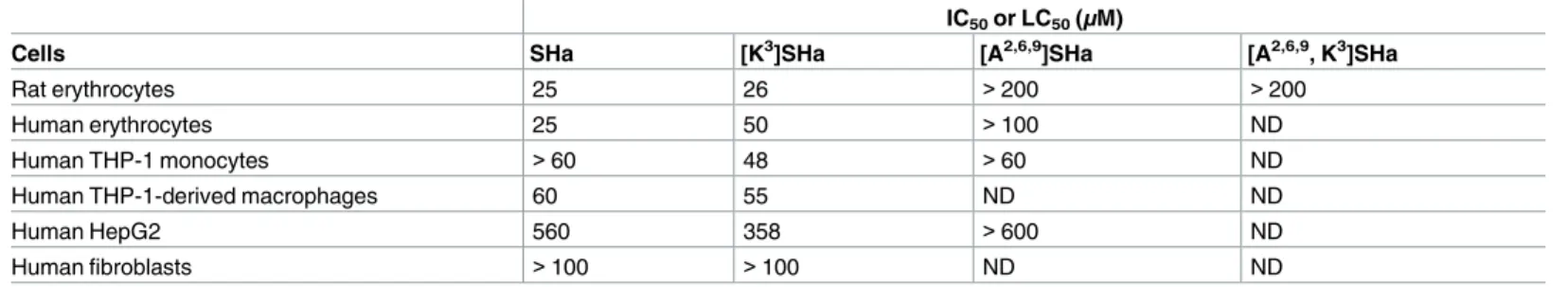
The cytotoxicity of temporins was evaluated toward rat and human erythrocytes, and several human cell lines (Table 5). At antimicrobial concentrations (3–6μM for bacteria and virtually
all yeast strains; 5–15μM for parasites), low cytotoxicity (human erythrocytes, THP-1
mono-cytes and THP-1-derived macrophages) or no cytotoxicity (HepG2 cells and fibroblasts) was observed for [K3]SHa. For SHa, similar results were obtained, with a 2-fold higher hemolytic activity on human erythrocytes (25μM) compared to [K3]SHa. However, this 2-fold higher activity was also found for both peptides on rat erythrocytes. These results indicate that intra-macrophagic amastigotes can be killed by temporins without damage to the host cell at leish-manicidal concentrations. As for bacteria and parasites, the analogs [A2,6,9]SHa and [A2,6,9, K3] SHa were inactive toward erythrocytes, and no cytotoxicity has been detected when [A2,6,9] SHa was also tested on THP-1 monocytes (IC50> 60μM) and HepG2 cells (IC50> 600μM)
(Table 5). In contrast, the positive control, dermaseptin B2, was highly cytotoxic toward THP-1 monocytes (IC50~ 7μM).
To evaluate the cell selectivity (microorganisms versus human cells), the therapeutic indices of [K3]SHa and SHa were calculated as the ratio of IC50or LC50values for the different human
cells over the MICGMvalues obtained for Gram-negative bacteria, Gram-positive bacteria and
yeasts/fungi. The results showed inTable 6reveal that [K3]SHa displays a better therapeutic index with values 1.37-fold to 8.12-fold higher than those of SHa depending on the type of microorganisms and cells considered. The therapeutic indices of [K3]SHa were also better when they were calculated with LC50values against rat erythrocytes (indicated inTable 5) and Table 4. Antiprotozoal activity of temporin-SHa and [K3]temporin-SHa.
IC50(μM)
SHa [K3]SHa
Leishmania promastigotes
L. infantuma 18 (8)b 10 (5)b
L. infantum (antimony resistant) 14 9
L. major 13 10 L. braziliensis 7 5 L. amazonensis 13 8 Leishmania amastigotes L. infantum (axenic) 20 20 L. infantum (intramacrophagic) 9 5 Trypanosoma epimastigotes T. brucei gambiense 14 16 T. cruzi 17 10
a[A2,6,9]SHa and [A2,6,9, K3]SHa were inactive against L. infantum promastigotes (IC
50>200μM). bvalues in parentheses were obtained in serum-free medium.
Fig 3. Time-kill curves of temporins against L. infantum. Parasites (2 x 10⁶cells/ml) were incubated in
HBSS with various concentrations (3, 6 and 12μM) of synthetic SHa (A) and [K3]SHa (B). HBSS without
peptide (w/o peptide) or containing 96μM [A2,6,9, K3]SHa was used as a negative control (C). The data are
shown as the means±SEM of one representative experiment obtained from three independent experiments carried out in duplicate.
https://doi.org/10.1371/journal.pone.0174024.g003
MICGM: Gram-negative bacteria, T.I.(SHa) = 1.7, T.I.([K3]SHa) = 6.8 (4-fold higher);
Gram-positive bacteria, T.I.(SHa) = 4.2, T.I.([K3]SHa) = 9.3 (2.21-fold higher); yeasts/fungi, T.I. (SHa) = 0.8, T.I.([K3]SHa) = 3.4 (4.25-fold higher). Therefore, the overall results indicate greater antimicrobial specificity for the analog [K3]SHa compared to the parent peptide.
[K
3]SHa alters more efficiently the integrity of the bacterial and parasite
plasma membrane
The effects of temporins on the membrane integrity of bacteria and parasites were investigated by two complementary approaches (ONPG and SYTOX Green assays) to analyze membrane permeabilization. First, we incubated SHa and [K3]SHa withE. coli ML-35p, a bacterial strain
constitutively expressing cytoplasmicβ-galactosidase, and we measured the time-dependent hydrolysis of the small chromogenic substrateo-nitrophenyl-β-D-galactopyranoside (ONPG)
intoo-nitrophenol (ONP) by cytoplasmic β-galactosidase (Fig 4). As indicated inFig 4A and 4B, both peptides were able to permeabilize the bacterial cytoplasmic membrane. However, [K3]SHa induced a more rapid and potent permeabilization of the Gram-negative strainE. coli
ML-35p compared to the parent peptide. Interestingly, at 10μM, this 13-residue analog was as
efficient as the two positive controls used in our study, dermaseptin B2 (33 residues) and
Table 5. Cytotoxic activity of temporins SHa and analogs against human cells and rat erythrocytes. IC50or LC50(μM)
Cells SHa [K3]SHa [A2,6,9]SHa [A2,6,9, K3]SHa
Rat erythrocytes 25 26 >200 >200
Human erythrocytes 25 50 >100 ND
Human THP-1 monocytes >60 48 >60 ND
Human THP-1-derived macrophages 60 55 ND ND
Human HepG2 560 358 >600 ND
Human fibroblasts >100 >100 ND ND
Values were determined with GraphPad Prism 6.0 software and are the means of three independent experiments performed in triplicate. IC50, half maximal
inhibitory concentration; LC50, half maximal lytic concentration (erythrocytes and macrophages).
ND: not determined.
https://doi.org/10.1371/journal.pone.0174024.t005
Table 6. Therapeutic indices (T.I.) of [K3]SHa and SHaa.
T.I.b Human THP1- THP1-derived HepG2 Fibroblasts
[K3]SHa (SHa) erythrocytes monocytes macrophages
Gram-negative strains 13.1 (1.7) 12.6 (8.3) 14.5 (4.2) 94.2 (38.9) 52.6 (13.9) 7.70c 1.52 3.45 2.42 3.78 Gram-positive strains 17.8 (4.2) 17.1 (20) 19.6 (10) 127.8 (93.3) 71.4 (33.3) 4.24 0.85 1.96 1.37 2.14 Yeast/fungal strains 6.5 (0.8) 6.2 (3.8) 7.1 (1.9) 46.5 (18) 26 (6.4) 8.12 1.63 3.37 2.58 4.06 a
Therapeutic indices of SHa are indicated in parentheses.
b
Ratio of IC50or LC50values (for human cells fromTable 5) over the geometric mean of MIC values (fromTable 3and corresponding to Gram-negative,
Gram-positive, and yeast/fungal strains). When IC50or LC50values were higher than the maximum concentration tested, a minimal 2-fold concentration
value was used to calculate the therapeutic index because IC50or LC50values were determined by carrying out serial 2-fold dilutions (for example, LC50>
100 was considered as 200).
c
Values in bold represent the fold improvement in the therapeutic index of [K3]SHa compared to SHa.
Fig 4. Temporin-induced membrane permeabilization of E. coli ML-35p. Bacteria were incubated with different concentrations of SHa (A) or [K3]SHa (B). The leakage kinetics were measured as the production of ONP at 405 nm resulting from hydrolysis of ONPG by the
cytoplasmic bacterialβ-galactosidase. C, comparison of the membrane leakage of temporins (SHa and [K3]SHa), dermaseptin B2 (B2) and
melittin at the same concentration (10μM). The negative control without peptide is also indicated (w/o peptide). D, no permeabilization was observed with [A2,6,9]SHa (2, 4, 6, 8 and 10μM). E, Extracellular release of cytoplasmicβ-galactosidase after 60 min incubation of bacteria
with 10μM peptide followed by centrifugation (1,000 x g, 10 min, 4˚C) to measure ONP production (A405) in the supernatant. The results are
melittin (26 residues), with identical kinetics of permeabilization (Fig 4C). Under the same conditions, the level of permeabilization of SHa was 1.6-fold lower than that of [K3]SHa. In contrast, the analog [A2,6,9]SHa was not able to permeabilize the cytoplasmic membrane ofE. coli ML-35p (Fig 4D). This is consistent with its lack of antimicrobial activity. To assess the extent of membrane damage caused by temporins and to determine whether these peptides act via pore formation or a detergent-like effect, we investigated the release of intracellular β-galactosidase (Mw ~ 540 kDa, Stokes radius ~ 70Å) from E. coli ML-35p bacteria upon incu-bation with 10μM peptide. As shown inFig 4E, the leakage induced by [K3]SHa led to extra-cellularβ-galactosidase activity that was comparable to dermaseptin B2 but 29-fold higher than melittin (a pore-forming peptide) [43]. This leakage was 1.7-fold higher than that induced by SHa. Thus, these results suggest that temporins and dermaseptin B2 act through similar mechanisms, i.e., a detergent-like effect [42], and also confirm the potent bactericidal activity of [K3]SHa by inducing a more efficient disruption of the bacterial membrane.
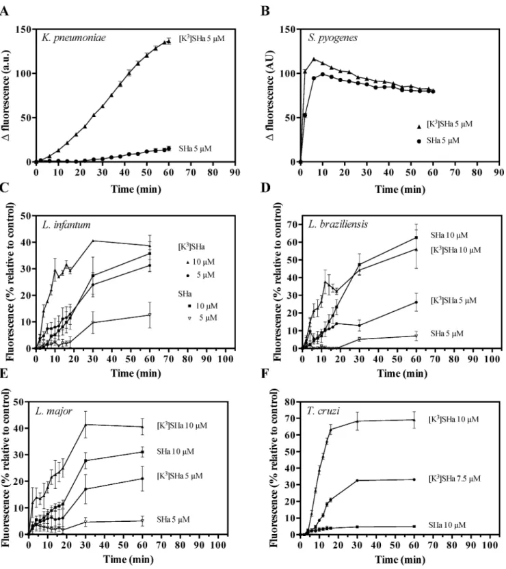
In the second approach to analyze membrane permeabilization, we used two clinically relevant strains (S. pyogenes and K. pneumoniae), and we tested the ability of the vital dye
SYTOX Green (SG, ~ 600 Da), which requires lesions in the membrane, to enter into the cell. For the Gram-positiveS. pyogenes, SHa and [K3]SHa showed rapid and similar SG influx kinetics at 5μM (Fig 5A). However, for the Gram-negative strainK. pneumoniae, the [K3]SHa analog more efficiently permeabilized the cell membrane compared to SHa (Fig 5B). These results are consistent with the ONPG experiments and confirm the high antibacterial effi-ciency of the analog [K3]SHa against Gram-negative and Gram-positive strains. To elucidate the antiparasitic mechanism of temporins, we analyzed the permeabilization of theLeishmania
andTrypanosoma cytoplasmic membranes. As shown inFig 5C–5F, parasite permeabilization occurred in a distinct curve-shaped manner, with a more rapid and potent SG influx into
Leishmania and T. cruzi parasites for [K3]SHa compared to the parent peptide. Differences were clearly observed after 60 min incubations with the two peptides, particularly at 5μM,
where 12% (L. infantum), 7% (L. braziliensis) and 5% (L. major) of the maximum
permeabili-zation were reached after incubation with SHavs 31% (L. infantum), 26% (L. braziliensis) and
21% (L. major) with [K3]SHa. Moreover, the influx of SG intoT. cruzi was 14-fold higher after
incubation of the parasites with 10μM [K3]SHa (69% of permeabilization at 60 min, only 5% for SHa) (Fig 5F). This was consistent with the enhanced antitrypanosomal activity of the ana-log (Table 4).
When we used propidium iodide (PI), another nucleic acid stain, [K3]SHa was also more efficient in affecting the integrity of theL. infantum membrane at 10 and 20 μM (Fig 6A). At 40μM, similar PI staining curves were obtained for both temporins (85% of permeabilized
cells). Using the same methodology for bacteria, we investigated the damage toL. infantum
promastigotes caused by temporins by evaluating the leakage of intracellular components, such as luciferase (~ 61 kDa). Peptide-induced leakage of luciferase was detected by measuring enzyme activity in the supernatant at various times (Fig 6B). The curves were similar to those obtained with PI staining.
Temporins were tested for their ability to dissipate the membrane potential using the fluo-rescent cyanine dye DiSC3(5) (3,30-dipropylthiadicarbocyanine iodide). We observed that the
addition of the peptides caused an instantaneous depolarization of theS. aureus and L. infan-tum membranes, resulting in increased fluorescence (Fig 7A and 7B). This effect was more potent for [K3]SHa, as indicated by the fact that a plateau with an increased magnitude was
expressed as the means±SEM after subtraction of the negative control values (no peptide) from the test values and correspond to one representative experiment carried out in triplicate.
Fig 5. Temporin-induced SYTOX Green (SG) influx into the bacteria K. pneumoniae ATCC 13883 (A) and S. pyogenes ATCC 19615 (B), and the parasites L. infantum (C), L. braziliensis (D), L. major (E), and T. cruzi (F). Bacteria (106cfu/ml) and parasites (2.5 x 10⁶cells/
ml) were preincubated with 1μM SG, and peptides (SHa or [K3]SHa) were added after fluorescence stabilization. Membrane alteration is
correlated with the fluorescence of the DNA fluorescent probe (λexcitation= 485 nm andλemission= 520 nm). For bacteria, the data are expressed as the means±SEM after subtraction of the negative control values (w/o peptide) from the test values. For parasites, the results (mean±SEM) were plotted as a percentage of the fluorescence relative to that of parasites fully permeabilized by 0.1% Triton X-100. The curves correspond to one experiment carried out in triplicate and are representative of two independent experiments.
https://doi.org/10.1371/journal.pone.0174024.g005
reached within 1.5 min (S. aureus) or 2.5 min (L. infantum). Membrane depolarization was
also detected forL. amazonensis and T. cruzi, although to a lesser extent for the latter (Fig 7C and 7D).
Altogether, the killing and permeabilization/depolarization experiments are consistent with membrane impairment and the release of cellular components, two concomitant events of the primary mode of action of temporins that kill bacteria and parasites.
We used atomic force microscopy (AFM) and field emission gun-scanning electron micros-copy (FEG-SEM) imaging to obtain high-resolution images of the effects of temporins on bac-terial and parasite morphology. As shown inFig 8A and 8B, when the pathogenic Gram-negative bacteriaP. aeruginosa was in contact with 50 μM SHa for 1 h, severe membrane
dam-age with drastic morphological changes of the bacteria were observed (Fig 8B) compared to the untreated control bacteria (Fig 8A).Fig 8Cindicates thatP. aeruginosa cells were also Fig 6. Dose- and time-dependent propidium iodide (PI) staining (A) and luciferase release in the extracellular medium (B) of L. infantum parasites upon addition of temporins. L. infantum promastigotes (106cells/ml) were incubated with different concentrations (10, 20 and 40μM) of SHa or [K3]SHa for different
times. PI-positive cells were counted by flow cytometry after adding PI (1μg/ml) to the parasites. The luciferase activity in the extracellular medium was determined after centrifugation of the parasites and measurement of the luminescence using the Steady-Glo®Luciferase Assay System (Promega). The data are expressed as the means±SEM of two experiments carried out in triplicate.
severely damaged by [K3]SHa at a very low concentration (6μM), as revealed by the presence
of flattened bacteria. In this selected area of the glass surface, bacteria with a normal shape were also present. This is likely due to the low peptide concentration (6μM, corresponding to
the MIC) relative to the high cell concentration (4 x 10⁷cells/ml, i.e., a bacterial density 40-fold higher than that used for MIC determination). Incubation ofL. infantum promastigotes or T. cruzi epimastigotes with 5 μM [K3]SHa for 30 min led to loss of the morphological integrity of the parasites (Fig 8D–8G). AFM imaging revealed temporin-induced damage of the cell body and the flagellum, with alterations of the parasite shape (Fig 8E and 8G) compared to untreated parasites (Fig 8D and 8F). FEG-SEM analysis ofL. infantum showed that 5 μM [K3]SHa caused permeabilization/lysis of the parasite membrane, likely with leakage of the cellular contents, leading to a large, flattened leaf-shaped parasite (Fig 8H and 8I). Thus, microscopic analysis indicated that the primary killing effect of temporins is similar for bacteria and trypanosoma-tids and occurs through a membranolytic mechanism.
Fig 7. Changes in the membrane potential of bacteria and parasites upon addition of temporins. S. aureus ATCC 25923 (A), L.
infantum (B), L. amazonensis (C) and T. cruzi (D) were equilibrated with DiSC3(5) (1μM for S. aureus and 2.5μM for parasites). SHa or [K3]
SHa was then added (t = 0) at a concentration of 5μM (bacteria) or 50μM (parasites), and changes in the fluorescence were monitored for 15 min (bacteria) or 20 min (parasites) atλexcitation= 622 nm andλemission= 670 nm. The curves correspond to a single experiment representative of three independent experiments.
https://doi.org/10.1371/journal.pone.0174024.g007
Fig 8. AFM and FEG-SEM visualization of the effect of temporins-SH on P. aeruginosa bacteria (A–C) and parasites (L. infantum
promastigotes and T. cruzi epimastigotes; D–I). A–C, AFM imaging of P. aeruginosa: A, untreated control bacteria; B, bacteria after 1 h incubation with 50μM SHa; C, bacteria treated for 1 h with 6μM [K3]SHa. Bacteria were severely damaged by temporins compared to the control. D–G, AFM imaging of L. infantum promastigotes and T. cruzi epimastigotes: D and E, L. infantum untreated (D) or treated with 5μM [K3]SHa (E); F and G, T.
cruzi without peptide (F) or with 5μM [K3]SHa (G). H and I, FEG-SEM visualization of L. infantum promastigotes without peptide (H) or with 5μM [K3]
SHa. Morphological changes were observed for parasites that were incubated with peptides (E, G and I).
SHa and its analog induce apoptotic-like death in Leishmania infantum
promastigotes
Because temporins permeabilize the parasite membrane even at doses below lytic concentra-tions, we investigated whether these peptides could induce cell death by apoptosis. As a first step, we used the fluorescence probe tetramethylrhodamine ethyl ester (TMRE) to monitor the mitochondrial membrane potential inL. infantum promastigotes (Fig 9). As indicated by the negative index of variation (IV) values, SHa (Fig 9A) and [K3]SHa (Fig 9B) were able to depolarize the mitochondrial membrane compared to the positive (CCCP: carbonyl cyanide m-chlorophenylhydrazone, a well-known uncoupling agent) and negative (no peptide) con-trols. However, while both peptides showed similar depolarization profiles at 6μM, the
mito-chondrial membrane potential was not affected at a 2-fold lower concentration of SHa, unlike [K3]SHa.
Fig 9. Kinetics of mitochondrial membrane depolarization of L. infantum promastigotes. Parasites were incubated for 3 h at 26˚C with different concentrations of peptide (3 and 6μM, final concentrations). Mitochondrial membrane potential was monitored by flow cytometry using the fluorescence probe TMRE. A, index of variation for SHa. B, index of variation for [K3]SHa. Negative and positive controls were assayed
without peptides or with 500μM CCCP, respectively. The index of variation is expressed in arbitrary units (a. u.). The curves were obtained from a single experiment representative of three independent experiments.
https://doi.org/10.1371/journal.pone.0174024.g009
Next, we analyzed DNA fragmentation, another hallmark of apoptosis. For this assay, para-sites were incubated with antileishmanial compounds for 48 h, and DNA fragmentation was monitored by TUNEL (terminal deoxynucleotidyl transferase-mediated dUTP nick-end label-ing) assay (Fig 10A) or by cell cycle analysis (Fig 10B–10D). As shown inFig 10A, whenL. infantum promastigotes were treated with 50 μM miltefosine (hexadecylphosphocholine), a
drug used for leishmaniasis treatment that is known to induce apoptosis [44], approximately 65% of DNA fragmentation was observed. In contrast, at the same concentration, [A2,6,9, K3] SHa did not cause any fragmentation. In the presence of SHa, a dose-dependent effect was observed, with a significant DNA fragmentation (approximately 25%) at 50μM. A similar
level of DNA fragmentation was observed when parasites were treated with a 2-fold lower con-centration of [K3]SHa (25μM) and was maintained at 50 μM (Fig 10A). These results were confirmed by cell cycle analysis ofL. infantum promastigotes (Fig 10B–10D). Indeed, flow cytograms revealed a sub-G1 peak after treatment with SHa (50μM) or [K3]SHa (25 and 50
μM) (Fig 10C and 10D) compared to the negative controls (Fig 10B), indicating a significant sub-G1 population (Table 7). At a concentration of 50μM, similar percentages of sub-G1 cells
were observed for miltefosine (29%) and SHa (26%). For [K3]SHa, the proportion of sub-G1 cells was concentration-dependent, with percentages of 18% and 47% at 25 and 50μM,
respec-tively (Table 7). At concentrations above the IC50, significant apoptosis-like death ofL. infan-tum promastigotes was observed following incubation with SHa and [K3]SHa.
Multipassage resistance selection reveals that temporins display no
propensity to promote bacterial resistance, unlike ampicillin
To determine whether bacterial resistance may emerge against temporins, we performed experiments on long-term cultures to select resistant mutants using SHa and [K3]SHa. The more stable analog D-SHa (enantiomeric SHa) with all residues in the D-configuration was also tested because it is potentially less prone to induce bacterial resistance. We usedE. coli
ATCC 25922, and we selected ampicillin as a conventional antibiotic for comparison. The MIC values of temporins and ampicillin were as follows: SHa, 12.5μM; [K3]SHa, 3μM;
D-SHa, 12.5μM; ampicillin, 12.5 μM. In our resistance selection assay (seeMaterials and Methods),E. coli ATCC 25922 was exposed to increasing concentrations of temporins or
ampicillin from MIC/16 to MIC (50 passages, 10 bacterial lineages with MIC/16, MIC/8, MIC/ 4, MIC/2 and MIC) after 5 initial passages in unsupplemented Mueller-Hinton (MH) medium. A control with MilliQ H2O instead of an antimicrobial agent was also performed using the
same conditions. After selection, the MICs of temporins and ampicillin were determined against the adapted lineages originating from different last passages (passage 5,E. coli with no
antimicrobial agent; 15,E. coli with a concentration of antimicrobial agent equal to MIC/16;
25, MIC/8; 35, MIC/4; 45, MIC/2; 55, MIC) (Fig 11A), and also against control bacteria (H2O)
corresponding to the same passages that were not subjected to antibacterial agent adaptation (Fig 11B). During the 55 passages, we observed a constant MIC value for [K3]SHa (3μM)
against control bacteria (Fig 11B). SHa and D-SHa displayed similar profiles, with an approxi-mate 2-fold increase in the MIC value at the end of the selection. However, unlike temporins, bacteria became naturally less susceptible to ampicillin. Indeed, an increase in the MIC value occurred from day 35 to reach a 4-fold greater value until day 55 (Fig 11B). Interestingly, we observed no more than a 2-fold increase in the MIC when temporins were tested against adaptedE. coli lineages (Fig 11A). Because resistance is defined as a > 4-fold increase in MIC [45,46], this indicates that temporin resistance did not develop during the 55 passages in our conditions. This was not the case for ampicillin, where a 6-fold increase over the initial MIC was detected at the end of the selection (Fig 11A).
Fig 10. DNA fragmentation (A) and cell cycle analysis (B–D) of L. infantum promastigotes. Parasites were treated with different concentrations (12.5, 25 and 50μM, final concentrations) of SHa or [K3]SHa. Miltefosine (hexadecylphosphocholine, 50μM), a drug used for the treatment of leishmaniasis that is known to induce apoptosis, and [A2,6,9, K3]SHa (50μM) were used as positive and negative controls,
respectively. A, DNA fragmentation was assessed by TUNEL assay, and fluorescence values were corrected (subtraction of negative control fluorescence value) and converted into a histogram that represents the percentage of FITC-positive cells. Parametric data were analyzed by a one-way ANOVA and Dunnett’s post-test using GraphPad Prism 5.0.*, p<0.05;**, p<0.01;***, p<0.001. B-D, L. infantum promastigotes were stained with propidium iodide and analyzed by flow cytometry. Flow cytograms are shown: B, parasites untreated or treated with 50μM of [A2,6,9, K3]SHa or miltefosine; C, parasites treated with SHa (12.5, 25μM or 50μM); D, parasites treated with [K3]SHa (12.5, 25μM or 50μM).
The sub-G1 peak is shown with an arrow. Flow cytograms correspond to a single experiment representative of three independent experiments and were obtained using FlowJo vX.0.7 software.
https://doi.org/10.1371/journal.pone.0174024.g010
[K
3]SHa forms an amphipathic
α
-helix oriented parallel to the lipid
membrane and binds selectively to anionic lipids with a higher affinity
than SHa
We previously demonstrated by circular dichroism (CD) and nuclear magnetic resonance (NMR) spectroscopy that SHa adopts anα-helical conformation in water/2,2,2-trifluoroetha-nol mixtures and in sodium dodecyl sulfate detergent micelles [15]. The secondary structure of SHa and its analogs was investigated by CD in the presence of dimyristoyl phosphatidylcholine (DMPC) / dimyristoyl phosphatidylglycerol (DMPG) 3:1 (mol:mol) anionic large unilamellar vesicles (LUVs) as a bacterial membrane mimic (Fig 12A). SHa, [K3]SHa and [A2,6,9]SHa adoptα-helical ordered structures, as evidenced by the characteristic double minima at 208 and 222 nm, with global helical contents of 57%, 61% and 44%, respectively. The conforma-tions of the two membrane-active peptides were next studied by NMR. We chose phospholipid bicelles made of short-chain zwitterionic DHPC (dihexanoyl phosphatidylcholine) and long-chain anionic DMPG as a more reliable membrane mimic than micelles [47]. Indeed, bicelles are disc-shaped assemblies that have an advantage over detergent micelles as they can form a planar phospholipid bilayer. A long-chain to short-chain phospholipidq ratio of 0.5 was
cho-sen to form small, isotropically tumbling bicelles in solution. The1H resonance linewidths of temporins in bicelles were larger than in detergent micelles, as expected for the higher molecu-lar weight peptide-bicelle complex, but they were still compatible with high-resolution solution NMR studies. The1H chemical shifts of SHa and the [K3]SHa analog (S1andS2Tables) were assigned using 2D TOCSY and 2D NOESY experiments optimized for the selective detection of amide/aromatic protons in the acquisition dimension, as the non-deuterated lipids dis-played strong signals in the aliphatic region. The conformation of both peptides was analyzed at the residue level using the chemical shift deviation (CSD) of Hα protons (Fig 12B). CSDs are defined as the difference between experimental and random coil chemical shifts and are good indicators of secondary structure formation [48]. Both peptides had large negative values of Hα CSDs that are characteristic of helical conformation. The [K3
]SHa analog displayed slightly more negative CSD values in the first turn of the helix, indicating that the Ser to Lys substitution marginally stabilized the helix structure, consistent with the CD results. Both peptides also exhibited a large number of HN-HN and Hα-HN nuclear Overhauser effects (NOEs) that are characteristic ofα-helical conformations, namely strong intraresidual Hαi
-HNiand sequential HNi-HNi+1NOE correlations, sequential Hαi-HNi+1and medium-range Table 7. Percentage of L. infantum promastigotes in the sub-G1 phase of the cell cycle.
Compound Sub-G1 phase (% of cells)
Control No peptide 4.9 [A2,6,9, K3]SHa 50μM 0.85 Miltefosine 50μM 29.1 SHa 12.5μM 5.68 25μM 8.81 50μM 26.5 [K3]SHa 12.5μM 6.61 25μM 18.3 50μM 46.9 https://doi.org/10.1371/journal.pone.0174024.t007
HNi-HNi+2, Hαi-HNi+3and Hαi-HNi+4NOEs. The sequence of both temporins contains 3
Gly, residues known to have a weak helical propensity. Although the CSDs of the Gly Hα pro-tons dropped to zero, helical-type NOEs were still observed on either side of the Gly residues, suggesting that the helical structures are not interrupted. The use of non-deuterated lipids enabled us to detect intermolecular NOEs between peptides and phospholipids. The aromatic side chain protons of Phe1and Phe13form NOEs with the protons at 1.25 ppm, corresponding to the methylenic groups (C4 to C12) of lipid acyl chains, indicating that the temporins pene-trate through their hydrophobic face into the hydrocarbon bilayer. Phe1, but not Phe13, also form NOEs with the glycerol backbone protons of phospholipids, indicating that it adopts
Fig 11. Multipassage resistance selection. A, plot of MICs against E. coli lineages adapted to increasing concentrations of temporins or ampicillin. B, control: MICs against lineages grown in the same conditions without antimicrobial agents (MilliQ water). The following temporins were tested: SHa, D-SHa (SHa with all residues in D-configuration), and [K3]SHa. The conventional antibiotic ampicillin was also used for comparison.
E. coli ATCC 25922 was continuously re-cultured in the presence of doubling concentrations of antimicrobial
agents from 1/16 of the MIC until adaptation to the MIC (50 passages, 10 bacterial lineages with 1/16 MIC, 1/8 MIC, 1/4 MIC, 1/2 MIC, and MIC) (seeMaterials and Methods). The MIC of the antimicrobial agent was determined against the adapted E. coli lineages originating from different last passages: passage 5 (E. coli with no antimicrobial agent), 15 (E. coli with a concentration of antimicrobial agent equal to 1/16 MIC), 25 (1/8 MIC), 35 (1/4 MIC), 45(1/2 MIC), and 55 (MIC). MIC values were obtained in triplicate and represent the average of three independent experiments. Curves representing the MIC as a function of the passage number were obtained from the means±SEM of MIC values of at least three independent experiments.
https://doi.org/10.1371/journal.pone.0174024.g011
orientations that are more exposed in the membrane than Phe13. We also used a lipophilic paramagnetic probe to more precisely analyze the orientation of temporins relative to the bilayer. The addition of 2% 1-palmitoyl-2-stearoyl-(12-doxyl)-sn-glycero-3-phosphocholine
(12-doxylPC) in bicelles led to differential broadening of the proton resonances. The most
Fig 12. CD and NMR investigation of temporins. A, CD spectra of SHa, [K3]SHa and [A2,6,9]SHa (30μM) in
DMPC:DMPG 3:1 (mol:mol) LUVs (1 mg/ml in PBS). No ordered structure was found in PBS. CD measurements are reported as the dichroic increment (Δε) per residue. The relative helix content was deduced as the percent of helix = [Δε222x –10], whereΔε222 nmis the dichroic increment at 222 nm. B, NMR chemical shift deviations (CSDs) of Hαprotons of SHa and [K3]SHa in 50 mM DHPC/25 mM DMPG bicelles. C, Residual peak volume
after addition of 2% 1-palmitoyl-2-stearoyl-(12-doxyl)-sn-glycero-3-phosphocholine (12-doxylPC) paramagnetic probe. For each residue, 1 to 3 cross-peaks corresponding to HN-Hαand HN-HβNOE correlations were integrated. The HN protons of residues 1 and 2 were not detected. The standard deviation of peak volumes integrated for each residue is indicated.
affected resonance corresponds to the Phe13Hz proton in both peptides (10% residual inten-sity), indicating a close proximity to the paramagnetic probe. The paramagnetic relaxation enhancements were monitored at the residue level by measuring residual volumes of the HN-Hα and HN-Hβ correlations on 2D NOESY spectra (Fig 12C). Periodic variations of the residual amplitudes could be detected: residues Ile5, Val6and Leu9on the hydrophobic face were the most affected, while residues Gly4, Gly7, Gly10and Lys11on the polar face were the least affected. These paramagnetic waves clearly indicate that both SHa and [K3]SHa adopt parallel orientations with respect to the bilayer surface. The Ser to Lys substitution induced dif-ferences in paramagnetic relaxation enhancements. Lys3was less affected and therefore more exposed than Ser3in the lipid bilayer. It is likely that a closer positioning to the surface is induced by favored electrostatic interactions between the Lys side chain and anionic lipid headgroup.
To obtain further insight into the mechanism of interaction by which [K3]SHa exerts its membranolytic effect, we used differential scanning calorimetry (DSC). Changes in the ther-modynamics of lipid interactions and lipid phase transitions were monitored to assess the abil-ity of the peptide to interact with and disrupt the lipid acyl chain packing compared to the parent peptide SHa. For this study, we used negatively charged DMPG multilamellar vesicles (MLVs) as a model for bacterial membranes and zwitterionic DMPC MLVs as a model for mammalian cell membranes. As shown inS1 Fig, [K3]SHa, similar to SHa, abolished the pre-transition of the DMPG (S1A Fig) and DMPC (S1B Fig) MLVs, starting at the lowest concen-tration (peptide/lipid ratio = 1:100). The weakly energetic pretransition peak of DMPG and DMPC occurs near 13˚C due to the conversion of the Lβ0
(ordered lamellar gel phase with tilted hydrocarbon chains) to the Pβ0phase (ordered rippled gel phase) [49]. The disappear-ance of the transition peak suggests that electrostatic interactions occur between the peptide and the lipid headgroups, resulting in the abolition of hydrocarbon chain tilt in the gel phase bilayer. Very different perturbing effects were detected on the main lipid-phase transition of negatively charged and zwitterionic MLVs, a strongly energetic and highly cooperative transi-tion with a peak appearing near 23˚C (DMPG) or 24˚C (DMPC) (conversion of the rippled gel phase to the fluid lamellar liquid-crystalline phase Lα) [49] (S1A and S1B Fig). The main phase transition (chain melting) is essentially due totrans-gauche isomerization of the acyl chains
(i.e., a decrease in the acyl chain packing of lipid molecules), which increases the fluidity of the membrane. We observed that binding of a low concentration of [K3]SHa (peptide/lipid ratio = 1:100) to negatively charged MLVs led to a marked decrease in the melting temperature (Tm) and enthalpy (ΔH) of the main phase transition, together with an enhanced broadening
of the peak (ΔT½) (S1A Fig). Because the change in the value ofΔH upon peptide addition
reflects the disruption of van der Waals interactions between the hydrocarbon chains, this shows that [K3]SHa is able to intercalate between the fatty acid chains, reducing the coopera-tivity of the transition (increase in theΔT1/2). Moreover, the decrease in the melting
tempera-ture (Tm) of the main transition indicates stabilization of the fluid lamellar liquid-crystalline
phase by hydrophobic interactions between the peptide and lipid acyl chains [50–52]. Interest-ingly, an increased peptide amount (peptide/lipid ratio = 1:50) led to a complete abolishment of the main phase transition, indicating that [K3]SHa, compared to SHa, severely perturbs anionic bilayer membranes by interacting with the polar headgroups and acyl region of the phospholipids, disrupting the acyl chain packing of the bilayer. In contrast, [K3]SHa and SHa only slightly affected the main transition of DMPC (S1B Fig). This indicates that these peptides interact “atmospherically” at the headgroup level without penetrating and perturbing the hydrophobic core of the zwitterionic vesicles, consistent with the low cytotoxic activity of the peptide. Altogether, the DSC results demonstrate that [K3]SHa, similar to SHa, selectively interacts with anionic membranes. However, compared to SHa, [K3]SHa induces stronger
perturbations in the lipid chain packing that are consistent with deep penetration of the hydro-phobic region of the peptide helix into the fatty acyl chains of the lipid bilayer.
The membrane-binding affinity of temporins was determined by surface plasmon reso-nance (SPR) using negatively charged LUVs (DMPC/DMPG 3:1). When [K3]SHa or SHa (5
μM) was injected directly onto a L1sensor chip, we observed non-specific binding of the
pep-tide (SHa: 197 RU, [K3]SHa: 240 RU) to the carboxymethylated dextran containing covalently attached alkyl chains (Fig 13A). Immobilization of bovine serum albumin (BSA, 0.2 mg/ml) on the sensor chip surface prior to temporin injection led to complete abolition of this non-specific binding (Fig 13B and 13C). We therefore followed an optimized SPR procedure, shown inFig 13D, to determine the temporin-binding equilibrium dissociation constant KD.
Interaction kinetics were obtained using a range of temporin concentrations (Fig 13E and 13F). To optimize the calculation of the kinetic constants, the response was limited below 100 RU (resonance units) for the peptide (0–300 nM range). SHa (Fig 13E) and [K3]SHa (Fig 13F) both displayed high binding affinity (KDrange: 10−7to 10−8M) toward the negatively charged
bacteria membrane mimetic model. However, the affinity of the analog [K3]SHa was approxi-mately 4-fold higher (KD= 3.1± 0.7 x 10⁻⁸M; n = 3) than the natural peptide (KD= 1.3± 0.4 x
10−7M; n = 3). Chi-square (χ2
) values were lower than 10 (SHa:χ2
= 3.7± 1.3; [K3]SHa:χ2
= 3.2± 0.7), indicating the reliable quality of the fit (1:1 Langmuir binding model). The better affinity of [K3]SHa is in agreement with its more potent antibacterial activity and its higher net positive charge, which may enhance electrostatic interactions with the anionic membrane. The selective interaction of temporins with anionic membranes (DMPG LUVs) revealed by DSC experiments was confirmed by SPR. When 500 nM of SHa (Fig 13G) or [K3]SHa (Fig 13H) was injected on DMPG LUVs immobilized on the L1 sensor chip precoated with BSA, the binding plateau of both temporins remained during the dissociation phase (end of injection). In contrast, this phenomenon was not observed with DMPC LUVs, indicating a selective and strong interaction of temporins with the negatively charged DMPG vesicles and a probable peptide insertion into the membrane.
Discussion
The increasing prevalence of microbial drug resistance is a major public health concern that threatens the effective prevention and treatment of infections caused by various microorgan-isms. According to the WHO [53], antibiotic resistance has reached alarming levels in many countries of the world, with few, if any, treatment options in some cases. This is particularly true for multidrug-resistant Gram-negative bacteria that produce extended-spectrum β-lacta-mases (ESBLs), such asE. coli, K. pneumoniae, P. aeruginosa and A. baumannii. Drug resistance
is also found in parasites. This is the case forLeishmania, the causative agent of leishmaniases, a
group of neglected tropical diseases endemic in 98 countries, with an estimated 1.3 million new cases and 20,000–50,000 deaths reported every year [54,55]. In most developing countries, the main treatment for leishmaniasis is antimonials (first-line drugs) [56,57]. However, in addition to the toxic effects and the need for parenteral administration, this therapy leads to the emer-gence of resistance [35].
Due to their small size and simple composition, temporins are attractive compounds for the development of a new class of peptide-based anti-infective drugs. Among temporins-SH isolated from the North African ranid frogPelophylax saharicus, SHa has emerged as a potent
AMP, with broad-spectrum activity against Gram-positive and Gram-negative bacteria (MICs in the range of 3–50μM), yeasts/fungi, and also against Leishmania infantum [15,22]. Increas-ing the net positive charge of SHa yielded a highly potent analog, [K3]SHa. This analog is more efficient than the parent peptide against a wide range of clinically relevant bacterial species,
with MICs in the range 1–6μM against all bacteria and yeasts/fungi tested, except for C. parapsi-losis (MIC = 25 μM). The higher antimicrobial specificity of [K3]SHa was attested by its higher therapeutic index than SHa, which was determined from IC50or LC50values obtained for
vari-ous human cells and from the geometric mean of MIC values obtained against several strains of Gram-negative bacteria, Gram-positive bacteria, and yeast/fungi. Substitution of Ser3with Lys in the sequence of SHa retains theα-helical structure and enhances the electrostatic interactions with the membrane and consequently improves antimicrobial activity. The Schiffer-Edmund-son helical wheel projections presented inFig 1show that the helix is amphipathic, with a nar-row polar face containing Lys and Gly residues and a wide apolar face consisting of bulky hydrophobic residues (Leu, Phe). The high number of Gly residues does not appear to be detri-mental to helix stability. This is likely linked to the exclusive distribution of Gly over the hydro-philic face, as is also observed for Gly-rich plasticin antimicrobial peptides [58], with helix stabilization promoted by favorable interactions of bulky side chains on the hydrophobic face.
The [K3]SHa helix is more amphipathic (<μH> = 0.74) and less hydrophobic (<H> =
0.84) than SHa (<μH> = 0.69, <H> = 0.91), which may explain the 2-fold decrease in its
hemolytic activity. This is consistent with previous studies indicating the importance of the net positive charge and hydrophobicity for antimicrobial and hemolytic activity, respectively [59–
62]. The difference of hemolytic activity of [K3]SHa observed between rat and human erythro-cytes might be directly related to the wide variation in the phosphatidylcholine (PC) and sphyngomyelin (SM) content of the erythrocyte membrane in different mammalian species. Indeed, the different hemolytic susceptibility of erythrocytes from various mammalian species to amphipathic cationic peptides was related to the differences in the PC/SM ratio and then to the membrane fluidity [63]. The analogs [A2,6,9]SHa and [A2,6,9, K3]SHa were designed to decrease the hydrophobicity and consequently the hemolytic character. Nevertheless, an ala-nine insertion on the apolar face of the parent peptide led to a complete loss of antimicrobial activity as well as hemolytic/cytotoxic activity. This phenomenon was also observed for tem-porin-Ta [64]. Structural changes may explain the inactivity of the two alanine-substituted analogs against prokaryotes because we observed a lowerα-helical content for [A2,6,9]SHa upon interaction with anionic model membranes by CD. This effect can be ascribed to the large reduction in side chain hydrophobicity and confirms that the helical folding of temporins is predominantly driven by hydrophobic side chain interactions with lipid acyl chains.
Time-kill studies revealed that both SHa and [K3]SHa were able to killS. aureus bacteria
within 5 min at a concentration 2-fold above the MIC. However, the killing effect of [K3]SHa
Fig 13. Surface plasmon resonance (SPR) analysis of temporin binding to negatively charged DMPC/DMPG 3:1 (mol:mol) LUVs. A, binding of temporins directly to the L1 sensor chip surface. SHa and [K3]SHa injected at a concentration of 5μM (20μl during 1 min) interact with the carboxymethylated dextran containing covalently attached alkyl chains, as indicated by the significant amount of temporin non-specific binding (SHa: 197 RU, [K3]SHa: 240 RU) remaining on the sensor chip surface after the end of
peptide injection. B and C, binding of SHa (B) and [K3]SHa (C) after injection of BSA. In contrast, no peptide interaction was observed after binding of 0.2 mg/ml BSA (15μl injected during 3 min) to the sensor chip surface followed by injection of SHa or [K3]SHa (5μM). D, complete SPR cycle used for the binding of temporins. In the example, 0.2 mg/ml BSA was first injected (15μl during 3 min) on the L1 surface to prevent non-specific binding of temporins and was followed by an injection (2μl during 2 min) of 0.2 mg/ml DMPC/ DMPG LUVs and then of peptide (300 nM of SHa in the example; 20μl during 1 min). Complete regeneration of the surface was obtained using 40 mM of the detergent n-octyl-β-D-glucopyranoside (OG) (30μl injected during 1 min). E and F, determination of the binding affinity of temporins SHa (E) and [K3]SHa (F). Peptides diluted in HBS-N buffer were tested at different concentrations (0 to
300 nM) for their binding to DMPC/DMPG LUVs. The baseline corresponds to HBS-N alone. The following KDvalues were calculated
by BIAevaluation software analysis: KD(SHa) = 1.3±0.4 x 10⁻⁷M,χ2= 3.7±1.3 (n = 3); KD([K3]SHa) = 3.1±0.7 x 10⁻⁸M,χ2=
3.2±0.7 (n = 3). Chi2(χ2
) values below 10 indicate a good fit of the Langmuir (1:1) binding model. G and H, selective SPR binding of temporins SHa (G) and [K3]SHa (H) toward anionic model membranes. Negatively charged DMPG or zwitterionic DMPC LUVs were
injected onto the L1 sensor chip precoated with BSA (0.2 mg/ml). Temporins (500 nM) were then injected, and binding to the DMPG (solid line) and DMPC (dashed line) LUVs was monitored. RU: resonance units; SI: start of injection; EI: end of injection. The curves correspond to a single experiment representative of three different experiments.


![Fig 2. Time-killing curves of SHa and its analog [K 3 ]SHa against S. aureus ST1065 (A) and E](https://thumb-eu.123doks.com/thumbv2/123doknet/13656221.428994/8.918.299.652.116.648/fig-time-killing-curves-sha-analog-sha-aureus.webp)
![Table 4. Antiprotozoal activity of temporin-SHa and [K 3 ]temporin-SHa.](https://thumb-eu.123doks.com/thumbv2/123doknet/13656221.428994/9.918.298.863.132.400/table-antiprotozoal-activity-temporin-sha-k-temporin-sha.webp)
![Fig 3. Time-kill curves of temporins against L. infantum. Parasites (2 x 10 6 cells/ml) were incubated in HBSS with various concentrations (3, 6 and 12 μM) of synthetic SHa (A) and [K 3 ]SHa (B)](https://thumb-eu.123doks.com/thumbv2/123doknet/13656221.428994/10.918.299.649.111.926/curves-temporins-infantum-parasites-incubated-various-concentrations-synthetic.webp)
![Fig 4. Temporin-induced membrane permeabilization of E. coli ML-35p. Bacteria were incubated with different concentrations of SHa (A) or [K 3 ]SHa (B)](https://thumb-eu.123doks.com/thumbv2/123doknet/13656221.428994/12.918.135.866.112.964/temporin-induced-membrane-permeabilization-bacteria-incubated-different-concentrations.webp)