HAL Id: hal-01388873
https://hal.archives-ouvertes.fr/hal-01388873
Submitted on 27 Oct 2016
HAL is a multi-disciplinary open access
archive for the deposit and dissemination of
sci-entific research documents, whether they are
pub-lished or not. The documents may come from
teaching and research institutions in France or
abroad, or from public or private research centers.
L’archive ouverte pluridisciplinaire HAL, est
destinée au dépôt et à la diffusion de documents
scientifiques de niveau recherche, publiés ou non,
émanant des établissements d’enseignement et de
recherche français ou étrangers, des laboratoires
publics ou privés.
Carbon-Hydrogen Bond Breaking and Making in the
Open-Shell Singlet Molecule Cp* 2 Yb(4,7-Me 2 phen)
Grégory Nocton, Corwin Booth, Laurent Maron, Louis Ricard, Richard
Andersen
To cite this version:
Grégory Nocton, Corwin Booth, Laurent Maron, Louis Ricard, Richard Andersen.
Carbon-Hydrogen Bond Breaking and Making in the Open-Shell Singlet Molecule Cp* 2 Yb(4,7-Me 2 phen).
Organometallics, American Chemical Society, 2014, 33 (23), pp.6819-6829. �10.1021/om500843z�.
�hal-01388873�
Carbon-Hydrogen Bond Breaking and Making in the Open-Shell
Singlet Molecule, Cp*
2
Yb(4,7-Me
2
phen)
Grégory Nocton,*
†‡Corwin H. Booth,
§Laurent Maron,
┴
Louis Ricard,
†and Richard A. Andersen.*
‡§† Laboratoire de Chimie Moléculaire, CNRS, Ecole Polytechnique, Palaiseau, France.
‡ Department of Chemistry, University of California, Berkeley, California 94720
§Chemical Sciences Division, Lawrence Berkeley National Laboratory, Berkeley, CA 94720 ┴ LPCNO, UMR 5215, Université de Toulouse-‐CNRS, INSA, UPS, Toulouse, France
Supporting Information Placeholder
ABSTRACT: The adducts formed between the 4,7-‐Me2, 3,4,7,8-‐Me4, 3,4,5,6,7,8-‐Me6-‐phenanthroline ligands and Cp*2Yb are shown
to have open-‐shell singlet ground states by magnetic susceptibility and LIII-‐edge XANES spectroscopy. Variable temperature XANES
data show that two singlet states are occupied in each adduct that are fit to a Boltzmann distribution for which ΔH = 5.75 kJ.mol-‐1
for the 4,7-‐Me2phen adduct. The results of a CASSCF calculation for the 4,7-‐Me2phen adduct indicates that three open-‐shell singlet
states, SS1, SS2, and SS3 lie 0.44 eV, 0.06 EV and 0.02 eV, respectively, below the triplet state. These results are in dramatic con-‐ trast to those acquired for the phenanthroline and 5,6-‐Me2phen adducts that are ground state triplets (J. Am Chem. Soc., 2014, 136,
8626). A model that accounts for these differences is traced to the relative energies of the LUMO and LUMO+1 orbitals that depend on the position the methyl group occupies in the
phenanthroline ligand. The model also accounts for the difference in reactivity of Cp*2Yb(3,8-‐Me2phen)
and Cp*2Yb(4,7-‐Me2phen); the former forms a σ C-‐
C bond between C(4)C(4’) and the latter undergoes C-‐H bond cleavage at the methyl group on C(4) and leads to two products that co-‐crystallize: Cp*2Yb(4-‐
(CH2),7-‐Mephen), which has lost an hydrogen atom
and Cp*2Yb(4,7-‐Me2-‐4H-‐phen), which has gained an hydrogen atom.
INTRODUCTION.
The ground state electronic structure of the neutral 2,2'-‐ bipyridine and some 1,10-‐phenanthroline adducts of Cp*2Yb,
Cp*2Yb(diimine), has been studied with the goal of under-‐
standing the fundamental nature of how the spin on the f13
fragment couples with the electron in the diimine LUMO of these paramagnetic compounds. The bipyridine adducts are open-‐shell singlet molecules that result when an open-‐shell singlet configuration couples with the closed-‐shell singlet configuration, driving the energy of the resultant singlet state below the triplet state, in violation of Hund’s maximum multi-‐ plicity rules. These states are multiconfigurational, the spins are antiferromagnetically coupled, and the valence of ytterbi-‐ um is intermediate, i.e. it lies between Yb(II), f14, and Yb(III),
f13.1-‐5 In contrast, when the diimine is a phenanthroline ligand,
x,x’-‐Me2phen, where x,x’ is H,H, 3,8-‐Me2 or 5,6-‐Me2, the
ground state is a spin triplet in which the spins on the indi-‐ vidual fragments are ferromagnetically coupled and the va-‐ lence is fully trivalent, Yb(III), f13.6 In these phenanthroline
adducts, when x,x’ is H,H or 3,8-‐Me2, the monomeric units
couple by forming a σ C-‐C bond between the C(4)C(4’) atoms and the Cp*2Yb(III), f13, fragments are isolated paramagnetic
Cp*2Yb+ groups bridged by the resulting diamagnetic dianion.
The C‒C bonds in these dimers are weak and in solution a dimer – monomer equilibrium exists in which ΔG ≈ 0 when x,x’ is 3,8-‐Me2.
In this article, it is shown that the ground state electronic structure of the 4,7-‐Me2phen adduct of Cp*2Yb is an open-‐
shell singlet like those for bipyridine, which is in contrast to the triplet ground state when x,x’-‐phen is H,H, 3,8Me2 or 5,6-‐
Me2. The different behavior of substituted phenantholines has
it origin in the ground state of their solvent-‐separated radical anions, as determined by analysis of their EPR spectra. Thus, the radical anions of bipy·-‐, 4,4'-‐Me2bipy·-‐, and 5,5'-‐Me2bipy·-‐,7
phen·-‐,7 2,9-‐Me2phen·-‐,8 4,7-‐Me2phen·-‐7 and 5,6-‐Me2phen·-‐8
have 2B1 ground states but 3,4,7,8-‐Me4phen·-‐ has a 2A2 ground
state.9 The influence of the electronic structure of the diimine
radical anion on the physical and chemical properties is illus-‐ trated by the following example: Cp2Ti(bipy) is a spin equilib-‐
rium adduct in which the ground state singlet is in equilibri-‐ um with a triplet excited state with -‐2J ≈ 600 cm-‐1.10 Although
the spin state of Cp2Ti(phen) is unknown, the 3,4,7,8-‐Me4phen
derivative disproportionates according to the reaction illus-‐ trated in Scheme 1.11 These titanocene results set the stage for
the decamethylytterbocene chemistry that follows.
Scheme 1.11
2
Table 1. Solid state properties of the compounds 1-‐6. The atom numbering scheme for the phenanthroline is in scheme 2.
Compound color m.p (°C) IR (cm-‐1) µ
eff (300K)a)
Cp*2Yb(2,9-‐Me2phen) (1) deep purple 313-‐317 1622, 1592, 1505, 848 1.62
Cp*2Yb(4,7-‐Me2phen) (2) deep purple 265-‐268 1622, 1577, 1520, 849 1.98
Cp*2Yb(3,4,7,8-‐Me4phen) (3) dark brown 264-‐266 1611, 1567, 1518, 810 1.79
Cp*2Yb(3,4,5,6,7,8-‐Me6phen) (4) dark brown 270-‐272 -‐ 1.67
Cp*2Yb(2,9-‐Me2-‐4,7-‐Ph2phen) (5) dark purple 288-‐292 1622, 1569,1547, 880 2.05
[Cp*2Yb(4,7-‐Me2phen)]I (6) gold yellow 214-‐218 1631, 1534, 1430, 728 4.44
Cp*2Yb(5,6-‐Me2phen)6 deep purple 285-‐287 1605, 1584, 1480, 804 3.68
a) In µB per Yb(III), calculated from a plot of χT vs T, where μeff = 2.828 (χT)½-‐at T = 300 K.
RESULTS.
Synthesis and Physical Properties. The substituted phenan-‐ throline adducts are prepared as illustrated in eq. 1. Some physical properties of the adducts prepared in this article are given in Table 1.
All the adducts are soluble in either toluene or diethyleth-‐ er from which they are purified, in most cases, by crystalliza-‐ tion at low temperature. The good solubility of the neutral adducts is in contrast to the solubility of Cp*2Yb(x-‐phen),
where x is H,5 3-‐Me, 4-‐Me, 5-‐Me, which are only sparingly
soluble in these solvents.6 All of the adducts are high melting
point solids that are thermally stable in solution, except for the 4,7-‐Me2, 3,4,7,8-‐Me4, and 3,4,5,6,7,8-‐Me6 phenanthroline
adducts that are best prepared and isolated at 0 °C or below; the thermal rearrangement of these adducts is described below.
Scheme 2.
The effective magnetic moments of these adducts in the solid state at 300 K are substantially lower than in the x-‐phen ad-‐ ducts mentioned above and in Cp*2Yb(5,6-‐Me2phen),6 and
lower than expected for two uncorrelated spins of Yb(III), f13
and a radical anion of 2F7/2 and 2S1/2, respectively, for which a
µeff value of 4.85 µB is expected. However, the value for the
cation, [Cp*2Yb(4,7-‐Me2phen)]I, 6, is as expected for an isolat-‐
ed 2F7/2, Yb(III) paramagnetic compound of 4.54 µB. The lower
effective magnetic moments at 300 K for the adducts given in Table 1 clearly show that the spins are correlated and that antiferromagnetic coupling is greater when two methyl groups occupy the 4 and 7-‐positions in the phenanthroline ring, in contrast to when they occupy the 3,8-‐ or 5,6-‐ posi-‐ tions.
The temperature dependence of the effective magnetic mo-‐ ments in the solid state is shown in Figure 1, the plots of χ, χ-‐1
and χT vs. T are available in SI. These plots clearly illustrate the reduced moments of the neutral adducts compared to Cp*2Yb(5,6-‐Me2phen) and [Cp*2Yb(4,7-‐Me2phen)]I, 6. In all
cases, the general shape of the curves is similar for the neutral adducts, µeff decreasing rapidly below ca. 25 K, indicating
either a change in population of the crystal field states, since the crystal field splitting is small, and/or an intermediate valent nature of the ground state, as observed in the neutral methyl substituted bipyridine adducts of Cp*2Yb for which the
relative f13 to f14 configuration fractions depend on tempera-‐
ture.3
Figure 1. Temperature dependent magnetic data for 1-‐6 and Cp*2Yb(5,6-‐Me2phen).6
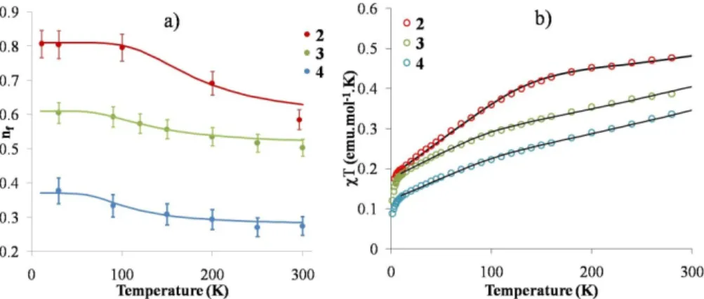
Table 2. Thermodynamic constants derived for the SS1 ⇌ SS2 equilibrium from nf vs. T and χ vs. T plots (Figure 3).
Compound nf(gs) nf(ex) χ(gs)a) χ (ex) a) ΔH(nf) b) ΔH(χ)b) ΔS(nf) c) ΔS(χ)c) %imp
Cp*2Yb(4,7-‐Me2phen) (2) 0.81 0.58 0.00182 0.00060 5.75 5.75 31 25 7.0
Cp*2Yb(3,4,7,8-‐Me4phen) (3) 0.61 0.50 0.00125 0.00060 3.2 3.6 21 21 6.8
Cp*2Yb(3,4,5,6,7,8-‐Me6phen) (4) 0.37 0.27 0.000108 0.00062 3.0 3.9 25 21 4.7
3
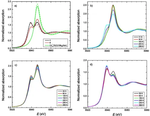
Figure 2: Yb LIII-‐edge XANES data for the complexes a) Cp*2Yb(2,9-‐Me2phen), 1, Cp*2Yb(2,9-‐Me2,4,7-‐Ph2phen), 5, and
Cp*2Yb(5,6-‐Me2phen)6 at 300 K (dashed) and 30 K (solid). b) Cp*2Yb(4,7-‐Me2phen), 2, at various temperatures, c) Cp*2Yb(3,4,7,8-‐
Me4phen), 3, at various temperatures and d) Cp*2Yb(3,4,5,6,7,8-‐Me6phen), 4, at various temperatures.
The LIII-‐edge XANES spectra are able to distinguish between
these two possibilities. In contrast to Cp*2Yb(5,6-‐Me2phen),
which shows only one temperature-‐independent f13 feature at
8946 eV (Figure 2a), Cp*2Yb(4,7-‐Me2phen), 2, presents two
different features, in agreement with the presence of both f14
and f13 contributions, for which the intensity ratio changes
with the temperature (Figure 2b). This situation was ob-‐ served in previous work with some substituted bipy adducts3
when the ligand can accept the electron from Yb in Cp*2Yb
into several accessible π* orbitals (π*1, π*2, ...), which results in
several possible open-‐shell singlets in which both the relative π* contribution and the f13:f14 ratio are different, accounting
for the temperature dependence in the LIII-‐XANES spectra. For
[Cp*2Yb(4,7-‐Me2phen)], 2, the f13 configuration fraction (also
known as the relative f13 occupancy or the f-‐hole occupancy,
nf) varies from 0.81 at low temperature to 0.58 at 296 K. This
situation also accounts for the low magnetic moment of Cp*2Yb(4,7-‐Me2phen)], 2. When the number of methyl groups
is increased in the phenanthroline ligand, the f13:f14 ratio
decreases and the spectra continue to show a temperature dependence.
In the Cp*2Yb adducts with substituted bipyridine ligands, the
temperature dependence is due to the presence of several open-‐shell singlet states (SS) that develop below the triplet (T) and it is therefore possible to determine their relative population using a Boltzmann equation.3 A similar methodol-‐
ogy is used in this work for Cp*2Yb(4,7-‐Me2phen), 2;
Cp*2Yb(3,4,7,8-‐Me4phen), 3; Cp*2Yb(3,4,5,6,7,8-‐Me6phen), 4
and the fits are presented in Figure 3, while the parameters are reported in Table 2. Only two singlet states, SS1 and SS2, are used in all three complexes. In the same manner, the magnetic curves are fit (Figure 3) with a Boltzmann distribution of two open-‐shell singlet states below the triplet and the enthalpy and entropy changes obtained from both fits (XANES and magnetism) agree well. The enthalpy change for Cp*2Yb(4,7-‐Me2phen), 2, is 5.75 kJ.mol-‐1 and lies in the
reported range for the enthalpy changes in substituted bipyridine adducts of Cp*2Yb. As an additional note, the
percentage of 2F7/2 impurties used for the fit of the magnetic
data is relatively high, compared to previous work, but is rationalized by the thermal instability of 2-‐4 (see Reactivity Section). The LIII-‐edge XANES data give nf values of 0.41 and
0.55 at 30 K for Cp*2Yb(2,9-‐Me2phen) 1, and Cp*2Yb(2,9-‐Me2-‐
4,7-‐Ph2phen) 5, respectively, and show no temperature
dependence for Cp*2Yb(2,9-‐Me2phen), 1, while nf decreases to
0.37 at 300 K for Cp*2Yb(2,9-‐Me2-‐4,7-‐Ph2phen), 5. The reason
for the low nf values and variable temperature behavior of
these two complexes is not addressed in a quantitative manner in this work but the general behavior is similar to that of Cp*2Yb(6,6'-‐Me2bipy) and Cp*2Yb(6-‐Mebipy), respectively;3
the presence of a methyl group(s) in the α position to nitrogen destabilizes the f13 configuration, presumably
because of the steric hindrance. The low values of nf for 1 and
5 also account for the low values of effective moments (see Table 1, nf = 0 indicates diamagnetic f14) and for the chemical
shift in their solution 1H NMR spectra.
4
Figure 3: a) Plot of nf vs. T (K) for 2-‐4. The best fits are obtained from a Boltzmann distribution between two singlet states using
the parameters reported in Table 2. b) Plot of χT vs. temperature for complexes 2-‐4. The black lines for each compound show the fit to the data using the parameters reported in Table 3.
X-‐ray crystallography. The ORTEPs of Cp*2Yb(4,7-‐Me2phen),
2, and Cp*2Yb(2,9-‐Me2-‐4,7-‐Ph2phen), 5, are shown in Figure 4
and Figure 5, respectively. Some bond lengths and angles are given in Table 3 and crystal data are in the Experimental Section and SI. It is clear that these two adducts are monomers in the solid state, in contrast to the phenanthroline and 3,8-‐Me2phen adducts.6 The average Yb-‐C(Cp*) distance in
Cp*2Yb(4,7-‐Me2phen) of 2.642 ± 0.007 Å is 0.032 Å and 0.020
Å longer than the equivalent distances in Cp*2Yb(phen)
monomer and in the four molecules in the two crystal structures of Cp*2Yb(5,6-‐Me2phen), respectively. The Yb-‐N
distances follow a similar pattern; the distance in Cp*2Yb(4,7-‐
Me2phen) is 0.035 Å and 0.030 Å longer than the equivalent
distances in Cp*2Yb(phen) monomer and Cp*2Yb(5,6-‐
Me2phen), respectively. The Yb-‐C(Cp*) and Yb-‐N distances in
Cp*2Yb(2,9-‐Me2-‐4,7-‐Ph2phen), 5, are 0.098 Å and 0.16 Å,
respectively, much longer than equivalent distances in Cp*2Yb(4,7-‐Me2phen), 2, and close to the equivalent distances
in Cp*2Yb(py)2 of 2.74 Å and 2.565 ± 0.005 Å, respectively.12
The longer Yb-‐C(Cp*) and Yb-‐N distances at 173 K in 2 and 5, relative to these distances in Cp*2Yb(phen) and Cp*2Yb(5,6-‐
Me2phen)6, indicate that the population of the Yb(II, f14)
configuration is greater in the former adducts, consistent with the lower values of nf.
Table 3. Selected bond distances (Å) and angles (°) at 173 K for complexes 2 and 5.
Cp*2Yb(4,7-‐Me2phen) (2) Cp*2Yb(2,9-‐Me2-‐4,7-‐Ph2phen) (5)
Yb-‐C (distances range) 2.630(4) – 2.653(4) 2.699(10) – 2.781(7) Yb-‐C(ring) (mean) 2.642 ± 0.007 2.74 ± 0.02 Yb-‐Ct (mean)a 2.35 2.46 Ct-‐Yb-‐Ct a 139.5 140.0 Yb-‐N(mean) 2.35 ± 0.01 2.51 ± 0.002 C(23)-‐Cexob 1.486(6) 1.496(9) C(28)-‐Cexo c 1.507(5) 1.506(9) torsion angle N-‐C-‐C-‐N 1 3 torsion angle C-‐C-‐C-‐C 1 1
a) Ct is the centroid of the C5Me5 ring b) Cexo in 2 is C(33) and C(35) in 5 c) Cexo is C(34) in 2 and C(41) in 5
Figure 4. ORTEP for the complex Cp*2Yb(4,7-‐Me2phen) (2).
(Thermal ellipsoids are at 50% level)
Figure 5. ORTEP for the complex Cp*2Yb(2,9-‐Me2-‐4,7-‐
5
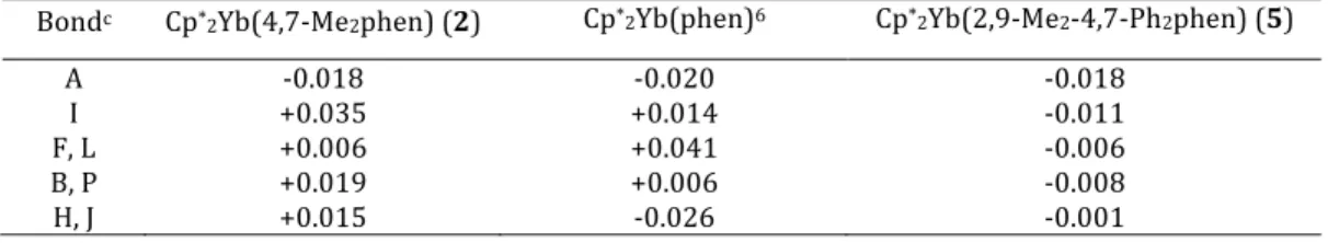
Table 4. Bond Lengths (Å) Changes Δa,b in 2, 5 and Cp*2Yb(phen).
Bondc Cp*2Yb(4,7-‐Me2phen) (2) Cp*2Yb(phen)6 Cp*2Yb(2,9-‐Me2-‐4,7-‐Ph2phen) (5)
A -‐0.018 -‐0.020 -‐0.018
I +0.035 +0.014 -‐0.011
F, L +0.006 +0.041 -‐0.006
B, P +0.019 +0.006 -‐0.008
H, J +0.015 -‐0.026 -‐0.001
a) Δ is the bond length in the adduct minus that in the free ligand in Å. b) Free 4,7-‐Me2phen, ref 13 and for free 2,9-‐Me2-‐4,7-‐Ph2phen, ref 14.
c) The letter refers to the C-‐C or C-‐N bond label in Scheme 2. Although the Yb-‐C(Cp*) and Yb-‐N distances in Cp*2(4,7-‐
Me2phen), 2, are longer that those in Cp*2Yb(phen), the C‒C
and C‒N distances in the phenanthroline ligands are con-‐ sistent with occupation of the b1 and a2 symmetry orbitals..
Using the method outlined in an earlier article,6 the distances
labeled A, I, F and L, B and P, H and J (Scheme 2) change in opposite directions depending on whether the b1 or a2 orbit-‐
als are populated (See Scheme 1 and Table 4 of ref6). The
bond length alteration for Cp*2(4,7-‐Me2phen), 2, and
Cp*2(phen), monomer, indicate that both of these symmetry
orbitals are populated but the relative values of Δ are differ-‐ ent, implying that the relative energy of these orbitals depend on the position of the methyl groups in the ring. Although these bond length changes are only qualitative, they point towards population of the a2 orbital in which the coefficient
for the carbon pπ-‐orbitals in the 4,7-‐positions in the ring are greater than that in b1; this difference has ramifications for
the reactivity at these positions, see below.
Solutions properties. The UV-‐Vis spectra at 20 °C in toluene for the 4,7-‐Me2phen and related adducts are shown in Figure
6. The absorption around 550 nm is due to the π−π* transition in the radical-‐anion.15
Figure 6: UV-‐Vis spectra of Cp*2Yb(4,7-‐Me2phen), 2, (green),
Cp*2Yb(3,4,7,8-‐Me4phen) (red), 3, Cp*2Yb(3,4,5,6,7,8-‐
Me6phen), 4, (blue).
These adducts also have an intense absorption around 900 nm, which was assigned to a f-‐f transition in Cp*2Yb(phen).15
Although this transition is forbidden, its intensity is attributed to the formation of a ligand-‐to-‐metal charge transfer state,
1LMCT, in which intensity is stolen.15
The solution 1H NMR spectra in C6D6 at 20 °C for the dime-‐
thylphenanthroline adducts are listed and assigned in Table 5. The assignments are made by the changes that results when a pair of hydrogens is replaced by a pair of methyl groups at a given position in the phenanthroline ring. Three patterns emerge from the changes in the chemical shift values. (i) The values of δ2,9 are strongly shifted downfield, consistent with
the dipolar contribution dominating the chemical shift tensor. This is expected since the dipolar contribution is related to the geometry of the hydrogen atoms and H2,9 are proximal to
the paramagnetic center. The chemical shifts of δ2,9 also aligns
the magnetic z-‐axis collinear with the N-‐N vector and perpen-‐ dicular to Ct-‐Yb-‐Ct. (ii) The values of δ4,7 and δ3,8 are less
strongly shifted downfield and when the H4,7 or H3,8 are re-‐
placed by methyl groups, the sign of the chemical shift chang-‐ es showing that the contact contribution to the chemical shift tensor dominates the dipolar contribution.16 (iii) The δ5,6
values are the farthest upfield and the sign does not change when H5,6 is replaced by methyl groups.
In the neutral adducts, the order of chemical shift is δ2,9 > δ4,7 >
δ3,8 > δ5,6 but in the cation, the order of δ4,7 and δ3,8 exchange
places consistent with the dipolar contribution dominating the chemical shift tensor in the cation. The value of δCp* in the
neutral and cation adducts, is around 4 ppm in all cases. It is informative to compare the chemical shifts of the neutral bipyridine and the neutral phenanthroline adducts of Cp*2Yb
using the graphic in Scheme 3. Scheme 3.
Table 5. 1H NMR chemical shifts (δ) of the phenanthroline adducts at 300K.(a)
Compound 2,9 4,7 3,8 5,6 Cp* ref
Cp*2Yb(phen)-‐monomerb) 139.9 47.9 14.0 0.47 4.11 ref6
Cp*2Yb(4,7-‐Me2phen) b) 109.1 -‐21.4(Me) 16.7 3.15 3.63 This work
Cp*2Yb(3,8-‐Me2phen) b) 95.5 51.1 -‐10.0 (Me) 3.83 3.36 ref6
Cp*2Yb(5,6-‐Me2phen) b) 137.4 44.1 14.7 0.03 (Me) 3.95 ref6
[Cp*2Yb(4,7-‐Me2phen)]I c) 301 8.4 (Me) 53.4 -‐3.5 3.73 This work
N N
γ
b N Nγ
pβ
pα
pβ
bα
b6
[Cp*2Yb(phen)]I c) 281 9.5 52.4 -‐2.5 3.82 ref6a) δ value in ppm relative to Me4Si b) in C6D6 or C7D8 c) in CD2Cl2
The labels α, β, γ are the hydrogen on the alpha, beta and gamma carbon atoms relative to N and b is for bipyridine and p for phenanthroline. The values of the chemical shift at 20 °C in C6D6 for Cp*2Yb(bipy)4,5 and Cp*2Yb(phen)6 are, to the
nearest whole number, αb, 160; βb, 6; γb, 26, in the bipyridine
complexes and αp, 140; βp, 14; γp, 48 in the phenanthroline
complexes. In the bipyridine and phenanthroline adducts δα >> δβ < δγ, with δα in each adduct approximately equal but δβ
and δγ in the phenantholine complexes being about twice the
value of δβ and δγ in the bipyridine complexes. Since the chemi-‐
cal shift of the hydrogens on the β and γ sites are largely de-‐ termined by the contact contribution, this implies that the unpaired spin density at the γ site is greater than that at the β site, and is greater in the phenantholine complexes than in the bipyridine complexes; as mentioned above, this difference plays a role in the reactivity at these positions, see later Calculations. As in earlier articles on the bipyridine adducts, the CASSCF methodology is used to determine the ground state electronic structure of the phenanthroline adducts of Cp*2Yb.1-‐4,6 The phenanthroline adduct of Cp*2Yb is calculated
to consist of a pair of nearly degenerate triplets, T1 and T2, which are 2.1 eV below the open-‐shell singlet exited state.6
When two methyl groups are attached to the 3,8 and 5,6 positions in the phenanthroline ligand, the ground state of the adducts is still a triplet but the open-‐shell singlet excited state is now only 0.08 and 0.09 eV above the triplet. In these compounds, the f13 configuration is the only active one and
the ytterbium atoms are fully trivalent; the computational results are in agreement with the magnetic and XANES experiments for these adducts.6 In contrast, the calculated
ground state of the 4,7-‐Me2phen adduct is not a triplet but
consists of three open-‐shell singlets, SSI, SS2 and SS3 that are lower in energy relative to the triplet by 0.44 eV, 0.06 eV and 0.02 eV, respectively. The three open-‐shell singlets have different contribution of f13 and f14 from Yb and π*1 and π*2
contributions from the radical anionic ligands and both are therefore multiconfigurational singlets. The SS1, SS2, and SS3 states are given by 0.92 f13 + 0.08 f14, 0.34 f13 + 0.66 f14 and
0.63 f13 + 0.37 f14, respectively. The π*1 and π*2 contribution to
SS1 is 0.89 π*1 + 0.11 π*2 and for SS2 and SS3, the
contributions are 0.35 π*1 + 0.65 π*2 and 0.68 π*1 + 0.32 π*2,
respectively. The most stable state, SS1, is dominated by f13
and π*1 configurations and in the nearly degenerate SS2 and
SS3 states, these configurations are less dominant. For SS1, the nf value is 0.92, the value of c12 in Ψ = c1|Yb(III, f13)(phen•-‐
) > + c2|Yb(II, f14)(phen)0 >, such that nf = 1 when c1=1 and c2 =
0 and nf = 0 when c1 = 0 and c2 = 1. The calculated value of nf
for SS1 of 0.92 is in reasonable agreement with the experi-‐ mental value of 0.81 at low temperature, assuming that only SS1 is populated. As the temperature is increased the nf value
of the excited state decreases to 0.58, in reasonable agree-‐ ment with the calculated values of SS2 and SS3, where the average values in these two states is 0.48.
Reactivity. As mentioned in the Synthesis Section above, the yield of Cp*2Yb(4,7-‐Me2phen), 2, is higher and purification is
easier when the temperature is kept at 0 °C. In the synthesis of the other two adducts of Me4phen and Me6phen the tem-‐
perature must be maintained at ‒70 °C during the synthesis stage. It was not clear why the temperature is important until the thermal stability of isolated Cp*2Yb(4,7-‐Me2phen), 2, was
monitored by 1H NMR spectroscopy as a function of time. At
60 °C, the resonances of Cp*2Yb(4,7-‐Me2phen), 2, in C7D8
disappear while a new set appears, a transformation that is complete over 15 days. The 1H NMR spectrum is complex but
two different set of resonances can be identified, labeled A1
and A2, since the ligand resonances are asymmetric, and the
Cp* resonances appear as a broad feature at ~ 3.7 ppm. The spectrum is available in SI. Although the resonances cannot be conclusively assigned, they may be tentatively assigned as due to a hydrogen atom transfer reaction analogous to that illus-‐ trated in Scheme 1, as shown in the Experimental Section, Scheme 7. This attribution is supported by the following ex-‐ periments.
1. The thermal reaction conducted on a synthetic scale yields brown crystals isolated by crystallization from toluene solu-‐ tion (referred to as 7). The 1H NMR spectrum contains the
resonances referred to as A1 and A2, above. A brown crystal
was selected and an X-‐ray data set was collected (see Figure 7). Analysis of the X-‐ray data indicated the presence of two different molecules that co-‐crystallize in a 72:28 ratio. The major one consists of a dimethylphenanthroline group that has lost a H atom from a methyl group leading to a terminal methylene represented in pink in Figure 7 (C28 and C34) (Scheme 4). The minor molecule consists of a dimethylphe-‐ nanthroline to which a H atom is added to the phenanthroline ring at carbon C28. This leads to a C(Me)(H) group in two possible positions at C28 (up and down). This position disor-‐ der is represented in orange in Figure 7 (Scheme 4) (C28A-‐ C34A and C28B-‐C34B). Details of the crystallographic solution are given in the Experimental Section and in SI.
Figure 7. ORTEP for the brown crystal obtained from toluene in the thermal reaction of Cp*2Yb(4,7-‐Me2phen), 2 (thermal
ellipsoids are at 50% level) and referred to as "compound" 7. The carbon atoms represented in orange are due to a posi-‐ tional disorder of a methyl group connected to the phenan-‐ throline ring and the carbon atom represented in pink is due to the methylene group.
7
In both cases, a pyridyl ring has lost its aromaticity that re-‐sults in a shorter Yb-‐N(2) distance of 2.286(3) Å compared to Yb-‐N(1) distance of 2.363(3) Å, a consequence of the localiza-‐ tion of negative charge on the nitrogen N(2) (Figure 7).Washing the brown crystals with pentane results in a pur-‐ ple solution and a green residue. Cooling the pentane solution (-‐20 °C) afforded very small crystals (too small for conven-‐ tional X-‐ray crystallographic use), but whose 1H NMR spec-‐
trum in C7D8 is identical to those labeled as A1, with the excep-‐
tion that the Cp* resonances are resolved as two singlets at δ = 3.94 ppm and 3.58 ppm due to 15 protons each.
2. Hydrolysis (H2O) of the products of thermal transformation,
on a NMR tube scale, results in an asymmetric set of 1H reso-‐
nances due to H2A1, that is , 4,7-‐Me2phen+2H’s, which evolve
into resonances due to 4,7-‐Me2phen over one day. No reso-‐
nances attributable to A2 are identified. Examination of the
solution by GCMS showed Cp*H and 4,7-‐Me2phen. When the
thermal transformation is monitored by 1H NMR spectroscopy
in C7D8 in presence of excess dihydroanthracene or 1,4-‐
cyclohexadiene, neither anthracene nor benzene are detected in the spectrum and the t1/2 of the reaction is not altered by
these two free radical traps. Monitoring the transformation in an atmosphere of D2 also did not alter the rate but HD is in-‐
deed observed. These experiments do not identify A1 and A2
with confidence but allow us to ascribe them as resulting from a disproportionation reaction analogous to that shown in Scheme 1.
Discussion. The key point that emerges when the bipyridine and phenanthroline adducts of Cp*2Yb are compared is that
the bipyridine adducts are all open-‐shell singlets but the phe-‐ nanthroline adducts are either open-‐shell multiconfiguration-‐ al singlets in Cp*2Yb(4,7-‐Me2phen) 2, or triplets in the phe-‐
nanthroline, 3,8-‐Me2phen, and 5,6-‐Me2phen adducts. Thus,
two methyl groups in different positions on the phenantholine ligand change the magnetic properties of the resulting adduct. A qualitative MO model was presented to account for the difference between Cp*2Yb(bipy) and Cp*2Yb(phen), which
are ground state singlets and triplets, respectively.6 The mod-‐
el is straightforward for bipyridine, since only the b1-‐orbital is
low enough in energy to accept an electron in the charge transfer ground state resulting in a singlet state. In the phe-‐ nanthroline radical anion, the LUMO and LUMO + 1 orbitals of b1 and a2 symmetry (in C2v symmetry) are low enough in
energy to be populated in the charge-‐transfer ground state of the adducts resulting in a triplet state, as shown in the left-‐ hand side of the diagram in Figure 8. The spin state of Cp*2Yb(x,x'-‐phen) is qualitatively determined by the energy
that separates the b1 and a2 symmetry orbitals. When the
separation is on the same order or smaller than in phenan-‐ throline, a triplet state results when x,x' is 3,8 or 5,6, as shown
in the left-‐hand side of the diagram in Figure 8, since the symmetry of the SOMOs (singly occupied molecular orbitals) is different and the spins of the electrons in these orbitals can have the same sign. When the separation is larger, the spin arrangement approaches that of Cp*2Yb(bipy), resulting in a
singlet ground state, as shown in the right-‐hand side of Figure 8, since the symmetry of the SOMOs is the same and the elec-‐ trons in them cannot have the same sign.
A model to account for the energy separation between the b1
and a2 orbitals, based on the CI calculations, is presented as a
guide to understand how the methyl groups can control the spin state in the Cp*2Yb(x,x'-‐phen). Figure 9 shows a cartoon
energy-‐level diagram that is modified from Figure 8 in ref 4.
The diagram illustrates how the open-‐shell singlet, OSS, and the closed-‐shell singlet, CSS, energy levels mix under configu-‐ ration interaction. The diagram is idealized since it only shows a singly occupied f-‐electron interacting with a single electron in a π* orbital and how these two orbitals change when CI is turned on as the triplet state energy is held con-‐ stant.
The energy separation, ΔSS, on the left-‐hand side of Figure 9
represents the large separation in OSS and CSS in Cp*2Yb(phen). Replacing two hydrogens by methyl groups on
the phenanthroline ligand reduces ΔSS since the methyl group is a σ-‐donor relative to hydrogen. This raises the energy of the HOMO of the ligand, moving it closer to the energy of the empty d-‐parentage orbital of Cp*2Yb(II, f14) as the bond de-‐
velops, and stabilizing the configuration. In addition, the me-‐ thyl substituents raise the LUMO of the ligand, driving up the π* orbital energy, as shown by the increased value of E1/2,
relative to Fc0/+. The values in thf for phen and 3,4,7,8-‐
Me4phen are -‐2.55 and -‐2.68 V, respectively.9
Scheme 5.6
8
Figure 8. Qualitative MO diagram comparing bonding in Cp*2Yb(3,8-‐Me2phen) (a) and Cp*2Yb(4,7-‐Me2phen). The 4f-‐parentage
orbitals in the center of the MO-‐diagram are associated with the Cp*2Yb-‐fragment in C2v symmetry; the two sigma bonding and
antibonding orbitals used in bonding of Cp*2Yb to the diimine ligands are are not shown, but the three empty d-‐parentage orbitals
are shown. The orbitals on the extreme left-‐and and right-‐hand side are the LUMO and LUMO + 1 of the diimine ligands. The orbit-‐ als shown in (a) and (b) are the two possible electron configurations in the charge-‐transfer state of the adducts.
Figure 9. Cartoon energy-‐level diagram demonstrating the effect of singlet -‐ triplet configurations; close-‐shell singlet, CSS, open-‐ shell singlet, OSS, and triplet, T, on a singly occupied f orbital coupled with one unpaired electron in a ligand π* orbital. The orbital energies are only qualitative, but they reflect the CASSCF energies.
The reduction potential for the other dimethylphenanthro-‐ lines are not available in the literature. The σ-‐donor substitu-‐ ent destabilizes OSS with the net effect that ΔSS gets smaller,
when CI is turned on, driving the energy separation between the singlet and triplet state energies, ΔST, closer to each other,
as in the middle illustration in Figure 9. The qualitative pic-‐ ture agrees with the calculated separation between the sin-‐ glet-‐triplet state in the phen, 3,8-‐Me2phen, and 5,6-‐Me2phen,
with the triplet state lower in energy. As ΔSS continues to
contract, the multiconfigurational state, composed of the OSS and CSS configurations, falls below the triplet state, as found in the calculation for Cp*2Yb(4,7-‐Me2phen), 2. A qualitative
picture for these energy changes is provided by inspection of the relative coefficients on the carbon-‐pπ orbitals on the b1-‐
and a2-‐symmetry orbitals. The coefficient on the carbon pπ
orbitals in the 5,6-‐positions of b1-‐orbital are small, the a2-‐
orbital has a node in the 3,8-‐positions, and the coefficient in the 4,7-‐positions in both orbitals is large, Scheme 5. The net effect is that methyl groups occupying the 3,8-‐ and 5,6-‐ positions destabilize the OSS state. The destabilization is substantially larger in both orbitals when methyl groups
occupy the 4,7-‐positions, lowering the ΔSS and driving the multiconfigurational singlet state below the triplet when CI is turned on. As more methyl groups replace hydrogens in the phenanthroline ligand, the stabilization continues, consistent with the experimental results in Table 2.
The change in the electronic structure of the adducts plays a key role in determining the site of reactivity in the phenan-‐ throline adducts. Thus, Cp*2Yb(phen) and Cp*2Yb(3,8-‐
Me2phen) are dimers in the solid state due to formation of a σ-‐
C‒C bond between the carbon atoms in the 4-‐position of the phenanthroline ligand.6 The 3,8-‐Me2phen adducts exists in a
dimer ⇌ 2 monomer equilibrium in solution but the insolubil-‐ ity of the phen adduct prevents the acquisition of an equilibri-‐ um constant. In the solid state structures of these dimers, the C‒C bond length is longer and presumably weaker in the 3,8-‐ Me2phen dimer than in the unsubstituted phen dimer, likely
due to steric repulsion between the two monomeric units in the dimer. Steric repulsion is presumably a factor contributing to the monomeric solid state structure of Cp*2Yb(5,6-‐
Me2phen) as well as the distribution of the unpaired spin
density at the 4-‐carbon –pπ orbital.6 b1 a1+a2 +b1 a1+b1 b1* a2* a2* b1* a2 a1+b1 a2* b1* a2 b1* a2* b1 5d -parentage 4f-parentage
3,8-Me2phen a) Cp*2Yb b) 4,7-Me2phen
2 a1+ a2 + 2 b1+ 2 b2 b1 b1 CSS OSS T ΔSS f π* Cp*2Yb(phen) ΔST CI turne d on f π* Cp*2Yb(x,x'-Me2phen) x,x' = 3,8 or 5,6 ΔST f π* Cp*2Yb(x,x'-Me2phen) x,x' = 4,7
9
When two methyl groups are located on the 4,7-‐positions, therepulsion between the methyl groups in a hypothetical dimer is larger than the stabilization resulting from σ-‐C‒C bond formation. The increase in spin density on the carbon pπ-‐
orbitals at the 4,7-‐positions is relieved by delocalization of the spin density in the pπ-‐orbital into the σ*-‐C‒H bonds of a me-‐
thyl group. This delocalization results in weakening the C‒H bond of the methyl group and strengthening the C=C bond as the negative charge in the pyridyl ring rearranges forming A1
and A2 in the Cp*2Yb adducts, as well in the Cp2Ti adducts.11
The qualitative picture that is outlined above also fits the experimental observation that Cp*2Yb(4,4'-‐bipy) is a thermal-‐
ly stable monomer but Cp*2Yb(4,7-‐Me2phen) is not, even
though both have open-‐shell singlet multiconfigurational ground states and a similar value of nf. Inspection of the b1-‐
symmetry orbitals in both ligands provides a possible answer: the coefficients of the pπ-‐carbon orbitals on the pπ C-‐4 position
in each radical anion is smaller in bipy·-‐ than in phen·-‐. This difference results in localization of less spin density on a given carbon atom in bipy·-‐ and a smaller driving force for C‒C bond formation or C‒H bond breaking. This conjecture, viz., the spin density distribution in the radical anions means that deter-‐ mining the experimental spin density map in these radical anions is the next step in unraveling the physics and chemis-‐ try of these molecules.
Three possible mechanisms may be considered for the reac-‐ tion illustrated in Scheme 1: (i) an intramolecular free radical process, (ii) an intermolecular radical-‐pair process, and (iii) an intermolecular proton transfer process. A free radical mechanism is ruled out by the results of the radical trapping experiments, which leaves the intermolecular pathways in which a hydrogen atom (ii) or a proton (iii) is transferred. These two processes are illustrated in Scheme 6. The path-‐ ways differ by the nature of the σ-‐C‒C bond between the two radical anions; in (ii) the spins are weakly correlated as in a pair of radicals and in (iii) the spins are more strongly corre-‐ lated as in a bond. The experimental information does not allow a distinction to be made, but the formation of HD when D2 is present seems to favor the proton transfer mechanism.
Scheme 6.
Conclusion. The ground state electronic structure of the paramagnetic charge-‐transfer diimine adducts of Cp*2Yb
range from open-‐shell singlets to triplets depending on the identity of the diimine and the number and position of methyl substituents in the diimine. The different electronic structures are perhaps, surprising, since the neutral ligands have similar donor and acceptor properties17 and the magnetic properties
are determined by the symmetry and energy of the SOMO orbital(s) in the radical anion. All of the 2,2'-‐bipyridine ad-‐ ducts studied have open-‐shell singlet ground states whose wave function is described by Ψ = c1|Yb(III, f13)(bipy•-‐) > +
c2|Yb(II, f14)(bipy)0 >.4 All of the methyl and dimethyl bipy
adducts are open-‐shell singlets in which the coefficients c1 and
c2 depend on their position in the ring.3 In contrast, the vari-‐
ous phenanthroline adducts range from a triplet, c2 = 0, when
1,10-‐phenanthroline is the diimine,6 to open-‐shell singlets in
the 4,7-‐Me2phen adduct. The origin of this difference is traced
to the energy separation between the open-‐shell singlet (Yb↑
L↓) and the close-‐shell singlet (Yb↑↓ L0) and therefore their
extent of configuration-‐interaction mixing, which depends on the number and position of the methyl groups in the ring. The electronic structure of these phenanthroline adducts alters their reactivity patterns. For example, the 3,8-‐Me2phen adduct
exists in a dimer-‐monomer equilibrium in solution in which the σ-‐C‒C bond that connects the monomeric halves in the dimer is weak; the BDE is ca. 8 kcal-‐1mol-‐1.6 The 4,7-‐Me2phen
adduct undergoes an irreversible disproportionation in solu-‐ tion in which one C‒H is broken while another one is formed, Scheme 6. A tentative suggestion is offered for the different reactivity patterns that involves the extent of unpaired spin density in the carbon pπ-‐orbital on the 4 and 7-‐positions. This
postulate, if true, provides a way to control the reactivity patterns in these organometallic compounds.
EXPERIMENTAL SECTION.
General considerations. All reactions were performed using standard Schlenk-‐line techniques or in a drybox (MBraun). All glassware was dried at 150 °C for at least 12 h prior to use. Toluene, pentane, and diethyl ether were dried over sodium and distilled while CH2Cl2 was purified by passage through a
column of activated alumina. Toluene-‐d8 and CH2Cl2-‐d2 were
dried over sodium. All the solvents were degassed prior to use. 1H NMR spectra were recorded on Bruker AVB-‐400 MHz,
DRX-‐500 MHz, AVB-‐600 MHz and Advance 300 MHz spec-‐ trometers. 1H chemical shifts are in δ units relative to TMS,
and coupling constants (J) are given in Hz. Infrared spectra were recorded as Nujol mulls between KBr plates on a Ther-‐ mo Scientific Nicolet IS10 spectrometer. Samples for UV-‐Vis-‐ NIR spectroscopy were obtained in a Schlenk-‐adapted quartz cuvette and obtained on a Varian Cary 50 scanning spectrom-‐ eter. Melting points were determined in sealed capillaries prepared under nitrogen and are uncorrected. Elemental analyses were determined at the Microanalytical Laboratory of the College of Chemistry, University of California, Berkeley. X-‐ray structural determinations were performed at CHEXRAY, University of California, Berkeley. Magnetic susceptibility measurements were made for all samples at 1, 5 and 40 kOe in a 7 T Quantum Design Magnetic Properties Measurement System that utilizes a superconducting quantum interference device (SQUID). Sample containment and other experimental details have been described previously.18 Sample integrity
was verified by observing the absorption spectra of an oxygen “canary” that is always loaded into one slot of each multislot sample holder, typically a divalent ytterbocene such as Cp*2Yb(OEt2). Diamagnetic corrections were made using
Pascal’s constants. The samples were prepared for X-‐ray ab-‐ sorption experiments as described previously and the same methods were used to protect the air-‐sensitive compounds from oxygen and water.4 X-‐ray absorption measurements
were made at the Stanford Synchrotron Radiation Lightsource on beamline 11-‐2. The samples were prepared and loaded into a liquid helium-‐flow cryostat at the beamline as described previously.4 Data were collected at temperatures ranging
from 30 to 300 K, using a Si(220) double-‐crystal monochrom-‐ ator. Fit methods were the same as described previously.4
Low temperature (ca. 2 K) EPR spectra were obtained with a Varian E-‐12 spectrometer equipped with an EIP-‐547 micro-‐ wave frequency counter and a Varian E-‐500 gaussmeter, which was calibrated using 2,2-‐diphenyl-‐ 1-‐picrylhydrazyl (DPPH, g= 2.0036).
Calculations. The ytterbium center was treated with a small-‐ core relativistic pseudopotential (RECP) ([Ar] + 3d)19 in com-‐