HAL Id: hal-01430580
https://hal.archives-ouvertes.fr/hal-01430580
Submitted on 7 Mar 2018HAL is a multi-disciplinary open access archive for the deposit and dissemination of sci-entific research documents, whether they are pub-lished or not. The documents may come from teaching and research institutions in France or abroad, or from public or private research centers.
L’archive ouverte pluridisciplinaire HAL, est destinée au dépôt et à la diffusion de documents scientifiques de niveau recherche, publiés ou non, émanant des établissements d’enseignement et de recherche français ou étrangers, des laboratoires publics ou privés.
Structural and EELS studies on Doped Carbon
Nanostructures for Cold Field Emission
Rongrong Wang, Raul Arenal, Aurélien Masseboeuf, David Neumeyer, Marc
Monthioux, Alejandro Lopez-Bezanilla
To cite this version:
Rongrong Wang, Raul Arenal, Aurélien Masseboeuf, David Neumeyer, Marc Monthioux, et al.. Struc-tural and EELS studies on Doped Carbon Nanostructures for Cold Field Emission. Microscopy and Microanalysis, Cambridge University Press (CUP), 2016, 22, pp.60–61. �10.1017/S1431927616000490�. �hal-01430580�
Structural and EELS studies on Doped Carbon Nanostructures for Cold Field
Emission
Rongrong Wang1,3, Raul Arenal1,2, Aurélien Masseboeuf3, David Neumeyer3, Marc Monthioux3,
Alejandro Lopez-Bezanilla4
1. Laboratorio de Microscopias Avanzadas, INA Zaragoza, Zaragoza - Spain 2. Fundacion ARAID, Zaragoza - Spain
3. CEMES - CNRS, Toulouse – France
4. Materials Science Div., Argonne National Lab. Illinois, USA
Along with the development of cold field emission technology, more and more microscopes equipped with cold field emission gun (C-FEG) are applied in scientific research. Thanks to the lower working temperature, the service time of emitter has been extended. In the meantime, this technology allows to emit electrons from smaller emission radius compared to thermionic emission and Schottky emission [1]. Based on this property, higher current density, higher brightness, better special coherence and lower energy distribution of the emission can be expected, which also means that a better electron beam can be obtained [2]. For the further improvement of cold field emission, new emitter materials have been explored. As a rolled-up graphene sheet, carbon nanotubes (CNTs) have the similar property, for instance, low work function, high mechanic performance, high melting point, high carrier mobility [3-4]. In addition, CNTs have small apex radius curvature for its quasi-one-dimensional character at nanoscale. Therefore, CNTs are capable of working under the operating condition of cold field emission [5]. Furthermore, carbon nanocones are considered as candidate as well [6]. Significant improvements of such kind of carbon nanocones are demonstrated by replacing the regular tungsten tip in transmission electron microscope [7]. For a higher performance of the cold field emission application, the electronic band structure of carbon nano-object can be modulated through the introduction heteroatoms and a lower work function can be expected [8,9]. Our work is devoted to the doping of these carbon nanostructures by nitrogen and/or boron, and to the evaluation of these doped carbon nano-objects by a series characterization for the cold field emission application.
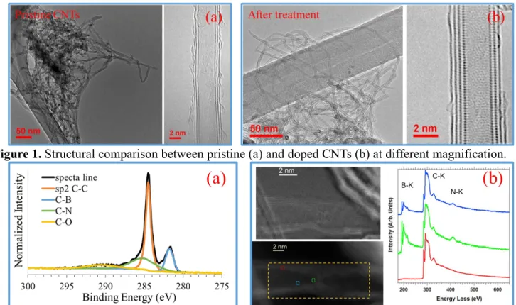
We started our doping study from the CNTs. The doping of CNTs was carried out in a tubular furnace from 1300°C to 1500°C under different atmosphere. Doped CNTs were characterized by transmission electron microscopy: high resolution imaging (HRTEM) and spatially resolved electron energy loss spectroscopy (EELS) in scanning (STEM) mode. Those studies were developed using 2 different aberrations corrected TEMs (FEI-Titan), working at low acceleration voltage (80 kV) in order to prevent nanotubes from the damage caused by electronic irradiation. In addition X-ray photoelectron spectroscopy (XPS, Kratos Analytical) has also been carried out. By these characterizations, structural modification after thermal treatment can be studied and the incorporation of hetero elements can be identified at local and macro scale. The pristine CNTs are compared with doped CNTs (fig. 1), this comparison illustrates that there is non-significant structural modification after the thermal treatment. The formations of C-B bonding and C-N bonding are identified in the spectra at macro scale by XPS (fig. 2 (a)). The presence of boron and nitrogen inside CNTs at local scale is confirmed by EELS (fig. 2 (b)). All these results will be discussed in depth in this contribution [10].
References:
[1] A. V. Crewe et al, The Review of Scientific Instruments 39 (1968), p. 576-583. [2] N. De Jonge et al, Nature 420 (2002), p. 393-395.
[3] J. W. Mintmire, B. I. Dunlap and C. T. White, Physical Review Letters 68 (1992), p. 631–634. [4] J.N. Coleman et al, Carbon 44 (2006), p. 1624–1652.
[5] J.-C. Charlier et al, Nano Letters 2 (2002), p. 1191–1195. [6] L. Ronald, M. Montioux, Nature 385 (1997), p. 211-212. [7] F. Houdellier et al, Carbon 50 (2012), p. 2037-2044.
[8] P. Ayala et al, Reviews of modern physics 82 (2010), p. 1843-1885. [9] R. Arenal, X. Blase, A. Loiseau, Advances in Physics 59, 101 (2010).
[10] This work was supported by the project ANR LASCAR (ANR-13-BS04-0007), the project ESTEEM2 (Integrated Infrastructure Initiative - I3, Grant Agreement 312483), the Spanish MINECO (FIS2013-46159-C3-3-P), and international associated laboratory TALEM (CNRS - U. of Zaragoza). TEM studies were developed in the Advanced Microscopy Laboratory (LMA) of Institute of Nanoscience of Aragon (INA) - U. of Zaragoza (Spain).
Figure 1. Structural comparison between pristine (a) and doped CNTs (b) at different magnification.
Figure 2. (a) XPS spectra of carbon C1sbinding energy. (b) Selection of EEL spectra extracted from the
spectra image; blue, red and green spectra correspond to the pixels outlined in blue, red and green in bottom left image.