HAL Id: hal-00917720
https://hal.archives-ouvertes.fr/hal-00917720
Submitted on 12 Dec 2013
HAL is a multi-disciplinary open access
archive for the deposit and dissemination of
sci-entific research documents, whether they are
pub-lished or not. The documents may come from
teaching and research institutions in France or
abroad, or from public or private research centers.
L’archive ouverte pluridisciplinaire HAL, est
destinée au dépôt et à la diffusion de documents
scientifiques de niveau recherche, publiés ou non,
émanant des établissements d’enseignement et de
recherche français ou étrangers, des laboratoires
publics ou privés.
Crystallisation of a highly metastable hydrated calcium
pyrophosphate phase
Pierre Gras, Sébastien Teychené, Christian Rey, Cédric Charvillat, Béatrice
Biscans, Stéphanie Sarda, Christèle Combes
To cite this version:
Pierre Gras, Sébastien Teychené, Christian Rey, Cédric Charvillat, Béatrice Biscans, et al..
Crystalli-sation of a highly metastable hydrated calcium pyrophosphate phase. CrystEngComm, Royal Society
of Chemistry, 2013, vol. 15, pp. 2294-2300. �10.1039/C2CE26499D�. �hal-00917720�
O
pen
A
rchive
T
OULOUSE
A
rchive
O
uverte (
OATAO
)
OATAO is an open access repository that collects the work of Toulouse researchers and
makes it freely available over the web where possible.
This is an author-deposited version published in :
http://oatao.univ-toulouse.fr/
Eprints ID : 9381
To link to this article :
DOI:10.1039/C2CE26499D
URL :
http://dx.doi.org/10.1039/C2CE26499D
To cite this version :
Gras, Pierre and Teychené, Sébastien and Rey, Christian and
Charvillat, Cédric and Biscans, Béatrice and Sarda, Stéphanie and
Combes, Christèle Crystallisation of a highly metastable hydrated
calcium pyrophosphate phase. (2013) CrystEngComm, vol. 15 (n°
12). pp. 2294-2300. ISSN 1466-8033
Any correspondance concerning this service should be sent to the repository
administrator:
staff-oatao@listes-diff.inp-toulouse.fr
Crystallisation of a highly metastable hydrated calcium
pyrophosphate phase
Pierre Gras,aSe´bastien Teychene´,bChristian Rey,aCe´dric Charvillat,aBe´atrice Biscans,b Ste´phanie Sardacand Christe`le Combes*a
A simple and fast synthesis method was set up to obtain pure hydrated calcium pyrophosphate (CPP) phases of biological interest. This work focused on a specific phase synthesised at 25 uC and pH 4.5 in a stirred tank reactor. Powder X-ray diffraction, FTIR spectroscopy, scanning electron microscopy and thermogravimetric analyses revealed that the phase is unknown but presents similarities with a monoclinic tetrahydrated CPP phase (Ca2P2O7?4H2O, m-CPPT b phase) synthesised under the same conditions of pH
and temperature. Characterisation of the unreferenced phase (u-CPP) has been performed, especially to better identify its composition, structure and stability, as well as its possible relation to the m-CPPT b phase or to other hydrated CPP phases.
Introduction
Osteoarthritis (OA) is the most common form of rheumatic disease, leading ultimately to chronic pain and disability for the patient. Calcium pyrophosphate (CPP) dihydrate crystals (CPPD, Ca2P2O7?2H2O) have been observed in the
inter-articular spaces of arthritic patients and appear to induce an inflammatory response. Up to now current treatments are not able to inhibit the formation of CPP crystals.
Two polymorphs of CPPD have been identified in vivo: a triclinic form (t-CPPD)1and a monoclinic form (m-CPPD).2In
addition to m- and t-CPPD phases, other hydrated calcium pyrophosphate phases have been prepared in vitro, including a third dihydrated supposedly hexagonal polymorph (h-CPPD)2
and a dimorphic monoclinic tetrahydrate (CPPT, Ca2P2O7? 4H2O) referred to as m-CPPT a3and m-CPPT b.4
The syntheses reported in the scientific literature are based on dissolution of an intermediate, amorphous CPP (a-CPP) or calcium dihydrogen pyrophosphate (CaH2P2O7), and on
crystallisation using different synthesis conditions (pH, temperature, concentration, agitation). These parameters have been described as having a great influence on the phase and the size of the synthesised crystals.2,5–7 The solubility
constants of CPP compounds have been reported to be low (pKs (m-CPPT b) = 17.1),8 leading to solutions with high
supersaturation rates during the synthesis. Thus, the addition of reactants and their mixing during the synthesis play
important roles in homogenising the solution and controlling the different steps of the precipitation: nucleation and crystal growth.9–11 However, to the best of our knowledge on CPP
compounds preparation, agitation mode has not been studied as a parameter influencing the phase synthesised.
There is an interest in studying the conditions of formation of all these hydrated CPP phases and in finely characterising them in order to better understand the formation of the hydrated CPP phases encountered in vivo (direct formation or hydrolysis and evolution of a metastable precursor phase).
Among these phases, the present work will focus on the preparation and characterisation of a metastable hydrated CPP phase formed at 25 uC and acidic pH (4.5) in a stirred tank reactor. Comparisons are made with a close pyrophosphate mineral, m-CPPT b, produced under the same concentration and temperature conditions.
Experimental section
MaterialsHydrated calcium pyrophosphate phases were synthesised by double decomposition mixing a potassium pyrophosphate solution and a calcium nitrate solution.
Anhydrous tetrapotassium pyrophosphate (K4P2O7) was
obtained by heating 100 g of dipotassium phosphate (K2HPO4, VWR, 100% purity) in a muffle furnace at 400 uC
for 3 h.
Calcium nitrate tetrahydrate salt (Ca(NO3)2?4H2O, Carlo
Erba, 97% purity), acetic acid solution (VWR, 100% purity) and ammonia solution (VWR, 30%) were used as received without further purification.
All solutions were prepared using de-ionised water.
aCIRIMAT, INPT-CNRS-UPS, Universite´ de Toulouse, ENSIACET, Toulouse, France.
E-mail: christele.combes@ensiacet.fr; Fax: +33 534323498; Tel: +33 534323409
bLaboratoire de Ge´nie Chimique, INPT-CNRS-UPS, Universite´ de Toulouse,
ENSIACET, Toulouse, France
c
CIRIMAT, INPT-CNRS-UPS, Universite´ de Toulouse, Universite´ Paul Sabatier, Toulouse, France
Experimental devices
The experiments were carried out in a 1 L round-bottomed Pyrex glass crystalliser. The double-jacketed crystalliser was equipped with a lid, four baffles, a condenser and a Pt-100 resistance thermometer (Fig. 1). The temperature of the crystalliser was controlled by a programmable thermostatic bath. Mixing was ensured by a marine propeller. For all the experiments, the stirring rate was 400 rpm.
A peristaltic pump (Minipuls 3, Gilson Inc.) with 2.79 mm diameter Tygon tubing was used to add the reactants during the synthesis (Fig. 1).
Synthesis
A buffer solution at pH 4.5 was prepared by adding 12 mL of acetic acid and 25 ml of ammonia solution to 400 mL of water. Two reagent solutions were obtained by adding K4P2O7(5.00
g, i.e. 1.51 6 1022mol) and Ca(NO
3)2?4H2O (7.16 g, i.e. 3.03 6
1022 mol) to 200 ml of water, respectively. Solutions were
stirred vigorously at room temperature until the solid was completely dissolved.
The double decomposition of the reagent solutions was performed in a stirred tank reactor at 25 uC. The reagent solutions were added continuously and simultaneously into the buffer solution at a constant volumetric flow rate (4.5 mL min21) using a peristaltic pump.
After addition was completed, the precipitate formed was left in solution during 10 min at 25 uC, then filtered using a Bu¨chner funnel and washed 3 times with de-ionised water before finally being dried overnight in an oven at 37 uC.
The samples were kept in a sealed vial at room temperature; no evidence of phase alteration was noticed over 1 month.
Similar experiments were carried in a 1 L Erlenmeyer flask with the same synthesis protocol and are considered in this paper as reference.
Characterisation
Powder X-ray diffraction (XRD) measurements were carried out using a PANanalytical X-ray generator with a Co Ka radiation (Co Ka1l= 1.78901 Å and Co Ka2 l= 1.79290 Å), a graphite
monochromator and a curved position-sensitive detector Inel CPS 120 diffractometer. The XRD patterns were obtained after 3 h of recording in the 2h range: 10u–120u at 298 K.
FTIR spectra were recorded with a Thermo Nicolet 5700 Fourier-transform infrared spectrometer in the 4000–400 cm21
wavelength range with 64 scans accumulation and 4 cm21
resolution using powder samples in KBr pellets (2 mg sample per 300 mg KBr).
Thermogravimetric analyses (TGA–DTA) were performed using a Setaram Instrumentation Setsys evolution system, with a heating rate of 5 uC per minute, starting at 30 uC up to 500 uC.
Scanning electron microscopy (SEM) micrographs were obtained using a Leo 435 VP microscope at an accelerating voltage of 7 kV. Sample were silver plated before observation. Phosphate concentrations were measured using a standard spectrophotometric determination of the yellow phosphova-nadomolybdic acid complex using a Hitachi U-1100 spectro-meter set at 460 nm. Pyrophosphate concentrations were determined after hydrolysis of pyrophosphate ions into phosphate ions at 100 uC in acidic medium. Then phosphate ions were titrated as described above.
Crystallographic lattice constants were determined using Jana2006 software (version 01/07/201112). The pseudo-Voigt
shape function was assumed and the background was determined manually. Refinement of the parameters was performed in the following order: scale factor, zero shift, cell parameters, profile parameter and asymmetry parameter. The total number of variables refined was 9.
Results and discussion
SynthesisAll reagents were stable for more than 6 months if stored in a dry place; they were easily soluble in water. An XRD diagram of anhydrous tetrapotassium pyrophosphate showed no evidence of dipotassium phosphate traces and characteristic vibrational modes of P2O742ions were observed at 882.0 cm21on FTIR
spectra (data not presented).
The synthesis in aqueous buffered medium we set up was based on the following chemical reaction equation (1) in water:
2Ca2++ P
2O742A Ca2P2O7?nH2O (1)
Whether performed in a stirred tank reactor or in an Erlenmeyer flask, the synthesis of the hydrated CPP phase was carried out without intermediates and led to a white precipitate with a reaction yield greater than 90%.
The pH was measured before and after the synthesis and found to be stable for each experiment. The temperature and the volumetric flow rate of reagents were kept stable during all the experiments. The influence of these three parameters was studied and several calcium pyrophosphate compounds were synthesised using different experimental conditions; the full study on the effect of these parameters will be presented in another paper.
Characterisation
The products were characterised ‘‘as synthesised’’ whether the syntheses were performed in a stirred tank reactor or in an Erlenmeyer flask. The phase produced in the Erlenmeyer flask
Fig. 1 a) Standard configuration of the precipitation devices. b) Positions of the marine propeller (1), the feeding (2) and the baffles (3) in the reactor, top view.
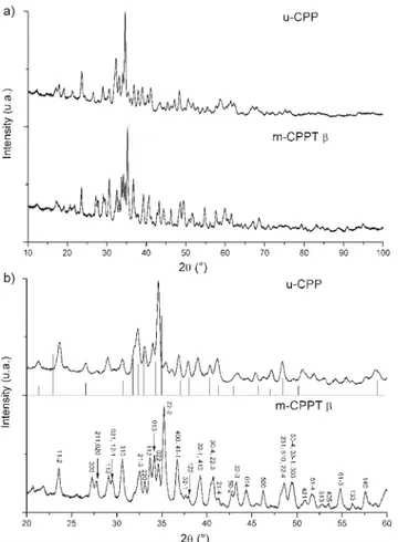
was identified by powder X-ray diffraction analysis as being pure m-CPPT b phase, whereas the diagram scattered by the phase produced within the stirred tank reactor did not match the patterns of any of the 25 calcium pyrophosphate compounds referenced (Fig. 2a).7 This unreferenced phase
will be referred to as u-CPP in this article. If we focus in the 2h = 20–60u domain (Fig. 2b), we notice that among the known CPP phases, the diagram of the u-CPP phase presents some analogies with that of the h-CPPD phase. h-CPPD has been described by Mandel et al.,2as a phase obtained from m-CPPT
bby dehydration under vacuum.
The method set up for m-CPPT b phase synthesis is faster than the methods already published: syntheses achieved within 45 min compared to 24 h to 1 week for previously published methods.2,7,8The same conditions of concentration
and temperature were already known to lead to the m-CPPT b phase (temperature below 50 uC, pH above 5) but the influence of the agitation has not been elucidated.
In order to further characterise this unknown u-CPP phase, several complementary analytical techniques were used to provide information about the new phase structure.
It shall be recalled that the hydrolysis of pyrophosphate ions might lead to the formation of orthophosphate anions
(HPO422 and PO432) according to the following reversible
chemical reaction (2):
P2O742+ H2O = 2HPO422 (2)
This reaction could occur for hydrated CPP phases in two different ways as a crystalline process or in the synthesis solution. Water molecules held in the structure could generate an internal hydrolysis at high temperature,13but the
phenom-enon is negligible at ambient temperature. External hydrolysis in acidic aqueous medium has been also reported and could occur in significant amounts during synthesis or titration of CPP phases even without another catalyst than H3O+.14
Nevertheless, phosphate titration results indicated that pyr-ophosphate hydrolysis either did not occur or occurred in insufficient quantity to be detected: 1% of the total P2O742
ions were titrated as being hydrolysed, which corresponds to the detection limit of the method. Indeed, a portion of the pyrophosphate could have been hydrolysed during the sample preparation used for the titration of orthophosphate ions, as a consequence of dissolution of the sample carried out in acidic medium. The titration of pyrophosphate indicated 20.2 wt% of phosphorus in the as-synthesised u-CPP phase, and 19.1 wt% of phosphorus in the as-synthesised m-CPPT b phase which correspond closely to the theoretical content (respectively 20.1 wt% and 19.0 wt%) (Table 1).
In the absence of catalyst, the hydrolysis reaction of pyrophosphate ions occurs extremely slowly at low tempera-ture, and the present synthesis conditions (temperature and duration) do not allow significant advancement of this reaction.
Fig. 3 shows the particle morphologies of the two phases synthesised. Both m-CPPT b and u-CPP crystals are assembled as oval shaped plates. Platelets of the u-CPP phase (Fig. 3b) are smaller, with size up to 4 mm, thinner and more agglomerated than those observed for m-CPPT b, leading to a sand rose flower organisation.
The difference in morphology between the two phases could probably be attributed to the mixing conditions during synthesis. The morphology and size of m-CPPT b crystals have already been reported to vary widely, depending on the protocol for their synthesis. However the plate-like morphol-ogy of this phase was related to the layered structure of m-CPPT b determined by Balic´-Zˇunic´ et al.;4 this feature
suggests there could be a link between the structures of these two phases.
The different structures of the calcium pyrophosphate hydrates elucidated indicate significant differences in the environment of the pyrophosphate ions.1,3,4In particular, the
P–O–P angle of the anion could change from 123.1u for the
Fig. 2 XRD patterns of the two hydrated calcium pyrophosphate phases synthesised using two different setups: a) in the 2h = 10–100u domain; b) in the 2h = 20–60u domain (bars under u-CPP diagram correspond to the diagram of h-CPPD phase reported by Mandel2).
Table 1 Phosphorus content (as P2O742or PO432ions; wt%) in the
as-synthesised hydrated CPP phases
P as P2O742(wt%) P as orthophosphate (wt%)
u-CPP 20.2 0.22 m-CPPT b 19.1 0.23
t-CPPD phase to 134u for m-CPPT b. Differences are also observed for the P–O distances.
FTIR spectroscopy could also be an important characterisa-tion technique for calcium pyrophosphate hydrates, with the geometry of the ion affecting the bands observed on the spectrum. Each compound provides a specific spectrum. However, FTIR spectroscopy analysis did not allow us to substantially distinguish the two CPP phases synthesised: the P–O and P–O–P vibration bands between 1250 and 500 cm21
are similar, although a faint difference can be noticed in their water O–H vibration domains between 2800 cm21and 3700
cm21(Fig. 4a).
The lack of a vibration band for the P–O bond beyond 1200 cm21 indicate the absence of HP
2O732 ions in the crystal.
HP2O732ions exists in solution and could possibly have been
coprecipitated with pyrophosphate. Comparison with CaH2P2O7 indicates that n1PO, a vibration specific for the P–
O bond symmetric elongation, would be detected around 1220 cm21(Fig. 4b).15
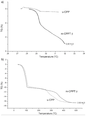
A thermogravimetric analysis was performed to study the stability of the m-CPPT b and u-CPP phases at low temperature (around 30 uC) and at higher temperature (up to 500 uC).
A weight loss approximately corresponding to one H2O
molecule (0.9H2O) was observed for the m-CPPT b phase,
during the preliminary plateau at 30 uC, whereas no significant weight loss was measured for the u-CPP phase at low temperature (Fig. 5a). However, at higher temperature, the two phases synthesised showed quite similar thermogravi-metric curves up to 500 uC, with the same weight loss: 3H2O
molecules, approximately (Fig. 5b).
The weight loss between 80 and 110 uC corresponds to 2H2O
whereas the one between 250 and 500 uC corresponds to 1H2O
(two steps). These ranges of temperature are consistent with the data already published.8
Finally, the total loss of water was around 4H2O as expected
for m-CPPT b and 3H2O for u-CPP. It appears that u-CPP is
potentially a trihydrated calcium pyrophosphate; the simila-rities observed with m-CPPT b during the second part of the experiment reinforce the assumption that there could be a link between the two phases.
Fig. 6 shows the evolution of the XRD diagram of the m-CPPT b phase when treated for 1 h at 50 uC. We observed that after this treatment the pattern corresponds to that of u-CPP (see Fig. 2 and 6) and there is no structural difference
Fig. 3 SEM micrographs of the two hydrated calcium pyrophosphate phases synthesised under the same conditions but using two different setups: a) m-CPPT b; b) u-CPP.
Fig. 4 FTIR spectra of the two hydrated calcium pyrophosphate phases synthesised in the same conditions but using two different setups: m-CPPT b and u-CPP: a) 4000–400 cm21domain; b) 1300–500 cm21domain.
observed between the u-CPP phases obtained by dry synthesis (dehydration of m-CPPT b) and wet synthesis, direct precipita-tion, and no trace of other compounds was found by comparison between the two diagrams.
This experiment confirms the link between the u-CPP and m-CPPT b phases synthesised using the same conditions of temperature and pH. As an explanation, a more efficient and powerful stirring in the tank reactor could influence the size and the organisation of the crystals9and explain the difference
in hydration state of the precipitated phases. According to the width of XRD peaks observed on u-CPP phase, the crystals synthesised in a stirred tank reactor were thinner or had a lower crystallinity compared to the crystals obtained by dehydration. These conditions could shift the temperature of dehydration, which was reported close to room temperature. A similar mechanism could explain the differences observed by TGA around 300 uC. The second step of dehydration started at lower temperature for u-CPP phase even if at this temperature these two phases were similar.
However the main difference between the two u-CPP phases prepared either in dry state by dehydration of m-CPPT b or by direct aqueous precipitation in stirred tank reactor is their stability at room temperature and atmosphere. When the u-CPP phase obtained by dry synthesis is left at ambient temperature, we can observe that after 15 min (Fig. 6) it is
reversibly transformed into the m-CPPT b phase, probably by capturing ambient air humidity to rehydrate whereas the u-CPP phase obtained in wet media can be kept for more than 3 months without alteration. The smaller crystal size of the u-CPP crystals obtained by wet synthesis would suggest an inverse reactivity and the experimental observation related to the re-hydration properties of these phases which highlights the effect of the conditions of synthesis on the crystals properties, has not yet received a satisfactory explanation.
The u-CPP phase can be considered as highly metastable, consequently small changes in the morphology could greatly influence its reactivity. Crystals formed in joints are reported to have various sizes depending on the joint type: from around 1 mm up to 20 mm for knee and 0.06 to 0.3 mm for temporomandibular joint.16,17Even if m-CPPT b crystals were
never observed in vivo, they could be formed as metastable intermediate and the dehydration described in the present paper could occur during the treatment of the synovial fluid specimen and may affect the identification of the CPP phases. These preliminary results on the characterisation of the two phases synthesised suggest that u-CPP phase could be a trihydrated calcium pyrophosphate metastable phase derived from the tetrahydrated m-CPPT b.
In order to compare with another m-CPPT b derived compound, attempts were made to synthesise h-CPPD, a phase produced by dehydration of m-CPPT b.2 However the
product obtained was characterised as a monohydrate CPP, with a XRD diagram different from h-CPPD and u-CPP patterns. In present study, no comparison could therefore be established between h-CPPD and u-CPP.
Calculation
Further investigations were performed to better define the u-CPP phase. Calculations on the structure of the u-CPP phase were made using the m-CPPT b structure as reference structure. The structure of the m-CPPT b phase was elucidated by Balic´-Zˇunic´ et al.4 This phase presents a layered structure
with the water molecules on the surface of layers. The platelet morphology observed for m-CPPT b crystals (Fig. 3a) is in
Fig. 5 TGA of the two hydrated calcium pyrophosphate phases (m-CPPT b and u-CPP) synthesised under the same conditions but using two different setups: a) at low temperature (up to 32 uC) and b) at high temperature (up to 500 uC).
Fig. 6 XRD patterns of m-CPPT b phase as synthesised, after heating 1 h at 50 uC and then 15 min at 20 uC.
accordance with this organisation. In addition, it has been reported that one of the four water molecules of the m-CPPT b phase, referred as OW4, does not coordinate any Ca atom and is held in the structure only by weak hydrogen bonding to other O atoms. This position suggests that weaker bonds hold this molecule in the structure and could explain the loss of one H2O molecule below 50 uC (Fig. 5a) observed by TGA. This
conclusion was previously suggested by Balic´-Zˇunic´ et al. The preliminary results of calculations of the cell para-meters for the u-CPP phase are reported in Table 2 and the corresponding XRD data for u-CPP are presented in Table 3 with the assumed indexing.
u-CPP has the same crystal symmetry as m-CPPT b, with nearly equal b and c lattice parameters. A larger difference is observed for the b angle and the a parameter. The difference is probably a consequence of the dehydration and reorganisation of the remaining water molecules. Indeed, the labile water molecule and two other structural water molecules are contained on a (100) plane of the m-CPPT b structure, which represents also the most extended surface plane of the crystals.4
We can thus expect a larger impact of dehydration and the reorganisation phenomenon on the a-axis than on the other lattice parameters. Moreover according to the FTIR spectro-copy analyses, the environments of pyrophosphate ions seem slightly affected by this reorganisation.
Although the atomic configuration of u-CPP phase was not determined with enough accuracy, a first approximation was to consider its structure as a modified m-CPPT b structure without the oxygen atom referred as OW4 by Balic´-Zˇunic´ et al.4
It should be noted that several structures of calcium pyropho-sphates are not resolved, including m-CPPD, a phase encountered in some arthritic joints.
The reorganisation of pyrophosphate and calcium ions in the structure could change the phlogistic potential of calcium pyrophosphate crystals. A proposed mechanism of inflamma-tion in crystal-induced diseases is dependent on the rupture of the lysosome phospholipid membrane and induced by pyrophosphate species on the surface of the crystals.18,19
Thus, the structure of the crystals as well as the surface is important in determining the inflammatory potential.20
Even if the m-CPPT b phase has never been observed in vivo, its role as precursor of m-CPPD and t-CPPD has been described in vitro.2 As well as m-CPPT b, u-CPP could be an
intermediate of CPPD formation under specific conditions. Finally a description of the u-CPP structure could increase the knowledge about calcium pyrophosphate interactions in vivo.
Conclusions
A method was set up to prepare pure m-CPPT b phase at 25 uC by double decomposition between a potassium pyrophosphate solution and a calcium nitrate solution in an aqueous buffered solution (pH 4.5) magnetically stirred in an Erlenmeyer flask. This method is fast, reliable and reproducible compared with methods of preparation already published.
For the first time, we showed that these conditions (temperature, pH) can also enable obtaining a pure unknown hydrated calcium pyrophosphate phase (u-CPP), just by changing the reaction vessel (stirred tank reactor). Fine characterisation of the u-CPP phase using complementary analytical techniques and calculations suggest that it is a highly metastable trihydrated monoclinic calcium pyropho-sphate derived from the structure of m-CPPT b. However the structure could not be determined and the structural refine-ment of the powder diffraction pattern is still under investigation.
This work contributes to clarifying the mechanism by which CPP crystals form in vitro and in vivo, and this metastable trihydrated CPP phase (u-CPP) could be an intermediate of CPPD crystal deposit formation in weight-bearing articulation.
Acknowledgements
The authors thank the Institut National Polytechnique de Toulouse (PRECIPYCA project – BQR INPT 2011) and the Centre National de la Recherche Scientifique (CalArthros project – ‘‘Longe´vite´ et Vieillissement 2010’’ CNRS interdisci-plinary program) for supporting this research work.
References
1 N. S. Mandel, Acta Crystallogr., Sect. B: Struct. Crystallogr.
Cryst. Chem., 1975, 31, 1730–1734.
Table 2 Calculated cell parameters (Jana200612) of u-CPP and m-CPPT b phases
compared to published data for cell parameters of m-CPPT b4
a(Å) b(Å) c(Å) b(u) a= c (u) u-CPP 11.399(6) 7.409(2) 10.859(3) 110.16(1) 90.00 m-CPPT
bsynthesised 12.290(4) 7.518(4) 10.774(6) 112.54(1) 90.00 m-CPPT b4 12.287(6) 7.511(3) 10.775(5) 112.54(1) 90.00
Table 3 Peak positions and indexing of u-CPP powder diffraction data (most intense peaks with intensities greater than 10%)
d(Å) Intensity hkl 10.7099 1000 100 5.7643 121 11-1 4.3608 181 11-2/210 3.5745 153 112/300 3.3905 140 31-1 3.2561 128 11-3 3.2182 283 310/31-2 3.1418 151 21-3 3.0538 168 12-2/220 3.0027 543 022 2.8281 213 311 2.7497 106 122 2.6804 201 400 2.5437 164 302/11-4 2.1830 140 41-4/22-4
2 G. S. Mandel, K. M. Renne, A. M. Kolbach, W. D. Kaplan, J. D. Miller and N. S. Mandel, J. Cryst. Growth, 1988, 87, 453–462.
3 N. L. Davis, G. S. Mandel, N. S. Mandel and R. E. Dickerson,
J. Crystallogr. Spectrosc. Res., 1985, 15, 513–521.
4 T. Balic´-Zˇunic´, M. R. Christoffersen and J. Christoffersen,
Acta Crystallogr., Sect. B: Struct. Sci., 2000, 56, 953–956.
5 K. P. H. Pritzker, Calcium pyrophosphate crystal formation and dissolution in Calcium Phosphate in Biological and
Industrial Systems, Kluwer Academic Publishers, 1998, pp. 296–301.
6 P. J. Grove, R. M. Wilson, P. A. Dieppe and R. P. Shellis, J.
Mater. Sci.: Mater. Med., 2007, 18, 1355–1360.
7 E. H. Brown, J. R. Lehr, J. P. Smith and A. W. Frazier, J.
Agric. Food Chem., 1963, 11(3), 214–222.
8 M. R. Christoffersen, T. Balic´-Zˇunic´, S. Pehrson and J. Christoffersen, J. Cryst. Growth, 2000, 212, 500–506. 9 N. Be´net, H. Muhr, E. Plasari and J. M. Rousseaux, Powder
Technol., 2002, 128, 93–98.
10 J. W. Mullin, Crystallization, ButterworthsLondon, 2nd edn, 1972.
11 C. Herman, V. Gelbgras, V. Halloin and B. Haut,
Proceedings of European Congress of Chemical Engineering, 2007.
12 V. Petricek, M. Dusek and L. Palatinus, Jana2006, The
crystallographic computing system, Institute of Physics, Praha, Czech Republic, 2006.
13 C. Slater, D. Laurencin, V. Burnell, M. E. Smith, L. M. Grover, J. A. Hriljac and A. J. Wright, J. Mater. Chem., 2011, 21, 18783–18791.
14 R. B. Stockbridge and R. Wolfenden, J. Biol. Chem., 2011, 286(21), 18546–18546.
15 J. L. Savio, PhD thesis, Contribution a` l’e´tude des
pyropho-sphates de calcium hydrates et de leurs proprie´te´s phlogo-ge`nes, Institut National Polytechnique de Toulouse,
Toulouse, 1982.
16 N. N. Kohn, R. E. Hughes, D. J. McCarty and J. S. Faires,
Ann. Intern. Med., 1962, 56, 738–745.
17 L. C. Dijkgraaf, R. S. B. Liem, L. G. M. de Bont and G. Boering, Osteoarthritis Cartilage, 1995, 3, 35–45. 18 N. S. Mandel, Arthritis Rheum., 1976, 19, 439–445.
19 A. Wierzbicki, P. Dalal, J. D. Madura and H. S. Cheung, J.
Phys. Chem. B, 2003, 107, 12346–12351.
20 M. Roch-Arveiller, R. Legros, B. Chanaud, O. Muntaner, S. Strzalko, A. Thuret, D. A. Willoughby and J. P. Giroud,