Characterization in Cochlea of KCTD12/PFET1,
An Intronless Gene with Predominant Fetal Expression
by
Sharon Fan Kuo
MASSACHUSETTS INSTrrTE
OF TECHNOLOGY
MAR 3 0 2006
.
LIBRARIES
LIBRARIES
B.S. Biochemistry, Tufts University, 1997
B.M. Piano Performance, New England Conservatory of Music, 1997 ARCH E
Submitted to the Harvard-MIT Division of Health Sciences and Technology in partial fulfillment of the requirements for the degree of
Doctor of Philosophy
in the Program of Speech and Hearing Bioscience and Technology
Massachusetts Institute of Technology Cambridge, Massachusetts
December 2005
Signature of Author...
Speech anl Hearing Bioscience and Technology
f
.13,
Po
6
Certified
by...
...
... ... Cynthia C. Morton
William Lambert Richardson Professor of Obstetrics, Gynecology and Reproductive Biology and Professor of Pathology, Harvard Medical School Thesis Supervisor
Accepted by ... .. ...
M~ tha L. Gray, Ph.D.
Edward Hood Taplin Professor on Medical & Elctrical Engineering Director, Harvard-MIT Division of Health Sciences and Technology
© 2005 by Sharon Fan Kuo
All rights reserved.
Characterization in Cochlea of KCTD12/PFET1,
An Intronless Gene with Predominant Fetal Expression
by
Sharon Fan Kuo
Submitted to the Harvard-MIT Division of Health Sciences and Technology in December 2005 in partial fulfillment of the requirements for the degree of
Doctor of Philosophy in Speech and Hearing Bioscience and Technology
Abstract
The prevalence of severe to profound bilateral congenital hearing loss is estimated at 1 in 1000 births, at least half of which can be attributed to a genetic cause. To date, mutations in
at least 67 genes have been associated with hearing loss. Discovery of these genes has revealed fundamental processes within the ear, and enabled diagnosis and implementation of genetic counseling in affected patients. As a part of the continuing effort to study genes important for hearing and deafness, a novel cochlear transcript with predominantly fetal expression containing a single tetramerization domain (PFET1, HUGO-approved symbol
KCTD12) was identified from the Morton fetal cochlear cDNA library. KCTD12/Kctdl]2 is an evolutionarily conserved intronless gene encoding a 6 kb transcript in human and three transcripts of approximately 4, 4.5 and 6 kb in mouse. The protein, pfetin, is predicted to
contain a voltage-gated potassium channel tetramerization (T1) domain. This thesis reports characterization of this novel human gene and its encoded protein pfetin in relation to its role in auditory function. Experimental data from tissue and cellular expression profiling, and genetic and functional analyses suggests KCTD12 and its orthologs playing a crucial role in
the developmental of the auditory sense organ.
Thesis Supervisor: Cynthia C. Morton
Title: William Lambert Richardson Professor of Obstetrics, Gynecology and Reproductive Biology and Professor of Pathology, Harvard Medical School; Director of Cytogenetics, Brigham and Women's Hospital
ACKNOWLEDGEMENTS
I would like to thank my thesis committee: David Corey, Douglas Cotanche, Stefan Heller, and Charles Liberman for their guidance and support over the last few years.
My heartfelt thanks to Cynthia Morton my research advisor for her unwavering support and encouragement through my exploration in the field of genetic hearing loss and my pursuit of clinical certification in speech and language pathology.
I was fortunate through this joint program to have directly worked with so many wonderful individuals from different areas of expertise. This thesis would not have been possible with out their generosity and kindness.
To my parents, thank you for your love, understanding and support through this incredible journey.
SK's Interaction and Functional Domain
AP: Arti Pandya ~:. Andria Schibler • Amanda Smith AT: Andrew Tucker
I:
Charles Lee : Charles Liberman CM: Cynthia Morton CU: Chinweike Ukomadu .: David Corey DC: Douglas Cotanche GA: Gil A1terovitz ~: Jessica ApplerIll: Joe Adams JG: Joshua Gamse JM: Jarema Malicki LG: Lisa Goodrich MH: Marnie Halpern
fII: Mingqian Huang MR: Marco Ramoni MT: Motokazu Tsujikawa MW: Melissa Wood RH: Robert Hillman SH: Stefan Heller WL: Weining Lu YO: Yoshihiro Omori ZYC: Zheng-Yi Chen
1-.-.-_1: Mom +Dad
SHBT:
Speech and Hearing Bioscience and Technology
TABLE OF CONTENTS
Abstract...iii
Acknowledgments ... v
Table of Contents ... vi
List of Figures and Tables ... vii
Chapter 1 ... 1
Introduction
Chapter
2...23
Isolation from Cochlea of a Novel Human Intronless Gene with Predominant Fetal Expression Chapter 3...65
Functional Characterization of KCTD12 in Zebrafish Chapter 4...94
Genomic and Proteomic Characterization of KCTD12 and Pfetin Chapter 5...112
Summary
Appendix
...
119
LIST OF FIGURES AND TABLES
2-1 ... 52
Nucleotide sequence of human PFET1 cDNA and its deduced amino acid sequence.
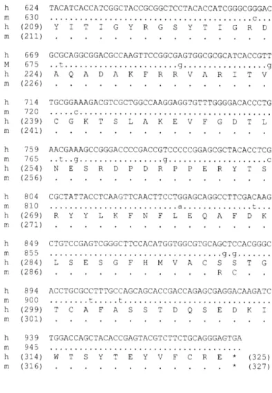
2-2 ... 53 Alignment of the complete deduced sequence of the open reading frames of the human and mouse Pfetl genes.
2-3 ... 54 Nucleotide sequence of mouse Pfrtl cDNA and its deduced amino acid sequence.
2-4 ... 55 Alignment of the consensus sequence for the tetramerization domain of the voltage-gated K+ channel family, deduced amino acid sequence of the tetramerization domain from the human and mouse PFET1 genes, and various potassium channel tetramerization domains. 2-5 ... 56
Autoradiographs of Northern blots of total human RNAs hybridized with radiolabeled
PFET1 fragments and schematic of PFET1 gene
2-6 ... 58 Northern blot analysis of mouse RNA samples hybridized with mouse Pfetl radiolabeled fragments and schematic of the mouse Pfetl gene.
2-7 ... 60
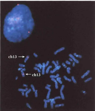
Chromosomal localization by fluorescence in sitit hybridization of a human PAC containing the entire PFETI gene.
2-8 ... 61
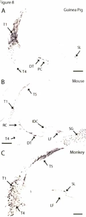
Immunohistochemical staining using pfetin antibody on FG-fixed guinea pig, FG-fixed mouse, and FA-fixed monkey cochleas.
2-9 ... 62 Immunohistochemical staining using pfetin antibody on formalin-fixed adult human cochlea.
2-10 ... 63
Immunostained formalin plus glutaraldehyde fixed mouse and formalin acetic fixed guinea pig cochlea.
2-11 ... 64 Pfetin immunostaining of vestibular tissue of 20-week human fetus (formalin-fixed), guinea pig (FG-fixed), and mouse (FG-fixed).
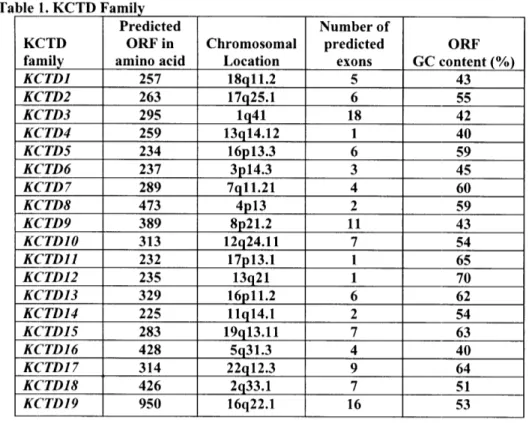
3 -T ... ... .... .... .. .... 7 4
KCTD Family
3-1 ... 87 FISH of zebrafish metaphase chromosomes for localization of leftover and right on.
3-2 ... 88 RNA expression levels of leftover and right on in RT-QPCR.
3-3 ... 89 Localization of KCTD12 zebrafish orthologs, leftover and right on via whole-mount in
situt hybridization.
3-4 ... 90 Expression pattern of ron riboprobe in zebrafish sections.
3-5 ... 91
Expression pattern of Ron and Isll in the otic vesicle.
3-6 ... 93
Phylogenetic tree of KCTD family members.
4-T ... 105
Putative Interactors of Pfetin
4-1 ... 110
Expression of Pfitl/Kctdl2 in Affymetrix Mouse Genome 430 Array.
4-2
...
111
Network graph of 38 cluster genes from Affymetrix Chip analysis.
Appendix ... 122
Hearing loss is the most common sensory disorder in the human population with the
incidence of congenital hearing loss estimated at 1 in 1000 births (Morton 1991). It is estimated that 50% to 75% of all childhood deafness is due to hereditary causes (Gorlin 1995).
The remainder is due to environmental factors including acoustic trauma, ototoxic drugs (e.g., aminoglycosides), bacterial and viral infections. Of the hearing disorders with a genetic contribution, roughly 70% are classified as nonsyndromic and 30% as syndromic, depending
on the presence or absence of other clinical features (http://webhost.ua.ac.be/hhh/). To date,
mutations in at least 67 genes have been associated with hearing loss. Discovery of these genes has revealed fundamental processes within the ear, and enabled diagnosis and implementation of more accurate genetic counseling for families and affected patients.
The auditory apparatus consists of three major compartments, the external, middle and
inner ear. The external ear consists of the auricle and the external auditory canal. It is bounded
at the external-middle ear junction by the tympanic membrane. The middle ear is an air-filled cavity containing a chain of three ossicles (malleus, incus, stapes). The inner ear resides in the temporal bone of the skull, and is a complex membranous labyrinth filled with endolymph housed inside a perilymph-filled bony labyrinth. The snail-like portion of this labyrinth is
called the cochlea, the auditory sense organ. It resides in the inner ear along with the vestibule.
The human cochlea detects sound frequencies between 20 Hz and 20 kHz and the vestibule (saccule, utricle, three semicircular canals) responds to linear and angular accelerations. The cochlea and the vestibule both derive embryologically from the otic placode (Fekete 1996) and share several structural and functional features.
Briefly, the mammalian hearing process functions as follows. Sound waves travel through the external ear canal to the tympanic membrane (TM) where sound pressure sets the
TM into vibration. The ossicular chain, attached at one end to the TM, contacts the cochlea
via the oval window. Vibration creates a traveling wave in the endolymph down the length of
the cochlea, and the traveling wave sets into motion the basilar membrane (on which rests a
cellular layer known as the organ of Corti lined with auditory hair cells), inducing movement of these hair cells relative to the tectorial membrane. Deflection of the hair cells leads to their depolarization, triggering neurotransmitter release, and creating an action potential traveling along the auditory nerve to the brain which encodes different characteristics of the sound stimulus including intensity, time course, and frequency.
Genes Involved in Hearing Disorders
Gene mutations have been found affecting almost every part of the auditory system
from the shape of the ear lobe, to the middle ear and the auditory neurons in the brain.
However, many encoded protein of genes responsible for deafness have been found to be expressed in the cochlea. The cochlea is a most intricate and complex organ consisting of dozens of cell types and specialized regions required for normal auditory function. Therefore, genes underlying the molecular development, structure, function and maintenance of these
cell types and regions are crucial to the hearing process. As mentioned previously, mutations in at least 67 genes have been identified to show association with hearing loss. These genes
encode a wide variety of proteins, including gap junctions, ion channels, extracellular matrix components and transcription factors.
Hair Cell Organization and Function
One of the most important structures in the cochlea is the auditory hair cells. These hair cells are highly organized and convert mechanical vibration into nerve impulses, and require precise structural maintenance for proper function. Mouse mutant studies have been instrumental in identifying a number of deafness-associated genes that are required for the proper organization and maintenance of hair cell stereocilia such as Myo7a, Myo6, Cdh23,
Pcdh15, Itga8, Espn and Tmie. Most of the human orthologs when mutated result in hearing
disorders: MY07A for DFNB2, DFNA11 and USH1B (Liu et al. 1997); MY015A for DFNB3
(Wang et al. 1998); CDH23 for both DFNB12 and USH1D (Bolz et al. 2001); PCDH15 for
USH1F (Ahmed et al. 2001); ESPN for DFNB36 (Naz et al. 2004); and TMIE for DFNB6 (Naz et al. 2002). In addition, mutation in MY06 results in the disorganization and fusion of stereocilia and accounts for the nonsyndromic autosomal dominant hearing loss in an Italian family (Melchionda et al. 2001). A defect in harmonin, a PDZ domain-containing protein expressed in the inner ear sensory hair cells, underlies USH1C (Verpy et al. 2000) and defects in whirlin a PDZ domain molecule involved in stereocilia elongation, cause deafness in the
whirler mouse and families with DFNB31 (Mburu et al. 2003).
Extracellldar Matrix
The extracellular matrix (ECM) is a complex structural entity surrounding and supporting cells that are found within mammalian tissues. The ECM is composed of three major classes of biomolecules: structural proteins like collagen and elastin; specialized proteins such as fibrillin, fibronectin, and laminin; and proteoglycans.
Collagens are crucial for the structural integrity of many organ systems including the inner ear. Several collagens responsible for deafness include COL4A5, COL4A3 and COL4A4 for both X-linked and autosomal forms of Alport syndrome; COLIA] and COLIA2 for
osteogenesis imperfecta (OI); COL2AJ for Stickler syndrome type I (STLI1); COLIIA2 for
Stickler syndrome type II (STL2) and DFNA13; COLllAJ for Stickler syndrome type III
(STL3) (reviewed in Resendes et al. 2001 and Smith et al. 2005). Mutations in COL9A3 have
recently been identified in patients with nonsyndromic hearing impairment with moderate progressive bilateral sensorineural hearing loss in all frequencies (Asamura et al. 2005). Other extracellular matrix proteins like usherin that is responsible for Usher syndrome type 2A (USH2A) (Eudy et al. 1998) contain both laminin epidermal growth factor and fibronectin type III motifs, which are most commonly observed in proteins comprising components of the
basal lamina and extracellular matrixes and in cell adhesion molecules. The tectorial
membrane is also an extracellular matrix of the inner ear that contacts the stereocilia bundles of specialized sensory hair cells. Mutations in TECTA causing defects in the membrane are etiologic for hearing disorders DFNA8 and DFNA12 (Verhoeven et al. 1998) and DFNB21
(Mustapha et al. 1999). The cause of DFNA9 is mutations in COCH (Robertson et al. 1998)
which encodes a secreted protein that becomes part of the extracellular matrix. The mutant
form of the protein leads to the loss of cells in the spiral ligament and limbus and
accumulation of acidophilic deposits in the nerve channels and supporting tissues of the organ of Corti.
Ion Homeostasis
For proper auditory signal transduction, it is critical to maintain ion homeostasis, especially regarding the high potassium concentration in endolymph. Potassium recycling begins with an efflux of potassium from the outer hair cells through the potassium channel.
Ions migrate to the stria vascularis through gap junctions between supporting cells and are
finally pumped back into the endolymph.
Potassium channels KCNQ4, KCNQI, and KCNE1 have all been shown to play fundamental roles in the potassium recycling pathway. Mutations in these genes lead to the
hearing disorders DFNA2, Jervell and Lange-Nielsen syndrome (loci and 2) (Schulze-Bahr et al. 1997, Neyroud et al. 1997). Mutations in an anion transporter protein like pendrin,
encoded by SLC(26A4, are postulated to disrupt transport of negatively charged particles, thus
upsetting fluid balance in the inner ear, and causing Pendred syndrome and DFNB4 (Li et al. 1998).
Gap junction subunits are called connexins and four connexins have been implicated
in at least six types of both dominant and recessive nonsyndromic hearing loss, and they are
connexin 26 (JB2), connexin 31 (GJB3), connexin 30 (GJB6), and connexin 43 (GJA1).
GJB2 alone is estimated to account for 50% or more of recessive congenital nonsyndromic hearing loss in some populations (Rabionet et al. 2000). In addition to ion recycling, ion
concentration mnust be strictly maintained within the two separate compartments of cochlea, one containing perilymph and the other endolymph. Tight junctions in the cochlea are thought to compartmentalize endolymph by controlling the permeability of the paracellular pathway. Mutations in the tight junction gene CLDN14 may cause loss of endolymphatic potential
leading to degeneration of cochlear hair cells, and is etiologic in the autosomal recessive hearing loss DFNB20 (Wilcox et al. 2001).
Transcription Factors
Transcription factors regulate the spatio-temporal expression of thousands of genes and the control of cellular proliferation, differentiation, and regulation of cellular function to ensure proper development and functioning of an organism, and the inner ear is no exception. Four transcription factor genes have been identified (POU3F4, POU4F3, EYA4, and
TFCP2L3) to be etiologic for the nonsyndromic hearing disorders DFN3, DFNA15, DFNA10 and DFNA28, respectively (de Kok et al. 1995, Vahava et al. 1998, Wayne et al. 2001, Peters et al. 2002).
POU3F4 is a member of a larger family of genes called POU domain genes, which play a role in determining cell types in the central nervous system during early development and are likely to be involved in the development of the middle and inner ear. Mutations in or
near POU3F4 probably lead to insufficiency of POU3F4 protein, thus disrupting the normal development of structures in the middle and inner ear and leading to hearing loss (de Kok et al.
1995). POU4F3, a class IV POU domain transcription factor, has a central function in the
development of all hair cells in the human and mouse inner ear sensory epithelia. Mutations in
POU4F3 affect protein stability, localization, and transcriptional activity (Hertzano et al.
2004).
Mutations in EYA4 lead to production of abnormal EYA4 protein, lacking some or all
of the Eya domain and thus impairing interactions with other proteins. Impaired protein interactions probably disrupt control of gene activities that are important for the development
of the inner ear and maintenance of normal hearing (Wayne et al. 2001). Transcription factor
TFCP2L3 was shown to be highly expressed in epithelial cells lining the cochlear duct. The
predicted translation product of TFCP2L3 has sequence similarity to a group of proteins comprising the transcription factor cellular promoter 2 (TFCP2) family. However, its exact function remains to be elucidated (Peter et al. 2002).
Often times, transcription factors work in synchrony during the development and maintenance of mammalian inner ear (Corey and Breakefield 1994, Cantos et al. 2000), an example being the interactions between MITF, PAX3, and SOX10. Mutations in these genes result in the various subtypes of Waardenburg syndrome: PAX3 (Waardenburg syndrome types I, III and craniofacial-deafness-hand syndrome), MITF (Waardenburg syndrome type 11), and SOXlO 0 (Waardenburg syndrome type IV). It has been shown that SOXlO 0 and PAX3
synergistically regulate MITF expression in transfection assays and mutant SOX10 and PAX3 proteins failed to bind to the MITF promoter region to commence induction (Bondurand et al.
2000).
Modifiers
In addition to genes that directly causing hearing loss, there also exist modifier genes
that can contribute to hearing disorders through their influence on the expression or function of other genes. Notable examples include tub (tubby) and moth], dwf and mdfw in mice and DFNB26 and DRNM in humans. Tub encodes a transcription factor with expression in the outer and inner hair cells and the spiral ganglion cells. Moth], a modifier of tubby hearing, can either worsen or prevent the hearing impairment in tubby, depending on the type of moth]
encodes an ATPase pump that is necessary for maintenance of low cytosolic calcium ions
(Kozel et al. 1998; Street et al. 1998). Mdfw as a modifier of dfw (deaf wobbler) can protect dw, heterozygotes from hearing loss with one allele and is permissive of hearing loss with the
other (Noben-Trauth et al. 1997). In the autosomal recessive, nonsyndromic sensorineural hearing loss DFNB26, a dominant modifier gene (DFNM1) has been mapped to lq24 and is thought to suppress deafness in individuals with DFNB26 (Riazuddin et al. 1999).
Approaches to Gene Discovery and Characterization in the Auditory System
The traditional method for identification of genes involved in deafness begins by collection of DNAs from kindreds segregating a hearing impairment. It is then followed by genetic linkage analysis to identify the region of genome in which a gene involved in hearing
is likely to reside. Once the region is discovered, positional cloning is then performed. When successful, it can reveal the identity of the gene involved in the deafness. This process has
been very successful in identifying a number of human deafness genes such as NDP, TCOF1,
DDP, SLC26A4, USH2A and DFNA5 (Morton 2002). However due to the complex genetic
nature of deafness, linkage analysis is a less than optimum method in gene discovery efforts for hearing disorders. Successful use of genetic linkage for mapping hearing disorders in autosomal recessive nonsyndromic loci has been fruitful largely in consanguineous kindreds or populations in which there has been limited admixture. Even in families in which a heritable hearing disorder is successfully mapped, there may be insufficient numbers of
recombination events to narrow a chromosomal interval, resulting in a candidate region in the megabase scale.
A complementary method to the genetic linkage analysis for gene identification is one
that utilizes tissues or organ-specific cDNA libraries to provide candidate genes (Hedrick et
al. 1984, Jones et al. 1989, Gurish et al 1992). Presently, cochlear libraries are available for
human, mouse, rat and chicken. These libraries have provided valuable tools for gene discovery in hearing and deafness. Almost 15,000 human (Morton fetal cochlear cDNA library) and 1,600 mouse (Soares mouse NMIE cDNA library) inner ear ESTs are currently available in the GenBank (http://www.ncbi.nlm.nih.gov/gquery/gquery.fcgi). ESTs from the human cochlear cDNA clones have already elucidated thousands of potential positional candidate genes for hearing disorders. Some of the human ESTs are genes already known to be involved in deafness: COL4A5 (Alport syndrome), EDNRB (Waardenburg syndrome, type
IV), EYA1A (130R syndrome), GJB2 (DFNB1 and DFNA3), GJB6 (DFNA3), KVLQT1 (Jervelle and L.ange-Nielsen syndrome), and MY06 (DFNA22). Several genes preferentially expressed in the cochlea, namely COCH (Robertson et al. 1997), OTOR (Roberton et al. 2000) and KCTD12/P-FET (Resendes and Kuo, 2004) have been identified from the human fetal
cochlea cDNA library. COCH was further shown to be responsible for the sensorineural
deafness and vestibular disorder, DFNA9 (Robertson et al. 1998). Using a similar approach, a
number of genes implicated in murine auditory function have been identified from mouse inner ear transcripts, such as Otog, Ocn95, Fdp (mouse homolog of OTOR), Strc and Ushlc, and (Morton 2002). The human ortholog of Strc was found to be etiologic in DFNB 16 (Verpy et al. 2001) and mutations found in the human ortholog of Ushlc underlie USHIC and
DFNB 18 (Ahnmed et al. 2002).
In addition to inner ear cDNA libraries, microarray technology offers a rapid and
very little starting material. It was instrumental in the identification of CRYM (Abe et al. 2003)
and COL9A3 (Asamura et al. 2005), which were then studied as candidate genes for
nonsyndromic deafness. With the completion of the DNA sequence of the human and mouse
genomes, the sequence can be used to determine the expression pattern of thousands of genes from the inner ear. Cross-tissue comparisons facilitate identification of genes that are
preferentially expressed in the inner ear. Microdissection and subtraction between data sets
can help identify cell-type specific and structural-specific genes important for the function of the inner ear and expedite identification of genes that interact with deafness genes (Corey and
Chen, 2002).
Model Organisms for Hearing and Deafness
Genes discussed in the previous section constitute just a fraction of what is involved in the process of hearing. Animal models have been invaluable in understanding how mutations or changes in these genes affect the function and development of the ear. Model organisms, genetically altered or pharmacologically treated, have been employed in the study of human diseases for many years. Animal models have made available a wide variety of studies including gene expression during development, anatomical and physiological experimentation, and various kinds of genetic manipulation that are not possible or ethical with human subjects. These have helped elucidate important questions such as whether candidate genes actually cause disease and by what mechanisms a gene mutation underlies a disorder.
Many considerations are taken into account when choosing an animal model, such as a researcher's familiarity with and accessibility to the animal. In addition, the time required to produce an animal model is critical, which is determined partly by the animal's generation
time, and in part by the ease of disease gene modification (lower eukaryotes being faster than higher eukaryotes). The potential resemblance of the phenotype observed in the animal model compared to that in human is an important criterion; this choice is dependent upon the disease gene under study and the affected organ. In general, the lowest eukaryote containing an ortholog of the human disease gene and the organ system is the preferred model organism.
Mouse Models
The mouse is a great model for studying human genetic deafness and genes essential for the auditory system because the anatomy, function, and hereditary abnormalities of the mouse ear have been shown to be highly similar to that of humans (Probst and Camper 1997). There are two general approaches to the utilization of mouse models in the analysis of human
disease, one being the disease-driven directed genetic approach and the other mutagenesis-driven non-directed approach. In the direct approach, the disease-causing genes are already identified and the mutation characterized in the human population. Then, the mouse ortholog is identified and manipulation performed for functional gene alteration. Specifically, genetic modification may be introduced via transgenic animals where multiple copies of a foreign
mutant gene were are inserted into its genome, or gene targeting where one or both normal
alleles of the animal's ortholog are mutated typically via homologous recombination. In the non-directed approach, the process begins with the genetic modification and phenotype characterization in the mice followed by genetic and molecular studies of the disease. This is exemplified by chemically induced mutations via N-ethyl-N-nitrosourea (ENU) where mice are treated with ENU and screened for hearing and balance phenotypes. There are multiple centers performing mouse ENU mutagenesis including Harwell in the UK; Neuherberg in
Germany; Ricken in Japan; and Brigham and Women's Hospital, The Jackson Laboratory, Baylor College of Medicine, McLaughlin Research Institute and Oak Ridge National Laboratory in the United States. Both spontaneous and chemically induced mutations provide an array of naturally occurring and randomly induced mouse mutations to study. Advantages and limitations are associated with each approach and technique (reviewed in Chapter 15 of
Current Protocols in Human Genetics, and Brown and Hardisty, 2003).
In addition to these genetically engineered and chemically induced mutations, there are also mutations that spontaneously arise in the mice (Johnson 2001). Spontaneous mutations have occurred naturally in large inbred populations of mice and they constitute a large portion of the mouse mutations now being used as models for human deafness. These mutations are identified often through behavioral abnormalities. Mice displaying hyperactivity, head bobbing, and circling behavior typical of vestibular dysfunction are frequently found to be deaf or hearing impaired. Other abnormalities in pigmentation and
development can be associated with a hearing deficit as well. Certain inbred strains of mice
exhibit a late-onset, progressive hearing loss, providing valuable models for the study of human age-related hearing loss (AHL), or presbycusis. In both humans and mice, AHL occurs
earliest at high frequencies with loss of sensory hair cells from the base to the apex of the cochlea. Thus far, three AHL loci have been mapped, Ahl3(AHL-resistant) (Nemoto et al.
2004), Ahl and Ahl2 (AHL-sensitive) (Johnson and Zheng 2002).
Deaf mouse mutants are posted on the Sanger Institute website (http://www.sanger.ac.uk/PostGenomics/mousemutants/deaf/). This site contains a current listing of genes or loci identified thus far known to be involved in deafness and/or balance
system. Numerous human hearing disorders with mouse models can be found at The Jackson Laboratory website (http://wwwjax.org/hmr/models.html). There are currently at least 22 nonsyndromic and 30 syndromic human deafness genes with corresponding mouse mutations.
Zebrafish Models
Zebrafish (Danio rerio) has become an important model system for the study of development and function of the vertebrate inner ear. The vertebrate inner ear is a complex system involving many sensory organs. It can be divided into two major compartments, dorsal and ventral. The dorsal part containing the utricle macula and three cristae for the semicircular canals is highly conserved structurally and functionally amongst all vertebrates. The ventral part becomes more specific to each vertebrate class and contains the macular organs of the saccule and lagena, which function in balance, audition or both. In the case of zebrafish, the primary auditory organ is the saccule. Sound is detected through the fish's air-filled swim bladder and transmitted via a series of bones called the Weberian ossicles connecting the
swim bladder to the sensory patch in the saccule. There are also additional auditory inputs
from the lagena and macula neglecta that are developed later during the juvenile stage (Fekete and Wu 2002). In addition to the inner ear, zebrafish also possess another sensory organ called the lateral line that allows for the detection of low-frequency stimuli such as water movements.
There are several advantages to using zebrafish as a model for studying hearing. Embryogenesis is rapid, and monitoring and manipulation of auditory organs in vivo during development are possible. Although zebrafish do not possess the equivalent of the mammalian cochlea, the organization and morphology of the zebrafish inner ear sensory epithelium cristae
and maculae are highly similar to that of higher vertebrates, including human. Genetic mechanisms governing the development and function of the zebrafish ear also appear well conserved (Whitfield et al. 2002). Nonetheless, there are exceptions including the formation of the otocyst by cavitation in zebrafish rather than by invagination as in chicks and mice.
Utilizing the classical forward genetic approach, many random mutations have been generated in zebrafish through ENU mutagenesis. Mutants can be screened initially for defects in morphology. Behavioral assays are also employed to search for mutants with aberrant swimming patterns that are indicative of vestibular defects. Functional assays such as generation of microphonic potentials and apical endocytosis, which is highly active in normal
hair cells, are also effective screening tools. One may also measure the startle response of
adult mutants to an auditory stimulus. This is a high throughput and automated screening that can elicit a rapid tail-flip from zebrafish with "normal" hearing in response to a tone burst. It can detect subtle behavioral defects that might have been missed by human observation. In
situ hybridization may also be used as a high-throughput method to identify genes with specific or restricted temporal and spatial expression patterns, which is ideal for studying
organ specific processes.
Reverse genetic approaches have also been useful in determining the importance of individual genes or a combination of genes' involvement in development and function of the inner ear via suppression of expression for targeted genes (morpholino knockdown), overexpression of a targeted gene (ectopic expression), and fluorescent reporting of targeted genes (transgenesis). At least 22 genes have been identified so far to be necessary for inner ear development and function in zebrafish (Nicolson 2005). Mutations in these genes can cause defects affecting many aspects of normal hair cell specification, survival and function,
development of otic induction and formation of the otic vesicle. At least five forms of syndromic or nonsyndromic human hearing disorders are known to have zebrafish models (Whitfield 2002).
In the past decade, tremendous progress has been made in auditory research. With the sequence completion of both human and mouse genomes and the zebrafish genome over three-fifths complete, gene discovery and functional analysis shall proceed at an ever rapid pace facilitated by advances in new genomic and proteomics technologies. To this end, we are ever closer to an enhanced understanding of the hearing process that will assist in better treatment, prevention and diagnosis of this complex disease.
REFERENCES
Abe S, Katagiri T, Saito-Hisaminato A, Usami S, Inoue Y, Tsunoda T, Nakamura Y (2003) Identification of CRYM as a candidate responsible for nonsyndromic deafness, through cDNA microarray analysis of human cochlear and vestibular tissues. Am J Hum Genet 72(1):73-82
Ahmed ZM, Riazuddin S, Bernstein SL, Ahmed Z, Khan S, Griffith AJ, Morell RJ, Friedman TB, Riazuddin S, Wilcox ER (2001) Mutations of the protocadherin gene PCDH15 cause Usher syndrome type F. Am J Hum Genet 69(1):25-34
Ahmed ZM, Smith TN, Riazuddin S, Makishima T, Ghosh M, Bokhari S, Menon PS,
Deshmukh D, Griffith AJ, Riazuddin S, Friedman TB, Wilcox ER (2002) Nonsyndromic
recessive deafness DFNB 18 and Usher syndrome type IC are allelic mutations of USHIC.
Hum Genet 110(6):527-31
Asamura K, Abe S, Fukuoka H, Nakamura Y, Usami S (2005) Mutation analysis of COL9A3,
a gene highly expressed in the cochlea, in hearing loss patients. Auris Nasus Larynx 32(2):113-7
Bolz H, von Brederlow B, Ramirez A, Bryda EC, Kutsche K, Nothwang HG, Seeliger M, del
C-Salcedo Cabrera M, Vita MC, Molina OP, Gal A, Kubisch C (2001) Mutation of
CDH23, encoding a new member of the cadherin gene family, causes Usher syndrome type D. Nat Genet 27(1):108-12
Bondurand N, Pingault V, Goerich DE, Lemort N, Sock E, Caignec CL, Wegner M, Goossens
M (2000) Interaction among SOX10, PAX3 and MITF, three genes altered in
Waardenburg syndrome. Hum Mol Genet 9(13):1907-17
Brown SD, Hardisty RE (2003) Mutagenesis strategies for identifying novel loci associated with disease phenotypes. Semin Cell Dev Biol 14(1): 19-24
Cantos R, Cole LK, Acampora D, Simeone A, Wu DK (2000) Patterning of the mammalian cochlea. Proc Natl Acad Sci USA 97(22):11707-13
Corey DP, Breakefield XO (1994) Transcription factors in inner ear development. Proc Natl Acad Sci USA 91(2):433-6
Chen ZY, Corey DP (2002) Understanding inner ear development with gene expression profiling. J Neurobiol 53(2):276-85
de Kok YJ, Merkx GF, van der Maarel SM, Huber I, Malcolm S, Ropers HH, Cremers FP
(1995) A cluplication/paracentric inversion associated with familial X-linked deafness (DFN3) suggests the presence of a regulatory element more than 400 kb upstream of the POU3F4 gene. Hum Mol Genet 4(11):2145-50
Eudy JD, Weston MD, Yao S, Hoover DM, Rehm HL, Ma-Edmonds M, Yan D, Ahmad I,
Cheng JJ, Ayuso C, Cremers C, Davenport S, Moller C, Talmadge CB, Beisel KW, Tamayo M. Morton CC, Swaroop A, Kimberling WJ, Sumegi J (1998) Mutation of a gene
encoding a protein with extracellular matrix motifs in Usher syndrome type IIa. Science 280(5370):1753-7
Fekete DM (1996) Cell fate specification in the inner ear. Curr Opin Neurobiol 6(4):533-41 Fekete DM, Wu DK (2002) Revisiting cell fate specification in the inner ear. Curr Opin
Neurobiol 12(1):35-42
Gorlin RJ, Toriello HV, Cohen MM (1995) Hereditary hearing loss and its syndromes. Oxford, Oxford University Press.
Gurish MF, Bell AF, Smith TJ, Ducharme LA, Wang RK, Weis JH (1992) Expression of
murine beta 7, alpha 4, and beta integrin genes by rodent mast cells. J Immunol
149:1964-72
Hedrick SM, Cohen DI, Nielsen EA, Davis MM (1984) Isolation of cDNA clones encoding T
cell-specific membrane-associated proteins. Nature 308:149-53
Hertzano R, Montcouquiol M, Rashi-Elkeles S, Elkon R, Yucel R, Frankel WN, Rechavi G,
Moroy T, Friedman TB, Kelley MW, Avraham KB (2004) Transcription profiling of inner ears from Pou4f3(ddl/ddl) identifies Gfil as a target of the Pou4f3 deafness gene. Hum Mol Genet 3(18):2143-53
Ikeda A, Zheng QY, Rosenstiel P, Maddatu T, Zuberi AR, Roopenian DC, North MA,
Naggert JK., Johnson KR, Nishina PM (1999) Genetic modification of hearing in tubby mice: evidence for the existence of a major gene (moth I) which protects tubby mice from hearing loss. Hum Mol Genet 8(9):1761-7
Johnson KR (2001) Mouse Models of Human Hearing Disorders. Curr Genomics 2:55-69
Johnson KR, Zheng QY (2002) Ahl2, a second locus affecting age-related hearing loss in
mice. Genomics 80(5):461-4
Jones DT, Reed RR (1989) Golf: an olfactory neuron specific-G protein involved in odorant
signal transduction. Science 244:790-5
Kozel PJ, Friedmnan RA, Erway LC, Yamoah EN, Liu LH, Riddle T, Duffy JJ, Doetschman T, Miller ML, Cardell EL, Shull GE (1998) Balance and hearing deficits in mice with a null
mutation in the gene encoding plasma membrane Ca2+-ATPase isoform 2. J Biol Chem
273(30): 18693-6
Li XC, Everett LA, Lalwani AK, Desmukh D, Friedman TB, Green ED, Wilcox ER (1998) A
Liu XZ, Walsh J, Mburu P, Kendrick-Jones J, Cope MJ, Steel KP, Brown SD (1997)
Mutations in the myosin VIIA gene cause non-syndromic recessive deafness. Nat Genet
16(2):188-90
Mburu P, Mustapha M, Varela A, Weil D, El-Amraoui A, Holme RH, Rump A, Hardisty RE,
Blanchard S, Coimbra RS, Perfettini I, Parkinson N, Mallon AM, Glenister P. Rogers MJ, Paige AJ, Moir L, Clay J, Rosenthal A, Liu XZ, Blanco G, Steel KP, Petit C, Brown SD
(2003) Defects in whirlin, a PDZ domain molecule involved in stereocilia elongation, cause deafiless in the whirler mouse and families with DFNB31. Nat Genet 34(4):421-8
Melchionda S Ahituv N, Bisceglia L, Sobe T, Glaser F, Rabionet R, Arbones ML, Notarangelo A, Di Iorio E, Carella M, Zelante L, Estivill X, Avraham KB, Gasparini P
(2001) MYO6, the human homologue of the gene responsible for deafness in Snell's waltzer mice, is mutated in autosomal dominant nonsyndromic hearing loss. Am J Hum Genet 69(3):635-40
Morton NE (1991) Genetic epidemiology of hearing impairment. Ann N Y Acad Sci
630:16-31
Morton CC (2002) Genetics, genomics and gene discovery in the auditory system. Hum Mol Genet 11 (10):1229-40
Mustapha M, Weil D, Chardenoux S, Elias S, El-Zir E, Beckmann JS, Loiselet J, Petit C
(1999) An alpha-tectorin gene defect causes a newly identified autosomal recessive form of sensorineural pre-lingual non-syndromic deafness, DFNB21. Hum Mol Genet
8(3):409- 12
Naz S, Giguere CM, Kohrman DC, Mitchem KL, Riazuddin S, Morell RJ, Ramesh A, Srisailpathy S, Deshmukh D, Riazuddin S, Griffith AJ, Friedman TB, Smith RJ, Wilcox ER (2002) Mutations in a novel gene, TMIE, are associated with hearing loss linked to the DFNB6 locus. Am J Hum Genet 71(3):632-6
Naz S, Griffith AJ, Riazuddin S, Hampton LL, Battey JF Jr, Khan SN, Riazuddin S, Wilcox ER, Friedman TB (2004) Mutations of ESPN cause autosomal recessive deafness and vestibular dysfunction. J Med Genet 41(8):591-5
Nemoto M, Morita Y, Mishima Y, Takahashi S, Nomura T, Ushiki T, Shiroishi T, Kikkawa Y,
Yonekawa H, Kominami R (2004) Ahl3, a third locus on mouse chromosome 17
affecting age-related hearing loss. Biochem Biophys Res Commun 324(4): 1283-8
Neyroud N, Tesson F, Denjoy I, Leibovici M, Donger C, Barhanin J, Faure S, Gary F,
Coumel P, Petit C, Schwartz K, Guicheney P (1997) A novel mutation in the potassium channel gene KVLQTI causes the Jervell and Lange-Nielsen cardioauditory syndrome. Nat Genet 15(2):186-9
Nicolson T (2005) The genetics of hearing and balance in zebrafish. Annu Rev Genet 39:9-22
Noben-Trauth K, Zheng QY, Johnson KR, Nishina PM (1997) mdfw: a deafness
susceptibility locus that interacts with deaf waddler (dfw). Genomics 44(3):266-72
Peters LM, Anderson DW, Griffith AJ, Grundfast KM, San Agustin TB, Madeo AC,
Friedman TB, Morell RJ (2002) Mutation of a transcription factor, TFCP2L3, causes progressive autosomal dominant hearing loss, DFNA28.
Hum Mol Genet 11(23):2877-85
Probst, F.J., and Camper, S.A. 1999. The role of mouse mutants in the identification of human hereditary hearing loss genes. Hear Res 130:1-6
Rabionet R, Gasparini P, Estivill X (2000) Molecular genetics of hearing impairment due to mutations in gap junction genes encoding beta connexins. Hum Mutat 16(3): 190-202
Resendes BL, Williamson RE, Morton CC (2001) At the speed of sound: gene discovery in
the auditory system. Am J Hum Genet 69(5):923-35
Resendes BL, Kuo SF, Robertson NG, Giersch AB, Honrubia D, Ohara 0, Adams JC, Morton CC (2004) Isolation from cochlea of a novel human intronless gene with predominant fetal expression. J Assoc Res Otolaryngol 5(2):185-202
Riazuddin S, Castelein CM, Friedman TB, Lalwani AK, Liburd NA, Naz S, Smith TN,
Riazuddin S, Wilcox ER (1999) A novel nonsyndromic recessive form of deafness maps
to 4q28 and demonstrates incomplete penetrance. Am J Hum Genet Suppl 65:A101
Robertson NG, Skvorak AB, Yin Y, Weremowicz S, Johnson KR, Kovatch KA, Battey JF,
Bieber FR, Morton CC (1997) Mapping and characterization of a novel cochlear gene in
human and in mouse: a positional candidate gene for a deafness disorder, DFNA9. Genomics 46(3):345-54
Robertson NG, Lu L, Heller S, Merchant SN, Eavey RD, McKenna M, Nadol JB Jr,
Miyamoto RT, Linthicum FH Jr, Lubianca Neto JF, Hudspeth AJ, Seidman CE, Morton CC, Seidman JG (1998) Mutations in a novel cochlear gene cause DFNA9, a human nonsyndromic deafness with vestibular dysfunction. Nat Genet 20(3):299-303
Robertson NG, Heller S, Lin JS, Resendes BL, Weremowicz S, Denis CS, Bell AM, Hudspeth AJ, Morton CC (2000) A novel conserved cochlear gene, OTOR: identification,
expression analysis, and chromosomal mapping. Genomics 66(3):242-8
Schulze-Bahr E, Wang Q, Wedekind H, Haverkamp W, Chen Q, Sun Y, Rubie C, Hordt M, Towbin JA, Borggrefe M, Assmann G, Qu X, Somberg JC, Breithardt G, Oberti C, Funke H (1 997) KCNE1 mutations cause jervell and Lange-Nielsen syndrome.
Smith RJH, Green GE, Van Camp G (2005) Deafness and Hereditary Hearing Loss Overview. In: GeneReviews at GeneTests: Medical Genetics Information Resource (database online). Copyright, University of Washington, Seattle. 1997-2005. Available at http://www.genetests.org
Street VA, McKee-Johnson JW, Fonseca RC, Tempel BL, Noben-Trauth K (1998) Mutations in a plasma membrane Ca2+-ATPase gene cause deafness in deafwaddler mice. Nat Genet
19(4):390-4.
Vahava 0, Morell R, Lynch ED, Weiss S, Kagan ME, Ahituv N, Morrow JE, Lee MK, Skvorak AB, Morton CC, Blumenfeld A, Frydman M, Friedman TB, King MC, Avraham KB (1998) Mutation in transcription factor POU4F3 associated with inherited progressive hearing loss in humans. Science 279(5358):1950-4
Verhoeven K, Van Laer L, Kirschhofer K, Legan PK, Hughes DC, Schatteman , Verstreken
M, Van Hauwe P. Coucke P, Chen A, Smith RJ, Somers T, Offeciers FE, Van de Heyning P. Richardson GP, Wachtler F, Kimberling WJ, Willems PJ, Govaerts PJ, Van Camp G
(1998) Mutations in the human alpha-tectorin gene cause autosomal dominant non-syndromic hearing impairment. Nat Genet 19(1):60-2
Verpy E, Leibovici M, Zwaenepoel I, Liu XZ, Gal A, Salem N, Mansour A, Blanchard S, Kobayashi , Keats BJ, Slim R, Petit C (2000) A defect in harmonin, a PDZ domain-containing protein expressed in the inner ear sensory hair cells, underlies Usher syndrome type 1IC. Nat Genet 26(1):51-5
Verpy E, Masmoudi S, Zwaenepoel , Leibovici M, Hutchin TP, Del Castillo , Nouaille S,
Blanchard S, Laine S, Popot JL, Moreno F, Mueller RF, Petit C (2001) Mutations in a
new gene encoding a protein of the hair bundle cause non-syndromic deafness at the DFNB16 locus. Nat Genet 29(3):345-9
Wang A, Liang Y, Fridell RA, Probst FJ, Wilcox ER, Touchman JW, Morton CC, Morell RJ, Noben-Trauth K, Camper SA, Friedman TB (1998) Association of unconventional myosin MYO15 mutations with human nonsyndromic deafness DFNB3. Science
280(5368):1447-51
Wayne S, Robertson NG, DeClau F, Chen N, Verhoeven K, Prasad S, Tranebjarg L, Morton
CC, Ryan AF, Van Camp G, Smith RJ (2001) Mutations in the transcriptional activator
EYA4 cause late-onset deafness at the DFNA10 locus. Hum Mol Genet 10(3):195-200 Whitfield TT (2002) Zebrafish as a model for hearing and deafness. J Neurobiol 53(2):157-71 Whitfield TT, Riley BB, Chiang MY, Phillips B (2002) Development of the zebrafish inner
ear. Dev Dyn 223(4):427-58
Wilcox ER, Burton QL, Naz S, Riazuddin S, Smith TN, Ploplis B, Belyantseva , Ben-Yosef T, Liburd NA, Morell RJ, Kachar B, Wu DK, Griffith AJ, Riazuddin S, Friedman TB
(2001) Mutations in the gene encoding tight junction claudin-14 cause autosomal recessive deafness DFNB29. Cell 104(1):165-72
Isolation from Cochlea of a Novel Human Intronless Gene with
Predominant Fetal Expression
Barbara L. Resendes" 4, Sharon F. Kuol '
3*, Nahid G. Robertson', Anne B. S. Giersch2'4,
4, 5 7, 8 C.6 24
Dynio Honrubia4 5, Osamu Ohara 8, Joe C. Adams 6, Cynthia C. Morton' 24
'Departments of Obstetrics, Gynecology and Reproductive Biology, and 2Pathology, Brigham and Women's Hospital; 3Speech and Hearing Bioscience and Technology Program, Harvard-MIT Division of Health Sciences and Technology; 4Harvard Medical School, Boston, MA 02115; and 5Department of Neonatal Care, Children's Hospital, Boston, MA 02115;
6Massachusetts Eye and Ear Infirmary, Boston, MA 02114; 7Kazusa DNA Research Institute,
Chiba 292-0812 Japan; 8Laboratory of Immunogenomics, RIKEN Research Center for Allergy and Immunology, 1-7-22 Suehiro-cho, Tsurumi-ku, Yokohama, Kanagawa 230-0045,
Japan.
* Co-first authors
# Correspondence should be addressed to: Cynthia C. Morton, Ph.D.
Brigham and Women's Hospital
77 Avenue Louise Pasteur, Boston, MA 02115 Tel: (617) 525 4532
Fax: (617) 525 4533 Email: cmorton(&partners.org
Nucleotide sequences have been deposited in the GenBank database under accession numbers
AF359381 for human and AY267461 for mouse.
ACKNOWLEDGMENTS
We thank Steve Herrick for help with preparation of chromosome spreads and Dr. Charles Lee for assistance with the fluorescence in sitt hybridization and chromosome analysis. We thank Robert Blaustein for critical reading of this manuscript. This work was supported by the NIH Grants DC03402 (C.C.M.), DC00405 (B.L.R.), T32DC0019 (S.F.K.), and DC03929
ABSTRACT
We have cloned a novel human gene, designated PFET] (predominantly fetal expressed T domain) (HUGO approved symbol KCTD12 or Cl3orf2), by subtractive hybridization and differential screening of human fetal cochlear cDNA clones. Also, we have identified the mouse homolog, designated Pfetl. PFETI/PJ/tl encode a single transcript of
approximately 6 kb in human, and three transcripts of approximately 4, 4.5 and 6 kb in mouse with a 70% GC(-rich open reading frame (ORF) consisting of 978 bp in human and 984 bp in mouse. Both genes have unusually long 3' untranslated (3' UTR) regions (4996 bp in human PFET1, 3700 bp in mouse Pfetl) containing 12 and five putative polyadenylation consensus
sequences, respectively. Pfetin, the protein encoded by PFETJ/Pfetl, is predicted to have 325
amino acids in human and 327 amino acids in mouse and to contain a voltage-gated potassium
(K+) channel tetramerization (Tl) domain. Otherwise, to date these genes have no significant homology to any known gene. PFET1 maps to the long arm of human chromosome 13, in band q2l1 as shown by FISH analysis and STS mapping. Pt] maps to mouse chromosome 14 near the markers D 14Mit8, D 1 4Mit93 and D 14Mit 145.1. The human 6 kb transcript is present in a variety of fetal organs, with highest expression levels in the cochlea and brain and in stark contrast, is detected only at extremely low levels in adult
organs, such as brain and lung. Immunohistochemistry with a polyclonal antibody raised against a synthetic peptide to PFETI sequence (pfetin) reveals immunostaining in a variety of cell types in human, monkey, mouse, and guinea pig cochleas and the vestibular system, including type I vestibular hair cells.
KEYWORDS: novel gene, intronless, GC-rich, cochlea, predominant fetal expression,
INTRODUCTION
The prevalence of severe to profound bilateral congenital hearing loss is estimated at 1
in 1000 births (Gorlin et al. 1995). About 50% of congenital deafness is thought to be due to
environmental factors, such as acoustic trauma, ototoxicity (e.g., aminoglycoside antibiotics), and viral or bacterial infections (e.g., rubella, bacterial meningitis). The remaining 50% are
attributed to genetic causes and are categorized as syndromic or nonsyndromic hearing loss.
Approximately 77% of hereditary deafness is estimated to show autosomal recessive
inheritance, 22%/o is autosomal dominant, 1% is X-linked, and less than 1% segregates through
the maternal lineage via mitochondria mutations (Morton 1991). Hundreds of syndromes are recognized in which hearing loss is among the clinical findings (Gorlin et al. 1995); over 90 loci have been mapped for nonsyndromic hearing loss (51 autosomal dominant, 39 autosomal
recessive, 1 modifier, and 6 X-linked), and to date (Van Camp and Smith, 2003) mutations in at least 53 genes that cause deafness have been identified (Resendes et al. 2001).
We undertook an organ-specific cDNA library approach to identify genes important for hearing, a method that has been used successfully to identify various genes including
auditory genes (Hedrick et al. 1984; Jones and Reed 1989; Gurish et al. 1992; Cohen-Salmon et al. 1997; Soto-Prior et al. 1997; Heller et al. 1998; Jacob et al. 1998; Robertson et al. 1998). To this end, we made a human fetal cochlear cDNA library (Robertson et al. 1994) and have
used two complementary methods to identify genes within the cochlear library. The first strategy, sequencing of the cDNA library, resulted in over 14,000 ESTs, revealed the presence
of over greater than 1,200 known genes, over 2,200 EST clusters also expressed in other libraries, and 700 EST clusters unique to the cochlear library (Skvorak et al. 1999; Resendes et al. 2002). Analysis of the cochlear ESTs revealed 788 genetic loci some of which fall
within intervals of mapped deafness loci and represent positional candidates for deafness disorders (http://hearing.bwh.harvard.edu). This comparative sequence analysis led to the identification of the novel gene OTOR (Robertson et al, 2000). The alternative strategy combined the approaches of subtractive hybridization and differential screening of the cochlear library and led to identification of genes preferentially expressed in the cochlea
(Robertson et al. 1994). As a result of the latter strategy several auditory genes, namely ATQ1 and COCH, of which the latter is novel, have been identified from the cochlear cDNA library (Skvorak et al. 1997; Robertson et al. 2000). COCH was further shown to be
responsible for a sensorineural deafness and vestibular disorder, DFNA9 (Robertson et al.
1998).
Herein we present characterization of a novel human gene, PFET1, identified from the human fetal cochlear cDNA library by subtractive hybridization and differential screening, and the characterization of its mouse homolog, Pfetl. We describe expression analyses, chromosomal mapping and immunohistochemical analyses of the human and mouse genes.
MATERIALS AND METHODS
Differential screening of a subtracted cochlear cDNA library
Human PFET] was initially identified from a human fetal cochlear cDNA library by subtractive hybridization and differential screening techniques utilized to identify genes important for hearing (Robertson et al. 1994). The original partial cochlear cDNA was designated 2E9. Briefly, a human fetal cochlear cDNA library was subtracted with human fetal brain mRNAs by an avidin-biotin based procedure to enrich for cochlear-expressed
transcripts. Poly (A)+ RNAs from second trimester cochlea and brain cortex were isolated and reverse transcribed to generate 32P-labeled cDNA probes used for differential screening of
the subtracted clones to identify those clones expressed at higher levels in the cochlea.
Isolation of cDNA clones
The human PFET1 partial cDNA, which represents the 3'-most 848 bp of the length cDNA, was identified initially from the human fetal cochlear cDNA library. The full-length human PFET1 cDNA was obtained in two phases. During the first phase, 4.4 kb of the cDNA was obtained by using the insert from the original cochlear cDNA clone as a probe to screen 106 recombinant phage from a human fetal brain cDNA library cloned into Lambda
ZAP II (Stratagene, La Jolla, CA). Filters were prehybridized and then hybridized at 42°C
with a 32P-labeled random-primed (Feinberg and Vogelstein 1984) probe in 10% dextran sulfate, 4X SSC, 7 mM Tris-HCl (pH 7.6), 0.8X Denhardt's solution, and 20 ug/ml sonicated and denatured herring sperm DNA in 40% formamide and 0.5% SDS. Filters were washed in
0.1 X SSC in 0.10) SDS at 50°C prior to autoradiography using XAR-5 film (Eastman Kodak
Co., Rochester NY) and intensifying screens at -80°C. During the second phase, the remaining 1.7 kb of the 5' end was cloned through a computer search of the accumulated terminal sequence data of human long cDNA libraries of the Kazusa DNA Research Institute
(http://www.kazusa.or.jp/huge) (Ohara et al. 1997). The longest clone, which was 6.2 kb in
size, was isolated from an adult hippocampus library and is denoted as pg00707.
The mouse Pfetl sequence was isolated by using the open reading frame from the human PFET1 to search the GenBank EST database (http://www.ncbi.nlm.nih.gov/BLAST/, EST database). One EST (GenBank accession number AW230625) was identified with 95%
identity at the nucleotide level and contained 160 bp of the 3' end of the open reading frame
and 300 bp of the beginning of the 3' UTR. The AW230625 EST was derived from the 5' end of a mouse IMAGE clone (accession number IMAGE: 2647463) that was obtained from Research Genetics (now Invitrogen Life Technologies, Carlsbad, CA). Together with an overlapping mouse clone (accession number IMAGE: 5012249), the complete sequence of 3' UTR of mouse Pftl was determined. The remainder of the 70% GC-rich open reading frame was cloned from total adult mouse brain RNA using 5' rapid amplification of cDNA ends (RACE; Invitrogen Life Technologies, Carlsbad, CA). Because the ORF is 70% GC-rich, reverse transcription was performed at 50°C in the presence of PCRX Enhancer Solution (Invitrogen Life Technologies). For amplification of cDNA, the following PCR protocol was performed in the presence of PCRX Enhancer Solution: initial denaturation at 97°C for 3
minutes; 35 cycles of 96°C for 30 seconds, 62°C for 30 seconds, and 72°C for 2 minutes; and
final extension at 72°C for 7 minutes. PCR fragments were TA-cloned (Invitrogen Life Technologies) and sequenced.
Genomic clone
BLAST analysis of the PFET1 nucleotide sequence identified a 109 kb genomic clone
(GenBank accession no. AC000403) corresponding to RPCI-1 PAC clone 264J2, and this PAC was obtained from Research Genetics. PAC 264J2 contains the entire PFET gene.
Sequence analysis
Nucleotide sequence of partial cDNA clones was determined using an ABI PRISM dye-terminator cycle-sequencing system (PE Applied Biosystems, Foster City, CA).
Sequence analysis was performed using the University of Wisconsin Genetics Computer Group software (Devereux et al. 1984) and the Open Reading Frame (ORF) Finder program at the National Center for Biotechnology Information (NCBI; http://www.ncbi.nlm.nih.gov/). The cDNA insert of pg00707 was sequenced using the shotgun strategy according to procedures previously described (Ohara et al. 1997). For DNA sequencing, dye-primer or dye-terminator cycle sequencing reactions were performed using ABI PRISM cycle sequencing kits (PE Applied Biosystems) and the products were analyzed with ABI 373 or 377 DNA sequencers.
Northern blot analysis
Total cellular RNAs were extracted (Chirgwin et al. 1979) from second trimester human fetal organs, adult surgical specimens, and adult mouse tissues. All human organs and specimens were obtained in accordance with guidelines established by the Human Research Committee at Brigham and Women's Hospital. Ten g of each sample of RNA were electrophoresed in denaturing 1% agarose-formaldehyde gels and capillary-transferred
overnight in O10X SSC to GeneScreen Plus membranes (NEN Life Science Products, Inc.,
Boston, MA; Thomas 1980). Mouse aging brain and mouse embryonic Northern panels were
obtained from Seegene, Inc. (Seoul, Korea); each lane contains 20 ug of total RNA isolated
from either ICR strain whole mouse embryos at different stages or whole brain at different
ages.
Filters were prehybridized for 2 hours and hybridized overnight at 42°C as described above with either 32P-labeled random-primed probe or PCR-generated 32P-labeled probe.
XAR-5 film with intensifying screens at -80°C. A human 3' UTR probe was prepared via random labeling from the original 2E9 cochlear clone; a human 3' UTR internal region probe was amplified using the following primers and conditions: upper (5' TGCAAACATGCCAAGTATTTT 3') and lower (5' AGGCAACCAGGTCTCCTTCT 3');
initial denaturation at 97°C for 3 minutes; 35 cycles of 96°C for 30 seconds, 60°C for 30
seconds, and 72°C for 30 seconds; and final extension at 72°C for 7 minutes. To generate
radiolabeled PCR fragments representing the beginning (507 bp) and end (462 bp) of the human ORF, the following primers and the same conditions as above were used: upper (5'
CCTCTCTGTCATGGCTCTGG 3') and lower (5'TGTTCGGGCTCCGAGTAG 3'), and
upper (5' TCCTCTTCCGCTACATCCTG 3') and lower (5'
TTGAGGTAATAGCGCGAGGT 3'), respectively.
For generation of a mouse Pf/tl ORF 460 bp probe (contains 160 bp of the 3' region of the Pf/tl ORF and 300 bp of the beginning of the 3' UTR) from mouse clone AW230625,
the following primers and PCR conditions were used: upper (5'
CAGGCCTTCG(ATAAGCTGTC 3') and lower (5' CGACATCCTGACTCTTGCAT 3');
initial denaturation at 97°C for 3 minutes, 35 cycles of 96°C for 30 seconds, 58°C for 30 seconds, and 72°C for 30 seconds; and final extension at 72°C for 7 minutes. For generation
of the mouse Pf'etl 3' UTR probe 1 (400 bp), the following primers and PCR conditions were
used: upper (5' GGCTCATAGGACAGCACCTC 3') and lower (5'
GCATGGCTGCACATCAGATA 3'); initial denaturation at 97°C for 3 minutes, 35 cycles of
96°C for 30 seconds, 60°C for 30 seconds, and 72°C for 30 seconds; and final extension at 72°C for 7 minutes. For generation of the mouse Pfetl 3' UTR probe 2 (394 bp), the
GAGGGAATCGTTTTGATGTGA 3') and lower (5' CCCAGCAATTTATGGAGTTGA 3'); initial denaturation at 97°C for 2 minutes, 35 cycles of 96°C for 30 seconds, 60°C for 30
seconds, and 72°C for 30 seconds; and final extension at 72°C for 7 minutes.
Gene mapping
A human PAC (246J2, GenBank accession number AC000403) containing the entire
PFETI gene was obtained from Research Genetics and used to generate a biotin-labeled
probe for fluorescence in situ hybridization (FISH). About 3 pg of PAC DNA were labeled with dNTPs conjugated with biotin (Boehringer Mannheim, Indianapolis, IN) according to the manufacturer's protocol, precipitated with 6 ~tg of Cot-1 DNA (Gibco-BRL, Rockville, MD)
and resuspended in 30 tl hybridization buffer (50% formamide, 2X SSC). Hybridization of
metaphase chromosomes from peripheral blood lymphocytes obtained from a normal male was performed using 0.5 - 1 pg of labeled probe. The biotin-labeled probe was detected using Cy3 avidin (Amersham, Little Chalfont, Buckinghamshire, UK) and chromosomes were counterstained with 4,6-diamidino-2-phenylindole dihydrochloride (DAPI) (Vysis, Downers Grove, IL). The map position of the PFET1 gene was determined by visual inspection of the signal on the DAPI counterstained metaphase chromosomes. Chromosomes and signals were observed with an Olympus AX70 photomicroscope and photographs were captured using a Photonics CCD camera and Genus software (Applied Imaging, Santa Clara, CA). Mapping of the mouse Pfetl gene was performed by using the 3 kb mouse sequence to search for identical sequences in the Celera mouse genome database (Celera, Rockville, MD).
Tissue preparation for immunohistochemistry
Three mice (6-8 weeks), two guinea pigs (less than 3 months), one monkey (unknown
age), one adult human (68 years) and one fetal human (20 weeks) cochleas were used in this
study. Human fetal tissues were fixed in 4% paraformaldehyde in PBS at 4°C for 2-3 weeks
and then decalcified in 0.1 M EDTA in PBS at 4°C for approximately 2 weeks. The human
adult temporal bone was retrieved during autopsy; postmortem time is unknown. All tissues were prepared for paraffin sections in the following manner. Animals were anesthetized via intraperitoneal injection of urethane (1.5 g/kg), and exsanguinated through transcardial perfusion of saline with 0.01% sodium nitrite, followed by fixative. Fixatives used were
formalin acetic (FA: 10% formalin and 1% acetic acid in PBS) and formalin glutaraldehyde (FG: 10% formnalin and 0.1% glutaraldehyde in PBS). The bulla cavity of each animal was quickly exposed and 0.2-0.5 ml of fixative was injected slowly into the scala tympani through
the perforated round window. Specimens were kept overnight at 4C in their respective fixative followed by one week in 120 mM EDTA pH 7 for decalcification. For human
specimens, decalcification was performed up to a month. The decalcified specimens were
dehydrated in a series of ethanol solutions and xylene baths before embedding in paraffin
(Imamura and Adams 1996). Serial 8 ptm sections were cut and mounted on glass slides.
Human fetal tissues were obtained following guidelines established by the Human Research Committees at Brigham and Women's Hospital and the Massachusetts Eye and Ear Infirmary. The care and use of animals were in accordance with NIH's "Principles of
Laboratory Animal Care" and was approved by the institutional committee on animal care at
Immunohistochemical staining
Polyclonal antibody was raised in rabbits against a synthetic peptide corresponding to amino acid residues 256-280 of PFET1 human sequence, coupled to KLH (keyhole limpet hemocyanin) (Research Genetics). This region of PFET1 is highly conserved in mouse. Antisera were affinity purified using the pfetin peptide. In order to obtain pfetin as a positive control for Western analysis, the open reading frame of PFET1 was cloned into vector pET28a to express the protein (Novagen, Madison, WI). Pfetin was also extracted from various adult mouse organ tissues that were shown to contain PFET1 mRNA through Northern analysis. For a negative control, the pET28a vector only and the pET28a vector with another unrelated ORF were used. All protein was expressed in bacterial cell line BL21 (DES) (Stratagene, La Jolla, CA). Immunostaining of paraffin sections was performed with the biotinylated tyramine (BT) enhancement method (Adams 1992). Paraffin sections containing the cochlear regions were deparaffinzed, hydrated, and rinsed in deionized water and PBS. Sections were blocked with 5% normal horse serum (NHS) in PBS for 30 minutes and then incubated overnight with primary anti-PFET 1 antibody diluted between 1:1000 and 1:4000 in 1% NHS-PBS at room temperature in a humid chamber. Sections were rinsed with PBS and incubated for an hour in a 1:1000 dilution of biotinylated goat anti-rabbit IgG (Vector
Laboratories, Inc., Burlingame, CA) in 1% NHS-PBS. Sections were rinsed in PBS and
incubated with Vectastain ABC reagent (Vector Laboratories). After an hour, sections were
rinsed in PBS and incubated with BT diluted 1:100 in 0.01% H202 for 10 minutes, rinsed in
PBS, and incubated with ABC reagent for another 30 minutes. The primary antibody was visualized using 0.05% DAB (3,3' diaminobenzidine) in 0.01% H202 and 0.1 M phosphate